Contents. Review anatomy of the urinary tract Imaging modalities
|
|
|
- Ami Tyler
- 5 years ago
- Views:
Transcription


1
2 Contents Review anatomy of the urinary tract Imaging modalities
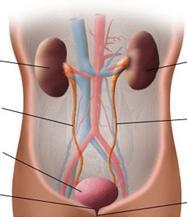
3 The Urinary Tract

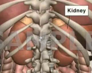
4 Kidneys
5
6 ตาแหน งไต (position) อย ใน retroperitoneum ระด บ T12-L3 โดยไต ขวาจะม ระด บตากว าไตซ ายเล กน อย ร ปร าง (shape) คล ายร ปถ ว ด านเว าห นเข าทาง medial ท ศทางการวางต ว Long axis ของไตเอ ยงขนานไปก บเงาของ (axis) psoas muscle ขนาด (size) ยาวประมาณ cm. กว าง 6 cm. หนา 4 cm. หร อประมาณ 3-4 lumbar vertebral bodies ขนาดของไตสองข างต างก นได ไม เก น 1.5 cm. ความหนาของ renal ประมาณ cm. cortex (cortical thickness) ขอบ (outline) ขอบเร ยบเสมอก น

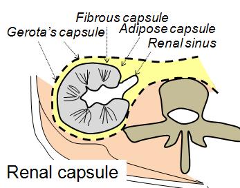
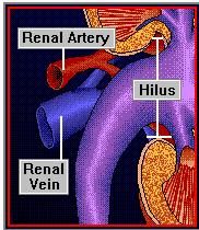
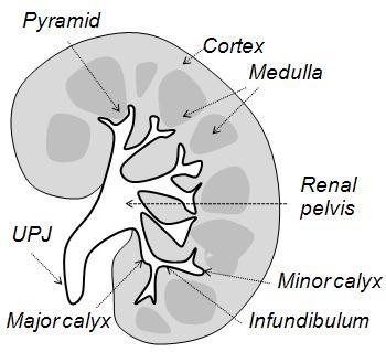
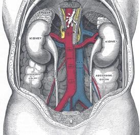
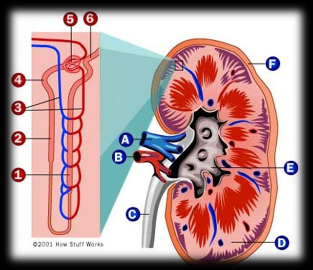
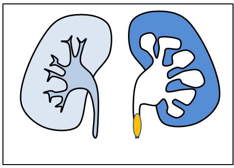
7 Kidneys 1. Renal capsule Gerota s capsule Adipose capsule Fibrous capsule 2. Renal parenchyma Cortex Medulla 3. Renal sinus Renal pelvis Artery, vein lymph, nerves Fat
8 Renal Capsule Renal capsule: 3 layers I. Gerota s capsule II. Adipose capsule III. Fibrous capsule
9 Renal Capsule Renal capsule: 3 layers I. Gerota s capsule II. Adipose capsule III. Fibrous capsule
10 Renal Capsule Renal capsule: 3 layers I. Gerota s capsule II. Adipose capsule III. Fibrous capsule
11

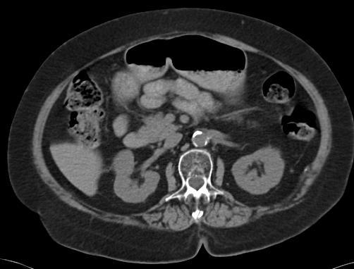
12 Renal Parenchyma

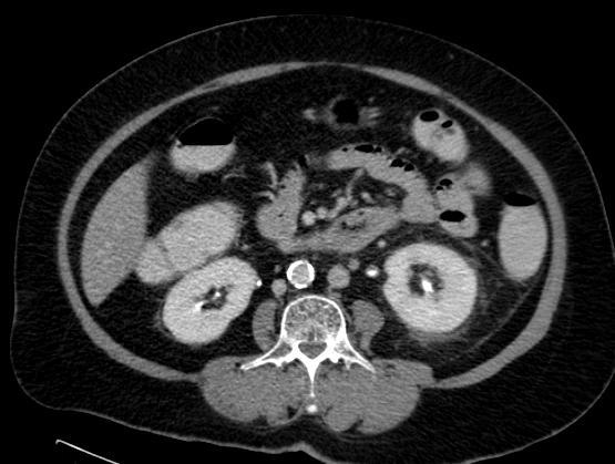
13 Renal Sinus
14
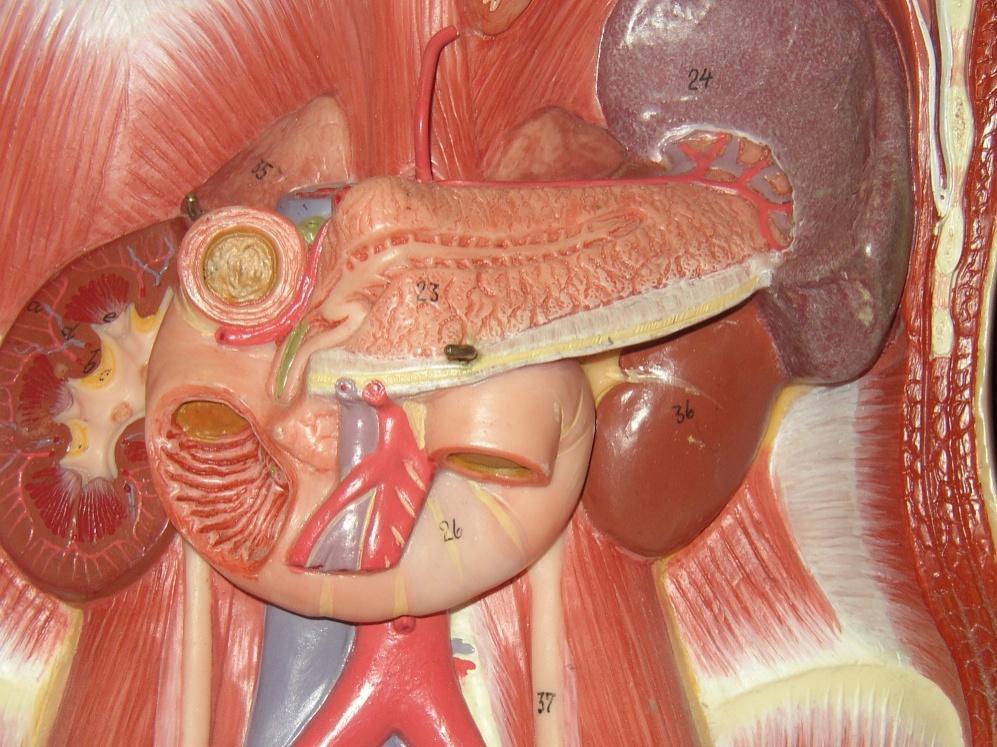
15 Associations

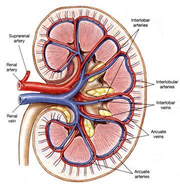
16 Renal Vessels
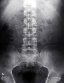
17 Film KUB

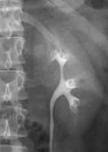
18 IVP 5 MINS IVP/IVU
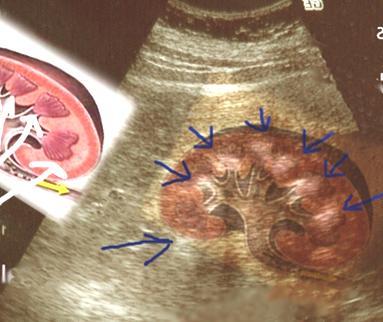
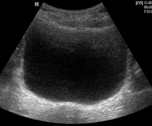
19 Ultrasound

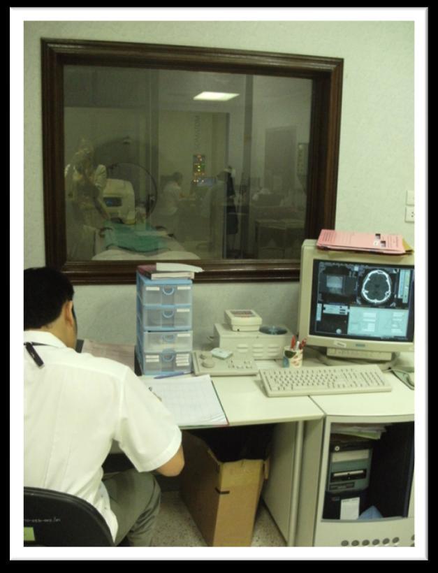
20 Computed Tomography (CT scan)

")
21 Magnetic resonance imaging (MRI)
22 Ureters
23 Ureters


24 Film KUB IVP 10 Minutes
 II.")

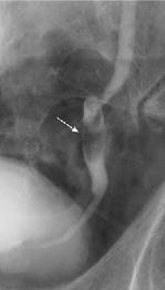
25 Physiologic narrowing of ureter I. Ureteropelvic junction (UPJ) II. Distal ureter that cross bifurcation of iliac vessels III. Ureterovesical junction (UVJ)

26 Physiologic Narrowing (1)
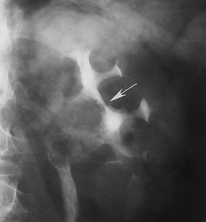
27 Physiologic Narrowing (2)

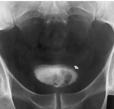
28 Physiologic Narrowing (3)
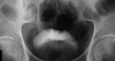
29 Urinary Bladder

30 Urinary Bladder

31 Urinary Bladder

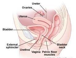
32 Urethra
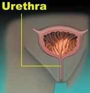
33 Urethra
34 Urethra


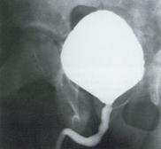
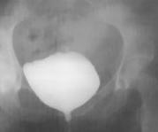
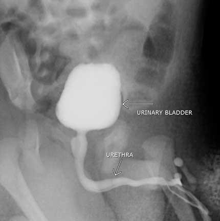
35 Normal male urethrogram
36
37 Investigations 1. Plain KUB 2. Intravenous pyelography (IVP) 3. Retrograde pyelography (RP) 4. Cystography 5. Voiding cystourethrography (VCUG) 6. Ultrasonography 7. CT scan 8. Magnetic resonance imaging 9. Renal angiogram 10. Renogram

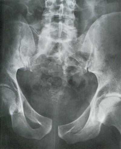
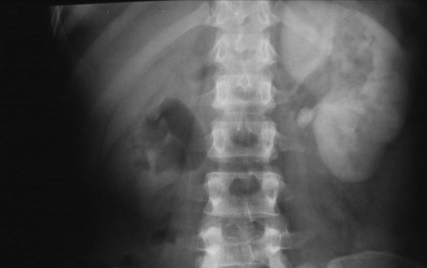
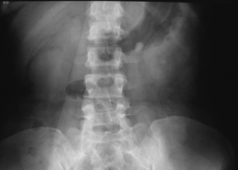
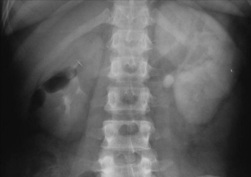
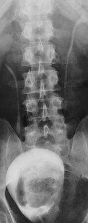
38 Plain KUB
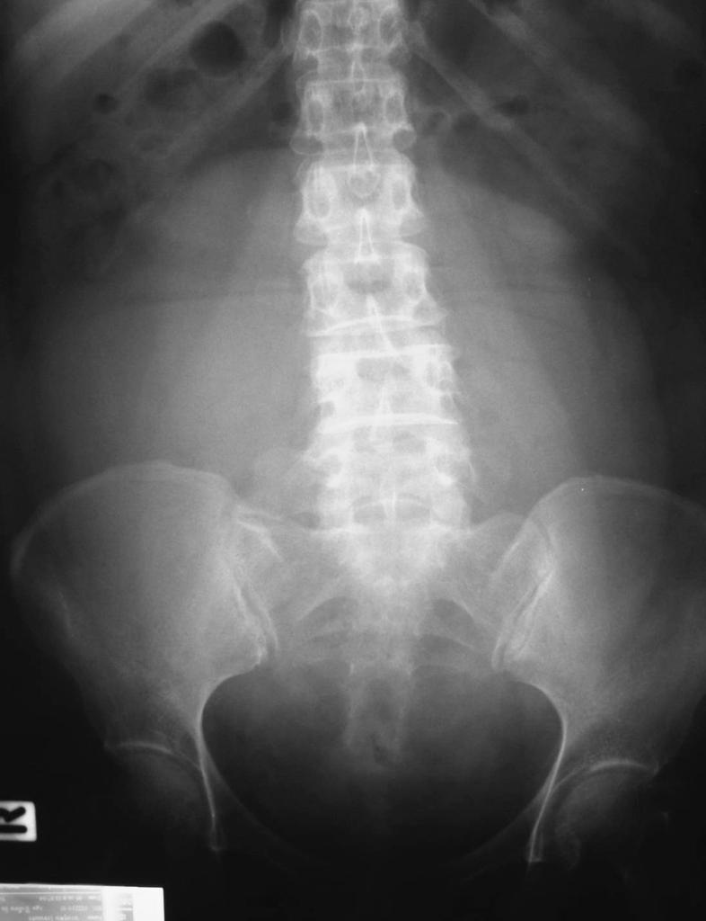
39 Plain KUB 1. Soft tissue shadow 2. Abnormal calcification 3. Free air or free fluid 4. Bony structure
40 Soft tissue shadows: Kidneys Psoas muscles Uterus Bladder Liver Spleen

41 Soft tissue shadows: Kidneys Psoas muscles Uterus Bladder Liver Spleen 1. Position 2. Shape 3. Axis 4. Size 5. Cortical thickness 6. Outline

42 Soft tissue shadows: Kidneys Psoas muscles Uterus Bladder Liver Spleen

43 Soft tissue shadows: Kidneys Psoas muscles Uterus Bladder Liver Spleen Retroperitoneal mass?

44 Right psoas abscess
45 Intraperitoneal mass
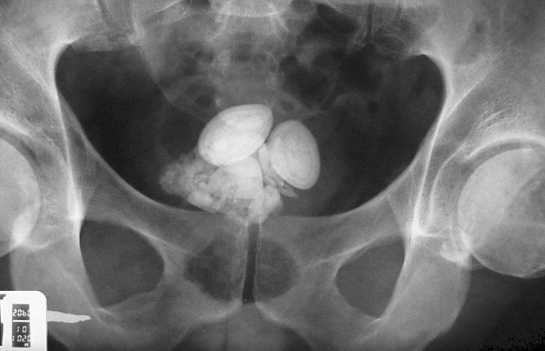
46 ? DDx. - Urinary tract stone - Calcified granuloma - Tumor - Gallstone - Appendicolith - Costochondral calcification - Atherosclerosis - Phlebolith - Prostatic calculi - Calcified uterine fibroid
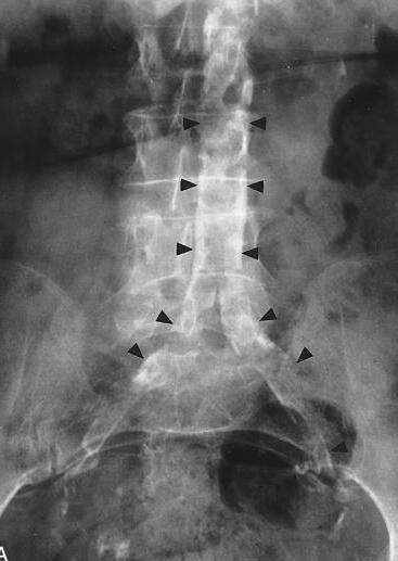
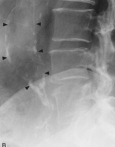
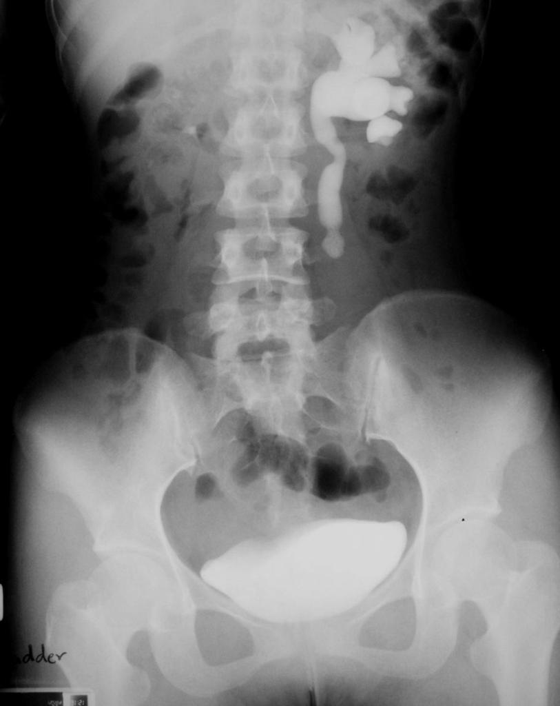
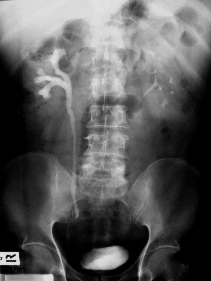
47 ? Example: There is an ovalshaped calcification at left L1-L2 paravertebral level, overriding lateral margin of left psoas muscle. These findings are suspected of left UPJ stone.
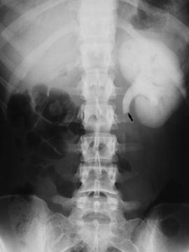
48 UPJ stone


49 Calyceal stone

50 Staghorn stone


51
52


53 Emphysematous pyelonephritis


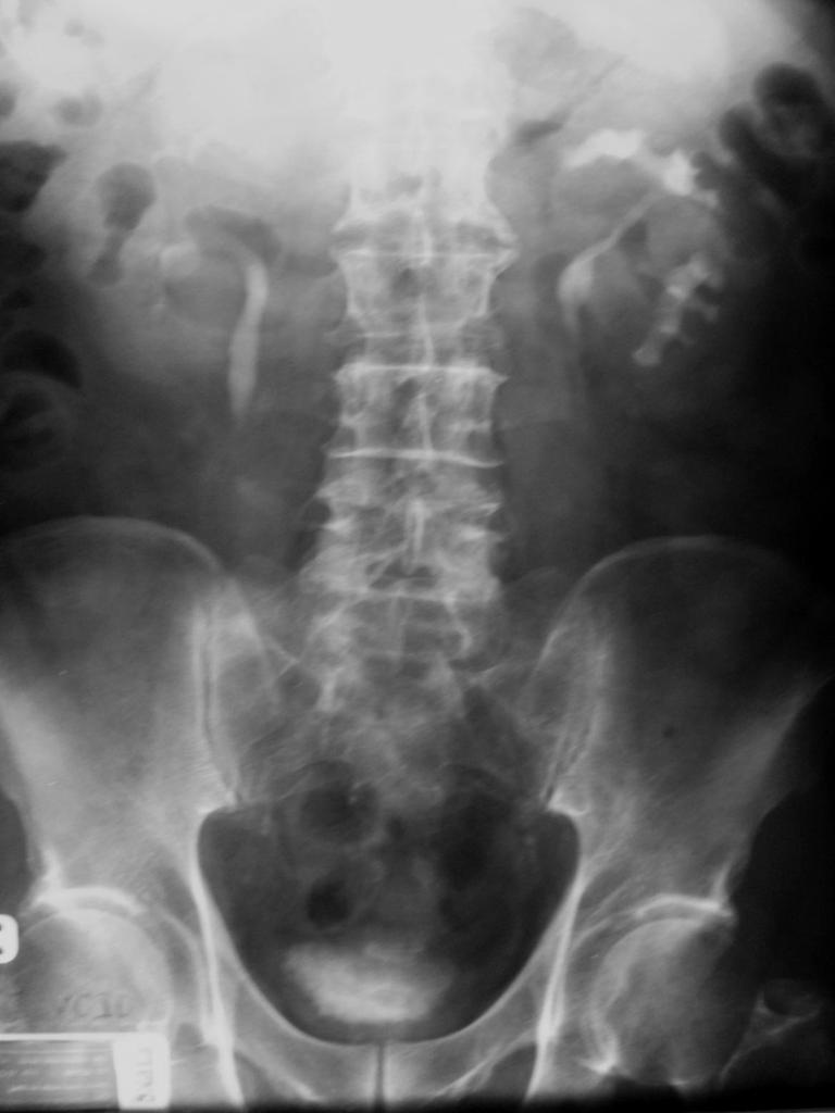
54 I. Spine II. Lower ribs III. Sacrum IV. Pelvis V. Hip & SI joints Look for - Fracture - Congenital bony defect - Bony destruction

55

56 สร ป: การอ าน Plain KUB 1. Soft tissue shadow 2. Abnormal calcification 3. Free air or free fluid 4. Bony structure


57 Intravenous Pyelography (IVP)
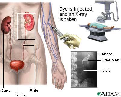
58
59 IVP *** Intravenous injection of iodinated contrast medium to evaluate for renal anatomy and function 1. Standard IVP (50 ml contrast medium) 2. Double dose IVP (100 ml contrast medium)


60 Factors 1. Kidney function GFR 2. Intrarenal concentration: State of hydration, osmotic diuresis 3. Exit of contrast from kidney (collecting system dynamics): Rate of urine flow, volume of collecting system, ureteral dynamics
61 IVP: Preparation Fluid restriction Increase visualization of contrast media Except for patients with poor renal function, DM, multiple myeloma, trauma, young child Bowel preparation To clear fecal content Not absolutely needed
 Pregnancy Note: Cr Clearance = (140-age)xBW(kg)")
62 IVP: Contraindication 1. Allergy to iodinated contrast media 2. Renal insufficiency (Creatinine Clearance < 30) Pregnancy Note: Cr Clearance = (140-age)xBW(kg) 72xserum Cr (mg/dl) If female (x0.85)


63 IVP: Evaluation 30 min, full bladder 3 min. Post void 10 min. 5 min.

64 Scout Film

65 Normal Nephrogram 3 min.

66 3 min: Nephrogram 1. Position 2. Shape 3. Axis 4. Size 5. Cortical thickness 6. Outline
67 Normal Excretion 3 min. 5 min.


68 5 min: Excretion 1. Minor/major calyx 2. Renal pelvis Abnormalities - Clubbing calyx/ hydronephrosis - Filling defect - Anomaly - Dense nephrogram

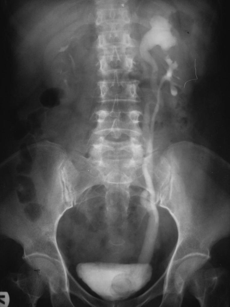
69 10 min: Pelvocalyeal system and ureter 5


70 Scout film Findings of UPJ obstruction 3 mins 5 mins 10 mins


71 Scout 5 min 10 min
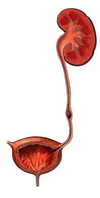
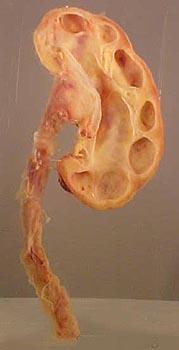
72 Hydronephrosis

73 Double collecting system


74 10 mins 30 mins

75 Right UVJ stone causing obstruction scout 10 mins post void


76 Bilateral hydronephrosis & hydroureters 1. Bladder cause 2. Urethra cause


77 Filling Defects


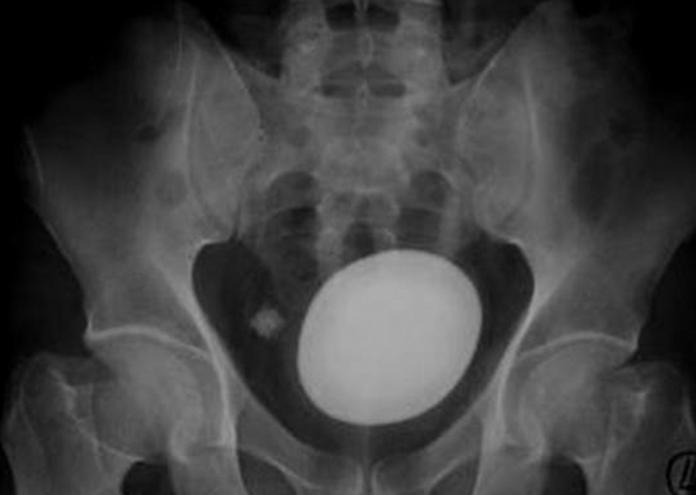
78 30 min: Full bladder 1. Size 2. Shape 3. Position 4. Architecture 5. Density


79 Irregular bladder outline Multiple bladder diverticulum Bladder trabeculation

80 Bladder filling defects

81 Post voiding film 1. Residual urine 2. Small tumor 3. Small stone
82 IVP: Indication 1. Renal and ureteric calculi 2. Colicky abdominal pain 3. Persistent or frank hematuria 4. Complicated urinary tract infection 5. Urothelial tumors 6. Abnormal US or renogram
83 IVP 1. Can evaluate renal function 2. Can demonstrate urothelium 3. Can visualize the ureter 1. Unable to identify renal parenchymal abnormality 2. Need IV contrast renal failure
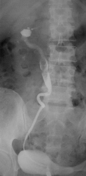
84 Retrograde Pyelography (RP)

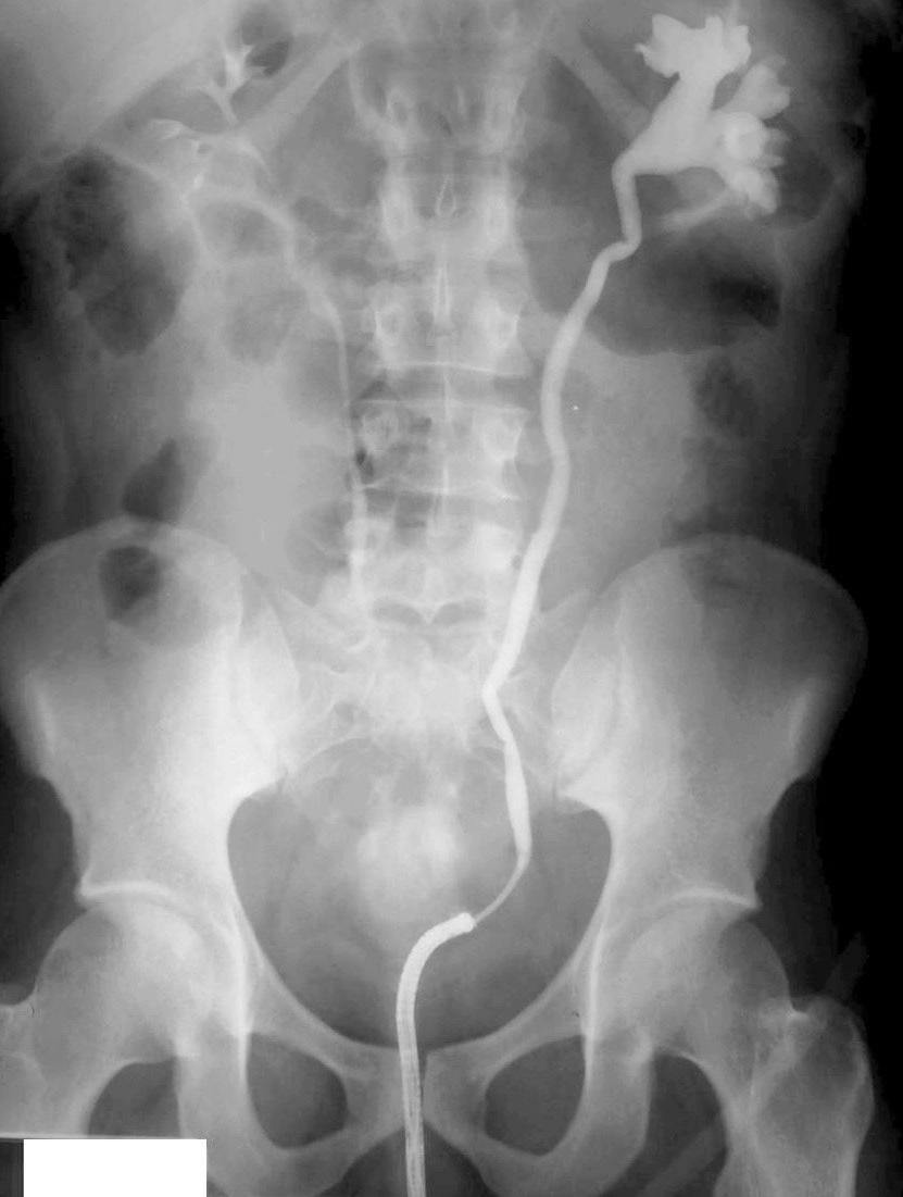
85 Retrograde Pyelography (RP)
86 RP: Indication 1. Poor kidney excretion 2. Evaluate pelvocalyceal system if uncertainty from IVP 3. Unexplained hematuria
87 RP: Contraindication 1. Urinary tract infection
88 RP: Complication 1. Infection 2. Trauma to urethra
89
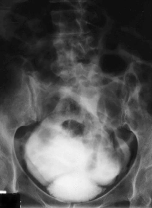
90 Cystography
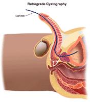
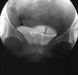
91 Retrograde Cystography

92 Cystography: Indication 1. Evaluate size and contour of urinary bladder 2. Bladder carcinoma 3. Trauma: rupture bladder 4. Low-pressure vesicoureteral reflux 5. Vesical fistula

93


94 Voiding Cystourethrography (VCUG)

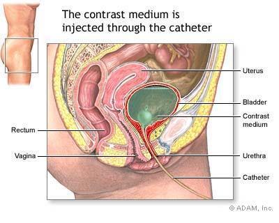
95 Voiding Cystourethrography (VCUG)
96

 2.")
97 VCUG: Indication 1. Urinary tract infection in children: - Urethral abnormality: posterior urethral valve - Vesicoureteral reflux (VUR) 2. Cause of urinary incontinence
98 Urethrography 1. Descending (antegrade) urethrography 2. Ascending (retrograde) urethrography

99 Descending Urethrography Urethrography was performed during voiding
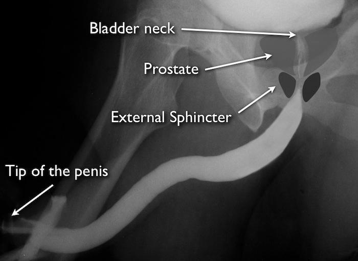
100 Ascending/Retrograde Urethrography


101 Urethral stricture at bulbous portion
102 Urethrograpy: Indication 1. Urethral trauma 2. Urethral stricture or anomaly

103 Ultrasonography (US)
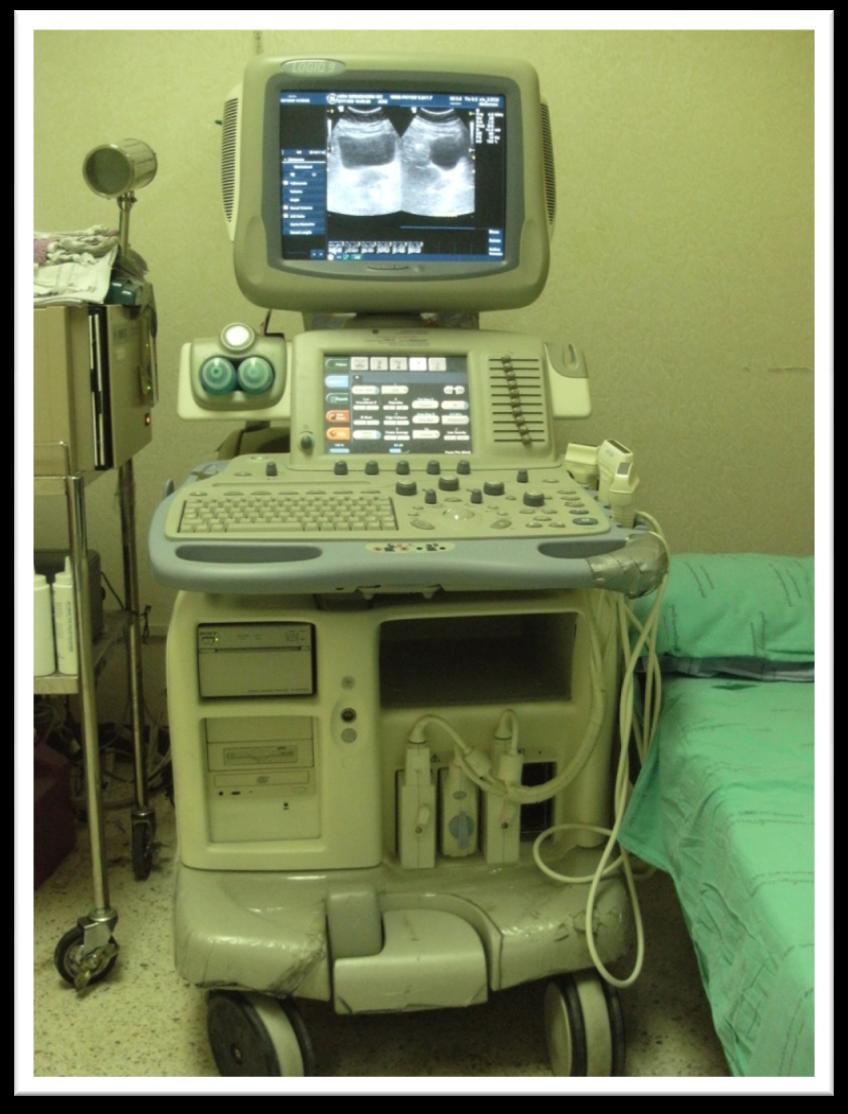
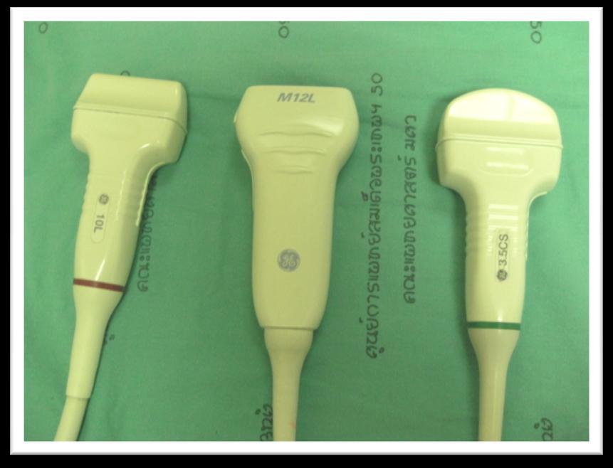
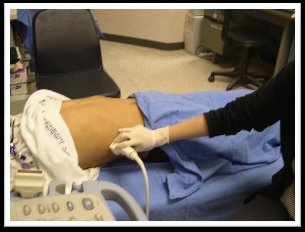
104


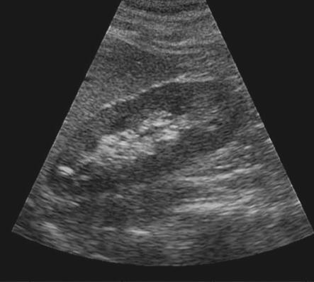
105 1. Liver 2. Right kidney 3. Right diaphragm 4. Hepatorenal pouch Longitudinal scan of right kidney


106 Normal kidney Liver parenchyma Normal renal parenchyma, slightly hypoechoic Hepatorenal pouch, no free fluid Normal renal fat = hyperechoic No dilatation of collecting system

107 Normal renal pyramids: medulla triangular-shaped, hypoechoic structures


108 Renal parenchymal disease Normal kidney Increased renal echogenicity


109 Obstruction hydronephrosis Renal parenchyma, hypoechoic Dilated collecting system, anechoic (fluid) Compressed renal fat, hyperechoic


110 Renal mass: solid / cystic


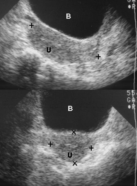
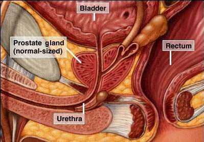
111 Bladder, uterus, prostate gland
112 US: Indication 1. Renal mass 2. Creatinine rising Renal disease or obstruction 3. Infection of kidney Renal abscess, perinephric abscess 4. Renal transplant patients 5. Urinary bladder lesion 6. Renal biopsy


113 US-KUB 1. No radiation 2. Can be used in pts with renal failure 3. No need to NPO 4. Image guided biopsy 1. Cannot evaluate renal function 2. Limit evaluation of ureter 3. Operator dependent

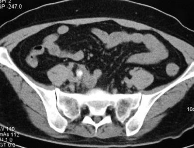
114 Computed Tomography (CT KUB)

115
116 CT Renal Protocol I. Precontrast phase II. Corticomedullary phase (30-60 sec) III. Nephrographic phase ( sec) IV. Excretory or delayed phase ( sec)
117 Non contrast
118 Corticomedullary phase
119 Nephrographic phase

120 Excretory phase
121 Ureter
122 Ureter

123 Ureter
124 Ureter
125 Ureter
126 Ureter
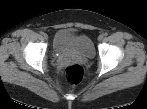
127 Bladder
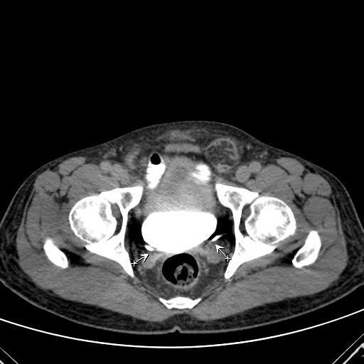
128 CT: Indication 1. Renal mass: diagnosis, staging 2. Tumor of urinary system 3. Renal trauma 4. Infection 5. Renal artery stenosis 6. Renal stone protocol

129 CT KUB 1. Can evaluate renal function and anatomy 2. Good detail study 1. Radiation 2. Contrast administration
130 Magnetic Resonance Imaging (MRI)

131 Magnetic Resonance Imaging (MRI)
132 MRI 1. No radiation 2. Risk of Gadolinium allergy < iodinated CM 3. Good for evaluating renal artery stenosis 1. Long scan time 2. Metallic / motion artifact 3. Not sensitive for stone or calcification 4. Expensive
133 Renal Angiography
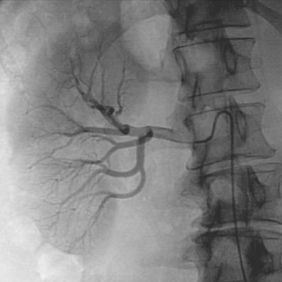
134 Renal Angiography
135 Renogram

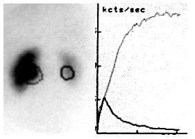
136 Renogram
137 Summary Normal radiographic anatomy Kidney, Ureter, Bladder, urethra Investigation of the KUB system Indication Contraindication Complication Interpretation

138
139 Thank You
Outline. Introduction to imaging modalities of the urinary system. Case base learning of common diseases in urinary tract
 Outline Introduction to imaging modalities of the urinary system Case base learning of common diseases in urinary tract Outline Introduction to imaging modalities of the urinary system Case base learning
Outline. Introduction to imaging modalities of the urinary system. Case base learning of common diseases in urinary tract
 Outline Introduction to imaging modalities of the urinary system Case base learning of common diseases in urinary tract Diagnostic Investigations in Urinary System PLAIN KUB EXCRETORY UROGRAPHY RETROGRADE
Excretory urography (EU) or IVP US CT & radionuclide imaging
 or IVP US CT & radionuclide imaging") Excretory urography (EU) or IVP US CT & radionuclide imaging MRI arteriography studies requiring catherization or direct puncture of collecting system EU & to a lesser extent CT provide both functional
Excretory urography (EU) or IVP US CT & radionuclide imaging MRI arteriography studies requiring catherization or direct puncture of collecting system EU & to a lesser extent CT provide both functional
PROFESSIONAL SKILLS 1 3RD YEAR SEMESTER 6 RADIOGRAPHY. THE URINARY SYSTEM Uz. Fatema shmus aldeen Tel
 PROFESSIONAL SKILLS 1 3RD YEAR SEMESTER 6 RADIOGRAPHY THE URINARY SYSTEM Uz. Fatema shmus aldeen Tel. 0925111552 Professional skills-2 THE URINARY SYSTEM The urinary system (review anatomy and physiology)
Urinary tract obstruction
 Urinary tract obstruction Common causes : stone, blood clot Radiographic findings depend on I. Level of obstruction II. Severity of obstruction : partial or complete III. Timing of obstruction Pathophysiology
Uroradiology For Medical Students
 Uroradiology For Medical Students Lesson 4: Cystography & Urethrography - Part 2 American Urological Association Review Cystography is useful in evaluating the bladder, the urethra and the competence of
US in non-traumatic acute abdomen. Lalita, M.D. Radiologist Department of radiology Faculty of Medicine ChiangMai university
 US in non-traumatic acute abdomen Lalita, M.D. Radiologist Department of radiology Faculty of Medicine ChiangMai university Sagittal Orientation Transverse (Axial) Orientation Coronal Orientation Intercostal
R adio logical investigations of urinary system
 R adio logical investigations of urinary system There are 4 main radiological Ix: 1 IVU: Intravenous urography. 2- U/S 3-CT scan 4-Radioisotope scan. Others (not frequently used): MRI, arteriography, antegrade
URINARY SYSTEM I. Kidneys II. Nephron Unit and Urine Formation
 URINARY SYSTEM I. Kidneys A. Location and Structure 1. Retroperitoneal 2. Between T12 and L3 3. Rt. kidney slightly lower 4. Two bean shaped organs 5. Adrenal gland 6. Internal construction a. Renal cortex
Uroradiology Tutorial For Medical Students
 Uroradiology Tutorial For Medical Students Lesson 3: Cystography & Urethrography Part 1 American Urological Association Introduction Conventional radiography of the urinary tract includes several diagnostic
Proceedings of the 34th World Small Animal Veterinary Congress WSAVA 2009
 www.ivis.org Proceedings of the 34th World Small Animal Veterinary Congress WSAVA 2009 São Paulo, Brazil - 2009 Next WSAVA Congress : Reprinted in IVIS with the permission of the Congress Organizers IMAGING
Acute flank pain in children: Imaging considerations
 Acute flank pain in children: Imaging considerations Carlos J. Sivit MD Rainbow Babies and Children s Hospital Case Western Reserve School of Medicine Flank pain Results from distention of ureter or renal
URINARY TRACT IMAGING - BASIC PRINCIPLES
 URINARY TRACT IMAGING - BASIC PRINCIPLES Clinical Radiology Every physician needs a basic understanding of diagnostic imaging to understand how to order the appropriate studies and to understand the resulting
Case MDCT 3D reconstructed features of posterior urethral valve
 Case 12688 MDCT 3D reconstructed features of posterior urethral valve Hidayatullah Hamidi Third year Resident of Radiology French medical institute for children Radiology Department; Kabul, Afghanistan;
Hydronephrosis. Nephrosis. Refers to the kidney
 What is hydronephrosis? Hydro Nephrosis Refers to water or fluid Refers to the kidney A build-up of fluid (urine) in the kidney is the medical term for a build-up of urine in the kidney. As the urine builds
Hydronephrosis. What is hydronephrosis?
 What is hydronephrosis? Hydronephrosis Hydronephrosis describes the situation where the urine collecting system of the kidney is dilated. This may be a normal variant or it may be due to an underlying
IMAGING OF THE UROGENITAL TRACT
 IMAGING OF THE UROGENITAL TRACT 1 A) URINARY TRACT There are many methods of imaging the urinary tract but plain abdominal X-ray and ultrasound scan are usually done first in most cases, especially in
Imaging the Urinary Tract
 Imaging the Urinary Tract Laura Armbrust, DVM, DACVR Gregory F. Grauer, DVM, MS, DACVIM Kansas State University Radiographic and ultrasound imaging in addition to history, physical examination, and clinicopathologic
My Patient Has Abdominal Pain PoCUS of the Biliary Tract and the Urinary Tract
 My Patient Has Abdominal Pain PoCUS of the Biliary Tract and the Urinary Tract Objectives PoCUS for Biliary Disease PoCUS for Renal Colic PoCUS for Urinary Retention Biliary Disease A patient presents
Human Anatomy Unit 3 URINARY SYSTEM
 Human Anatomy Unit 3 URINARY SYSTEM In Anatomy Today Components Kidneys Ureters Urinary bladder Urethra Functions Storage of urine Bladder stores up to 1 L of urine Excretion of urine Transport of urine
Pediatric Ure-Radiology*
 Pediatric Ure-Radiology* HERMAN GROSSMAN, M.D. Professor of Radiology and Pediatrics, Duke University Medical Center, Durham, North Carolina "Routine" radiologic studies do not, often enough, concentrate
Urinary system Ultrasound (Renal & Urinary bladder)
") Urinary system Ultrasound (Renal & Urinary bladder) Edited & Presented by ; Hussien A.B ALI DINAR. Msc.Phd ISRRT Associate Member Lecturer (National university) Reporting Sonographer (PHC) Objective By
Urinary system Ultrasound (Renal & Urinary bladder) Edited & Presented by ; Hussien A.B ALI DINAR. Msc.Phd ISRRT Associate Member Lecturer (National university) Reporting Sonographer (PHC) Objective By
Radiographic Procedures III (RAD 228)
") Radiographic Procedures III (RAD 228) Urinary System RADIOGRAPHIC EXAMINATIONS Urinary System Antegrade Exam IVU Functional test Hypertensive evaluation as per protocol Retrograde Exams Retrograde Urography
Radiographic Procedures III (RAD 228) Urinary System RADIOGRAPHIC EXAMINATIONS Urinary System Antegrade Exam IVU Functional test Hypertensive evaluation as per protocol Retrograde Exams Retrograde Urography
The Kidneys. (L., ren; Gk, nephros; hence the adjectives renal and nephric) & Suprarenal (Adrenal) Glands. Dr Maan Al-Abbasi PhD, MBChB
 & Suprarenal (Adrenal) Glands. Dr Maan Al-Abbasi PhD, MBChB") The Kidneys (L., ren; Gk, nephros; hence the adjectives renal and nephric) & Suprarenal (Adrenal) Glands Dr Maan Al-Abbasi PhD, MBChB Functions of Urinary System Regulate electrolytes (K+, Na+, etc) Regulate
The Kidneys (L., ren; Gk, nephros; hence the adjectives renal and nephric) & Suprarenal (Adrenal) Glands Dr Maan Al-Abbasi PhD, MBChB Functions of Urinary System Regulate electrolytes (K+, Na+, etc) Regulate
Abdominal Ultrasound : Aorta, Kidneys, Bladder
 Abdominal Ultrasound : Aorta, Kidneys, Bladder Nilam J. Soni, MD, MSc Associate Professor of Medicine Divisions of Hospital Medicine and Pulmonary/Critical Care Medicine Department of Medicine University
Acute renal colic Radiological investigation in patients with renal colic
 Acute renal colic Radiological investigation in patients with renal colic Mikael Hellström Professor Department of Radiology Sahlgrenska University Hospital Göteborg University 0.9-1.8/1.000 inhabitants
Obstetrics Content Outline Obstetrics - Fetal Abnormalities
 Obstetrics Content Outline Obstetrics - Fetal Abnormalities Effective February 2007 10 16% renal agenesis complete absence of the kidneys occurs when ureteric buds fail to develop Or degenerate before
Basic of Ultrasound Physics E FAST & Renal Examination. Dr Muhammad Umer Ihsan MBBS,MD, DCH CCPU,DDU1,FACEM
 Basic of Ultrasound Physics E FAST & Renal Examination Dr Muhammad Umer Ihsan MBBS,MD, DCH CCPU,DDU1,FACEM What is Sound? Sound is Mechanical pressure waves What is Ultrasound? Ultrasounds are sound waves
ASSESSING THE PLAIN ABDOMINAL RADIOGRAPH M A A M E F O S U A A M P O F O
 ASSESSING THE PLAIN ABDOMINAL RADIOGRAPH M A A M E F O S U A A M P O F O Introduction The abdomen (less formally called the belly, stomach, is that part of the body between the thorax (chest) and pelvis,
RADIOLOGY OF THE URINARY TRACT CHAPTER 9 239
 RADIOLOGY OF THE URINARY TRACT CHAPTER 9 239 in length. They lie cephalad to the kidneys, with the right just posterior to the inferior vena cava (IVC) and the left anteromedial to the upper pole of the
Role of imaging in evaluation of genitourinary i trauma Spectrum of GU injuries Relevance of imaging findings in determining management Focus on MDCT
 Genitourinary Tract Injuries 6 th Nordic Course Scott D. Steenburg, MD Assistant Professor University of Maryland Department of Radiology Division of Trauma and Emergency Radiology R Adams Cowley Shock
Genitourinary. Common Clinical Scenarios Protocoling Module. Patty Ojeda & Mariam Shehata
 The following training module was developed as a quality improvement project to serve as an educational tool for junior radiology residents. The following diagnostic radiology protocoling modules were
Lecture 56 Kidney and Urinary System
 Lecture 56 Kidney and Urinary System The adrenal glands are located on the superomedial aspect of the kidney The right diagram shows a picture of the kidney with the abdominal walls and organs removed
Obstructive Uropathy. PATHOPHYSIOLOGIC CHANGES UUO vs BUO. Arry Rodjani Urology Department Ciptomangunkusumo Hospital Jakarta
 Obstructive Uropathy PATHOPHYSIOLOGIC CHANGES UUO vs BUO Arry Rodjani Urology Department Ciptomangunkusumo Hospital Jakarta INTRODUCTION Obstructive uropathy refers to the functional or anatomic obstruction
Genitourinary Trauma Introduction GU Trauma overlooked
 Genitourinary Trauma Introduction GU Trauma overlooked 10-20% of all injured patients Long term morbidity Impotence Incontinence Life-threatening injuries first Urethral Injury Plan Bladder Injury Kidney
Indications and effectiveness of the open surgery in vesicoureteral reflux
 Indications and effectiveness of the open surgery in vesicoureteral reflux Suzi DEMIRBAG, MD Department of Pediatric Surgery, Gulhane Military Medical Academy, Ankara, TURKEY Vesicoureteral reflux (VUR)
Kidneys and Urinary Tract Content Outline. Anatomy Coverings. Location. (Effective February 2007) (16%-24%)
 (16%-24%)") Kidneys and Urinary Tract Content Outline (Effective February 2007) (16%-24%) Anatomy Coverings true capsule perirenal fat surrounds capsule Gerota s fascia separates perirenal from extraperitoneal fat
Kidneys and Urinary Tract Content Outline (Effective February 2007) (16%-24%) Anatomy Coverings true capsule perirenal fat surrounds capsule Gerota s fascia separates perirenal from extraperitoneal fat
ANATOMY OF PELVICAYCEAL SYSTEM -DR. RAHUL BEVARA
 1 ANATOMY OF PELVICAYCEAL SYSTEM -DR. RAHUL BEVARA 2 KIDNEY:ANATOMY OVERVIEW Kidneys are retroperitoneal, in posterior abdominal region, extending from T12 L3 Bean-shaped Right kidney is lower than left
Prenatal Hydronephrosis
 Prenatal Hydronephrosis What is hydronephrosis? Hydronephrosis is dilation of the kidney, specifically the renal pelvis (place where urine is stored after its production). This can be the result of an
Gross Anatomy of the Urinary System
 Gross Anatomy of the Urinary System Lecture Objectives Overview of the urinary system. Describe the external and internal anatomical structure of the kidney. Describe the anatomical structure of the ureter
Role of imaging in RCC. Ultrasonography. Solid lesion. Cystic RCC. Solid RCC 31/08/60. From Diagnosis to Treatment: the Radiologist Perspective
 Role of imaging in RCC From Diagnosis to Treatment: the Radiologist Perspective Diagnosis Staging Follow up Imaging modalities Limitations and pitfalls Duangkamon Prapruttam, MD Department of Therapeutic
Chapter 6: Genitourinary and Gastrointestinal Systems 93
 Chapter 6: Genitourinary and Gastrointestinal Systems 93 Chapter 6 Genitourinary and Gastrointestinal Systems Embryology Three sets of excretory organs or kidneys develop in human embryos: Pronephros:
Kidney & Urinary Tract Ultrasound. Fatina Fadel Hafez Bazaraa
 Kidney & Urinary Tract Ultrasound Fatina Fadel Hafez Bazaraa Ultrasonography Ultrasound Available Rapid Inexpensive Painless & no sedation needed No adverse effects/ complications Can be repeated Useful
General Anatomy of Urinary System
 General Anatomy of Urinary System URINARY SYSTEM ORGANS Kidneys (2) Ureters (2) Urinary bladder Urethra KIDNEY FUNCTIONS Control blood volume and composition KIDNEY FUNCTIONS Filter blood plasma, eliminate
Sex: 女 Age: 51 Occupation: 無 Admission date:92/07/22
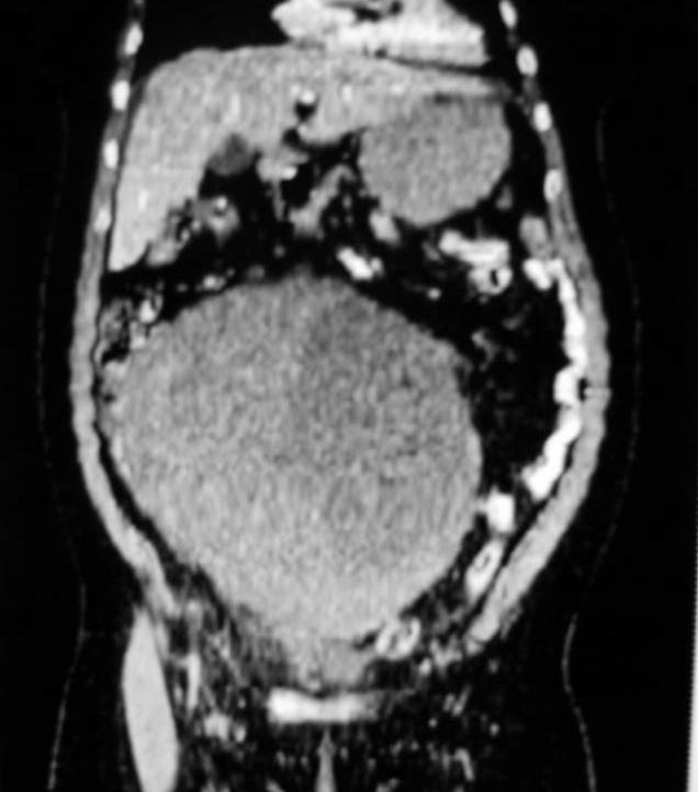
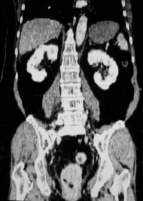
 Sex: 女 Age: 51 Occupation: 無 Admission date:92/07/22 Chief complaint Unknown fever for one month Hand tremor and left huge renal tumor was noted Present illness Suffered from fever for one month, hand
URINARY SYSTEM ANATOMY
 URINARY SYSTEM ANATOMY Adapted from Human Anatomy & Physiology Marieb and Hoehn (9 th ed.) OVERVIEW Metabolism of nutrients by the body produces wastes that must be removed from the body. Although excretory
UBC Department of Urologic Sciences Lecture Series. Urological Trauma
 UBC Department of Urologic Sciences Lecture Series Urological Trauma Disclaimer: This is a lot of information to cover and we are unlikely to cover it all today These slides are to be utilized for your
URINARY SYSTEM ANATOMY PART
 URINARY SYSTEM ANATOMY PART 1 DANIL HAMMOUDI.MD Urinary System Composed of kidneys, ureters, urinary bladder, and urethra Eliminates nitrogenous wastes from the body Regulates water, electrolyte, and ph
Anatomy of the renal system. Professor Nawfal K. Al-Hadithi
 Anatomy of the renal system Professor Nawfal K. Al-Hadithi Objectives To describe the posterior abdominal wall To identify the main anatomical landmarks of the kidneys & ureters To describe the suprarenal
Find Medical Solutions to Your Problems HYDRONEPHROSIS. (Distension of Renal Calyces & Pelvis)
") HYDRONEPHROSIS (Distension of Renal Calyces & Pelvis) Hydronephrosis is the distension of the renal calyces and pelvis due to accumulation of the urine as a result of the obstruction to the outflow of
HYDRONEPHROSIS (Distension of Renal Calyces & Pelvis) Hydronephrosis is the distension of the renal calyces and pelvis due to accumulation of the urine as a result of the obstruction to the outflow of
The Urinary System Pearson Education, Inc.
 26 The Urinary System Introduction The urinary system does more than just get rid of liquid waste. It also: Regulates plasma ion concentrations Regulates blood volume and blood pressure Stabilizes blood
Spectrum of Micturating Cystourethrogram Revisited: A Pictorial Assay
 603 International Journal of Collaborative Research on Internal Medicine & Public Health Spectrum of Micturating Cystourethrogram Revisited: A Pictorial Assay Abhinav Jain 1, Vivek Setia 1, Shweta Agnihotri
CASE REPORT RENAL TUBERCULOSIS CAUSE OF RENAL REPLACEMENT LIPOMATOSIS : A RARE ASSOCIATION
 CASE REPORT RENAL TUBERCULOSIS CAUSE OF RENAL REPLACEMENT LIPOMATOSIS : A RARE ASSOCIATION DR ANAND AARTI 1, DR CHANDAK PRIYA 2,DR SURESH PARVATHY 3 1. PROF AND HOD, DEPARTMENT OF RADIODIAGNOSIS, GOVERNMENT
Canadian Undergraduate Urology Curriculum (CanUUC): Genitourinary Trauma. Last reviewed June 2014
: Genitourinary Trauma. Last reviewed June 2014") Canadian Undergraduate Urology Curriculum (CanUUC): Genitourinary Trauma Last reviewed June 2014 Session Objectives 1. Recognize hematuria as the cardinal symptom of urinary tract trauma. 1. Outline the
Canadian Undergraduate Urology Curriculum (CanUUC): Genitourinary Trauma Last reviewed June 2014 Session Objectives 1. Recognize hematuria as the cardinal symptom of urinary tract trauma. 1. Outline the
Acute Pyelonephritis
 Acute Pyelonephritis Variant 1: Acute pyelonephritis. Uncomplicated patient (eg, no history of diabetes or immune compromise or history of stones or obstruction or prior renal surgery or lack of response
Radiology of the abdomen Lecture -1-
 Radiology of the abdomen Lecture -1- Objectives To know radiology modalities used in abdomen imaging mainly GI tract. To know advantages and disadvantages of each modality. To know indications and contraindications
What s Your Diagnosis??? Renée Fahrenholz, Class of 2012
 Renée Fahrenholz, Class of 2012 What s Your Diagnosis??? Signalment Emma, a 9 year old, Female, Spayed, Domestic Short Haired Feline Presenting Complaint Weight loss, vomited the morning of her visit,
Lab Monitor Images Dissection of the Abdominal Vasculature + Lower Digestive System
 Lab Monitor Images Dissection of the Abdominal Vasculature + Lower Digestive System Stomach & Duodenum Frontal (AP) View Nasogastric tube 2 1 3 4 Stomach Pylorus Duodenum 1 Duodenum 2 Duodenum 3 Duodenum
Urinary System. Chapter 17 7/19/11. Introduction
 7/19/11 Chapter 17 Urinary System Introduction A. The urinary system consists of two kidneys that filter the blood, two ureters, a urinary bladder, and a urethra to convey waste substances to the outside.
Abdominal ultrasound:
 Abdominal ultrasound: Non-traumatic acute abdomen Wittanee Na-ChiangMai, MD Department of Radiology ChiangMai University 26/04/2017 Contents Technique of examination Normal anatomy Emergency conditions
4 th Year Urology Core Objectives Keith Rourke (Revised June 1, 2007)
") 4 th Year Urology Core Objectives Keith Rourke (Revised June 1, 2007) I. Genitourinary Trauma: 1. Goal: The student will be able to demonstrate a basic clinical approach to the management & diagnosis of
4 th Year Urology Core Objectives Keith Rourke (Revised June 1, 2007) I. Genitourinary Trauma: 1. Goal: The student will be able to demonstrate a basic clinical approach to the management & diagnosis of
Guidelines, Policies and Statements D5 Statement on Abdominal Scanning
 Guidelines, Policies and Statements D5 Statement on Abdominal Scanning Disclaimer and Copyright The ASUM Standards of Practice Board have made every effort to ensure that this Guideline/Policy/Statement
Urinary tract infections, renal malformations and scarring
 Urinary tract infections, renal malformations and scarring Yaacov Frishberg, MD Division of Pediatric Nephrology Shaare Zedek Medical Center Jerusalem, ISRAEL UTI - definitions UTI = growth of bacteria
Renal Trauma: Management Options
 Renal Trauma: Management Options Immediate surgical repair Nephrectomy Conservative management Alonso RC et al. Kidney in Danger: CT Findings of Blunt and Penetrating Renal Trauma. RadioGraphics 2009;
Developmental Abnormalities of the Kidneys and GU System
 A5 Developmental Abnormalities of the Kidneys and GU System Erin Parilla, MD Neonatologist Pediatrix Medical Group, Tampa, FL The speaker has signed a disclosure form and indicated she has no significant
Urinary Tract Abnormalities
 Urinary Tract Abnormalities Dr Hennie Lombaard Senior Specialist Maternal and Fetal Medcine Department of Obstetrics and Gynecology Level 7 Pretoria Academic Hospital Pictures from The 18 to 23 weeks scan
L o o k L i s t e n F e e l S c a n. Your Pocus Cards For Your Every Day Scanning.
 L o o k L i s t e n F e e l S c a n Your Pocus Cards For Your Every Day Scanning E-FAST Extended Focused Assessment by Sonography in Trauma Subcostal Heart View Pleural Sliding on M-mode (Sea-shore sign)
Figure 26.1 An Introduction to the Urinary System
 Chapter 26 Figure 26.1 An Introduction to the Urinary System Components of the Urinary System Kidney Produces urine Ureter Transports urine toward the urinary bladder Urinary Bladder Temporarily stores
Urologic Surgical Complications In Renal Transplantation
 Urologic Surgical Complications In Renal Transplantation Chris Freise, MD Professor of Surgery UCSF Transplant Division Urologic Complications Review of Bladder Anastomosis Complications and Management
URINARY SYSTEM. These organs lie posterior or inferior to the. (membrane).
.") URINARY SYSTEM I. INTRODUCTION Each kidney is made up of about a million tiny tubules called nephrons. Each nephron individually filters the blood and makes urine and it does the job completely, from start
URINARY SYSTEM I. INTRODUCTION Each kidney is made up of about a million tiny tubules called nephrons. Each nephron individually filters the blood and makes urine and it does the job completely, from start
Perineal Sonography in Diagnosis of an Ectopic Ureteric Opening Into the Urethra
 Case Series Perineal Sonography in Diagnosis of an Ectopic Ureteric Opening Into the Urethra S. Boopathy Vijayaraghavan, MD, DMRD Objective. To study the role of perineal sonography in the diagnosis of
IVU ((INTRAVENOUSUROGRAM))
)") IVU ((INTRAVENOUSUROGRAM)) Anatomy The urinary system consists of : 2 kidneys, 2 ureters,1 bladder, 1 urethra Renal pelvis Minor calyx Major calyx Renal parenchyma Proximal ureter Pelvi-uretric junction
IVU ((INTRAVENOUSUROGRAM)) Anatomy The urinary system consists of : 2 kidneys, 2 ureters,1 bladder, 1 urethra Renal pelvis Minor calyx Major calyx Renal parenchyma Proximal ureter Pelvi-uretric junction
Abdomen and Retroperitoneum Ultrasound Protocols
 Abdomen and Retroperitoneum Ultrasound Protocols Reviewed By: Anna Ellermeier, MD Last Reviewed: March 2018 Contact: (866) 761-4200, Option 1 **NOTE for all examinations: 1. If documenting possible flow
Autosomal Dominant Polycystic Kidney Disease
 Case Studies [1] July 01, 2014 By Amar Udare, MBBS [2] Case History: 45-year-old female with vague pain in the abdomen. Case History: A 45-year-old female presented with vague pain in the abdomen. A USG
URINARY SYSTEM CHAPTER 28 I ANATOMY OF THE URINARY SYSTEM. Student Name
 Student Name CHAPTER 28 URINARY SYSTEM L iving produces wastes. Wherever people live or work or play, wastes accumulate. To keep these areas healthy, there must be a method of disposing of these wastes
Imaging findings in renal infections
 Imaging findings in renal infections Poster No.: C-0221 Congress: ECR 2013 Type: Educational Exhibit Authors: I. lópez blasco, D. Soriano Mena, R. Pastor Toledo, S. Paz Maya, A. M. Julve Parreño, J. Palmero
Urinary system. Urinary system
 INTRODUCTION. Several organs system Produce urine and excrete it from the body Maintenance of homeostasis. Components. two kidneys, produce urine; two ureters, carry urine to single urinary bladder for
Index. Note: Page numbers of article titles are in boldface type.
 Magn Reson Imaging Clin N Am 12 (2004) 587 591 Index Note: Page numbers of article titles are in boldface type. A Adenoma(s), adrenal, gadolinium-enhanced MR imaging in, 533 534 hyperfunctioning versus
RENAL SCINTIGRAPHY IN THE 21 st CENTURY
 RENAL SCINTIGRAPHY IN THE 21 st CENTURY 99m Tc- MAG 3 with zero time injection of Furosemide (MAG 3 -F 0 ) : A Fast and Easy Protocol, One for All Indications Clinical Experience Congenital Disorders PROTOCOL
Bladder Trauma Data Collection Sheet
 Bladder Trauma Data Collection Sheet If there was no traumatic injury with PENETRATION of the bladder DO NOT proceed Date of injury: / / Time of injury: Date of hospital arrival: / / Time of hospital arrival:
The functional anatomy of the urinary system. Human Anatomy Department Dr. Anastasia Bendelic
 The functional anatomy of the urinary system Human Anatomy Department Dr. Anastasia Bendelic Plan Development of the kidneys and their abnormalities Development of the urinary ways and their abnormalities
Lec-8 جراحة بولية د.نعمان
 4th stage Lec-8 جراحة بولية د.نعمان 11/10/2015 بسم هللا الرحمن الرحيم Ureteric, Vesical, & urethral stones Ureteric Calculus Epidemiology like renal stones Etiology like renal stones Risk factors like
Request Card Task ANSWERS
 Request Card Task ANSWERS Medical Student Workbook Author: Dr Sam Leach, SpR Case 1 What differential diagnoses are most likely? Which investigation is most appropriate? Case 1 The most likely diagnosis
Five Views of Transitional Cell Carcinoma: One Man s Journey
 September 2006 Five Views of Transitional Cell Carcinoma: One Man s Journey Amsalu Dabela, Harvard Medical School III Outline Overview: Renal Anatomy Our Patient s Story Diagnostic Imaging Studies Appearance
Fetal Renal Malformations: The Role of Ultrasound in Diagnosis & Management
 Fetal Renal Malformations: The Role of Ultrasound in Diagnosis & Management 12 weeks Alfred Abuhamad, M.D. Eastern Virginia Medical School 13 weeks 2nd trimester Medullary pyramids Renal Sinus Cortex 2nd
Abdominal Ultrasonography
 Abdominal Ultrasonography David A. Masneri, DO, FACEP, FAAEM Assistant Professor of Emergency Medicine Assistant Director, Emergency Medicine Residency Medical Director, Operational Medicine Division Center
DISEASES AFFECTING TUBULES AND INTERSTITIUM
 DISEASES AFFECTING TUBULES AND INTERSTITIUM Acute tubular injury (ATI) Pyelonephritis Drug-induced tubulointerstitial nephritis (TIN) Myeloma cast NP Renal stones Urinary outflow obstruction: hydronephrosis
Urogenital Injuries The role of radiology
 Urogenital Injuries The role of radiology NORDTER 7 th Nordic Trauma Radiology Course Helsinki, Finland May 21-24, 2012 Johann Baptist Dormagen, MD, PhD Oslo University Hospital, Norway Kidney injuries
Basic Urinary Tract Anatomy and Histology
 Basic Urinary Tract Anatomy and Histology The two kidneys are located in the retroperitoneum on either side of the vertebral bladder and the contraction of the detrusor muscle. Any mechanical barrier,
Clinical aspects in urogenital injuries
 Clinical aspects in urogenital injuries Rolf Wahlqvist Oslo Urological University Clinic Aker University Hospital Nordic Rad.2008 1 Urogenital injuries in trauma patients Renal injury Ureteral injury (infrequent/iatrogenic)
CYSTIC DISEASES of THE KIDNEY. Dr. Nisreen Abu Shahin
 CYSTIC DISEASES of THE KIDNEY Dr. Nisreen Abu Shahin 1 Types of cysts 1-Simple Cysts 2-Dialysis-associated acquired cysts 3-Autosomal Dominant (Adult) Polycystic Kidney Disease 4-Autosomal Recessive (Childhood)
A Z OF ABDOMINAL RADIOLOGY
 Z OF BDOMINL RDIOLOGY bdominal trauma to Z of bdominal Radiology Clinical characteristics general discussion, followed by organ-specific summaries, is given below. bdominal trauma is managed as part of
Radiology of hepatobiliary diseases
 GI cycle - Lecture 14 436 Teams Radiology of hepatobiliary diseases Objectives 1. To Interpret plan x-ray radiograph of abdomen with common pathologies. 2. To know the common pathologies presentation.
In Canada, there was a 25% reduction in incidence of genitourinary TB in the period compared with An interesting speculation
 Renal T B EPIDEMIOLOGY Young to middle age usually affected, rare in children Male : female ratio = 2:1 True prevalence and incidence not known as patients are usually asymptomatic With HIV pandemic, there
Body MRI from the Liver to the Bladder
 Body MRI from the Liver to the Bladder I Want You! Audience Participation Methodist Hospital Continuing Education Seminar Jordan Swensson, MD November 7, 2015 Objectives Observe the uses of MRI for organs
GU Ultrasound in First Trimester
 Fetal Renal Malformations: The Role of Ultrasound in Diagnosis & Management Outline 1. Renal Anomalies Urinary Tract Dilation Aberrant Early Development Defects Terminal Maturation Alfred Abuhamad, M.D.
Radiological Investigations of Abdominal Trauma
 76 77 Investigations of Abdominal Trauma Introduction: Trauma to abdominal organs is a common cause of patient morbidity and mortality among trauma patients. Causes of abdominal trauma include blunt injuries,
Imaging the Urogenital System
 maging the Urogenital System Tony Pease, DVM, MS, DACVR Assistant Professor of Radiology North Carolina State University Reading Thrall Chapters 42-46 Prostate Gland Not visible radiographically in normal
Effective Utilization of Imaging. John V. Roberts, M.D. Premier Radiology Abdominal Imaging
 Effective Utilization of Imaging John V. Roberts, M.D. Premier Radiology Abdominal Imaging Safety Contrast and Radiation What to order Abdomen/Pelvis Brain/Spine Chest Musculoskeletal Ob/Gyn Head and Neck
The urinary system consists of:
 Urinary system The urinary system consists of: - Two kidneys: this organ extracts wastes from the blood, balance body fluids and form urine. - Two ureters: this tube conducts urine from the kidneys to