Chapter 11: The Muscular System. Copyright 2009, John Wiley & Sons, Inc.
|
|
|
- Veronica Bryant
- 5 years ago
- Views:
Transcription
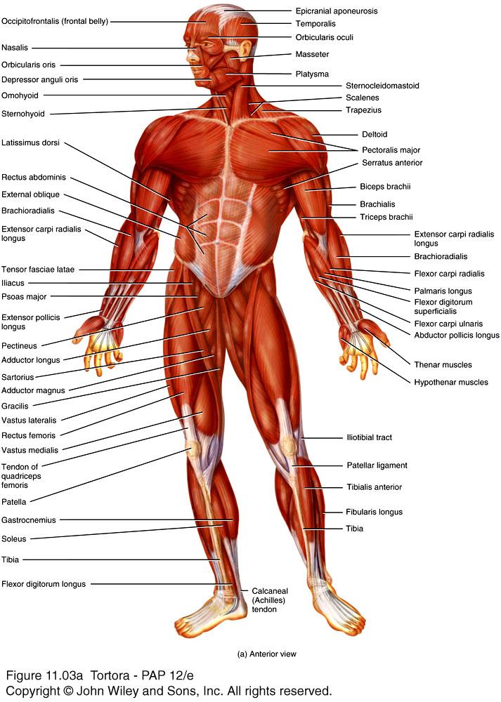
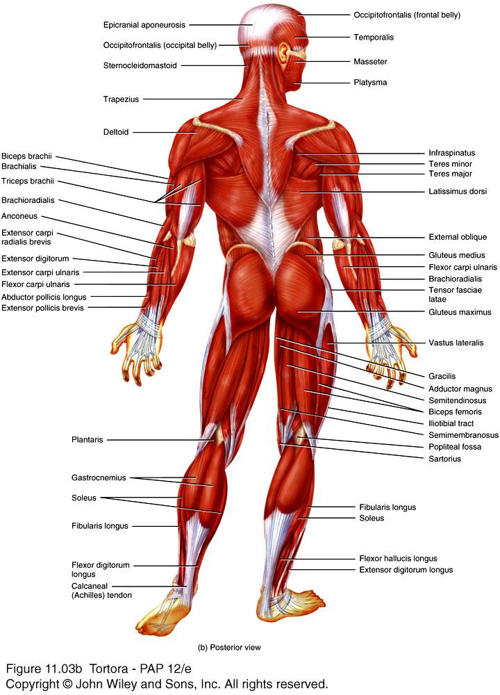
1 Chapter 11: The Muscular System
2 Muscle Attachment Sites: Origin & Insertion n Skeletal muscles cause movements by exerting force on tendons, which pulls on bones or other structures. n Articulating bones usually do not move equally in response to contraction. q q q the attachment of a tendon to the stationary bone is called the origin. the attachment of the muscle s other tendon to the movable bone is called the insertion. the action/s of a muscle are the main movements that occur during contraction (e.g., flexion or extension).

3 Relationship of skeletal muscles to bones
4 Lever Systems n A lever is a rigid structure that can move around a fixed point called a fulcrum. n A lever is acted on at two different points by two different forces: q q the effort, which causes movement, and the load or resistance, which opposes movement. n The effort is the force due to muscular contraction; the load is the weight that is moved or some resistance an object to being moved (e.g., weight of a book to be overcome before you can pick it up). n Motion occurs when the effort applied to the bone at the insertion exceeds the load.
5 Types of levers n There are 3 types of levers that differ on the positions of the fulcrum, effort, and load. q First-class levers are not common: the fulcrum is between the effort and the load. q Second-class levers are uncommon: the load is between the fulcrum and the effort. q Third-class levers are common: the effort is between the fulcrum and the load.

6 Types of levers
7 Effects of muscle fascicle arrangement n All muscle fibers are parallel to one another within a single fascicle. n Fascicles, however, form patterns with respect to the tendons. q Parallel q Fusiform q Circular q Triangular q Pennate
8 Effects of muscle fascicle arrangement n Muscle fascicles have a compromise that they must make. They must compromise between power and range of motion. q The longer the fibers in a muscle, the greater the range of motion it can produce. q The power of a muscle depends not on length but on its total cross-sectional area.
9 Coordination among muscles n It is common to attribute a specific action at a joint to a single muscle bundle, but remember that muscles do not work in isolation. n Movements usually result from several skeletal muscles acting as a group. Most skeletal muscles are arranged in opposing (antagonistic) pairs at joints (e.g., flexors vs. extensors). n In an opposing muscle pair, one is called the prime mover or agonist and is responsible for the action, while the other muscle called the antagonist stretches and yields to the effects of the agonist.
10 Coordination among muscles n To prevent unwanted movements at other joints or to otherwise aid the movement of the agonist, muscles called synergists contract and stabilize the intermediate joints. n Other muscles act as fixators, stabilizing the origin of the agonist so that the agonist is more efficiently. n Depending upon the movement required, many muscles may act as prime movers, antagonists, synergists, or fixators.
11
12
13 Muscles of facial expression n Muscles of facial expression q lie within the subcutaneous layer q usually originate in the fascia or skull bones & insert into the skin. n Because of their insertions, the muscles of facial expression move the skin rather than a joint when they contract.

14 Muscles of Facial Expression
15 Extrinsic Eye Muscles n Six extrinsic eye muscles control movements of each eyeball. They are called extrinsic because they originate on the outside of the eyeballs in the bony orbit and insert on the outer surface of the sclera. n Those muscles with the word rectus in their name have obvious actions (the inferior rectus muscle moves the eye inferiorly so that you would be looking downward). n The actions of the two oblique muscles cannot be deduced from their names. To understand how they move the eye, you must know the origin, insertion, and the unusual path that each follows (see 11.5b).

16 Muscles that move the eyeballs and the upper eyelid
17 Muscles that move the mandible n Four pairs of muscles move the mandible, and are known as muscles of mastication. n The masseter, temporalis, and medial pterygoid account for the strength of the bite. n The medial and lateral pterygoid muscles help to chew by moving the mandible from side to side. Additionally, these muscles protract (protrude) the mandible.

18 Muscles that move the mandible
19 Muscles of the anterior neck that help in swallowing and speech n There are two main muscle groups in the anterior neck: q suprahyoid muscles, are superior to the hyoid q infrahyoid muscles, are inferior to the hyoid. n Both groups of muscles stabilize the hyoid bone, allowing it to serve as a firm base on which the tongue can move.

20 Muscles of the anterior neck that help in swallowing and speech
21 Muscles of the neck that move the head n The head articulates with the vertebral column at joints formed by the atlas & occipital bone. n Balance and movement of the head involves several neck muscles. n An important landmark (the sternocleidomastoid muscle) divides the sides of the neck into two major triangles: anterior and posterior. q The triangles are important anatomically and surgically because of the structures that lie within their boundaries.

22 Muscles of the neck that move the head

23 Muscles of the Abdomen
24 Muscles of the abdomen that protect the viscera and move the vertebral column n The anterolateral abdominal wall includes the external oblique, internal oblique, and transversus abdominis muscles which form three protective layers around the abdomen. n The muscle fascicles of each layer extend in a different direction, conferring considerable protection to the abdominal viscera. n The aponeuroses of these 3 muscles form the rectus sheaths which enclose the rectus abdominis muscles. q The sheaths form the linea alba, a connective tissue band extending from the xiphoid process to the pubic symphysis.


25 Muscles of the Thorax that Assist in Breathing
26 Muscles of the Thorax that Assist in Breathing n Respiratory muscles alter the size of the thoracic cavity which affects the pressure in the lungs, and that determines whether we inhale or exhale. n The diaphragm is the most important respiratory muscle. n Other important respiratory muscles include the external and internal intercostal muscles. n There are also a number of accessory muscles useful in forced breathing.
27 Muscles of the Pelvic Floor n The levator ani and ischiococcygeus muscles, along with the fascia which covers them, form the pelvic diaphragm. n The pelvic diaphragm separates the pelvic cavity above from the perineum below.
28 Muscles of the Perineum n The perineum is a diamond-shaped area inferior to the pelvic diaphragm that extends from the pubic symphysis anteriorly, to the coccyx posteriorly, and to the ischial tuberosities laterally. n Perineal muscles are arranged in two layers; superficial and deep. n The deep muscles of the perineum assist in urination and ejaculation in males and urination and compression of the vagina in females.

29 Muscles of the Thorax that Move the Pectoral Girdle
30 Muscles of the Thorax that Move the Pectoral Girdle n Muscles that move the pectoral girdle must do so by stabilizing the scapula so it can function as a stable origin for the muscles that move the humerus. n Scapular movements increase the range of motion of the humerus. n Many humeral movements would not be possible without scapular movements accompanying those of the humerus (e.g., raising your arm above the head).
31 Muscles of the Thorax and Shoulder that move the Humerus n Seven of nine muscles that cross the shoulder joint originate on the scapula, except the pectoralis major and latissimus dorsi. n It is for this reason that the pectoralis major and latissimus dorsi are considered axial muscles. n Four deep shoulder muscles strengthen and stabilize the shallow shoulder joint, and act to join the scapula to the humerus. They form the rotator cuff, a nearly complete circle of tendons around the shoulder joint, like the cuff on a shirtsleeve.

32 Muscles of the Thorax and Shoulder that move the Humerus

33 Muscles of the Thorax that Move the Humerus
34 Muscles of the Arm that Move the Radius and Ulna n Most muscles that move the forearm cause flexion and extension at the elbow. n The biceps brachii, brachialis, and brachioradialis are flexors. The extensors are the triceps brachii and the anconeus. n Some muscles that move the forearm are involved in pronation and supination. The pronators are the pronator teres and pronator quadratus. Only the supinator can supinate. You use the powerful action of the supinator when you twist a corkscrew or turn a screw with a screwdriver.

35 Muscles of the Arm that Move the Radius and Ulna
36 Muscles of the Forearm that Move the Wrist, Hand, Thumb and Fingers n Muscles in this group are known as extrinsic muscles of the hand because they originate outside the hand and insert within it. n Based on location and function, these muscles are divided into an anterior, and a posterior compartment group. n The tendons of these muscles that continue into the hand are held close to the bones by strong fascial bands called retinacula.

37 Muscles of the Forearm that Move the Wrist, Hand, Thumb and Fingers

38 Muscles of the Palm that Move the Digits: Intrinsic Muscles of the Hand
39 Muscles of the Palm that Move the Digits: Intrinsic Muscles of the Hand n Intrinsic muscles of the hand produce weak but precise movements. n Intrinsic hand muscles are split into 3 groups: thenar, hypothenar, & intermediate. q q The thenar muscles plus the adductor pollicis form the thenar eminence. Hypothenar muscles act on the little finger and form the hypothenar eminence. n Movements of the thumb are defined in different planes compared to other digits because the thumb is positioned at a right angle to the other digits.
40 Muscles of the Neck and Back that Move the Vertebral Column n Muscles that move the backbone are quite complex having multiple origins/insertions with considerable overlap among them. n One way to simplify this is to group muscles based on the general direction of the muscle bundles and their lengths. n Many of these muscles are name for the position of the superior attachment site (e.g., splenius capitus is attached to the head).
41 Muscles of the Neck and Back that Move the Vertebral Column (continued) n n n Splenius muscles extend the head, and laterally flex and rotate the head. Erector spinae muscles include iliocostalis, longissimus, and spinalis groups. They are mainly responsible for extension of the backbone, but also can effect flexion, lateral flexion, and rotation. The transversospinales are so named because their fibers run from the transverse processes to the spinous processes of the vertebrae. q q The semispinalis muscles in this group are also named according to the region of the body with which they are associated. These muscles extend the vertebral column and rotate the head.


42 Muscles of the Neck and Back that Move the Vertebral Column
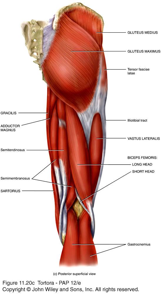
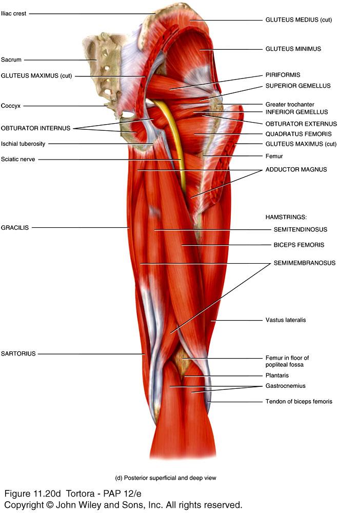
43 Muscles of the Gluteal Region that Move the Femur n Lower limb muscles function in stability, locomotion, and maintenance of posture. In contrast, upper limb muscles are characterized by versatility of movement. n Muscles of the lower limbs often cross two joints and can act equally on both. n Most muscles that move the femur originate on the pelvic girdle and insert on the femur. n Major muscle groups that move the thigh include the gluteals, and adductor muscles.


44 Muscles of the Gluteal Region that Move the Femur
45
46 Muscles of the Thigh n Deep fascia separate muscles that act on the femur, and tibia and fibula into medial, anterior, and posterior compartments. q medial (adductor) compartment of the thigh adduct the femur at the hip joint. q anterior (extensor) compartment of the thigh extend the leg (and flex the thigh). q posterior (flexor) compartment of the thigh flex the leg (and extend the thigh).
47 Muscles of the Leg that Move the Foot and Toes n Leg muscles, like those of the thigh, are divided by deep fascia into three compartments: anterior, lateral, and posterior. q Anterior compartment muscles dorsiflex the foot. q Lateral compartment muscles plantar flex & evert the foot. q Posterior compartment muscles are split between superficial (e.g., gastrocnemius) and deep (e.g., tibialis posterior) groups. Superficial muscles share a common tendon of insertion, the calcaneal tendon.


48 Muscles of the Leg that Move the Foot and Toes (Fig )
49 Intrinsic Muscles of the Foot that Move the Toes n These muscles are termed intrinsic because they originate & insert within the foot. n These muscles are limited designed for support and locomotion, and are split into dorsal and plantar groups. n There is only one dorsal muscle which extends toes 2 5 at the MTP joints. n Plantar muscles are arranged in four layers with the most superficial of these called the first layer, etc.

50 Intrinsic Muscles of the Foot that Move the Toes (Fig )
Chapter 11 The Muscular System. Copyright 2009, John Wiley & Sons, Inc.
 Chapter 11 The Muscular System Muscle Attachment Sites Skeletal muscles cause movements by exerting force on tendons, which pulls on bones or other structures. the attachment of a tendon to the stationary
Chapter 10: Muscular System: Gross Anatomy
 Chapter 10: Muscular System: Gross Anatomy I. General Principles A. General Terminology 1. Tendons attach 2. What is an aponeurosis? 3. The points of muscle attachment are called & 4. How is the "origin"
Lectures Muscular System 10-1
 Lectures 12-14 Muscular System 10-1 Properties of Muscle Ability of a muscle to shorten with force Capacity of muscle to respond to a stimulus Muscle can be stretched to its normal resting length and beyond
Prime movers provide the major force for producing a specific movement Antagonists oppose or reverse a particular movement Synergists
 Dr. Gary Mumaugh Prime movers provide the major force for producing a specific movement Antagonists oppose or reverse a particular movement Synergists Add force to a movement Reduce undesirable or unnecessary
The Muscular System PART C. PowerPoint Lecture Slide Presentation by Patty Bostwick-Taylor, Florence-Darlington Technical College
 PowerPoint Lecture Slide Presentation by Patty Bostwick-Taylor, Florence-Darlington Technical College The Muscular System 6 PART C Five Golden Rules of Skeletal Muscle Activity Table 6.2 Muscles and Body
The Muscular System Part A
 10 The Muscular System Part A Lecture Presentation by Lori Garrett Section 1: Functional Organization of the Muscular System Learning Outcomes 10.1 Describe the general function of the body s axial and
The muscular system I Muscles of the head neck and trunk
 The muscular system I Muscles of the head neck and trunk Dr. Nabil Khouri Dr. Nabil Khouri MD MSc, PhD Interactions of Skeletal Muscles Skeletal muscles work together or in opposition Muscles only pull
3/27/2012. Muscle Classification: Functional Groups. Interactions of Skeletal Muscles. Naming Skeletal Muscles. Naming Skeletal Muscles
 Interactions of Skeletal Muscles Skeletal muscles work together or in opposition Muscles only pull (never push) As muscles shorten, the insertion generally moves toward the origin Whatever a muscle (or
A. All movements require muscle which are organs using chemical energy to contract.
 Ch 8 Muscles Introduction: A. All movements require muscle which are organs using chemical energy to contract. B. The three types of muscle in the body are skeletal, smooth, and cardiac muscle. C. This
BIOH111. o Cell Module o Tissue Module o Skeletal system o Integumentary system o Muscle system o Nervous system o Endocrine system
 BIOH111 o Cell Module o Tissue Module o Skeletal system o Integumentary system o Muscle system o Nervous system o Endocrine system TEXTBOOK AND REQUIRED/RECOMMENDED READINGS o Principles of anatomy and
Muscles in the Shoulder, Chest, Arm, Stomach, and Back
 Muscles in the Shoulder, Chest, Arm, Stomach, and Back Shoulder Muscles Deltoid Supraspinatus Infraspinatus Teres Major Teres Minor Subscapularis Deltoid (Delts) Function: Raises the upper arm Origin:
BIOH111. o Cell Module o Tissue Module o Skeletal system o Integumentary system o Muscle system o Nervous system o Endocrine system
 BIOH111 o Cell Module o Tissue Module o Skeletal system o Integumentary system o Muscle system o Nervous system o Endocrine system TEXTBOOK AND REQUIRED/RECOMMENDED READINGS o Principles of anatomy and
Chapter 6 part 2. Skeletal Muscles of the Body
 Chapter 6 part 2 Skeletal Muscles of the Body Basic Principles 600 + muscles in the human body (you are required to learn 45, lucky kids)! Skeletal Muscles pull on bones Origin of a muscle = point of attachment
Cadaver Muscular System Practice Practical
 Cadaver Muscular System Practice Practical Station 1 Station 1 1. Specific structure 1. Rectus sheath 2. Red line 2. Linea alba Station 2 Station 2 3. Red muscle 1. Rectus abdominis 4. Red muscle actions
Temporalis Elevates & retracts mandible. Masseter Elevates mandible. Sternocleidomastoid Neck flexion. Trapezius Elevates & depresses shoulders
 Anterior Posterior Temporalis Elevates & retracts mandible Masseter Elevates mandible Sternocleidomastoid Neck flexion Trapezius Elevates & depresses shoulders Masseter Elevates mandible Temporalis Elevates
The Muscular System. Chapter 10 Part C. PowerPoint Lecture Slides prepared by Karen Dunbar Kareiva Ivy Tech Community College
 Chapter 10 Part C The Muscular System Annie Leibovitz/Contact Press Images PowerPoint Lecture Slides prepared by Karen Dunbar Kareiva Ivy Tech Community College Table 10.9: Muscles Crossing the Shoulder
Chapter 9. The Muscular System
 1 Chapter 9 The Muscular System 2 Introduction Skeletal muscles: movement in environment Smooth muscles: intestines, ureters, veins and arteries Cardiac muscle: pumps blood through heart and blood vessels
Figure 11-1: The lever-fulcrum principle is illustrated by flexion of the forearm.
 Chapter 11: The Muscular System Read pages 325 to 399 NAME Topic Outline And Objectives: A. How skeletal muscles produce movement, and naming muscles 1. Describe the relationship between bones and skeletal
SKELETAL MUSCLE ANATOMY
 SKELETAL MUSCLE ANATOMY OUTLINE I. Anatomical Terms of Motion II. Head, Face & Neck Muscles III. Anterior Torso Muscles IV. Posterior Torso Muscles V. Arm & Shoulder Muscles VI. Leg & Hip Muscles 2 ANATOMICAL
Lab Exercise #5 The Muscular System Student Performance Objectives
 Student Performance Objectives The material that you are required to learn in this exercise can be found in either the lecture text or the supplemental materials provided in lab. Prior to coming to class,
Certified Personal Trainer Re-Certification Manual
 Certified Personal Trainer Re-Certification Manual Section II 1 Anatomy & Physiology Terms Anatomy and physiology are closely related fields of study: anatomy is the study of form, and physiology is the
Chapter 11 Anatomy and Physiology Lecture
 1 THE MUSCULAR SYSTEM Chapter 11 Anatomy and Physiology Lecture THE MUSCULAR SYSTEM 2 Muscle Tissue - refers to all the contractile tissues of the body; skeletal, cardiac, and smooth muscle. Muscular System
Module 7 - The Muscular System Muscles of the Arm and Trunk
 Module 7 - The Muscular System Muscles of the Arm and Trunk This Module will cover the muscle anatomy of the arms and trunk. We have already seen the muscles that move the humerus, so this module will
Due in Lab weeks because of Thanksgiving Prelab #10. Homework #8. Both sides! Both sides!
 Lab 8 MUSCLES Due in Lab 10 2 weeks because of Thanksgiving Prelab #10 Both sides! Homework #8 Both sides! Refer to Muscles 22-23 Naming of muscles Origin Site of muscle attachment that doesn t move during
BLUE SKY SCHOOL OF PROFESSIONAL MASSAGE AND THERAPEUTIC BODYWORK. Musculoskeletal Anatomy & Kinesiology MUSCLES, MOVEMENTS & BIOMECHANICS
 BLUE SKY SCHOOL OF PROFESSIONAL MASSAGE AND THERAPEUTIC BODYWORK Musculoskeletal Anatomy & Kinesiology MUSCLES, MOVEMENTS & BIOMECHANICS MSAK101-I Session 7 Learning Objectives: 1. List the three types
The Human Muscular System Required reading before beginning this lab: Saladin, KS: Human Anatomy 5th ed (2017) Chapters 10, 11, 12 INTRODUCTION
 Chapters 10, 11, 12 INTRODUCTION") Biology 322: Human Anatomy The Human Muscular System Required reading before beginning this lab: Saladin, KS: Human Anatomy 5 th ed (2017) Chapters 10, 11, 12 INTRODUCTION We will use a number of lab periods
Biology 322: Human Anatomy The Human Muscular System Required reading before beginning this lab: Saladin, KS: Human Anatomy 5 th ed (2017) Chapters 10, 11, 12 INTRODUCTION We will use a number of lab periods
A&P 1 Muscle In-Lab Guide
 A&P 1 Muscle In-Lab Guide This lab guide includes a table with all the muscles you need to ID, along with their origins, insertions and actions Dashed lines means ignore. If several actions are listed,
Muscle fiber (cell) Blood vessel. Perimysium. Epimysium. Fascicle (wrapped by perimysium) Endomysium (between fibers) Tendon. Bone
 Blood vessel. Perimysium. Epimysium. Fascicle (wrapped by perimysium) Endomysium (between fibers) Tendon. Bone") Figure 6.1 Connective tissue wrappings of skeletal muscle. Blood vessel Muscle fiber (cell) Perimysium Epimysium Fascicle (wrapped by perimysium) Tendon Endomysium (between fibers) Bone Figure 6.15 Superficial
Figure 6.1 Connective tissue wrappings of skeletal muscle. Blood vessel Muscle fiber (cell) Perimysium Epimysium Fascicle (wrapped by perimysium) Tendon Endomysium (between fibers) Bone Figure 6.15 Superficial
Anatomy and Physiology 1 Chapter 11 self quiz Pro, Dima Darwish,MD.
 Anatomy and Physiology 1 Chapter 11 self quiz Pro, Dima Darwish,MD. 1) The attachment of a muscle s tendon to the stationary bone is called the ; the attachment of the muscle s other tendon to the movable
Scapula Spine Lateral edge of clavicle. Medial border Scapula. Medial border of Scapula, between superior angle and root of spine. Scapula.
 Muscle attachments and actions answer sheet Muscle Origins insertions Movements Joints crossed Trapezius Base of skull Spinous process of C7 Thoracic Spine Lateral edge of clavicle Elevation Retraction
Biology 218 Human Anatomy. Adapted from Martini Human Anatomy 7th ed. Chapter 10 The Muscular System Axial Musculature
 Adapted from Martini Human Anatomy 7th ed. Chapter 10 The Muscular System Axial Musculature Introduction The skeletal muscle of the body can be subdivided into: Axial musculature Muscles that position
Bio 113 Anatomy and Physiology The Muscles. Muscles of the Head and Neck. Masseter. Orbicularis occuli. Orbicularis oris. Sternocleidomastoid
 Bio 113 Anatomy and Physiology The Muscles Muscles of the Head and Neck Masseter Orbicularis occuli Orbicularis oris Sternocleidomastoid Temporalis BIO 113 Fall 2011 Muscles Page 1 of 5 Muscles of the
Muscular System: Skeletal Muscle Tissue and Organization
 Muscular System: Skeletal Muscle Tissue and Organization AFTER READING THE BOOK, use the following worksheet to link concepts from previous chapters with the concepts of muscle tissue. This worksheet will
Sports Medicine Part II : ANATOMY OF THE SPINE, ABDOMEN AND SHOULDER COMPLEX
 Sports Medicine 25 1.1 Part II : ANATOMY OF THE SPINE, ABDOMEN AND SHOULDER COMPLEX c.w.p. Wagner High School, Sports Medicine, A. Morgan, T. Morgan & A. Eastlake, 2008 Muscles of the Upper Limbs In this
Human Anatomy and Physiology I Laboratory
 Human Anatomy and Physiology I Laboratory Gross Anatomy of the Muscular System (Two weeks) 1 This lab involves study of the laboratory exercise Gross Anatomy of the Muscular System. Complete the Review
Bone Practical. Labs Muscle Labs. Final Practical. Divisions of the Muscular System. Quiz format
 Bone Practical Labs 17 + 18 Muscles Wed 7/11 @ 8am 40 50 stations About half axial, half appendicular bones Disarticulated bones: Skulls, partial skulls, vertebrae, ribs, skeletons, arm bones, leg bones,
Human Anatomy Lab #7: Muscles of the Cadaver
 Human Anatomy Lab #7: Muscles of the Cadaver Table of Contents: Expected Learning Outcomes.... 1 Introduction...... 1 Identifying Muscles on Yourself.... 2 Muscles of the Anterior Trunk and Arm.. 2 Muscles
Biology 2401 Muscles List for CPC models
 Biology 2401 List for CPC models Italicized muscles are dissect and similar in the cat = Dissect and note the differences in human and cat Major of the Human Head Facial Expression Epicranius frontalis
11/15/2018. Temporalis Elevates & retracts mandible. Masseter = Prime mover of jaw closure. Levator scapulae Supraspinatus Clavicle.
 Due in Lab 10 Lab 8 MUSCLES 2 weeks because of Thanksgiving Prelab #10 Both sides! Homework #8 Both sides! Refer to Muscles 22-23 Examples of Origin & Insertion Naming of muscles Origin Site of muscle
Muscles Built on the Maniken
 Muscles Built on the Maniken Facial Muscle Group 1. Temporalis O temporal fossa I anterior border of the ramus of the mandible A elevates the mandible (bite muscle) and holds jaw while at rest 2. Procerus
Anatomy and Physiology 141 Exam II November 6, Name Student Number
 Anatomy and Physiology 141 Exam II November 6, 2014 Name Student Number 1. In regards to the gross anatomy of muscle, which of the following is NOT TRUE? a. Perimysium is more superficial than the epimysium
Muscle Anatomy Review Chart
 Muscle Anatomy Review Chart BACK Superficial (5) Trapezius Transverse cervical a. Latissimus dorsi Thoracodorsal a. Rhomboideus major Dorsal scapular a. Rhomboideus minor Levator scapulae Intermediate
Lab Activity 11: Group I
 Lab Activity 11: Group I Muscles Martini Chapter 11 Portland Community College BI 231 Origin and Insertion Origin: The place where the fixed end attaches to a bone, cartilage, or connective tissue. Insertion:
Origins and Insertions Worksheet
 Origins and Insertions Worksheet First, understanding various boney markings and joint structure will help in determining movements that associated with each muscle. For example, if we are analyzing the
PRELIMINARY HSC PDHPE. CQ1 How do the musculoskeletal and cardiorespiratory systems of the body influence and respond to movement?
 PRELIMINARY HSC PDHPE CQ1 How do the musculoskeletal and cardiorespiratory systems of the body influence and respond to movement? How do the musculoskeletal and cardiorespiratory systems of the body influence
Improving swimming through Pilates
 Improving swimming through Pilates Cleia Ramsey 10 Mar 2017 2016 BASI Training Class Herndon, VA Introduction As a mother of two swimmers, I spend many hours at the pool deck and have learned a lot about
1. In regards to the bones of the face, which of the following is NOT TRUE?
 Anatomy and Physiology Fall Exam II: Form B Name: 1. In regards to the bones of the face, which of the following is NOT TRUE? A. The vomer bone articulates with the dorsal surface of the palatine process
Muscular System. IB Sports, exercise and health science 1.2
 Muscular System IB Sports, exercise and health science 1.2 Characteristics Common to Contractility-ability to shorten the muscles length Extensibility-ability to lengthen the muscles length Elasticity-muscle
The Muscular System Outline 10.1 For any movement, muscles can act in one of three ways (pp ) A. Muscles only pull; they never push, and as a
 A. Muscles only pull; they never push, and as a") The Muscular System Outline 10.1 For any movement, muscles can act in one of three ways (pp. 321 322) A. Muscles only pull; they never push, and as a muscle shortens, the insertion is pulled toward the
The Muscular System Outline 10.1 For any movement, muscles can act in one of three ways (pp. 321 322) A. Muscles only pull; they never push, and as a muscle shortens, the insertion is pulled toward the
Axial Muscles of the Abdominal Wall, and Thorax *
 OpenStax-CNX module: m46485 1 Axial Muscles of the Abdominal Wall, and Thorax * OpenStax This work is produced by OpenStax-CNX and licensed under the Creative Commons Attribution License 4.0 By the end
Epicranius (frontal belly) Zygomaticus minor. Zygomaticus major Buccinator
 Zygomaticus minor. Zygomaticus major Buccinator") Epicranius (frontal belly) Zygomaticus minor Zygomaticus major Buccinator Masseter Digastric (posterior belly) Stylohyoid Sternocleidomastoid Trapezius Scalenus Omohyoid (inferior belly) Orbicularis oris
Epicranius (frontal belly) Zygomaticus minor Zygomaticus major Buccinator Masseter Digastric (posterior belly) Stylohyoid Sternocleidomastoid Trapezius Scalenus Omohyoid (inferior belly) Orbicularis oris
Copy Right- Hongqi ZHANG-Department of Anatomy-Fudan University. Systematic Anatomy. Locomotor system - Part 6
 Systematic Anatomy Locomotor system - Part 6 Muscles of abdomen Muscles of the upper limb Dr.Hongqi Zhang ( 张红旗 ) Email: zhanghq58@126.com 1 Muscles of abdomen Muscles of the upper limb Muscles of abdomen
2. In regards to the bones of the arm and forearm, which of the following is TRUE?
 Anatomy and Physiology Fall Exam II: Form A Name: 1. Use the following table to answer Question 1. I II III IV V The nasal bone articulates with the frontal process of the maxilla The mastoid process is
Types of Muscle: Skeletal- muscle involved in movement of the skeleton. Striated, has alternating bands of light and dark due to overlapping
 Types of Muscle: Skeletal- muscle involved in movement of the skeleton. Striated, has alternating bands of light and dark due to overlapping filaments within the muscle cell. Skeletal muscle can be consciously
Anatomy & Physiology B. Chapter 6: Muscles
 Anatomy & Physiology B Chapter 6: Muscles Warm-up What are the three types of muscle tissue? Where are each located? Which are voluntary and which are involuntary? Which are striated which are unstriated?
Interactions of Skeletal Muscles, Their Fascicle Arrangement, and Their Lever Systems
 Interactions of Skeletal Muscles, Their Fascicle Arrangement, and Their Lever Systems Bởi: OpenStaxCollege To move the skeleton, the tension created by the contraction of the fibers in most skeletal muscles
The Muscular System. - composed of mostly skeletal muscle tissue, nervous tissue, blood and connective tissue
 The Muscular System Every action the body takes utilizes a muscular activity. Some of the muscles of the body are under voluntary control (skeletal muscles), and by using these muscle, you are able to
PHYSICAL EDUCATION. 4º E.S.O. 2nd TERM. The skeletal and muscular systems.
 PHYSICAL EDUCATION 4º E.S.O. 2nd TERM. The skeletal and muscular systems. PARTS OF THE BODY Head Torso / Trunk Dorsal: Back Ventral: Thorax y Abdomen Extremities Superior: Arm Forearm Hand Joint: Shoulder
A&P 1 Muscle Lab #1 - Muscle Tissue, Muscle Types, and Movement Terms Pre-lab Exercises
 1 A&P 1 Muscle Lab #1 - Muscle Tissue, Muscle Types, and Movement Terms Pre-lab Exercises Have someone in your group read the following out loud, while the others read along: In this "Lab Exercise Guide",
Connects arm to thorax 3 joints. Glenohumeral joint Acromioclavicular joint Sternoclavicular joint
 Connects arm to thorax 3 joints Glenohumeral joint Acromioclavicular joint Sternoclavicular joint Scapula Elevation Depression Protraction (abduction) Retraction (adduction) Downward Rotation Upward Rotation
Main Menu. Trunk and Spinal Column click here. The Power is in Your Hands
 1 The Trunk and Spinal Column click here Main Menu K.9 http://www.handsonlineeducation.com/classes/k9/k9entry.htm[3/27/18, 2:00:55 PM] The Trunk and Spinal Column Vertebral column complex 24 intricate
The Trunk and Spinal Column Kinesiology Cuneyt Mirzanli Istanbul Gelisim University
 The Trunk and Spinal Column Kinesiology Cuneyt Mirzanli Istanbul Gelisim University The Trunk and Spinal Column Vertebral column 24 articulating vertebrae 31 pairs of spinal nerves Abdominal muscles some
Class Outline: Posterior Anatomy
 Class Outline: Posterior Anatomy 5 minutes Breath of Arrival and Attendance 5 minutes Howdy Partner 35 minutes Posterior Anatomy using Power Point Presentation 5 minutes Overview of skeletal segments 5
Thoracolumbar Anatomy Eric Shamus Catherine Patla Objectives
 1 2 Thoracolumbar Anatomy Eric Shamus Catherine Patla Objectives List the muscular and ligamentous attachments of the thoracic and lumbar spine Describe how the muscles affect the spine and upper extremity
WTC II Term 3 Notes & Assessments
 Term 3 Notes & Assessments Planes of Motion/Axes The body moves in a number of various ways and directions. In the past you have learned about the terminology for movements at specific joints, for example,
Head & Neck The muscle names are followed by the chapter number
 Head & Neck The muscle names are followed by the chapter number. Splenius capitis (9) 2. Occipitalis (2) Temporalis () 3. Temporalis () 4. Semispinalis capitis (9) Facial / Scalp (2) 5. Temporalis () Facial
Location Terms. Anterior and posterior. Proximal and Distal The term proximal (Latin proximus; nearest) describes where the appendage joins the body.
 describes where the appendage joins the body.") HUMAN ANAT OMY Location Terms Anterior and posterior In human anatomical usage, anterior refers to the front of the individual. Similarly, posterior refers to the back of the subject. In standard anatomical
HUMAN ANAT OMY Location Terms Anterior and posterior In human anatomical usage, anterior refers to the front of the individual. Similarly, posterior refers to the back of the subject. In standard anatomical
The Role of Muscles in Movement
 The Role of Muscles in Movement Muscles can t push, they can only pull as they contract, so most often body movements are the result of the activity of pairs or teams of muscles acting together or against
The arm: *For images refer back to the slides
 The arm: *For images refer back to the slides Muscles of the arm: deltoid, triceps (which is located at the back of the arm), biceps and brachialis (it lies under the biceps), brachioradialis (it lies
or Everything you ever wanted to know about Muscles, but were afraid to ask!!!
 The Muscular System or Everything you ever wanted to know about Muscles, but were afraid to ask!!! Did you know that? - more than 50% of body weight is muscle! - And muscle is made up of proteins and water
Key Relationships in the Upper Limb
 Key Relationships in the Upper Limb This list contains some of the key relationships that will help you identify structures in the lab. They are organized by dissection assignment as defined in the syllabus.
ACTIVITIES 5 & 6: APPENDICULAR AND AXIAL MUSCLES
 ACTIVITIES 5 & 6: APPENDICULAR AND AXIAL MUSCLES Objectives: 1) How to get ready: Read Chapter 11 & 12, McKinley et al., Human Anatomy, 4e. All text references are for this textbook. Begin identifying
Muscular Nomenclature and Kinesiology - One
 Chapter 16 Muscular Nomenclature and Kinesiology - One Lessons 1-3 (with lesson 4) 1 Introduction 122 major muscles covered in this chapter Chapter divided into nine lessons Kinesiology study of human
Healing Hands School of Holistic Health. Advanced Circulatory & Sports Massage Class Handouts
 Class Handouts 1 Posterior Trepidations Torso Rock Torso Rock half-step Torso Rock both sides Torso Rock down body Torso Side Stretch Erector Rock Spinal Rock Lumbo Rock Cha Cha Leg Clay Snake Flop Leg
Systematic Anatomy (For international students)
") Systematic Anatomy (For international students) Department of Anatomy,Fudan University Teaching contents Muscles of abdomen & upper limbs Dr.Hongqi Zhang ( 张红旗 ) Email: zhanghq58@126.com 1 Muscles of abdomen
Systematic Anatomy (For international students) Department of Anatomy,Fudan University Teaching contents Muscles of abdomen & upper limbs Dr.Hongqi Zhang ( 张红旗 ) Email: zhanghq58@126.com 1 Muscles of abdomen
Human Anatomy Biology 351
 1 Human Anatomy Biology 351 Upper Limb Exam Please place your name on the back of the last page of this exam. You must answer all questions on this exam. Because statistics demonstrate that, on average,
ACE s Essentials of Exercise Science for Fitness Professionals TRUNK
 ACE s Essentials of Exercise Science for Fitness Professionals TRUNK Posture and Balance Posture refers to the biomechanical alignment of the individual body parts and the orientation of the body to the
Synergist Muscles. Shoulder (glenohumeral joint) Flexion Deltoid (anterior fibers) Pectoralis major (upper fibers) Biceps Brachii Coracobrachialis
 Flexion Deltoid (anterior fibers) Pectoralis major (upper fibers) Biceps Brachii Coracobrachialis") Synergist Muscles Dr Gene Desepoli DrGeneLMT@gmail.com Shoulder (glenohumeral joint) Deltoid (anterior fibers) Pectoralis major (upper fibers) Biceps Brachii Coracobrachialis Deltoid (posterior fibers)
Synergist Muscles Dr Gene Desepoli DrGeneLMT@gmail.com Shoulder (glenohumeral joint) Deltoid (anterior fibers) Pectoralis major (upper fibers) Biceps Brachii Coracobrachialis Deltoid (posterior fibers)
Exercise Science Section 3: The Muscular System
 Exercise Science Section 3: The Muscular System An Introduction to Health and Physical Education Ted Temertzoglou Paul Challen ISBN 1-55077-132-9 Major Functions of Muscles Movement Includes: breathing,
Upper Limb Muscles Muscles of Axilla & Arm
 Done By : Saleh Salahat Upper Limb Muscles Muscles of Axilla & Arm 1) Muscles around the axilla A- Muscles connecting the upper to thoracic wall (4) 1- pectoralis major Origin:- from the medial half of
Muscle. Dr. Carmen E. Rexach Anatomy 35 Mt San Antonio College
 Muscle Dr. Carmen E. Rexach Anatomy 35 Mt San Antonio College Functions Movements of bones and soft body parts Movements of fluids through a tube (blood, digestive) Functions Maintain posture Support soft
The Back. Anatomy RHS 241 Lecture 9 Dr. Einas Al-Eisa
 The Back Anatomy RHS 241 Lecture 9 Dr. Einas Al-Eisa The spine has to meet 2 functions Strength Mobility Stability of the vertebral column is provided by: Deep intrinsic muscles of the back Ligaments
STRUCTURAL BASIS OF MEDICAL PRACTICE EXAMINATION 5 October 6, 2006
 STRUCTURAL BASIS OF MEDICAL PRACTICE EXAMINATION 5 October 6, 2006 PART l. Answer in the space provided. (8 pts) 1. Identify the structures. (2 pts) B C A. _pisiform B. _ulnar artery A C. _flexor carpi
Fascial Compartments of the Upper Arm
 Fascial Compartments of the Upper Arm The upper arm is enclosed in a sheath of deep fascia and has two fascial septa: 1- Medial fascial septum (medial intermuscular septum): attached to the medial supracondylar
The Muscular System The Muscular System Muscles are responsible for all types of body movement Three basic muscle types are found in the body
 The Muscular System The Muscular System Muscles are responsible for all types of body movement Three basic muscle types are found in the body Skeletal muscle Cardiac muscle Smooth muscle Characteristics
medial half of clavicle; Sternum; upper six costal cartilages External surfaces of ribs 3-5
 MUSCLE ORIGIN INSERTION ACTION NERVE Pectoralis Major medial half of clavicle; Sternum; upper six costal cartilages Lateral lip of intertubercular groove of horizontal adduction Medial and lateral pectoral
CHAPTER 1: 1.1 Muscular skeletal system. Question - text book page 16. Question - text book page 20 QUESTIONS AND ANSWERS. Answers
 QUESTIONS AND ANSWERS CHAPTER 1: 1.1 Muscular skeletal system Question - text book page 16 Using the information on pages 12 to 14 above, complete the table below. joint joint type articulating bones associated
Biology 210 Chapter 10 Muscles. Types of Muscle Cells. Types of Muscle Cells
 Biology 210 Chapter 10 Muscles PowerPoint by John McGill Based on notes by Beth Wyatt & PowerPoint by Jack Bagwell Types of Muscle Cells Skeletal (Voluntary, Striated) Muscle these are muscles you control;
Upper limb Arm & Cubital region 黃敏銓
 Upper limb Arm & Cubital region 黃敏銓 1 Arm Lateral intermuscular septum Anterior (flexor) compartment: stronger Medial intermuscular septum Posterior (extensor) compartment 2 Coracobrachialis Origin: coracoid
Muscles of the Upper Limb
 Muscles of the Upper Limb anterior surface of ribs 3 5 coracoid process Pectoralis minor pectoral nerves protracts / depresses scapula Serratus anterior Subclavius ribs 1-8 long thoracic nerve rib 1 ----------------
Monday, November 13, 2017 A & P 2401
 Monday, November 13, 2017 A & P 2401 Today you will complete the following handouts. Study the last part of the handout for this will be on your quiz, which will be on Wednesday. It is titled steps of
OBJECTIVES. Unit 7:5 PROPERTIES OR CHARACTERISTICS OF MUSCLES. Introduction. 3 Kinds of Muscles. 3 Kinds of Muscles 4/17/2018 MUSCULAR SYSTEM
 OBJECTIVES Unit 7:5 MUSCULAR SYSTEM Compare the three main kinds of muscles by describing the action of each Differentiate between voluntary and involuntary muscles List at least three functions of muscles
Types of Body Movements
 Types of Body Movements Bởi: OpenStaxCollege Synovial joints allow the body a tremendous range of movements. Each movement at a synovial joint results from the contraction or relaxation of the muscles
YOGA ANATOMY. Part Three - Bones. Yoga Teacher Training Robin Bennett 200 RYT
 YOGA ANATOMY Yoga Teacher Training Part Three - Bones 2015 Robin Bennett 200 RYT THE HUMAN SKELETON BONE COMPOSITION A femur head with a cortex of compact bone and medulla of trabecular (spongy) bone OSTEOBLASTS
Anatomy and Physiology II. Review Shoulder Girdle New Material Upper Extremities - Bones
 Anatomy and Physiology II Review Shoulder Girdle New Material Upper Extremities - Bones Anatomy and Physiology II Shoulder Girdle Review Questions From Last Lecture Can you identify the following muscles?
Practical 1 Worksheet
 Practical 1 Worksheet ANATOMICAL TERMS 1. Use the word bank to fill in the missing words. reference side stand body arms palms anatomical forward All anatomical terms have a(n) point which is called the
Chapter 3: Applied Kinesiology. ACE Personal Trainer Manual Third Edition
 Chapter 3: Applied Kinesiology ACE Personal Trainer Manual Third Edition Introduction Kinesiology is the study of the body s infinite number of movements, positions, and postures and is grounded in the
The Arm and Cubital Fossa
 The Arm and Cubital Fossa Dr. Andrew Gallagher School of Anatomical Sciences University of the Witwatersrand Introduction The ARM (BRACHIUM) is the most proximal segment of the upper limb musculoskeletal
Practical 2 Worksheet
 Practical 2 Worksheet Upper Extremity BONES 1. Which end of the clavicle is on the lateral side (acromial or sternal)? 2. Describe the difference in the appearance of the acromial and sternal ends of the
Muscles of the Cat. N Deltoid MUSCLES OF THE CHEST. Pectoralis major. (This muscle is superior to Pectoralis minor) MUSCLES OF THE CHEST
 MUSCLES OF THE CHEST") MUSCLES OF THE CHEST Pectoralis major (This muscle is superior to Pectoralis minor) 1. MUSCLES OF THE CHEST Pectoralis minor (This muscle is inferior to Pectoralis major) 2. MUSCLES OF THE ARM Deltoid
MUSCLES OF THE CHEST Pectoralis major (This muscle is superior to Pectoralis minor) 1. MUSCLES OF THE CHEST Pectoralis minor (This muscle is inferior to Pectoralis major) 2. MUSCLES OF THE ARM Deltoid