Appendicular Skeleton. Prof. Abdulameer Al-Nuaimi
|
|
|
- Rudolf Wheeler
- 5 years ago
- Views:
Transcription

1 Appendicular Skeleton Prof. Abdulameer Al-Nuaimi

2 Hi Prof, It is great to hear from you, I really enjoyed your teaching last year. You taught me the hardest subject I have encountered so far in a manner that I understood and could remember. I hope you are enjoying your new job in Jordon. Are you still teaching anatomy? Many thanks for your teaching and best of luck in Jordon Sam Allison
3 Appendicular Skeleton Each upper limb skeleton consists of 32 bones, which form two distinct regions: The pectoral girdle. Clavicle. Scapula. The free upper limb. Humerus. Ulna Radius. Hand.

4 The clavicle is the anterior bone and the scapula is the posterior bone. The pectoral girdle
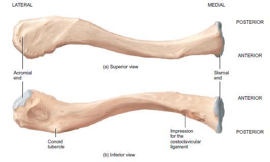
5 S - shaped bone. Clavicle Lies horizontally across the anterior part of the thorax superior to the first rib. The medial half of the clavicle is convex anteriorly and the lateral half is concave anteriorly. The medial end of the clavicle, called the sternal end, is rounded and articulates with the manubrium of the sternum to form the sternoclavicular joint. The broad, flat, lateral end, the acromial end, articulates with the acromion of the scapula at the acromioclavicular joint. The superior surface of the clavile (smooth), while the inferior surface (rough).
6 Clavicle
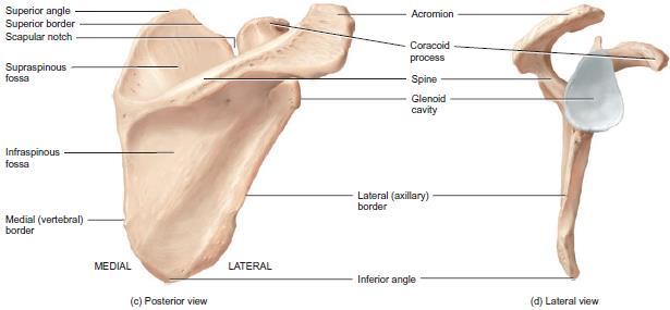
7 Scapula scapula or shoulder blade, is a large, triangular, flat bone with a ridge on its posterior surface. The scapula occupies the superior part of the posterior thorax between the levels of the second and seventh ribs a few finger breadths lateral to the vertebral column. A prominent ridge called the spine across the posterior surface of the scapula. The lateral end of the spine projects as a flattened, expanded process called the Acromion Inferior to the acromion is a shallow depression, the glenoid cavity, that accepts the head of the humerus to form the glenohumeral joint, or shoulder joint. The thin edge of the scapula closer to the vertebral column is called the medial (vertebral) border.

8 Scapula
9 Scapula The thick edge of the scapula closer to the arm is called the lateral (axillary) border. The medial and lateral borders join at the inferior angle. The superior edge of the scapula, called the superior border, joins the vertebral border at the superior angle. The scapular notch is a prominent indentation along the superior border. At the lateral end of the superior border of the scapula is the coracoid process. Superior and inferior to the spine are two fossae: the supraspinous fossa and the infraspinous fossa. On the anterior surface is a slightly hollowed-out area called the subscapular fossa.
10 Scapula

11 Humerus The humerus or arm bone, is the longest and largest bone of the upper limb. It has a ball-like proximal end with two prominent projections of bone at the base of the ball, a cylindrical, tubular shaft that makes up the majority of its length, and an expanded flattened distal end.
12 Humerus It articulates proximally with the scapula and distally with both the ulna and the radius to form the elbow joint. The proximal end of the humerus features a rounded head that articulates with the glenoid cavity of the scapula to form the glenohumeral (shoulder) joint. Distal to the head is the anatomical neck, which is visible as an oblique groove. The greater tubercle is a lateral projection distal to the anatomical neck. The lesser tubercle projects anteriorly. Between both tubercles runs an intertubercular sulcus. The surgical necek is a constriction in the humerus just distal to the tuercles. It is a common fracture site of the humerus (in contct posteriorly with the axillary nerve)
13 Humerus
14 Laterally, at the middle portion of the shaft, there is a roughened, V-shaped area called the deltoid tuberosity. This area serves as a point of attachment for the tendon of the deltoid muscle. The radial groove runs along the posterior surface of the humerus. This groove contains the radial nerve Humerus
15 Humerus at the distal end of the humerus, rounded knob on the lateral aspect of the bone that articulates with the head of the radius (capitulum). The radial fossa is an anterior depression above the capitulum that articulates with the head of the radius when the forearm is flexed. The trochlea, located medial to the capitulum, that articulates with the ulna. The coronoid fossa, anterior depression that receives the coronoid process of the ulna when the forearm is flexed. The olecranon fossa is the large posterior depression that receives the olecranon of the ulna when the forearm is extended.

16 Lower end of humerus

17 Humerus The medial epicondyle and lateral epicondyle are rough projections on either side of the distal end of the humerus to which the tendons of most muscles of the forearm are attached. The ulnar nerve passes posterior to the medial epicondyle in contact with The bone. Fractured hum. might injure these nerves Lat Epicond Ventral surface Med Epicond Dorsal surface

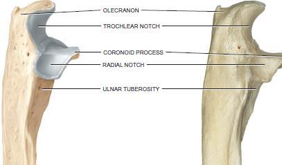
18 Ulna The ulna is located on the medial aspect of the forearm and is longer than the radius. The ulna is thick and notched at its proximal end, and its wide triangular shaft tapers to become more narrow and cylindrical distally. At the proximal end of the ulna is the olecranon, which forms the prominence of the elbow.
19 Ulna The coronoid process an anterior projection distal to a large notch, the trochlear notch. The trochlear notch on the anterior side of the olecranon, receives the trochlea of the humerus to form part of the elbow joint. The radial notch is a depression that is lateral and inferior to the trochlear notch and articulates with the head of the radius. Just inferior to the coronoid process is the ulnar tuberosity. The distal end of the ulna consists of a head that is separated from the wrist by a disc of fibrocartilage. A styloid process is located on the posterior side of the ulna s distal end.

20 Upper end of ulna
21 Radius The radius, the shorter of the two forearm bones, is located on the lateral aspect of the forearm. In contrast to the ulna, the radius is narrow at its proximal end and widens at its distal end. The proximal end of the radius has a disc-shaped head that articulates with the capitulum of the humerus and the radial notch of the ulna. Inferior to the head is the constricted neck. A roughened area inferior to the neck on the anteromedial side, called the radial tuberosity, is a point of attachment for the tendon of the biceps brachii muscle. The shaft of the radius widens distally to form a styloid process on the lateral side.

22 Radius and Ulna
23 Radius The ulna and radius articulate with the humerus at the elbow joint. The elbow articulation occurs in two places: where the head of the radius articulates with the capitulum of the humerus, and where the trochlear notch of the ulna receives the trochlea of the humerus. The ulna and the radius connect with one another at three sites. First, a broad, flat, fibrous connective tissue called the interosseous membrane, joins the shafts of the two bones. The ulna and radius articulate at their proximal and distal ends.
24 Radius Proximally, the head of the radius articulates with the ulna s radial notch, a depression that is lateral and inferior to the trochlear notch. This articulation is the proximal radioulnar joint. Distally, the head of the ulna articulates with the ulnar notch of the radius. This articulation is the distal radioulnar joint. Finally, the distal end of the radius articulates with three bones of the wrist the lunate, the scaphoid, and the triquetrum to form the radiocarpal (wrist) joint.

25 Upper and lower ends of Radius and Ulna
, Metacarpals. (5), Phalanges.")
26 Bones of the hand Carpals. (8), Metacarpals. (5), Phalanges. (14)
27 Carpals The carpus (wrist) is the proximal region of the hand and consists of eight small bones, the carpals, joined to one another by ligaments. Articulations between carpal bones are called intercarpal joints. The carpals are arranged in two transverse rows of four bones each.
28 Metacarpals The metacarpus or palm, is the intermediate region of the hand; it consists of five bones called metacarpals. Each metacarpal bone consists of a proximal base, an intermediate shaft, and a distal head. The metacarpal bones are numbered I to V (1-5), starting with the thumb, from lateral to medial. The bases articulate with the distal row of carpal bones to form the carpometacarpal joints. The heads articulate with the proximal phalanges to form the metacarpophalangeal (MP) joints.
29 Phalanges The phalanges or bones of the digits, make up the distal region of the hand. There are 14 phalanges in the five digits of each hand. Like the metacarpals, the digits are numbered I to V (or 1 5), beginning with the thumb from lateral to medial. Each phalanx consists of a proximal base, an intermediate shaft, and a distal head. The thumb has two phalanges, and there are three phalanges in each of the other four digits. In order from the thumb, these other four digits are commonly referred to as the index finger, middle finger, ring finger, and little finger.

30 Bones of the Hand
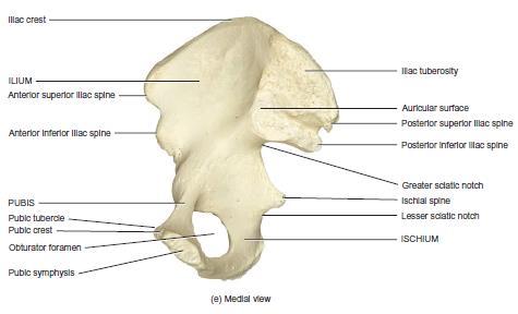
31 Lower Limb Each lower limb skeleton consists of 31 bones, which form two distinct regions: The pelvic girdle. consists of the two hip bones, also called coxal (pelvic) bones. The hip bones unite anteriorly at a joint called the pubic symphysis. They unite posteriorly with the sacrum at the sacroiliac joints. The complete cylinder composed of the hip bones, pubic symphysis, and sacrum forms a deep, basin like structure called the bony pelvis. pelvis also connects the bones of the free lower limbs to the axial.

32 Hip bones are parts of the pelvis

33 Each of the two hip bones of a newborn consists of three bones separated by cartilage: a superior ilium, an inferior anterior pubis, and an inferior posterior ischium. By age 23, the three separate bones fuse together.
, the 5 metatarsal bones in the metatarsus, and the 14 phalanges (bones of the digits) in the")
34 The free lower limb (extremity) has 30 bones in four locations: the femur in the thigh; the patella (knee cap); the tibia and fibula in the leg; the 7 tarsal bones in the tarsus (ankle), the 5 metatarsal bones in the metatarsus, and the 14 phalanges (bones of the digits) in the foot.
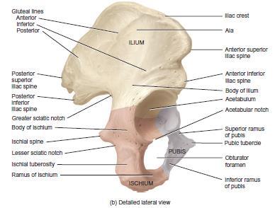
35 Ilium The ilium is the largest of the three components of the hip bone. It is thick near the hip joint and expands into a large curved plate of bone superiorly. A superior ala (wing) and an inferior body comprise the ilium. The body is one of the components of the acetabulum, the socket for the head of the femur. The superior border of the ilium, the iliac crest, ends anteriorly in a blunt anterior superior iliac spine. The medial surface of the ilium contains the iliac fossa, a concavity where the tendon of the iliacus muscle attaches.

36 Parts of the Hip bone
37 Ischium The ischium the inferior and posterior portion of the hip bone, is situated between the body of the ilium and the inferior ramus of the pubis. The ischium is a arched or U-shaped structure, with its concave, notched margin contributing to the posterior two thirds of the obturator foramen (the large hole on the hip bone). The ischium is comprised of a superior body and an inferior ramus.
38 Hip bone
39 Pubis The pubis is the inferior, anterior portion of the hip bone and, like the ischium, has the form of a sideways arch or a U- shape. It composed of a superior ramus, an inferior ramus, and a body between the rami. The anterior, superior border of the body is the pubic crest, and at its lateral end is a projection called the pubic tubercle. The pubic symphysis is the joint between the pubes of the two hip bones. The acetabulum is a deep fossa formed by the ilium, ischium, and pubis. It functions as the socket that contains the rounded head of the femur. Together, the acetabulum and the femoral head form the hip (coxal) joint.
40 Hip bone
41 Bony Pelvis The portion of the bony pelvis superior to the pelvic brim is the false (greater) pelvis. It is bordered by the lumbar vertebrae posteriorly, the upper portions of the hip bones laterally, and the abdominal wall anteriorly. It contains the superior portion of the urinary bladder and lower intestines in both genders and the uterus, ovaries, and uterine tubes of the female, The portion of the bony pelvis inferior to the pelvic brim is the true (lesser) pelvis. It is bounded by the sacrum and coccyx posteriorly, inferior portions of the ilium and ischium laterally, and the pubic bones anteriorly. It contains the rectum and urinary bladder in both genders, the vagina and cervix of the uterus in females, and the prostate in males.

42 Pelvis False Pelvis True pelvis
43 Bony Pelvis The superior opening of the true pelvis, bordered by the pelvic brim, is called the pelvic inlet; The inferior opening of the true pelvis is the pelvic outlet. difference between male and female pelves Oval and Wider transversely Heart shape Round Oval Shallow Cylindrical Long Funnel Rounded Angular

44 The femur, or thigh bone, is the longest, heaviest, and strongest bone in the body. Its proximal end articulates with the acetabulum of the hip bone. Its distal end articulates with the tibia and patella. The shaft of the femur directed medially and, as a result, the knee joints are closer to the midline than Femur

45 Femur The proximal end of the femur consists of a rounded head that articulates with the acetabulum of the hip bone to form the hip joint. The head contains a small, central depression called the fovea capitis. The neck of the femur is a constricted region distal to the head.
46 Femur The greater trochanter and lesser trochanter are projections that serve as points of attachment for the tendons of some of the thigh and buttock muscles. The greater trochanter is the prominence felt and It is a landmark commonly used to locate the site for intramuscular injections into the lateral surface of the thigh. The lesser trochanter is inferior and medial to the greater trochanter. Between the anterior surfaces of the trochanters is a narrow intertrochanteric line. A ridge called the intertrochanteric crest appears between the posterior surfaces of the greater trochanter and lesser trochanter.
47 Femur
48 Femur Inferior to the intertrochanteric crest on the posterior surface of the body of the femur is a vertical ridge called the gluteal tuberosity. It blends into another vertical ridge called the linea aspera. The expanded distal end of the femur includes the medial condyle and the lateral condyle. These articulate with the medial and lateral condyles of the tibia. A depressed area between the condyles on the posterior surface is called the intercondylar fossa. Superior to the condyles are the medial epicondyle and the lateral epicondyle.
49 Femur The patellar surface is located between the condyles on the anterior surface. Just superior to the medial epicondyle is the adductor tubercle, a roughened projection that is a site of attachment for the adductor magnus muscle.
50 Patella The patella (kneecap), is a small, triangular bone. located anterior to the knee joint. It is a sesamoid bone. The broad proximal end of the patella is called the base. The pointed distal end is the apex. The posterior surface contains two articular facets.
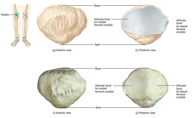
51 Patella
52 Tibia The tibia is the larger, medial, weight-bearing bone of the leg. The tibia is the second longest bone of the body. The tibia articulates at its proximal end with the femur and fibula and at its distal end with the fibula and the talus bone of the ankle. The tibia and fibula, are connected by an interosseous membrane. The proximal end of the tibia is expanded into a lateral condyle, medial condyle and Intercondylar Eminence Tibial condyles articulate with the condyles of the femur forming knee joint. The inferior surface of the lateral condyle articulates with the head of the fibula.
53 Tibia The tibial tuberosity on the anterior surface is a point of attachment for the patellar ligament. Inferior to and continuous with the tibial tuberosity is a sharp ridge that can be felt below the skin and is known as the anterior border. The medial surface of the distal end of the tibia forms the medial malleolus. This structure articulates with the talus of the ankle and forms the prominence that can be felt on the medial surface of the ankle. Laterally, the fibular notch articulates with the distal end of the fibula to form the distal tibiofibular joint.
54 Tibia and Fibula Fibular notch
55 Fibula The fibula is a slender bone that is slightly expanded at both ends. The fibula is parallel and lateral to the tibia, but is considerably smaller. Unlike the tibia, the fibula does not articulate with the femur and is non-weight-bearing, but it does help stabilize the ankle joint. The head of the fibula, the proximal end, articulates with the inferior surface of the lateral condyle of the tibia below the level of the knee joint to form the proximal tibiofibular joint. The distal end has a projection called the lateral malleolus that articulates with the talus of the ankle. This forms the prominence on the lateral surface of the ankle.
56 Tibia and Fibula

57 Fibula The fibula also articulates with the tibia at the fibular notch to form the distal tibiofibular joint. The fibula is a common source for bone grafting Tibia
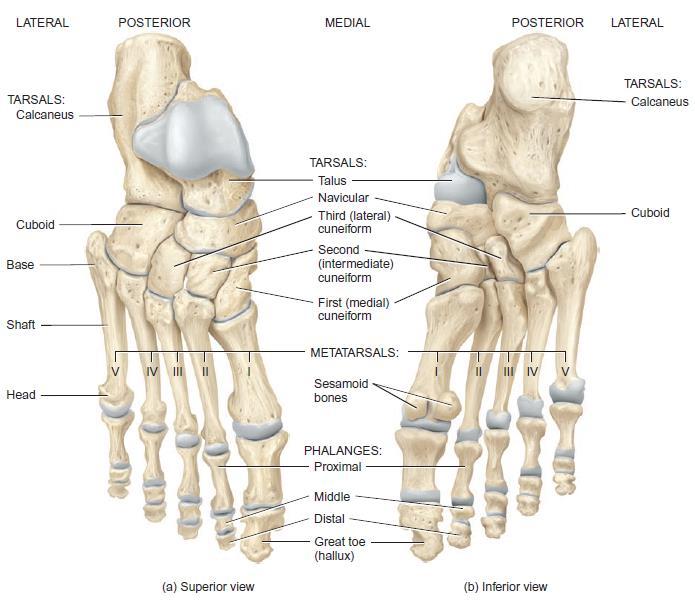
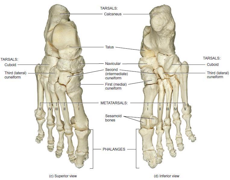
58 Tarsals The tarsus (ankle) is the proximal region of the foot and consists of seven tarsal bones. Joints between tarsal bones are called intertarsal joints. The tarsal bones include the talus and calcaneus, located in the posterior part of the foot. The calcaneus is the largest and strongest tarsal bone. The anterior tarsal bones are: Navicular bone. Three cuneiform bones called the lateral, intermediate and medial. Cuboid.
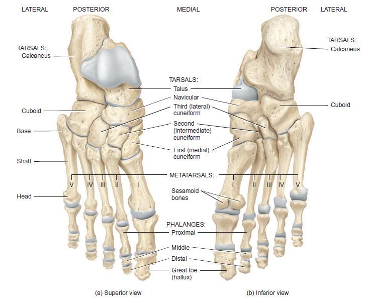
59 Foot
60 Metatarsals The metatarsus is the intermediate region of the foot and consists of five metatarsal bones They are convex dorsally and concave on their plantar surfaces. Like the metacarpals of the palm, each metatarsal consists of a proximal base, an intermediate shaft, and a distal head. The metatarsals articulate proximally with the first, second, and third cuneiform bones and with the cuboid to form the tarsometatarsal joints. Distally, they articulate with the proximal row of phalanges to form the metatarsophalangeal joints. The first metatarsal is thicker than the others because it bears more weight.
61 Foot
62 Phalanges The phalanges comprise the distal component of the foot. The toes are numbered I to V (or 1 5) beginning with the big toe (great toe) medially. Each phalanx consists of a proximal base, an intermediate, shaft, and a distal head. The great or big toe has two large, thick phalanges called proximal and distal phalanges. The other four toes each have three phalanges proximal, middle, and distal. Joints between phalanges of the foot, like those of the hand, are called interphalangeal joints.
63 Foot

64 Thank You Sun Set Malaysia 16 Feb. 2015
Pectoral (Shoulder) Girdle
 Girdle") Chapter 8 Skeletal System: Appendicular Skeleton Pectoral girdle Pelvic girdle Upper limbs Lower limbs 8-1 Pectoral (Shoulder) Girdle Consists of scapula and clavicle Clavicle articulates with sternum
Chapter 8 Skeletal System: Appendicular Skeleton Pectoral girdle Pelvic girdle Upper limbs Lower limbs 8-1 Pectoral (Shoulder) Girdle Consists of scapula and clavicle Clavicle articulates with sternum
The Appendicular Skeleton
 8 The Appendicular Skeleton PowerPoint Lecture Presentations prepared by Jason LaPres Lone Star College North Harris 8-1 The Pectoral Girdle The Pectoral Girdle Also called shoulder girdle Connects the
Biology 218 Human Anatomy
 Chapter 8 Adapted from Tortora 10 th ed. LECTURE OUTLINE A. Introduction (p. 203) 1. The appendicular skeleton contains 126 bones that form: i. two pectoral (shoulder) girdles two upper limbs i one pelvic
10/12/2010. Upper Extremity. Pectoral (Shoulder) Girdle. Clavicle (collarbone) Skeletal System: Appendicular Skeleton
 Girdle. Clavicle (collarbone) Skeletal System: Appendicular Skeleton") Skeletal System: Appendicular Skeleton Pectoral girdle Pelvic girdle Upper limbs Lower limbs 8-1 Pectoral (Shoulder) Girdle Consists of scapula and clavicle Clavicle articulates with sternum (Sternoclavicular
Skeletal System: Appendicular Skeleton Pectoral girdle Pelvic girdle Upper limbs Lower limbs 8-1 Pectoral (Shoulder) Girdle Consists of scapula and clavicle Clavicle articulates with sternum (Sternoclavicular
Chapter 8B. The Skeletal System: Appendicular Skeleton. The Appendicular Skeleton. Clavicle. Pectoral (Shoulder) Girdle
 Girdle") The Appendicular Skeleton Chapter 8B The Skeletal System: Appendicular Skeleton 126 bones Pectoral (shoulder) girdle Pelvic (hip) girdle Upper limbs Lower limbs Functions primarily to facilitate movement
The Appendicular Skeleton Chapter 8B The Skeletal System: Appendicular Skeleton 126 bones Pectoral (shoulder) girdle Pelvic (hip) girdle Upper limbs Lower limbs Functions primarily to facilitate movement
Chapter 8. The Appendicular Skeleton. Lecture Presentation by Lee Ann Frederick University of Texas at Arlington Pearson Education, Inc.
 Chapter 8 The Appendicular Skeleton Lecture Presentation by Lee Ann Frederick University of Texas at Arlington An Introduction to the Appendicular Skeleton The Appendicular Skeleton 126 bones Allows us
Biology 218 Human Anatomy. Adapted from Martini Human Anatomy 7th ed. Chapter 7 The Skeletal System Appendicular Division
 Adapted from Martini Human Anatomy 7th ed. Chapter 7 The Skeletal System Appendicular Division Introduction The appendicular skeleton includes: Pectoral girdle Shoulder bones Upper limbs Pelvic girdle
Chapter 8 The Skeletal System: The Appendicular Skeleton. Copyright 2009 John Wiley & Sons, Inc.
 Chapter 8 The Skeletal System: The Appendicular Skeleton Appendicular Skeleton The primary function is movement It includes bones of the upper and lower limbs Girdles attach the limbs to the axial skeleton
SKELETAL SYSTEM 206. AXIAL SKELETON 80 APPENDICULAR SKELETON 126 (see Figure 6.1) Clavicle. Clavicle. Pectoral girdles. Scapula. Scapula.
 Clavicle. Clavicle. Pectoral girdles. Scapula. Scapula.") SKELETAL SYSTEM 206 AXIAL SKELETON 80 APPENDICULAR SKELETON 126 (see Figure 6.1) Pectoral girdles 4 Clavicle Scapula 2 2 Clavicle Scapula Humerus 2 Humerus Upper limbs 60 Radius 2 Ulna Carpal bones Metacarpal
SKELETAL SYSTEM 206 AXIAL SKELETON 80 APPENDICULAR SKELETON 126 (see Figure 6.1) Pectoral girdles 4 Clavicle Scapula 2 2 Clavicle Scapula Humerus 2 Humerus Upper limbs 60 Radius 2 Ulna Carpal bones Metacarpal
Chapter 8 The Skeletal System: The Appendicular Skeleton. Copyright 2009 John Wiley & Sons, Inc.
 Chapter 8 The Skeletal System: The Appendicular Skeleton Appendicular Skeleton It includes bones of the upper and lower limbs Girdles attach the limbs to the axial skeleton The pectoral girdle consists
Copyright 2003 Pearson Education, Inc. publishing as Benjamin Cummings. Dr. Nabil khouri
 Dr. Nabil khouri Appendicular Skeleton The appendicular skeleton is made up of the bones of the upper and lower limbs and their girdles Two girdles: Pectoral girdles attach the upper limbs to the body
Principles of Anatomy and Physiology
 Principles of Anatomy and Physiology 14 th Edition CHAPTER 8 The Skeletal System: The Appendicular Skeleton The Appendicular Skeleton The 126 bones of the appendicular skeleton are primarily concerned
The Appendicular Skeleton
 8 The Appendicular Skeleton PowerPoint Lecture Presentations prepared by Jason LaPres Lone Star College North Harris An Introduction to the Appendicular Skeleton Learning Outcomes 8-1 Identify the bones
Figure 1: Bones of the upper limb
 BONES OF THE APPENDICULAR SKELETON The appendicular skeleton is composed of the 126 bones of the appendages and the pectoral and pelvic girdles, which attach the limbs to the axial skeleton. Although the
PRE-LAB EXERCISES. Before we get started, look up the definitions of these common bone marking terms: Canal: Condyle: Facet: Fissure:
 1 PRE-LAB EXERCISES When studying the skeletal system, the bones are often sorted into two broad categories: the axial skeleton and the appendicular skeleton. This lab focuses on the appendicular skeleton,
Lab Activity 9. Appendicular Skeleton Martini Chapter 8. Portland Community College BI 231
 Lab Activity 9 Appendicular Skeleton Martini Chapter 8 Portland Community College BI 231 Appendicular Skeleton Upper & Lower extremities Shoulder Girdle Pelvic Girdle 2 Humerus 3 Humerus: Proximal End
Biology 152 Appendicular Skeleton Anatomy Objectives
 Biology 152 Appendicular Skeleton Anatomy Objectives We will learn proper bone names, left/right/medial, and the parts of bones in this exercise. Start by learning the names of the bones. As you gain comfort
Figure 7: Bones of the lower limb
 BONES OF THE APPENDICULAR SKELETON The appendicular skeleton is composed of the 126 bones of the appendages and the pectoral and pelvic girdles, which attach the limbs to the axial skeleton. Although the
Chapter 8. The Pectoral Girdle & Upper Limb
 Chapter 8 The Pectoral Girdle & Upper Limb Pectoral Girdle pectoral girdle (shoulder girdle) supports the arm consists of two on each side of the body // clavicle (collarbone) and scapula (shoulder blade)
The Skeletal System THE APPENDICULAR SKELETON
 The Skeletal System THE APPENDICULAR SKELETON The appendicular skeleton consists of the girdles and the skeleton of the limbs. The upper (anterior) limbs are attached to the pectoral (shoulder) girdle
Chapter 7: Skeletal System: Gross Anatomy
 Chapter 7: Skeletal System: Gross Anatomy I. General Considerations A. How many bones in an average adult skeleton? B. Anatomic features of bones are based on II. Axial Skeleton A. Skull 1. Functionally
Dr.Israa H. Mohsen. Lecture 5. The vertebral column
 Anatomy Lecture 5 Dr.Israa H. Mohsen The vertebral column The vertebral column a flexible structure consisting of 33 vertebrae holds the head and torso upright, serves as an attachment point for the legs,
Amy Warenda Czura, Ph.D. 1 SCCC BIO130 Lab 7 Appendicular Skeleton & Articulations
 The Skeletal System II: Appendicular Skeleton and Articulations Exercises 11, 13 (begins: page 145 in 9 th and 10 th editions) Exercises 10, 11 (begins: page 147 in 11 th edition, page 149 in 12 th edition)
bio4165 lab quiz 1 Posterior View Anterior View Lateral View Anterior View bio fall.quarter lab.quiz.1...page.1 of 6
 B A Posterior View D C E Lateral View bio.4165...fall.quarter.2005...lab.quiz.1...page.1 of 6 F I G 35 Posterior View H bio.4165...fall.quarter.2005...lab.quiz.1...page.2 of 6 J Posterior View L K Inferior
Chapter 8 Outline. Pectoral Girdle Upper Limb Pelvic Girdle Lower Limb Aging of the Appendicular Skeleton Development of the Appendicular Skeleton
 Chapter 8 Outline Pectoral Girdle Upper Limb Pelvic Girdle Lower Limb Aging of the Appendicular Skeleton Development of the Appendicular Skeleton Figure 8.1 Appendicular Skeleton Pectoral Girdle Clavicle
Copyright 2003 Pearson Education, Inc. publishing as Benjamin Cummings. Dr. Nabil Khouri MD, MSc, Ph.D
 Dr. Nabil Khouri MD, MSc, Ph.D Pelvic Girdle (Hip) Organization of the Lower Limb It is divided into: The Gluteal region The thigh The knee The leg The ankle The foot The thigh and the leg have compartments
Chapter 7 Part C The Skeleton
 Chapter 7 Part C The Skeleton Part 2 The Appendicular Skeleton Consists of bones of the limbs and their girdles Pectoral girdle Attaches upper limbs to body trunk Pelvic girdle Attaches lower limbs to
Exercise Science Section 2: The Skeletal System
 Exercise Science Section 2: The Skeletal System An Introduction to Health and Physical Education Ted Temertzoglou Paul Challen ISBN 1-55077-132-9 Role of the Skeleton Protection Framework Attachments for
Bone Flashcards for 10a
 Bone Flashcards for 0a CLAVICLE (collar bone). Sternal extremity (end) flat end. Acromial extremity (end) rounded end. SCAPULA (shoulder blade). Right or left scapula?. Superior border (superior margin).
Bone List Anatomy
 1 Frontal Bone Skull 2 Parietal Bone Skull 3 Occipital Bone Skull 4 Temporal Bone Skull 5 Coronal Suture Skull 6 Sagittal Suture Skull 7 Squamous suture Skull 8 Lambdoid Suture Skull 9 Surpaorbital Ridge
An Introduction to the Appendicular Skeleton
 An Introduction to the Appendicular Skeleton The Appendicular Skeleton is composed of the 126 bones of the appendages (limbs) and the pectoral and pelvic girdles, which attach to the axial skeleton. Each
Axial skeleton bones and markings
 Axial skeleton bones and markings Skull Cranial bones Frontal x 1 Supraorbital foramen Occipital x 1 Foramen magnum Occipital condyles Superior nuchal line Inferior nuchal line Anterior cranial fossa External
Bones of Lower Limb. Dr. Heba Kalbouneh Associate Professor of Anatomy and Histology
 Bones of Lower Limb Dr. Heba Kalbouneh Associate Professor of Anatomy and Histology Bones of the lower limb Hip Bone Made up of 3 bones: 1) Ilium (flat), superior in position 2) Ischium (L), postero-inferior
BIOLOGY 113 LABORATORY Skeletal System
 BIOLOGY 113 LABORATORY Skeletal System Objectives Distinguish between the axial and appendicular skeleton. Distinguish between the cranium and facial skeleton. Locate and name the bones of the skull and
Exercise 11. The Appendicular Skeleton
 Exercise 11 The Appendicular Skeleton The Appendicular Skeleton The appendicular skeleton contains 126 bones. Consists of the upper and lower limbs, the pectoral girdles, and the pelvic girdles. The pectoral
Important Parts of Bones
 Important Parts of Bones For 2015 Know: Humerus (posterior) Clavical Femur (Anterior) Foot Hand Mandible Os Coxa Scapula Skull (Anterior, Inferior, Lateral) Sternum Humerus (posterior) A. olecranon fossa
Spring Written By: J. E. Sutton. Contents: I. Overview of the Skeleton: II. Appendicular Skeleton III. Axial Skeleton IV.
 Spring 2012 Written By: J. E. Sutton Contents: I. Overview of the Skeleton: II. Appendicular Skeleton III. Axial Skeleton IV. Articulations Overview of the Skeleton: I. Orientation to Human Skeleton: a.
THE SKELETAL SYSTEM. Focus on the Pectoral Girdle
 THE SKELETAL SYSTEM Focus on the Pectoral Girdle Appendicular Skeleton 126 bones Includes bones of the limbs (arms and legs) Pectoral girdle (shoulder) Pelvic girdle (hip) Pectoral Girdle (the shoulder)
It is formed by fusion of 3 bones: I. Ilium (superior bone). II. Pubis (antero-inferior bone). III. Ischium (postero-inferior bone).
. II. Pubis (antero-inferior bone). III. Ischium (postero-inferior bone).") It is formed by fusion of 3 bones: I. Ilium (superior bone). II. Pubis (antero-inferior bone). III. Ischium (postero-inferior bone). Pubis Acetabulum Ana (242 ) The three constituent of bones of the hip
It is formed by fusion of 3 bones: I. Ilium (superior bone). II. Pubis (antero-inferior bone). III. Ischium (postero-inferior bone). Pubis Acetabulum Ana (242 ) The three constituent of bones of the hip
Introduction. Skeletal Nomenclature. Lesson One: Bones of the Upper Extremity. Shoulder Girdle: Clavicle. Scapula: Bony Markings
 Chapter 14 Skeletal Nomenclature Susan G. Salvo Bones help locate muscles Can also be areas to avoid Chapter divided into 5 lessons covering bones, bony markings, and joints Introduction 1 2 Lesson One:
Perpendicular Plate Zygomatic Bone. Mental Foramen Mandible
 Glabella Frontal Middle Nasal Concha Nasal Lacrimal Perpendicular Plate Zygomatic Inferior Nasal Concha Maxilla Mental Mandible Skull (anterior view) Squamosal Suture Coronal Suture Frontal Parietal Nasal
C. Bones of the Pelvic Girdle
 C. Bones of the Pelvic Girdle 1. 2 coxal bones (a.k.a hip bones): -bony pelvis is made up of hip bones, sacrum, & coccyx -pelvic bones are large & heavy & attach to the axial skeleton via sacrum/coccyx
The skeleton consists of: Bones: special connective tissue, hard. Cartilage: special connective tissue, less hard than bones. Joints: joint is the
 The skeleton consists of: Bones: special connective tissue, hard. Cartilage: special connective tissue, less hard than bones. Joints: joint is the location at witch two bones make contact, whereas ligaments
CHAPTER 8 LECTURE OUTLINE
 CHAPTER 8 LECTURE OUTLINE I. INTRODUCTION A. The appendicular skeleton includes the bones of the upper and lower extremities and the shoulder and hip girdles. B. The appendicular skeleton functions primarily
Appendicular Skeleton. Dr. Carmen E. Rexach Anatomy 35 Mt. San Antonio College
 Appendicular Skeleton Dr. Carmen E. Rexach Anatomy 35 Mt. San Antonio College Pectoral girdle clavicle scapula Upper limb brachium antebrachium carpus manus Pelvic girdle oscoxae Lower limb femoral region
Biology 2401 The Skeletal System
 Biology 2401 The Skeletal System Purpose: The lab will describe the microscopic and gross anatomy of bone, identify bones of the body, and identify important bone markings. I. Overview of the Skeleton
TEST YOURSELF- Chapter 7
 TEST YOURSELF- Chapter 7 Cranial Bones 1. Give the name of the bone for each of the following markings. Some of the markings are found on more than one bone. List all that apply. Cranium a. Frontal squama:
the Skeletal System provided by Academic Web Services Grand Canyon University
 Anatomy Resource Center Study Guides the Skeletal System HEAD & NECK REGIONAL VIEW SKULL BONES CRANIUM FACE SKULL LANDMARKS ANTERIOR SIDE SUPERIOR/INFERIOR VERTEBRAL COLUMN VERTEBRAL REGIONS CERVICAL C1
Human Anatomy, First Edition McKinley & O'Loughlin
 Human Anatomy, First Edition McKinley & O'Loughlin Chapter 8 : Appendicular Skeleton 8-1 Appendicular Skeleton Includes the bones of the upper and lower limbs. The girdles of bones that attach the upper
BLUE SKY SCHOOL OF PROFESSIONAL MASSAGE AND THERAPEUTIC BODYWORK. Musculoskeletal Anatomy & Kinesiology I TERMINOLOGY, STRUCTURES, & SKELETAL OVERVIEW
 BLUE SKY SCHOOL OF PROFESSIONAL MASSAGE AND THERAPEUTIC BODYWORK Musculoskeletal Anatomy & Kinesiology I TERMINOLOGY, STRUCTURES, & SKELETAL OVERVIEW MSAK101-I Session 1 Learning Objectives: 1. Define
11/25/2012. Chapter 7 Part 2: Bones! Skeletal Organization. The Skull. Skull Bones to Know Cranium
 Chapter 7 Part 2: Bones! 5) Distinguish between the axial and appendicular skeletons and name the major parts of each 6) Locate and identify the bones and the major features of the bones that compose the
A. Incorrect! The appendicular skeleton includes bones of the shoulder, arm, hand, pelvis, leg and foot.
 Anatomy and Physiology - Problem Drill 08: The Skeletal System III No. 1 of 10 1. Which of the following statements about the appendicular skeleton is correct? A. The appendicular skeleton includes bones
The Lower Limb. Sevda LAFCI FAHRİOĞLU, MD.PhD.
 The Lower Limb Sevda LAFCI FAHRİOĞLU, MD.PhD. The Lower Limb The bones of the lower limb form the inferior part of the appendicular skeleton the organ of locomotion for bearing the weight of body stronger
Bones of the Lower Limb Bone Structure Description Notes. border of the superior ramus. inferolaterally from the pubic symphysis
 Bones of the Lower Limb Bone Structure Description Notes pubis an angulated bone the forms the anterior part of the pelvis one of three bones that form the os coxae: ilium, ischium, pubis; its forms 1/5
Anatomy and Physiology 2016
 Anatomy and Physiology 2016 O = Temporal line I = coronoid process (Mandible) A = elevates mandible (chewing) O = galea aponeurotica (layer of dense fibrous tissue which covers the upper part of the cranium)
Overview of the Skeleton: Bone Markings
 Name Overview of the Skeleton: Bone Markings Match the terms in column B with the appropriate description in column A. Column A 1. sharp, slender process* 2. small rounded projection* 3. narrow ridge of
CHAPTER 7, PART II (BONES)
") Anatomy Name: CHAPTER 7, PART II (BONES) Entry #: INSTRUCTIONS: 1) READ Chapter 7, pg. 140-161. 2) Using the outline, make a note card for each underlined bone name or phrase. 3) On each note card, put
Anatomy Name: CHAPTER 7, PART II (BONES) Entry #: INSTRUCTIONS: 1) READ Chapter 7, pg. 140-161. 2) Using the outline, make a note card for each underlined bone name or phrase. 3) On each note card, put
Anatomy & Physiology Skeletal System Worksheet
 1. Name the five functions of the skeleton. c) d) e) Anatomy & Physiology Skeletal System Worksheet 2. The term for the shaft of a bone is:. 3. The bony struts found in spongy bone are called. 4. In ossification,
First practical session. Bones of the gluteal region
 First practical session 2017 Bones of the gluteal region The Hip bone The hip bone is made of: 1 The ilium: superior in position 2 The ischium:postero-inferior in position 3 The pubis: antero-inferior
8 THE APPENDICULAR SKELETON
 CHAPTER 8 THE APPENDICULAR SKELETON 293 8 THE APPENDICULAR SKELETON Figure 8.1 Dancer The appendicular skeleton consists of the upper and lower limb bones, the bones of the hands and feet, and the bones
LAB Notes#1. Ahmad Ar'ar. Eslam
 LAB Notes#1 Ahmad Ar'ar Eslam 1 P a g e Anatomy lab Notes Lower limb bones :- Pelvic girdle: It's the connection between the axial skeleton and the lower limb; it's made up of one bone called the HIP BONE
Radiographic Positioning Summary (Basic Projections RAD 222)
") Lower Extremity Radiographic Positioning Summary (Basic Projections RAD 222) AP Pelvis AP Hip (Unilateral) (L or R) AP Femur Mid and distal AP Knee Lateral Knee Pt lies supine on table Align MSP to Center
Lower Extremity Radiographic Positioning Summary (Basic Projections RAD 222) AP Pelvis AP Hip (Unilateral) (L or R) AP Femur Mid and distal AP Knee Lateral Knee Pt lies supine on table Align MSP to Center
Riverside Community College Anatomy & Physiology 2B SPRING 2012 EXAM #1-ABC (Nervous System)
") Riverside Community College Anatomy & Physiology 2B SPRING 2012 EXAM #1-ABC (Nervous System) Name: 1) This vertebra is an example of a(n). 1) A) thoracic B) axis C) atlas D) lumbar E) sacral 1 2) W hich
Riverside Community College Anatomy & Physiology 2B SPRING 2012 EXAM #1-ABC (Nervous System) Name: 1) This vertebra is an example of a(n). 1) A) thoracic B) axis C) atlas D) lumbar E) sacral 1 2) W hich
Bones of Thorax (Rib Cage)
") Musculoskeletal System (Part A-2) Module 7 -Chapter 10 Overview Muscles Attachments Bones Bone types Surface features of bones Divisions of the skeletal system Joints or Articulations Susie Turner, M.D.
Musculoskeletal System (Part A-2) Module 7 -Chapter 10 Overview Muscles Attachments Bones Bone types Surface features of bones Divisions of the skeletal system Joints or Articulations Susie Turner, M.D.
PELVIS & SACRUM Dr. Jamila El-Medany Dr. Essam Eldin Salama
 PELVIS & SACRUM Dr. Jamila El-Medany Dr. Essam Eldin Salama Learning Objectives At the end of the lecture, the students should be able to : Describe the bony structures of the pelvis. Describe in detail
SUPERIEUR ARM AND HAND
 Pectoral girdle, SUPERIEUR ARM AND HAND Danil Hammoudi.MD The pectoral girdle is the set of bones which connect the upper limb to the axial skeleton on each side. It consists of the clavicle scapula in
GENERAL SCOPE AND USES OF PHYSICAL/BIOLOGICAL ANTHROPOLOGY. Paper No. & Title: B.A./B.Sc. (Honours) 2 dn semester. (Practical)
 2 dn semester. (Practical)") GENERAL SCOPE AND USES OF PHYSICAL/BIOLOGICAL ANTHROPOLOGY Course name: Physical Anthropology Paper No. & Title: B.A./B.Sc. (Honours) 2 dn semester (Practical) Topic No. & Title: 5/12 (Part-I) Drawing
GENERAL SCOPE AND USES OF PHYSICAL/BIOLOGICAL ANTHROPOLOGY Course name: Physical Anthropology Paper No. & Title: B.A./B.Sc. (Honours) 2 dn semester (Practical) Topic No. & Title: 5/12 (Part-I) Drawing
Sports Medicine Part I : ANATOMY OF THE SPINE, ABDOMEN AND SHOULDER COMPLEX
 Sports Medicine 25 1.1 Part I : ANATOMY OF THE SPINE, ABDOMEN AND SHOULDER COMPLEX c.w.p. Wagner High School, Sports Medicine, A. Morgan, T. Morgan 2008 Anatomy of the Upper Body In this section of the
Chapter 7: Skeletal System
 Chapter 7: Skeletal System The Skeletal System Introduction P. 182 Bone is an organ made up of tissues: It is made up of the following components. Cartilage Blood Nerves Bone Connective Bone Classification
Skeletal System - Prelab 1
 Skeletal System - Prelab 1 1. Which bones contain the paranasal sinuses? What function do the sinuses serve? 2. What two areas are separated from each other by the hard palate? Name the two bones that
Connects arm to thorax 3 joints. Glenohumeral joint Acromioclavicular joint Sternoclavicular joint
 Connects arm to thorax 3 joints Glenohumeral joint Acromioclavicular joint Sternoclavicular joint Scapula Elevation Depression Protraction (abduction) Retraction (adduction) Downward Rotation Upward Rotation
Located more distal and anterior together with Trapezoid, anterior to scaphoid Trapezium rarely to be fractured.
 The hand The hand consists of the 3 groups: Proximal part: carpals bones (8) Middle part: metacarpal bones (5) Distal part: fingers or phalanges bones (3 for each finger except for the thumb just 2 bones).
Axial Skeleton BONE TERMINOLOGY FEATURES
 Axial Skeleton BONE TERMINOLOGY FEATURES Tuberosity Rounded area on bone often roughened for muscle attachment. Tubercle Rounded projection on bone. This is called a tuberosity on the femur. Crest Ridgeline
Pectoral girdle, SUPERIEUR ARM AND HAND. Danil Hammoudi.MD
 Pectoral girdle, SUPERIEUR ARM AND HAND Danil Hammoudi.MD The pectoral girdle is the set of bones which connect the upper limb to the axial skeleton on each side. It consists of the clavicle scapula in
Pearson Education Limited Edinburgh Gate Harlow Essex CM20 2JE England and Associated Companies throughout the world
 Pearson Education Limited Edinburgh Gate Harlow Essex CM20 2JE England and Associated Companies throughout the world Visit us on the World Wide Web at: www.pearsoned.co.uk Pearson Education Limited 204
Bones of the Upper Limb *
 OpenStax-CNX module: m46368 1 Bones of the Upper Limb * OpenStax This work is produced by OpenStax-CNX and licensed under the Creative Commons Attribution License 3.0 By the end of this section, you will
AP1 Lab 4 - Appendicular Skeleton
 Project 1 Learn the Bone Names AP1 Lab 4 - Appendicular Skeleton Use Figure 7.1 and the hanging skeletons to learn the whole bones of the arms and legs. Don t learn the features of each bone yet just be
The Skeleton. Bone Markings. The Axial Skeleton
 The Skeleton Consists of bones, cartilage, joints, and ligaments Composed of 206 named bones grouped into two divisions Axial skeleton (80 bones) Appendicular skeleton (126 bones) Bone Markings Bone markings
Bones of the wrist and ankle Bones that form within tendons (e.g., patella)
") Skeletal System Review Surface Anatomy Dr. Gary Mumaugh Function of Bones Support form the framework that supports the body and cradles soft organs Protection provide a protective case for the brain, spinal
Skeletal System Review Surface Anatomy Dr. Gary Mumaugh Function of Bones Support form the framework that supports the body and cradles soft organs Protection provide a protective case for the brain, spinal
TRAINING LAB SKELETAL REMAINS: IDENTIFYING BONES NAME
 TRAINING LAB SKELETAL REMAINS: IDENTIFYING BONES NAME Background: Skeletal remains are important pieces of evidence. The flesh, muscle, and organs of a victim rapidly decompose; however, the victim s skeleton
Lab Unit One Flashcards
 CLAVICLE (collar bone). Sternal extremity (end) flat end. Acromial extremity (end) rounded end.. Conoid tubercle near round end SCAPULA (shoulder blade). Right or left scapula?. Superior border (superior
Copyright 2010 Pearson Education, Inc.
 E. VERTEBRAL COLUMN 1. The vertebral column extends from the skull to the pelvis and forms the vertical axis of the skeleton. 2. The vertebral column is composed of vertebrae that are separated by intervertebral
Bone Composition. Bone is very strong for its relatively light weight The major components of bone are:
 Human Bones Bone Composition Bone is very strong for its relatively light weight The major components of bone are: Calcium carbonate Calcium phosphate Collagen Water Cortical Bone Spongy Bone Medullary
Ch. 5 - Skeletal System
 Ch. 5 - Skeletal System Bones are living, ever-changing structures. This allows them grow and adapt to new situations that the body encounters. The functions of the skeletal system: 1) support bones are
Hole s Human Anatomy and Physiology
 Hole s Human Anatomy and Physiology 1 Chapter 7 Skeletal System Bone Classification Long Bones Short Bones Flat Bones Irregular Bones Sesamoid (Round) Bones 2 Parts of a Long Bone epiphysis distal proximal
Skeletal System Module 13: The Pelvic Girdle and Pelvis
 OpenStax-CNX module: m47993 1 Skeletal System Module 13: The Pelvic Girdle and Pelvis Donna Browne Based on The Pelvic Girdle and Pelvis by OpenStax College This work is produced by OpenStax-CNX and licensed
Introduction to Human Osteology Chapter 3: Hands and Feet
 Introduction to Human Osteology Chapter 3: Hands and Feet Roberta Hall Kenneth Beals Holm Neumann Georg Neumann Gwyn Madden Revised in 1978, 1984, and 2008 Bones of the Hand Eight carpal bones, in two
The Skeletal System. Mosby items and derived items 2010, 2006, 2002, 1997, 1992 by Mosby, Inc., an affiliate of Elsevier Inc.
 The Skeletal System Functions of Skeletal System Provides internal framework that supports the body Protects internal organs Helps fight disease by producing white blood cells 2 Functions of Skeletal System
In-Depth Foundations: Anatomy Terms to Know
 Be familiar with / able to identify and define all the following parts. The Spine Cranium Vertebrae Cervical, Thoracic, Lumbar Sacrum Coccyx Bones of Upper Body Cranium Mastoid process; Occipital condyle,
External Acoustic Meatus. Mastoid Process. Zygomatic Process. Temporal Bone
 Bone lab review 1. Frontal Bone 2. Supra-Orbital Foramen 3. Orbit (Orbital Cavity) 4. Superior Orbital Fissure 5. Inferior Orbital Fissure 6. Zygomatic Bone 7. Infra-Orbital Foramen 8. Maxilla 9. Mandible
A&P 1 Skeletal Lab Guide Week 2 - Appendicular Skeleton and Joints Lab Exercises: Pectoral Girdle
 A&P 1 Skeletal Lab Guide Week 2 - Appendicular Skeleton and Joints Lab Exercises: Pectoral Girdle PLEASE NOTE: Your group will need an articulated skeleton, a disarticulated skeleton, and the joint models
Chapter 6 & 7 The Skeleton
 Chapter 6 & 7 The Skeleton Try this Make clockwise circles with your RIGHT foot, while doing this, draw the number 6 in the air with you RIGHT hand what happens to your foot???? Bony Background Adult body
The Dance Hall by Vincent van Gogh,1888
 The Dance Hall by Vincent van Gogh,1888 Articulations of the pelvic girdle Lumbosacral joints, sacroiliac joints & pubic symphysis The remaining joints of the lower limb Hip joint Knee joint Tibiofibular
SKELETON FUNCTIONS OF BONE:
 SKELETON FUNCTIONS OF BONE: SKELETON: 1. Performs a mechanical function in forming the skeletal support of the body and in forming a leverage system whereby work and movement are possible. 2. Serves as
Anatomy and Physiology II. Review Shoulder Girdle New Material Upper Extremities - Bones
 Anatomy and Physiology II Review Shoulder Girdle New Material Upper Extremities - Bones Anatomy and Physiology II Shoulder Girdle Review Questions From Last Lecture Can you identify the following muscles?
Lab 1: body, skeletal and axial systems
 Lab 1: body, skeletal and axial systems Types of tissue Epithelia tissue - Sheets of tissue covering other tissues or linin cavities - Protective tissue e.g. skin - Cell types: squamous, cuboid, columnar
Pelvic Girdle
 ARTICULATIONS OF LOWER EXTREMITY Pages 429-437 Pelvic Girdle formed by connection of the hip bones and the sacrum Sacroiliac Joints compound joints synovial joint - anterior, between the auricular surfaces
The Lower Limb. Anatomy RHS 241 Lecture 2 Dr. Einas Al-Eisa
 The Lower Limb Anatomy RHS 241 Lecture 2 Dr. Einas Al-Eisa The bony pelvis Protective osseofibrous ring for the pelvic viscera Transfer of forces to: acetabulum & head of femur (when standing) ischial
Anatomy. Anatomy deals with the structure of the human body, and includes a precise language on body positions and relationships between body parts.
 Anatomy deals with the structure of the human body, and includes a precise language on body positions and relationships between body parts. Proper instruction on safe and efficient exercise technique requires
The os coxae or hip bone consists of three flat bones, ilium, ischium and pubis, which fuse together to form the acetabulum.
 The os coxae The os coxae or hip bone consists of three flat bones, ilium, ischium and pubis, which fuse together to form the acetabulum. The ilium extends from the acetabulum upwards forming the lateral