Appendicular Skeletal Trauma
|
|
|
- Lizbeth Claribel Crawford
- 5 years ago
- Views:
Transcription

1 Appendicular Skeletal Trauma Dr. Tudor H. Hughes M.D., FRCR Department of Radiology University of California School of Medicine San Diego, California
2 Types of cognitive error Satisfaction of search; Once a diagnostic finding is identified the search stops and additional potentially important finding is missed Alliterative error; Bias from prior reports influencing the current report Watching the grass grow; The finding is compared to only the most recent previous exam and not to older exams, thereby missing the slow growth/change over a long period of time Ascertainment bias; Personal bias toward certain patient characteristics Anchoring; Early determination of diagnosis and fitting the findings to the diagnosis Gambler s fallacy; Thinking that if one has recently seen several patients with a certain pathology, the chances that the next patient has the same pathology are slim Availability bias; Recent exposure to a diagnosis, especially a missed call, increases sensitivity to that abnormality, leading one to overcall this in the future Framing bias; Misdirection by clinical history Distraction; Distraction during the interpretation of a case can interfere with the search pattern and increase the chances of missing relevant findings.
3 Top 10 Missed Fractures 1. Base of skull 2. Odontoid process 3. Zygomatic arch and orbit 4. C7 Fracture dislocation 5. Posterior dislocation of humerus 6. Scaphoid, lunate and perilunar dislocation 7. Sacroiliac fractures 8. Undisplaced neck of femur 9. Dislocated hip with ipsilateral femoral fracture 10. Tibial plateau fractures Fulde GWO (1994) Emergency Medicine

4 Top 10 Missed Fractures 5. Posterior dislocation of humerus Fulde GWO (1994) Emergency Medicine

 Emergency")
5 Top 10 Missed Fractures 6. Scaphoid, lunate and perilunar dislocation Fulde GWO (1994) Emergency Medicine

")
6 Top 10 Missed Fractures 7. Sacroiliac fractures Fulde GWO (1994) Emergency Medicine
")
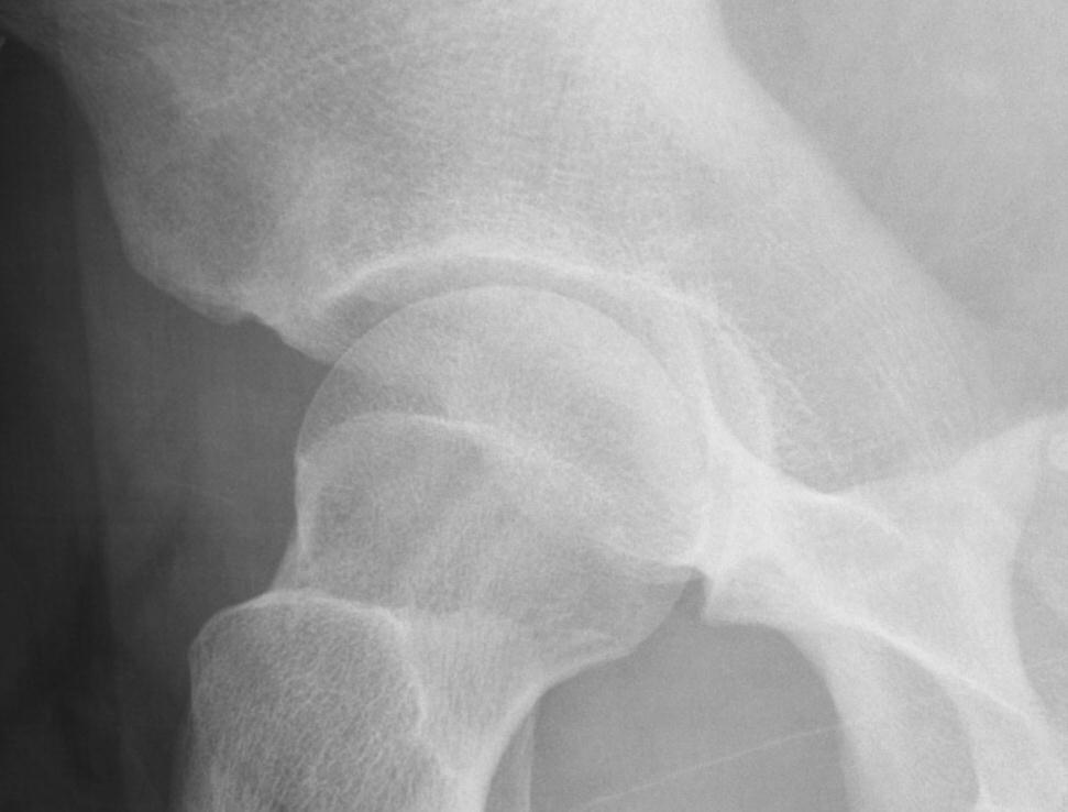
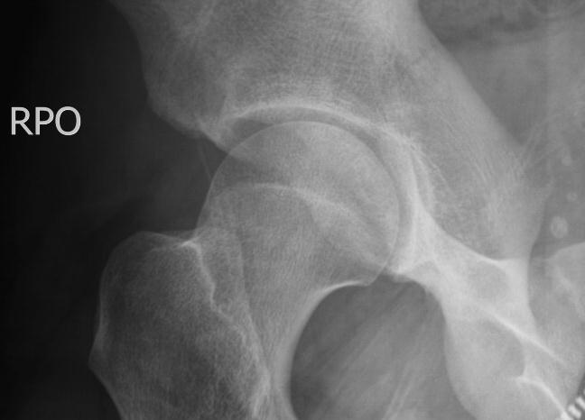
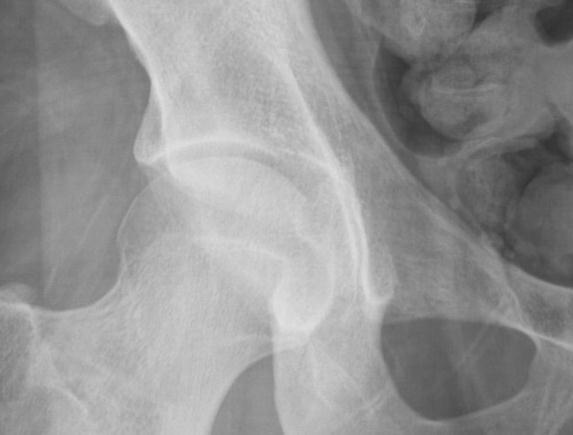
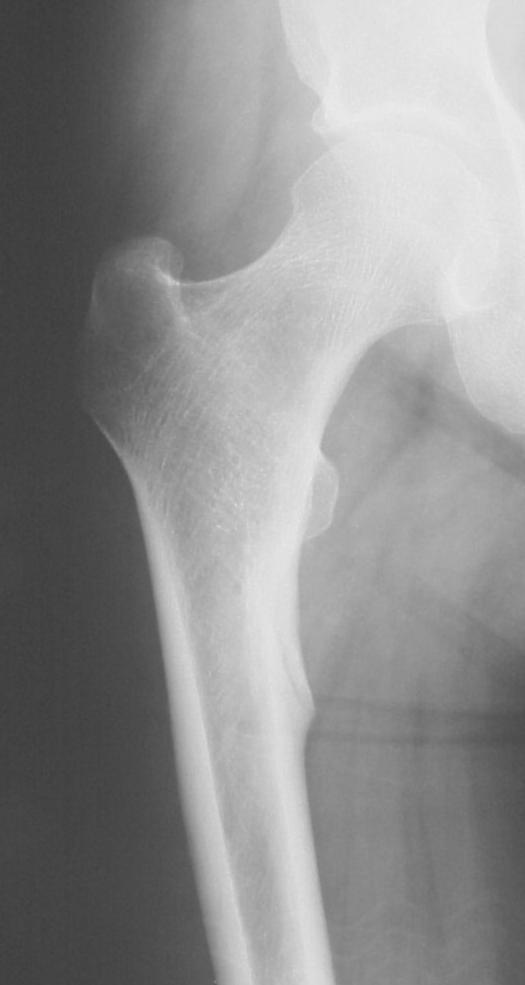
7 Top 10 Missed Fractures 8. Undisplaced neck of femur Fulde GWO (1994) Emergency Medicine

")
8 Top 10 Missed Fractures 9. Dislocated hip with ipsilateral femoral fracture Fulde GWO (1994) Emergency Medicine
9 Top 10 Missed Fractures 10. Tibial plateau fractures Fulde GWO (1994) Emergency Medicine
10 Reasons for Misses Simple miss Satisfaction of Search Inadequate study Not what was expected Corner of film finding Inappropriate history Working conditions
11 Reasons for Misses Satisfaction of Search One of the commonest reasons to miss injuries See most obvious injury Miss other (more significant) injury?


12 Reasons for Misses Satisfaction of Search? SH2 fracture distal tibia with base of 5 th MT fracture

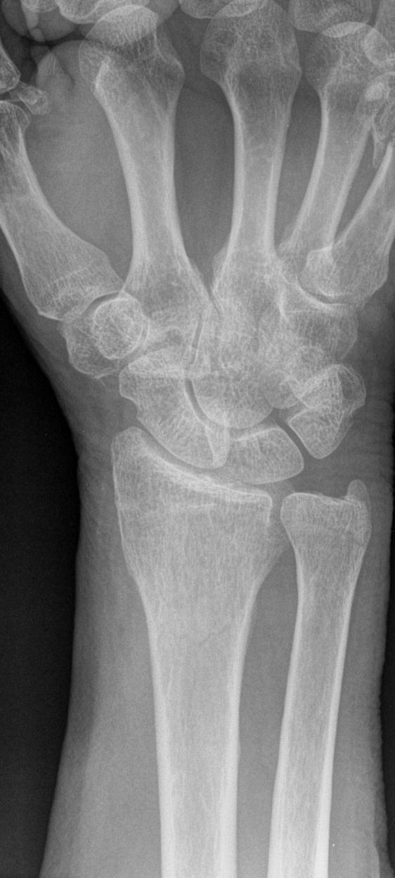

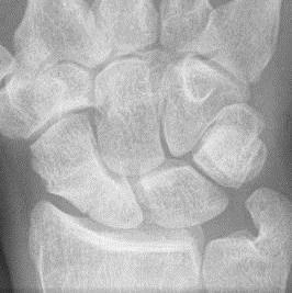
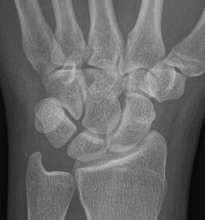
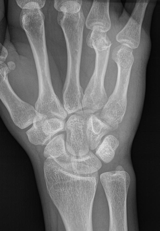
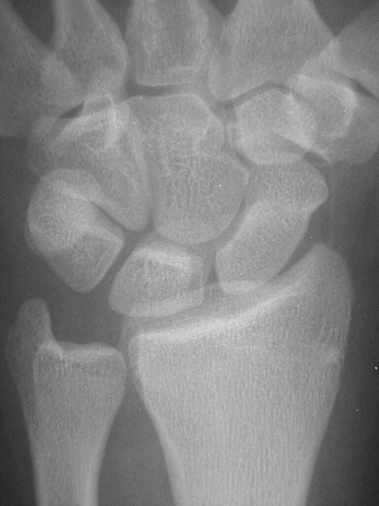
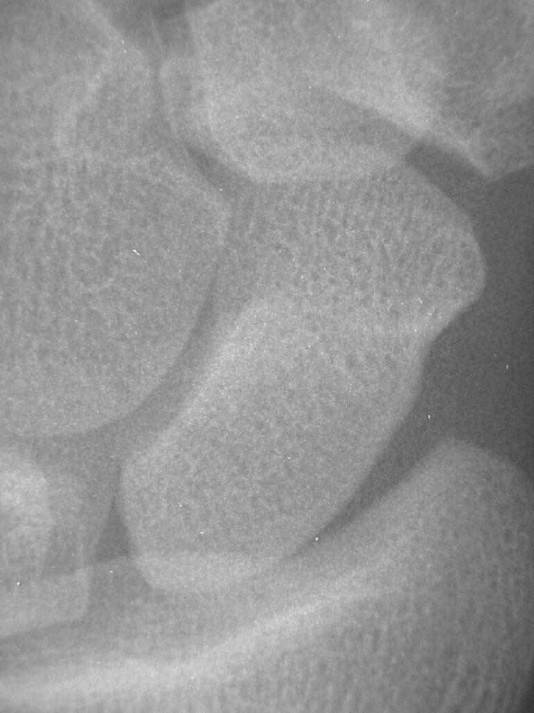
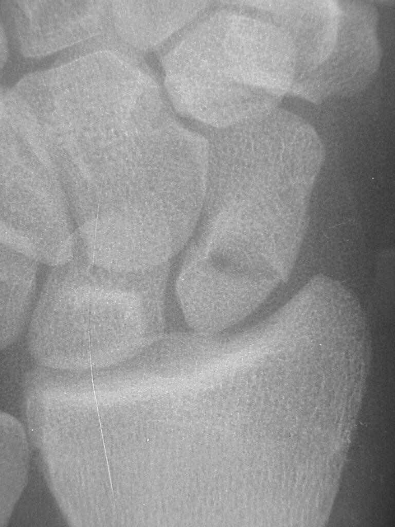
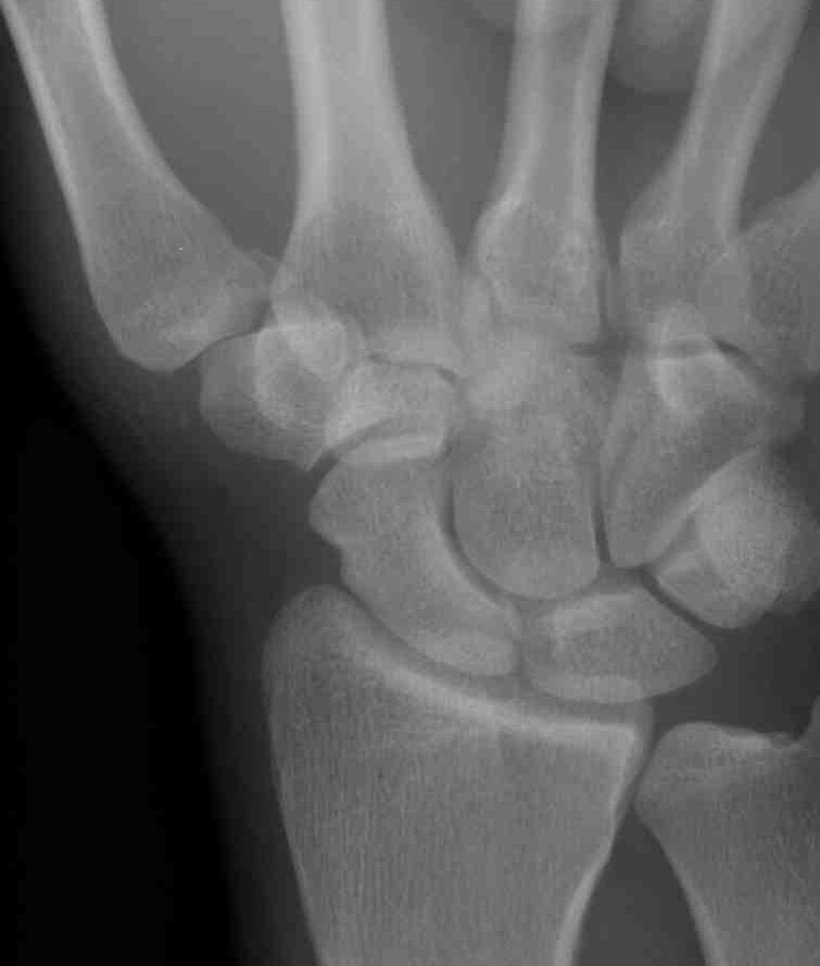
13 Reasons for Misses Satisfaction of Search? SOS wrist 68M


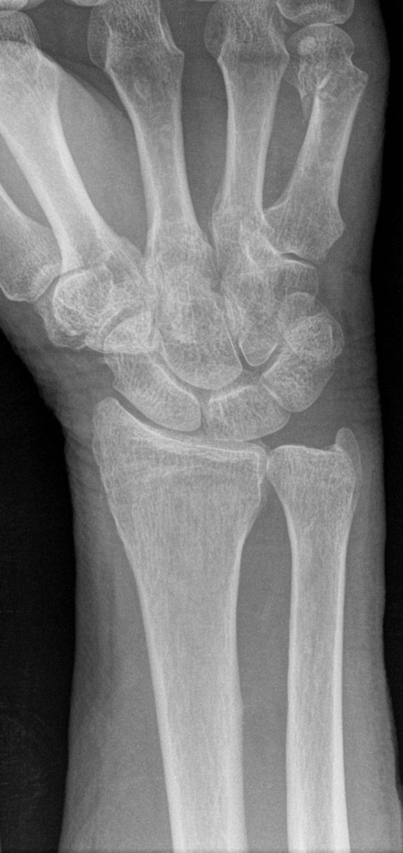
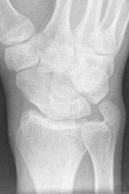
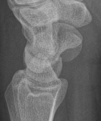
14 Reasons for Misses Satisfaction of Search Trans scaphoid and triquetral peri lunate 15M

15 Reasons for Misses Satisfaction of Search Simple miss Satisfaction of Search Inadequate study Not what was expected Corner of film finding Inappropriate history Working conditions? Talar neck fracture with subtalar dislocation

16 Reasons for Misses Satisfaction of Search Missed scaphoid fracture
17 Reasons for Misses Inadequate Study Need two or more views to assess for fracture or dislocation Need appropriate study Insist on good quality studies With empathy If equivocal, ask for more

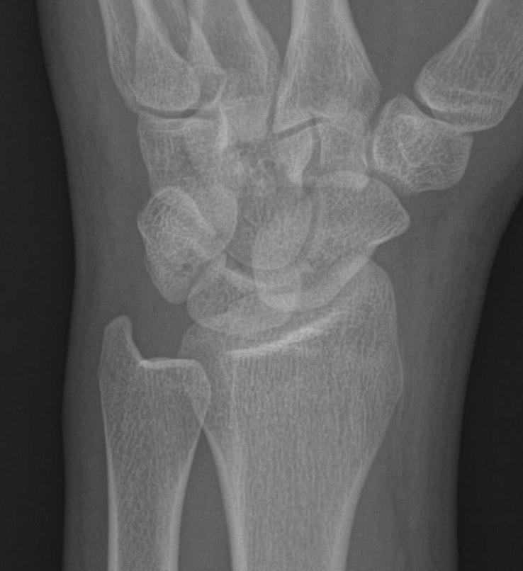
18 Reasons for Misses Inadequate Study Scaphoid fracture on ulna deviation view 19F
 Cone marks Appropriate exposure")
19 Reasons for Misses Inadequate Study All films need Patients name Patients number Date and time of study Side marker (lead, not added later) Cone marks Appropriate exposure


20 Reasons for Misses - Inadequate Study Two or more Views One view is never enough to assess for fractures Posterior process fracture of calcaneus
21 Reasons for Misses - Inadequate Study Two or more Views Circular saw injury

22 Reasons for Misses - Inadequate Study Two or more Views Car door

23 Reasons for Misses Not what was expected Dislocated right shoulder
24 Ways to Avoid Missing Fractures Look for fracture patterns Look at regions that should align Look for secondary signs of fracture Look for the common sites of fractures Have check lists for each region Special circumstances

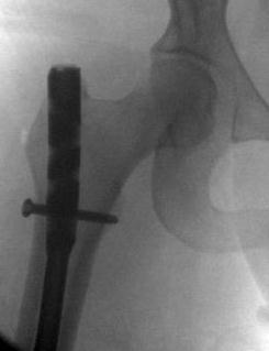
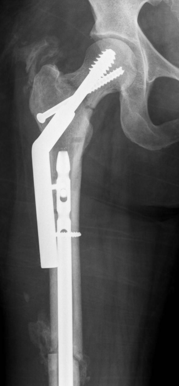
25 Fracture Patterns Patterns help us know where to look Transtriquetral / scaphoid perilunate fracture dislocation Maisonneuve Essex Lopresti Galeazzi Monteggia Waist of Scaphoid Femoral shaft and neck

26 Fracture Patterns Patterns help us know where to look Transtriquetral / scaphoid perilunate fracture dislocation Maisoneuve Essex Lopresti Galeazzi Monteggia Pelvic ring fractures Waist of Scaphoid Don Juan Femoral shaft and neck 27M


27 Fracture Patterns Patterns help us know where to look Transtriquetral / scaphoid perilunate fracture dislocation Maisoneuve Essex Lopresti Galeazzi Monteggia Pelvic ring fractures Waist of Scaphoid Don Juan Femoral shaft and neck

28 Fracture Patterns Patterns help us know where to look Transtriquetral / scaphoid perilunate fracture dislocation Maisoneuve Essex Lopresti Galeazzi Monteggia Pelvic ring fractures Waist of Scaphoid Don Juan Femoral shaft and neck


29 Fracture Patterns Patterns help us know where to look Transtriquetral / scaphoid perilunate fracture dislocation Maisoneuve Essex Lopresti Galeazzi Monteggia Pelvic ring fractures Waist of Scaphoid Don Juan Femoral shaft and neck
30 Fracture Patterns Patterns help us know where to look Transtriquetral / scaphoid perilunate fracture dislocation Maisoneuve Essex Lopresti Galeazzi Monteggia Pelvic ring fractures Waist of Scaphoid Don Juan Femoral shaft and neck


31 Fracture Patterns Patterns help us know where to look Transtriquetral / scaphoid perilunate fracture dislocation Maisoneuve Essex Lopresti Galeazzi Monteggia Pelvic ring fractures Waist of Scaphoid Don Juan Femoral shaft and neck Vertical Sacral fracture


32 Fracture Patterns Patterns help us know where to look Transtriquetral / scaphoid perilunate fracture dislocation Maisoneuve Essex Lopresti Galeazzi Monteggia Pelvic ring fractures Waist of Scaphoid Don Juan Femoral shaft and neck


33 Fracture Patterns Patterns help us know where to look Transtriquetral / scaphoid perilunate fracture dislocation Maisoneuve Essex Lopresti Galeazzi Monteggia Pelvic ring fractures Waist of Scaphoid Don Juan Femoral shaft and neck

34 Pattern Approach Occult basicervical fx NOF 32F

35 Pattern Approach Occult basicervical fx NOF 32F

36 Pattern Approach Occult basicervical fx NOF 32F


37 Pattern Approach 4th 5th MC Fx dis 24M


38 Pattern Approach 4th fx 5th dis 26M


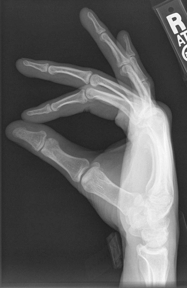
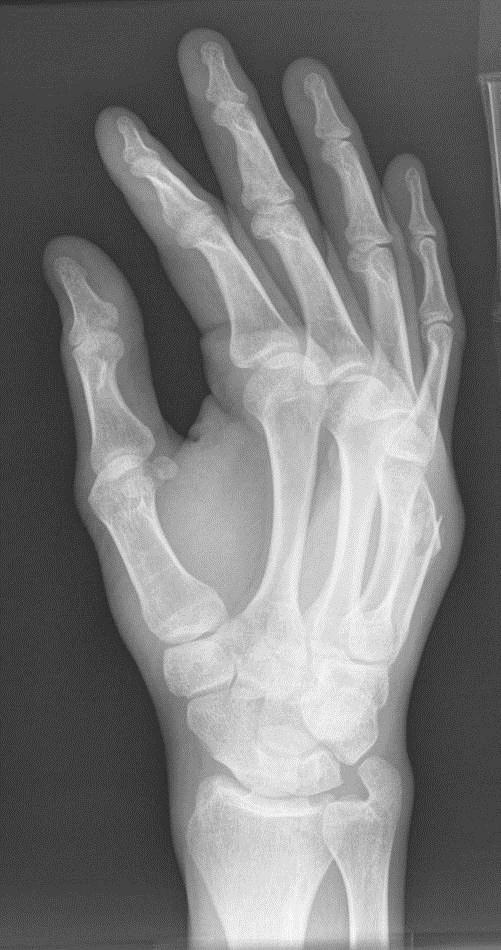
39 Pattern Approach Triple CMCJ dislocation 37M


40 Pattern Approach Volar Plate of Phalanx Adjacent structures

41 Pattern Approach Mallet Fracture
42 Ways to Avoid Missing Fractures Look for fracture patterns Look at regions that should align Look for secondary signs of fracture Look for the common sites of fractures Have check lists for each region Special circumstances


43 Alignment Rules These are helpful at various sites ACJ SCJ Lisfranc joint Elbow in children Carpal bones Also check for rotation


44 Alignment Rules These are helpful at various sites ACJ SCJ Lisfranc joint Elbow in children Carpal bones Also check for rotation

45 Alignment Rules These are helpful at various sites ACJ SCJ Lisfranc joint Elbow in children Carpal bones Also check for rotation If in doubt recommend weight bearing

46 Alignment Rules Childhood Elbow Fractures Medial epicondyle fracture With dislocation in children Can become trapped in joint Radial head dislocation Radiocapitellar line Should bisect capitellum Supracondylar fracture Anterior humeral line Middle 1/3 of capitellum Can Radiology Make Trauma Less Obscure CRITOE

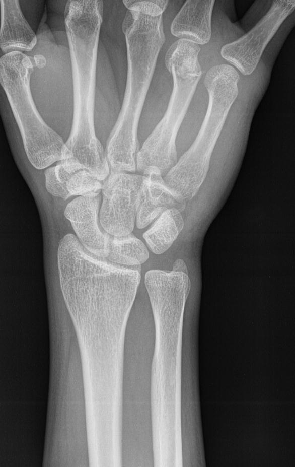
47 Alignment Rules These are helpful at various sites ACJ SCJ Lisfranc joint Elbow in children Carpal bones Also check for rotation Many distal radial fractures have a SCL injury

48 Alignment Rules These are helpful at various sites ACJ Lisfranc joint Elbow in children Carpal bones Also check for rotation

49 Alignment Rules These are helpful at various sites ACJ Lisfranc joint Elbow in children Carpal bones Also check for rotation

50 Alignment Rules - Rotation

51 4 Alignment Rules - Rotation
52 3 Alignment Rules - Rotation
53 2 Alignment Rules - Rotation
54 1 Alignment Rules - Rotation


55 4 Alignment Rules - Rotation
56 Ways to Avoid Missing Fractures Look for fracture patterns Look at regions that should align Look for secondary signs of fracture Look for the common sites of fractures Have check lists for each region Special circumstances

57 Secondary Signs Joint Effusion Secondary signs Joint effusion Lipohemarthrosis Gas in joint ST swelling Obliteration of fat planes Fat in joint on CT Bone edema on CT Intraosseous Vacuum Delayed resorption Delayed sclerosis The hinge joints

58 Secondary Signs Joint Effusion Secondary signs Joint effusion Lipohemarthrosis Gas in joint ST swelling Obliteration of fat planes Fat in joint on CT Bone edema on CT Intraosseous Vacuum Delayed resorption Delayed sclerosis The hinge joints

59 Secondary Signs Joint Effusion Secondary signs Joint effusion Lipohemarthrosis Gas in joint ST swelling Obliteration of fat planes Fat in joint on CT Bone edema on CT Intraosseous Vacuum Delayed resorption Delayed sclerosis The hinge joints


60 Secondary Signs Joint Effusion Acute 6w later

61 Secondary Signs Lipohemarthrosis Secondary signs Joint effusion Lipohemarthrosis Gas in joint ST swelling Obliteration of fat planes Fat in joint on CT Bone edema on CT Intraosseous Vacuum Delayed resorption Delayed sclerosis Tibial Plateau Fracture

62 Secondary Signs Lipohemarthrosis Secondary signs Joint effusion Lipohemarthrosis Gas in joint ST swelling Obliteration of fat planes Fat in joint on CT Bone edema on CT Intraosseous Vacuum Delayed resorption Delayed sclerosis Tibial Plateau Fracture

63 Secondary Signs Lipohemarthrosis Secondary signs Joint effusion Lipohemarthrosis Gas in joint ST swelling Obliteration of fat planes Fat in joint on CT Bone edema on CT Intraosseous Vacuum Delayed resorption Delayed sclerosis Tibial Plateau Fracture

64 Secondary Signs Lipohemarthrosis Secondary signs Joint effusion Lipohemarthrosis Gas in joint ST swelling Obliteration of fat planes Fat in joint on CT Bone edema on CT Intraosseous Vacuum Delayed resorption Delayed sclerosis Stellate patella fracture 61F
65 Secondary Signs Lipohemarthrosis Secondary signs Joint effusion Lipohemarthrosis Gas in joint ST swelling Obliteration of fat planes Fat in joint on CT Bone edema on CT Intraosseous Vacuum Delayed resorption Delayed sclerosis Stellate patella fracture 61F
66 Secondary Signs Lipohemarthrosis Secondary signs Joint effusion Lipohemarthrosis Gas in joint ST swelling Obliteration of fat planes Fat in joint on CT Bone edema on CT Intraosseous Vacuum Delayed resorption Delayed sclerosis 50M

67 Secondary Signs Lipohemarthrosis Secondary signs Joint effusion Lipohemarthrosis Gas in joint ST swelling Obliteration of fat planes Fat in joint on CT Bone edema on CT Intraosseous Vacuum Delayed resorption Delayed sclerosis 50M

68 Secondary Signs Gas in Joint Secondary signs Joint effusion Lipohemarthrosis Gas in joint ST swelling Obliteration of fat planes Fat in joint on CT Bone edema on CT Intraosseous Vacuum Delayed resorption Delayed sclerosis Open tibial plateau Fx

69 Secondary Signs Gas in Joint Secondary signs Joint effusion Lipohemarthrosis Gas in joint ST swelling Obliteration of fat planes Fat in joint on CT Bone edema on CT Intraosseous Vacuum Delayed resorption Delayed sclerosis Open tibial plateau Fx

70 Secondary Signs Gas in Joint Secondary signs Joint effusion Lipohemarthrosis Gas in joint ST swelling Obliteration of fat planes Fat in joint on CT Bone edema on CT Intraosseous Vacuum Delayed resorption Delayed sclerosis Open tibial plateau Fx
71 Secondary Signs Soft Tissue Swelling Secondary signs Joint effusion Lipohemarthrosis Gas in joint ST swelling Obliteration of fat planes Fat in joint on CT Bone edema on CT Intraosseous Vacuum Delayed resorption Delayed sclerosis 8M
72 Secondary Signs Soft Tissue Swelling Secondary signs Joint effusion Lipohemarthrosis Gas in joint ST swelling Obliteration of fat planes Fat in joint on CT Bone edema on CT Intraosseous Vacuum Delayed resorption Delayed sclerosis 8M


73 Secondary Signs Fat plane obliteration Secondary signs Joint effusion Lipohemarthrosis Gas in joint ST swelling Obliteration of fat planes Fat in joint on CT Bone edema on CT Intraosseous Vacuum Delayed resorption Delayed sclerosis Between the radial collateral ligament and APL/EPB Obliterated in Fx / Infection

74 Secondary Signs Fat in joint on CT Secondary signs Joint effusion Lipohemarthrosis Gas in joint ST swelling Obliteration of fat planes Fat in joint on CT Bone edema on CT Intraosseous Vacuum Delayed resorption Delayed sclerosis 14 Tibial plafond fracture

75 Secondary Signs Lipohemarthrosis Anterior recess of the posterior subtalar joint 3 Tibial plafond fracture


76 Secondary Signs Lipohemarthrosis Secondary signs Joint effusion Lipohemarthrosis Gas in joint ST swelling Obliteration of fat planes Fat in joint on CT Bone edema on CT Intraosseous Vacuum Delayed resorption Delayed sclerosis 78M

77 Secondary Signs Lipohemarthrosis Fat fluid level in iliopsoas bursa 61F


78 Secondary Signs Lipohemarthrosis 3 60 y/o female with pain s/p fall

79 Secondary Signs Lipohemarthrosis 60 y/o female with pain s/p fall


80 Secondary Signs Lipohemarthrosis 60 y/o female with pain s/p fall


81 Secondary Signs Lipohemarthrosis Radial head Fx 1 History of recent trauma.


82 Secondary Signs Lipohemarthrosis Sag PD Sag T2FS Bubbles may be more acute Lateral patella dislocation with patella Fx


83 Secondary Signs Lipohemarthrosis Cor T1 Cor PDFS Bubbles may be more acute 1+1 Lateral patella dislocation with patella Fx

84 Secondary Signs Lipohemarthrosis Ax PDFS Bubbles may be more acute 1 Lateral patella dislocation with patella Fx


85 Secondary Signs Bone edema on CT Secondary signs Joint effusion Lipohemarthrosis Gas in joint ST swelling Obliteration of fat planes Fat in joint on CT Bone edema on CT Intraosseous Vacuum Delayed resorption Delayed sclerosis


86 Secondary Signs Bone edema on CT Secondary signs Joint effusion Lipohemarthrosis Gas in joint ST swelling Obliteration of fat planes Fat in joint on CT Bone edema on CT Intraosseous Vacuum Delayed resorption Delayed sclerosis Sacral fx edema CT 98F

87 Secondary Signs Bone edema on CT Secondary signs Joint effusion Lipohemarthrosis Gas in joint ST swelling Obliteration of fat planes Fat in joint on CT Bone edema on CT Intraosseous Vacuum Delayed resorption Delayed sclerosis Sacral fracture edema CT 66F

88 Secondary Signs Lipohemarthrosis Secondary signs Joint effusion Lipohemarthrosis Gas in joint ST swelling Obliteration of fat planes Fat in joint on CT Bone edema on CT Intraosseous Vacuum Delayed resorption Delayed sclerosis Lipohem knee bone edema 62F

89 Secondary Signs Lipohemarthrosis Secondary signs Joint effusion Lipohemarthrosis Gas in joint ST swelling Obliteration of fat planes Fat in joint on CT Bone edema on CT Intraosseous Vacuum Delayed resorption Delayed sclerosis Lipohem knee bone edema 62F


90 Secondary Signs Intraosseous Vacuum Phenomenon y/o right hip pain s/p fall

91 Secondary Signs Intraosseous Vacuum Phenomenon y/o right hip pain s/p fall

92 Secondary Signs Intraosseous Vacuum Phenomenon y/o right hip pain s/p fall


93 Secondary Signs Intraosseous Vacuum Phenomenon Cor T1 Cor PDFS 1 93 y/o right hip pain s/p fall
94 Secondary Signs Delayed resorption Secondary signs Joint effusion Lipohemarthrosis Gas in joint ST swelling Obliteration of fat planes Fat in joint on CT Bone edema on CT Intraosseous Vacuum Delayed resorption Delayed sclerosis Acute 2w later
95 Delayed resorption 6w follow up
96 Secondary Signs Delayed sclerosis Secondary signs Joint effusion Lipohemarthrosis Gas in joint ST swelling Obliteration of fat planes Fat in joint on CT Bone edema on CT Intraosseous Vacuum Delayed resorption Delayed sclerosis Presentation 1 month follow up
97 Ways to Avoid Missing Fractures Look for fracture patterns Look at regions that should align Look for secondary signs of fracture Look for the common sites of fractures Have check lists for each region Special circumstances

98 Hill Sachs ER Neutral IR Stryker view supine hand on head


99 Bony Bankart Consider Westpoint

100 Greater Tuberosity of Humerus One of the most commonly missed fractures


101 Hook of Hamate Fracture Hook not seen on AP


102 Beak Ligament Avulsion Fracture

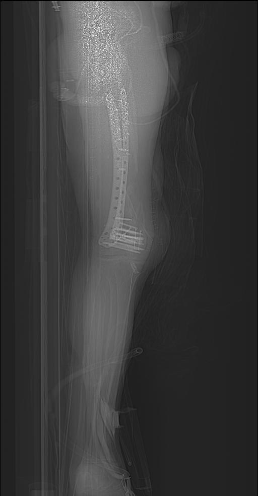
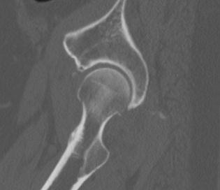
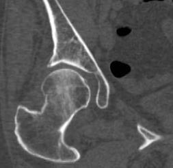
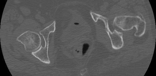
103 Acetabular fracture Discrete acetabular fx 21M


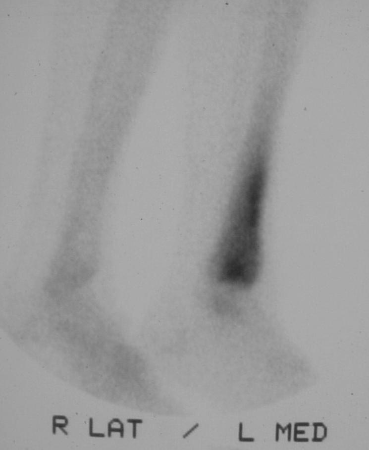
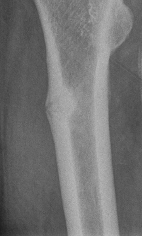
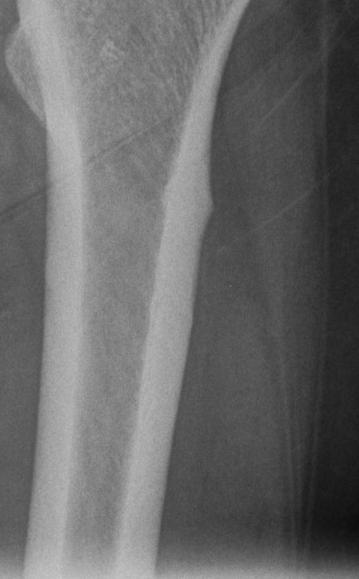
104 Femoral Neck Stress Fracture


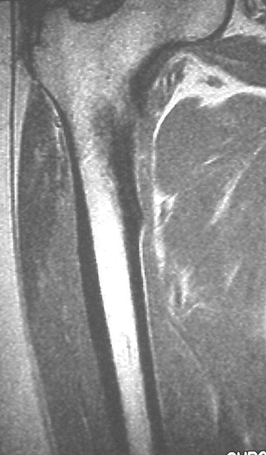
105 Proximal Femoral Stress Fracture


106 Tibial Plateau Fracture Fall with twist Ped V s MVA 50% > 50Y 80% lateral due to valgus Obliques useful MRI for diagnosis CT to stage Schatzker classification


107 Segond Fracture Segond fracture suggests the presence of significant pathology A small, vertically oriented, avulsed bony fragment Involves the lateral aspect of the proximal lateral tibia Nearly always associated with a tear of the anterior cruciate ligament in the older population Alternatively, an avulsion of the tibial spines is seen in younger patients


108 Proximal Fibula Fractures Can indicate an unstable posterolateral corner Ass. ACL injury

109 Deep lateral condylopatellar notch
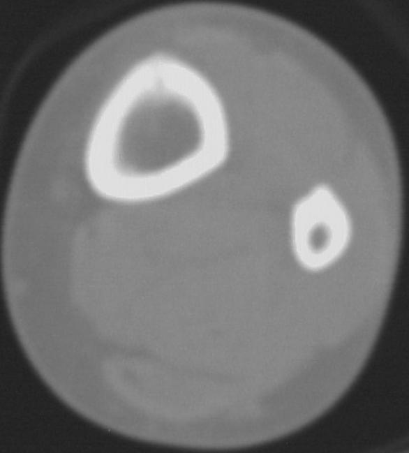
110 Longitudinal Stress Fracture

111 Toddler Fracture


112 Metatarsal Stress Fractures 2nd metatarsal neck stress fx 28F
113 Ways to Avoid Missing Fractures Look for fracture patterns Look at regions that should align Look for secondary signs of fracture Look for the common sites of fractures Have check lists for each region Special circumstances
114 Ankle Injury - Check List Malleoli Lateral process of Talus Talar dome Anterior process of Calcaneus EDB avulsion Base of 5 th metatarsal Jones fracture Does ankle fracture suggest Maisoneuve Dorsal chip fractures


115 Ankle Injury - Check List Malleoli Lateral process of Talus Talar dome Anterior process of Calcaneus EDB avulsion Base of 5 th metatarsal Jones fracture Does ankle fracture suggest Maisoneuve Dorsal chip fractures

116 Ankle Injury - Check List Malleoli Lateral process of Talus Talar dome Anterior process of Calcaneus EDB avulsion Base of 5 th metatarsal Jones fracture Does ankle fracture suggest Maisoneuve Dorsal chip fractures

117 Ankle Injury - Check List Malleoli Lateral process of Talus Talar dome Anterior process of Calcaneus EDB avulsion Base of 5 th metatarsal Jones fracture Does ankle fracture suggest Maisoneuve Dorsal chip fractures


118 Ankle Injury - Check List Malleoli Lateral process of Talus Talar dome Anterior process of Calcaneus EDB avulsion Base of 5 th metatarsal Jones fracture Does ankle fracture suggest Maisoneuve Dorsal chip fractures 29M recent trauma

119 Ankle Injury - Check List Malleoli Lateral process of Talus Talar dome Anterior process of Calcaneus EDB avulsion Base of 5 th metatarsal Jones fracture Does ankle fracture suggest Maisoneuve Dorsal chip fractures

120 Ankle Injury - Check List Malleoli Lateral process of Talus Talar dome Anterior process of Calcaneus EDB avulsion Base of 5 th metatarsal Jones fracture Does ankle fracture suggest Maisoneuve Dorsal chip fractures
121 Ankle Injury - Check List Malleoli Lateral process of Talus Talar dome Anterior process of Calcaneus EDB avulsion Base of 5 th metatarsal Jones fracture Does ankle fracture suggest Maisoneuve Dorsal chip fractures

122 Ankle Injury - Check List Malleoli Lateral process of Talus Talar dome Anterior process of Calcaneus EDB avulsion Base of 5 th metatarsal Jones fracture Does ankle fracture suggest Maisoneuve Dorsal chip fractures
123 Ways to Avoid Missing Fractures Look for fracture patterns Look at regions that should align Look for secondary signs of fracture Look for the common sites of fractures Have check lists for each region Special circumstances
124 Special circumstances Elderly Fractures often hard to see Degenerative changes obscure fractures Fatty marrow makes bone edema useful sign Fractures more often fatal If alters management, low threshold for MRI
125 Pelvic Insufficiency Fractures sacrum iliac wing superomedial ileum supracetabular symphysis pubic rami
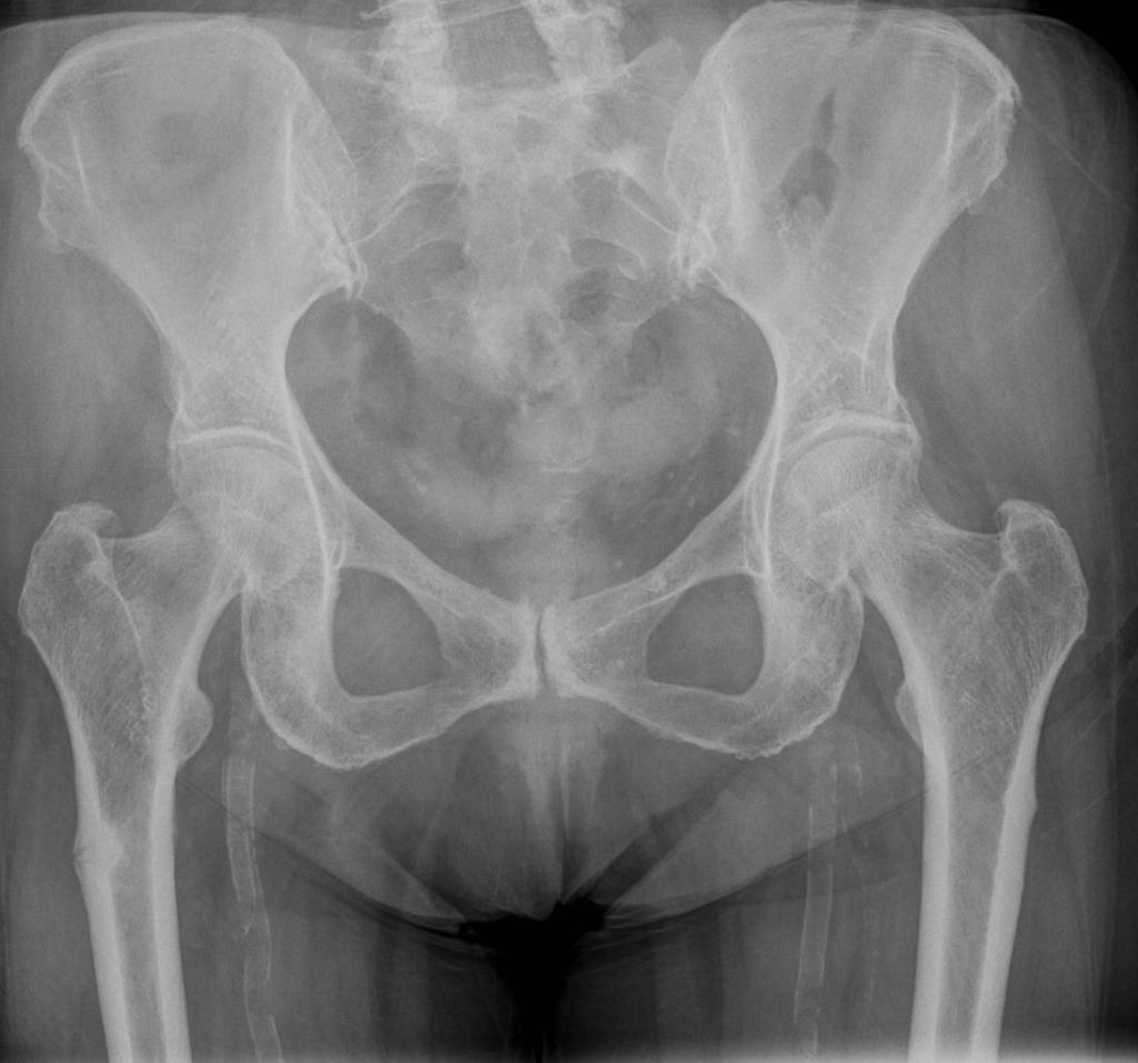
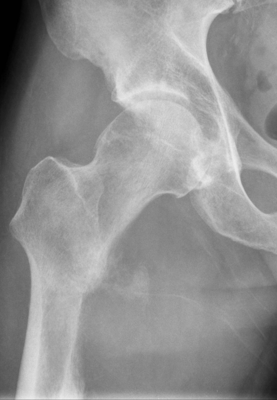
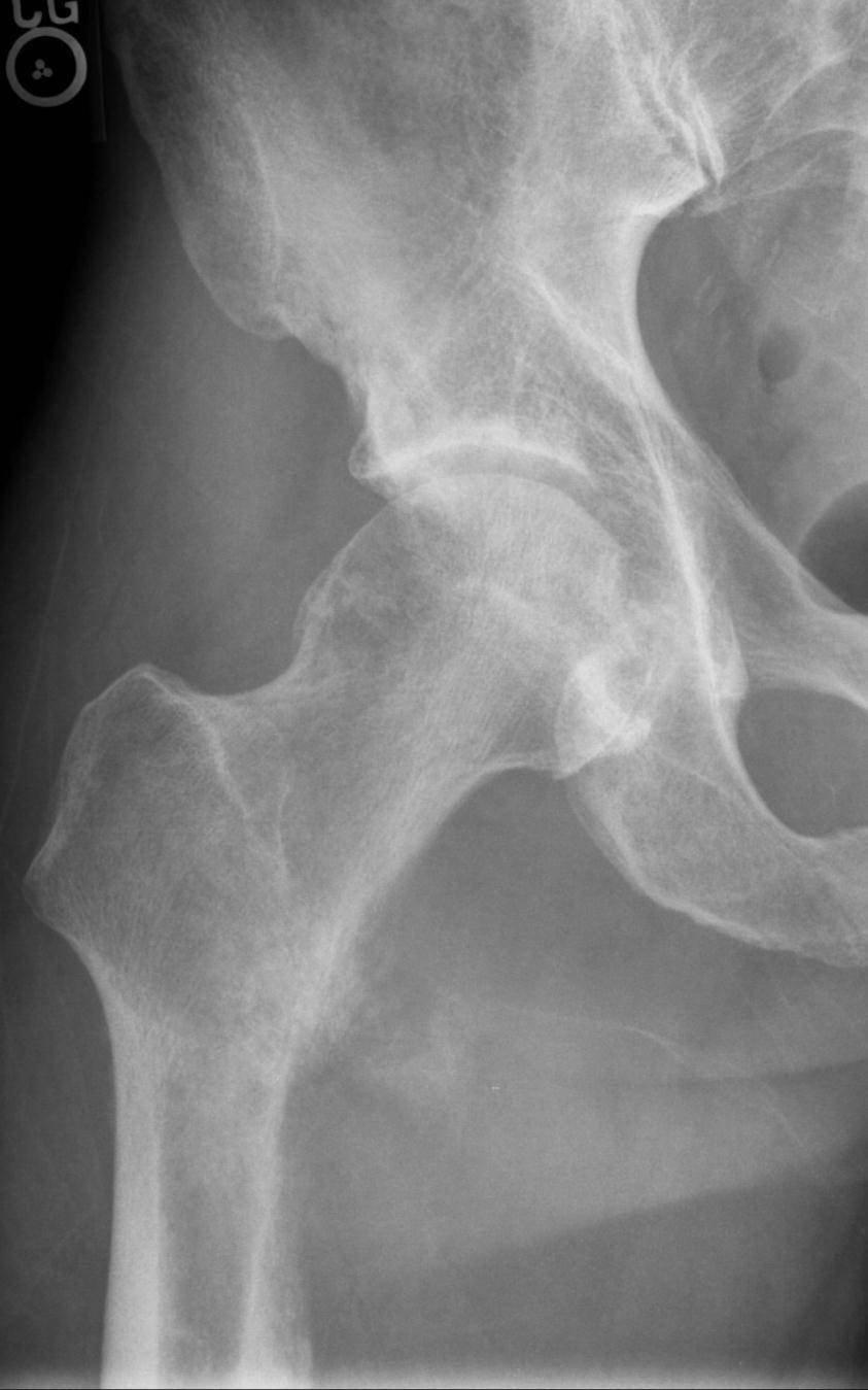
126 Bisphosphanate fractures proximal femur 84 year old female with bilateral hip pain
127 Pathologic lesser trochanteric avulsion


128 Special circumstances Childhood Fractures Tendons stronger than bone Apophyseal avulsion Fracture patterns different Salter Harris Incomplete fractures more common Plastic bowing Torus / Buckle Greenstick Remember NAI 11M ACL avulsion


129 Childhood Fractures Tendons stronger than bone Apophyseal avulsion Fracture patterns different Salter Harris Incomplete fractures more common Plastic bowing Torus / Buckle Greenstick Remember NAI

130 Childhood Fractures Tendons stronger than bone Apophyseal avulsion Fracture patterns different Salter Harris SCFE is Salter 1 fracture of femoral epiphysis Bilateral in 25% Best seen on frog-leg lateral Incomplete fractures more common Plastic bowing Torus / Buckle Greenstick Remember NAI

131 Childhood Fractures Tendons stronger than bone Apophyseal avulsion Fracture patterns different Salter Harris Incomplete fractures more common Plastic bowing Torus / Buckle Greenstick Remember NAI


132 Childhood Fractures Tendons stronger than bone Apophyseal avulsion Fracture patterns different Salter Harris Incomplete fractures more common Plastic bowing Torus / Buckle Greenstick Remember NAI

133 Childhood Fractures Tendons stronger than bone Apophyseal avulsion Fracture patterns different Salter Harris Incomplete fractures more common Plastic bowing Torus / Buckle Greenstick Remember NAI
134 Problem solving Repeat Oblique views Tibial plateau Radial head Dedicated views Scaphoid Radial head Single emulsion Periphery Tomography, CT, MRI, Scintigraphy
Commonly Missed Injuries of the Extremities
 Commonly Missed Injuries of the Extremities Dr. Tudor H. Hughes M.D., FRCR Department of Radiology University of California School of Medicine San Diego, California 1. Base of skull 2. Odontoid process
Basic Radiographic Principles Part II
 Basic Radiographic Principles Part II Kristopher Avant, D.O. October 19 th, 2016 I have no disclosures relevant to the material presented in this discussion. Good Stuff!!! 1 Really? Really! Musculoskeletal
4/28/2010. Fractures. Normal Bone and Normal Ossification Bone Terms. Epiphysis Epiphyseal Plate (physis) Metaphysis
 Metaphysis") Fractures Normal Bone and Normal Ossification Bone Terms Epiphysis Epiphyseal Plate (physis) Metaphysis Diaphysis 1 Fracture Classifications A. Longitudinal B. Transverse C. Oblique D. Spiral E. Incomplete
Fractures Normal Bone and Normal Ossification Bone Terms Epiphysis Epiphyseal Plate (physis) Metaphysis Diaphysis 1 Fracture Classifications A. Longitudinal B. Transverse C. Oblique D. Spiral E. Incomplete
PEM GUIDE CHILDHOOD FRACTURES
 PEM GUIDE CHILDHOOD FRACTURES INTRODUCTION Skeletal injuries account for 10-15% of all injuries in children; 20% of those are fractures, 3 out of 4 fractures affect the physis or growth plate. Always consider
Basic Principles of Fractures & Easily Missed Fractures. Mr Irfan Merchant Trauma & Orthopaedic Registrar Bedford Hospital, East of England
 Basic Principles of Fractures & Easily Missed Fractures Mr Irfan Merchant Trauma & Orthopaedic Registrar Bedford Hospital, East of England Objectives Types Fracture Patterns Fracture Healing Assessing
Index. Note: Page numbers of article titles are in boldface type.
 Note: Page numbers of article titles are in boldface type. A Abscess, epidural, 822 824 Achilles tendon rupture, 894 895, 981 982 Acromioclavicular separations, shoulder pain in, 751 753 Adhesive capsulitis,
Index. Note: Page numbers of article titles are in boldface type.
 Note: Page numbers of article titles are in boldface type. A Acetabular fractures, 462 464 Achilles tendon rupture, 389 Acromioclavicular dislocations, 302 Acromion fractures, 301 Ankle, anatomy of, 376
Pediatric Fractures. Objectives. Epiphyseal Complex. Anatomy and Physiology. Ligaments. Bony matrix
 1 Pediatric Fractures Nicholas White, MD Assistant Professor of Pediatrics Eastern Virginia Medical School Attending, Pediatric Emergency Department Children s Hospital of The King s Daughters Objectives
EMERGENCY PITFALLS IN ORTHOPAEDIC TRAUMA. Thierry E. Benaroch, MD, FRCS MCH Trauma Rounds February 9, 2009
 EMERGENCY PITFALLS IN ORTHOPAEDIC TRAUMA Thierry E. Benaroch, MD, FRCS MCH Trauma Rounds February 9, 2009 MORAL OF THE STORY Fracture distal radius and intact ulna W/O radius fracture will most likely
Surgical Care at the District Hospital. EMERGENCY & ESSENTIAL SURGICAL CARE
 Surgical Care at the District Hospital 1 18 Orthopedic Trauma Key Points 2 18.1 Upper Extremity Injuries Clavicle Fractures Diagnose fractures from the history and by physical examination Treat with a
Orthopedic X-Rays most commonly missed
 Orthopedic X-Rays most commonly missed Vukiet Tran, MD, MHSc, MBA University Health Network Toronto, Canada 1 COI Disclosure I am the current Medical Director for Best Doctors Canada. Presenter: Dr. Vu
Childhood Fractures. Incomplete fractures more common. Ligaments stronger than bone. Tendons stronger than bone. Fractures may be pathologic
 Childhood Fractures Incomplete fractures more common Plastic bowing Torus / Buckle Greenstick Ligaments stronger than bone Fracture patterns different Physeal injury, not dislocation Tendons stronger than
10/12/2010. Upper Extremity. Pectoral (Shoulder) Girdle. Clavicle (collarbone) Skeletal System: Appendicular Skeleton
 Girdle. Clavicle (collarbone) Skeletal System: Appendicular Skeleton") Skeletal System: Appendicular Skeleton Pectoral girdle Pelvic girdle Upper limbs Lower limbs 8-1 Pectoral (Shoulder) Girdle Consists of scapula and clavicle Clavicle articulates with sternum (Sternoclavicular
Skeletal System: Appendicular Skeleton Pectoral girdle Pelvic girdle Upper limbs Lower limbs 8-1 Pectoral (Shoulder) Girdle Consists of scapula and clavicle Clavicle articulates with sternum (Sternoclavicular
Radiographic Positioning Summary (Basic Projections RAD 222)
") Lower Extremity Radiographic Positioning Summary (Basic Projections RAD 222) AP Pelvis AP Hip (Unilateral) (L or R) AP Femur Mid and distal AP Knee Lateral Knee Pt lies supine on table Align MSP to Center
Lower Extremity Radiographic Positioning Summary (Basic Projections RAD 222) AP Pelvis AP Hip (Unilateral) (L or R) AP Femur Mid and distal AP Knee Lateral Knee Pt lies supine on table Align MSP to Center
Imaging the musculoskeletal system. An Introduction
 Imaging the musculoskeletal system An Introduction Objectives Discuss: commonly used imaging modalities in the musculoskeletal system normal imaging anatomy in the extremities fracture description Imaging
Appendicular skeleton: ABCs Image Interpretation Search strategy
 NOVEMBER 2013 volume 51 number 2 THE SOUTH AFRICAN RADIOGRAPHER peer reviewed ARTICLE OF INTEREST Appendicular skeleton: ABCs Image Interpretation Search strategy IJ Williams MSc in Medical Imaging; B
radiologymasterclass.co.uk
 http://radiologymasterclass.co.uk Hip X-ray anatomy - Normal AP (anterior-posterior) Shenton's line is formed by the medial edge of the femoral neck and the inferior edge of the superior pubic ramus Loss
Pectoral (Shoulder) Girdle
 Girdle") Chapter 8 Skeletal System: Appendicular Skeleton Pectoral girdle Pelvic girdle Upper limbs Lower limbs 8-1 Pectoral (Shoulder) Girdle Consists of scapula and clavicle Clavicle articulates with sternum
Chapter 8 Skeletal System: Appendicular Skeleton Pectoral girdle Pelvic girdle Upper limbs Lower limbs 8-1 Pectoral (Shoulder) Girdle Consists of scapula and clavicle Clavicle articulates with sternum
RADIOGRAPHY OF THE ANKLE and LOWER LEG
 RADIOGRAPHY OF THE ANKLE and LOWER LEG Patient Position: ANKLE AP Projection Part Position: True Slight to place foot s long axis Center to Central Ray: to IR Midway Note: Ankle joint is to tips of malleoli
The Appendicular Skeleton
 8 The Appendicular Skeleton PowerPoint Lecture Presentations prepared by Jason LaPres Lone Star College North Harris 8-1 The Pectoral Girdle The Pectoral Girdle Also called shoulder girdle Connects the
Imaging the Athlete s Knee. Peter Lowry, MD Musculoskeletal Radiology University of Colorado
 Imaging the Athlete s Knee Peter Lowry, MD Musculoskeletal Radiology University of Colorado None Disclosures Knee Imaging: Radiographs Can be performed weight-bearing or non-weight-bearing View options
Biology 218 Human Anatomy. Adapted from Martini Human Anatomy 7th ed. Chapter 7 The Skeletal System Appendicular Division
 Adapted from Martini Human Anatomy 7th ed. Chapter 7 The Skeletal System Appendicular Division Introduction The appendicular skeleton includes: Pectoral girdle Shoulder bones Upper limbs Pelvic girdle
Amy Warenda Czura, Ph.D. 1 SCCC BIO130 Lab 7 Appendicular Skeleton & Articulations
 The Skeletal System II: Appendicular Skeleton and Articulations Exercises 11, 13 (begins: page 145 in 9 th and 10 th editions) Exercises 10, 11 (begins: page 147 in 11 th edition, page 149 in 12 th edition)
Trauma Films for Upper Body. LCDR. Naruebade Rungrattanawilai RTN M.D., LL.B. FRCOST, DMOC
 Trauma Films for Upper Body LCDR. Naruebade Rungrattanawilai RTN M.D., LL.B. FRCOST, DMOC Objective A 42 year-old housekeeper with history of motorcycle accident. There was no external wound but she have
Shoulder Position: Supine arm in the neutral position. Collateral arm above head Indication: fracture humerus, fracture scapula
 Shoulder Position: Supine arm in the neutral position. Collateral arm above head Indication: fracture humerus, fracture scapula No instrumentation With metal or cast KV/ Effective mas/rotation time 140/300/1.0
Chapter 8. The Appendicular Skeleton. Lecture Presentation by Lee Ann Frederick University of Texas at Arlington Pearson Education, Inc.
 Chapter 8 The Appendicular Skeleton Lecture Presentation by Lee Ann Frederick University of Texas at Arlington An Introduction to the Appendicular Skeleton The Appendicular Skeleton 126 bones Allows us
Montreal Children s Hospital McGill University Health Center Emergency Department Fracture Guideline
 Montreal Children s Hospital McGill University Health Center Emergency Department Guideline Disclaimers This document is designed to assist physicians working in our emergency department in caring for
Chapter 8B. The Skeletal System: Appendicular Skeleton. The Appendicular Skeleton. Clavicle. Pectoral (Shoulder) Girdle
 Girdle") The Appendicular Skeleton Chapter 8B The Skeletal System: Appendicular Skeleton 126 bones Pectoral (shoulder) girdle Pelvic (hip) girdle Upper limbs Lower limbs Functions primarily to facilitate movement
The Appendicular Skeleton Chapter 8B The Skeletal System: Appendicular Skeleton 126 bones Pectoral (shoulder) girdle Pelvic (hip) girdle Upper limbs Lower limbs Functions primarily to facilitate movement
SKELETAL SYSTEM 206. AXIAL SKELETON 80 APPENDICULAR SKELETON 126 (see Figure 6.1) Clavicle. Clavicle. Pectoral girdles. Scapula. Scapula.
 Clavicle. Clavicle. Pectoral girdles. Scapula. Scapula.") SKELETAL SYSTEM 206 AXIAL SKELETON 80 APPENDICULAR SKELETON 126 (see Figure 6.1) Pectoral girdles 4 Clavicle Scapula 2 2 Clavicle Scapula Humerus 2 Humerus Upper limbs 60 Radius 2 Ulna Carpal bones Metacarpal
SKELETAL SYSTEM 206 AXIAL SKELETON 80 APPENDICULAR SKELETON 126 (see Figure 6.1) Pectoral girdles 4 Clavicle Scapula 2 2 Clavicle Scapula Humerus 2 Humerus Upper limbs 60 Radius 2 Ulna Carpal bones Metacarpal
Learning from Discrepancies Meetings - What we've learned from Musculoskeletal Diagnostic Errors in 2014
 Learning from Discrepancies Meetings - What we've learned from Musculoskeletal Diagnostic Errors in 2014 Poster No.: P-0104 Congress: ESSR 2015 Type: Scientific Poster Authors: B. Batohi, R. Chhabra, S.
A. Incorrect! The appendicular skeleton includes bones of the shoulder, arm, hand, pelvis, leg and foot.
 Anatomy and Physiology - Problem Drill 08: The Skeletal System III No. 1 of 10 1. Which of the following statements about the appendicular skeleton is correct? A. The appendicular skeleton includes bones
Lab Activity 9. Appendicular Skeleton Martini Chapter 8. Portland Community College BI 231
 Lab Activity 9 Appendicular Skeleton Martini Chapter 8 Portland Community College BI 231 Appendicular Skeleton Upper & Lower extremities Shoulder Girdle Pelvic Girdle 2 Humerus 3 Humerus: Proximal End
ORTHOSCAN MOBILE DI POSITIONING GUIDE
 ORTHOSCAN MOBILE DI POSITIONING GUIDE Table of Contents SHOULDER A/P of Shoulder... 4 Tangential (Y-View) of Shoulder... 5 Lateral of Proximal Humerus... 6 ELBOW A/P of Elbow... 7 Extended Elbow... 8 Lateral
Trauma-related Pediatric Orthopedic Emergencies. Javier Gonzalez del Rey, M.D. Professor Pediatrics Cincinnati Children s Hospital Medical Center
 Trauma-related Pediatric Orthopedic Emergencies Javier Gonzalez del Rey, M.D. Professor Pediatrics Cincinnati Children s Hospital Medical Center Room # 10 7 month old sick since birth Room # 11 5 y/o Fell
Upper Extremity Fractures
 Upper Extremity Fractures Ranie Whatley, RN,FNP-C David W. Gray, MD Skeletal Trauma 10 to 15 % of all Childhood Injuries Physeal (Growth Plate) Injuries are ~ 15% of all Skeletal Injuries Orthopaedic Assessment
Common Orthopaedic Injuries in Children
 Common Orthopaedic Injuries in Children Rakesh P. Mashru, M.D. Division of Orthopaedic Trauma Cooper University Hospital Cooper Medical School of Rowan University December 1, 2017 1 Learning Objectives
Bones of Lower Limb. Dr. Heba Kalbouneh Associate Professor of Anatomy and Histology
 Bones of Lower Limb Dr. Heba Kalbouneh Associate Professor of Anatomy and Histology Bones of the lower limb Hip Bone Made up of 3 bones: 1) Ilium (flat), superior in position 2) Ischium (L), postero-inferior
Radiologic Pitfalls. Pelvis/ Hip Hip DL Femoral neck Another ring fracture Sacrum Acetabulum
 Radiologic Pitfalls Michelle Lin, MD UCSF Associate Professor of Clinical Emergency Medicine San Francisco General Hospital (Michelle.Lin@emergency.ucsf.edu) ERRORS IN RADIOGRAPH INTERPRETATION Commonly
Chapter 8 The Skeletal System: The Appendicular Skeleton. Copyright 2009 John Wiley & Sons, Inc.
 Chapter 8 The Skeletal System: The Appendicular Skeleton Appendicular Skeleton It includes bones of the upper and lower limbs Girdles attach the limbs to the axial skeleton The pectoral girdle consists
Country Health SA Medical Imaging
 Country Health SA Medical Imaging REMOTE OPERATORS POSITIONING GUIDE Contents Image Evaluation Page 4 Positioning Guides Section 1 - THORAX 1.1 Chest Page 5 1.2 Bedside Chest Page 7 1.3 Ribs Page 8 Section
Anatomy of the Musculoskeletal System
 Anatomy of the Musculoskeletal System Kyle E. Rarey, Ph.D. Department of Anatomy & Cell Biology and Otolaryngology University of Florida College of Medicine Outline of Presentation Vertebral Column Upper
Radiology Positioning Practical Test #2 Table (By Jung Park):
:") Radiology Positioning Practical Test #2 Table (By Jung Park): (Lower Extremity): patient is fully gowned / no artifacts / properly shielded (exposure for femur and below : hold still, don t move ) (exposure
Radiology Positioning Practical Test #2 Table (By Jung Park): (Lower Extremity): patient is fully gowned / no artifacts / properly shielded (exposure for femur and below : hold still, don t move ) (exposure
PRE-LAB EXERCISES. Before we get started, look up the definitions of these common bone marking terms: Canal: Condyle: Facet: Fissure:
 1 PRE-LAB EXERCISES When studying the skeletal system, the bones are often sorted into two broad categories: the axial skeleton and the appendicular skeleton. This lab focuses on the appendicular skeleton,
Anatomy and Physiology 2016
 Anatomy and Physiology 2016 O = Temporal line I = coronoid process (Mandible) A = elevates mandible (chewing) O = galea aponeurotica (layer of dense fibrous tissue which covers the upper part of the cranium)
Appendicular Skeleton. Dr. Carmen E. Rexach Anatomy 35 Mt. San Antonio College
 Appendicular Skeleton Dr. Carmen E. Rexach Anatomy 35 Mt. San Antonio College Pectoral girdle clavicle scapula Upper limb brachium antebrachium carpus manus Pelvic girdle oscoxae Lower limb femoral region
OBJECTIVES: Define basic assessments skills needed to identify orthopedic injuries. Differentiate when an orthopedic injury is a medical emergency
 1 2 How to Triage Orthopaedic Care David W. Gray, M.D. OBJECTIVES: Define basic assessments skills needed to identify orthopedic injuries Differentiate when an orthopedic injury is a medical emergency
Lecture (10) Bone Fractures. Resources: - Lecture by dr.alboukai - Diagnostic imaging book
 Bone Fractures. Resources: - Lecture by dr.alboukai - Diagnostic imaging book") Lecture (10) Bone Fractures Hanan Alsalman Hanan Alrabiah Reem Aljurayyad Ayshah Almahboob Ghadeer Alwuhyad Khawlah AlOthman Dalal Alqadi Suliman Alshammari Maha AlKubaidan Rawabi Alghamdi Resources: -
Lecture (10) Bone Fractures Hanan Alsalman Hanan Alrabiah Reem Aljurayyad Ayshah Almahboob Ghadeer Alwuhyad Khawlah AlOthman Dalal Alqadi Suliman Alshammari Maha AlKubaidan Rawabi Alghamdi Resources: -
Principles of Anatomy and Physiology
 Principles of Anatomy and Physiology 14 th Edition CHAPTER 8 The Skeletal System: The Appendicular Skeleton The Appendicular Skeleton The 126 bones of the appendicular skeleton are primarily concerned
Copyright 2003 Pearson Education, Inc. publishing as Benjamin Cummings. Dr. Nabil Khouri MD, MSc, Ph.D
 Dr. Nabil Khouri MD, MSc, Ph.D Pelvic Girdle (Hip) Organization of the Lower Limb It is divided into: The Gluteal region The thigh The knee The leg The ankle The foot The thigh and the leg have compartments
Exercise Science Section 2: The Skeletal System
 Exercise Science Section 2: The Skeletal System An Introduction to Health and Physical Education Ted Temertzoglou Paul Challen ISBN 1-55077-132-9 Role of the Skeleton Protection Framework Attachments for
THE ELBOW. The elbow is a commonly injured joint in both children and adults.
 ABC of Emergency Radiology FIG i-lateral radiograph of elbow and line THE ELBOW D A Nicholson, P A Driscoll The elbow is a commonly injured joint in both children and adults. Interpretation of elbow radiographs
11/4/2018 SUBTLETIES OF LOWER EXTREMITY TRAUMA IMAGING SPEAKER DISCLOSURES
 SUBTLETIES OF LOWER EXTREMITY TRAUMA IMAGING Charles S. Resnik, M.D. Professor of Radiology University of Maryland School of Medicine Upon completion of this presentation, participants will be better able
CHAPTER 8: THE BIOMECHANICS OF THE HUMAN LOWER EXTREMITY
 CHAPTER 8: THE BIOMECHANICS OF THE HUMAN LOWER EXTREMITY _ 1. The hip joint is the articulation between the and the. A. femur, acetabulum B. femur, spine C. femur, tibia _ 2. Which of the following is
Goals. Initial management skeletal trauma. Physical Exam ABC OF PRIMARY CARE MEDICINE FRACTURE MANAGEMENT 12/4/2010
 ABC OF PRIMARY CARE MEDICINE FRACTURE MANAGEMENT Brian Feeley, MD UCSF Sports Medicine and Shoulder Surgery Goals Discuss common fractures and initial management, treatment guidelines Let your patients
bio4165 lab quiz 1 Posterior View Anterior View Lateral View Anterior View bio fall.quarter lab.quiz.1...page.1 of 6
 B A Posterior View D C E Lateral View bio.4165...fall.quarter.2005...lab.quiz.1...page.1 of 6 F I G 35 Posterior View H bio.4165...fall.quarter.2005...lab.quiz.1...page.2 of 6 J Posterior View L K Inferior
RADIOGRAPHY OF THE WRIST
 RADIOGRAPHY OF THE WRIST Patient Position: WRIST PA Projection, elbow in same plane Part Position: Hand ; fingers centered to IR Central Ray: Structures Shown: NOTE: Optional AP projection best demonstrates
Joints Dr. Ali Ebneshahidi
 Joints Dr. Ali Ebneshahidi Function of Joints 1. Serve as functional junctions between bones. 2. Bind bones, strokes, and other related tissues together. 3. Allow bone growth to occur. 4. Permit certain
The Appendicular Skeleton
 8 The Appendicular Skeleton PowerPoint Lecture Presentations prepared by Jason LaPres Lone Star College North Harris An Introduction to the Appendicular Skeleton Learning Outcomes 8-1 Identify the bones
Skeletal System. Supplementary Information
 Skeletal System Supplementary Information COMMON ANATOMICAL TERMS Planes run through the body side to side and front to back eg. median plane Surfaces of the body are also named eg. anterior surface This
Biology 218 Human Anatomy
 Chapter 8 Adapted from Tortora 10 th ed. LECTURE OUTLINE A. Introduction (p. 203) 1. The appendicular skeleton contains 126 bones that form: i. two pectoral (shoulder) girdles two upper limbs i one pelvic
Exercise 11. The Appendicular Skeleton
 Exercise 11 The Appendicular Skeleton The Appendicular Skeleton The appendicular skeleton contains 126 bones. Consists of the upper and lower limbs, the pectoral girdles, and the pelvic girdles. The pectoral
Biology 152 Appendicular Skeleton Anatomy Objectives
 Biology 152 Appendicular Skeleton Anatomy Objectives We will learn proper bone names, left/right/medial, and the parts of bones in this exercise. Start by learning the names of the bones. As you gain comfort
Other Upper Extremity Trauma. Inje University Sanggye Paik Hospital Yong-Woon Shin
 Other Upper Extremity Trauma Inje University Sanggye Paik Hospital Yong-Woon Shin Forearm Fractures Forearm fractures - the most common orthopaedic injuries in children - 30-50% of all pediatric fractures
Joints. Vi Michelle Austin
 Joints Vi Michelle Austin Joints Overview A joint, otherwise known as an articulation, is a point at which points connect. They are constructed to allow movement (except for skull bones) and provide mechanical
X-Ray Rounds: (Plain) Radiographic Evaluation of the Ankle.
 Radiographic Evaluation of the Ankle.") X-Ray Rounds: (Plain) Radiographic Evaluation of the Ankle www.fisiokinesiterapia.biz Anatomy Complex hinge joint Articulations among: Fibula Tibia Talus Tibial plafond Distal tibial articular surface
X-Ray Rounds: (Plain) Radiographic Evaluation of the Ankle www.fisiokinesiterapia.biz Anatomy Complex hinge joint Articulations among: Fibula Tibia Talus Tibial plafond Distal tibial articular surface
Chapter 8 The Skeletal System: The Appendicular Skeleton. Copyright 2009 John Wiley & Sons, Inc.
 Chapter 8 The Skeletal System: The Appendicular Skeleton Appendicular Skeleton The primary function is movement It includes bones of the upper and lower limbs Girdles attach the limbs to the axial skeleton
CASE ONE CASE ONE. RADIAL HEAD FRACTURE Mason Classification. RADIAL HEAD FRACTURE Mechanism of Injury. RADIAL HEAD FRACTURE Imaging
 CASE ONE An eighteen year old female falls during a basketball game, striking her elbow on the court. She presents to your office that day with a painful, swollen elbow that she is unable to flex or extend
Ch. 5 - Skeletal System
 Ch. 5 - Skeletal System Bones are living, ever-changing structures. This allows them grow and adapt to new situations that the body encounters. The functions of the skeletal system: 1) support bones are
Multiple Choice Identify the letter of the choice that best completes the statement or answers the question.
 RA202 positioning class three- EXM Multiple Choice Identify the letter of the choice that best completes the statement or answers the question. 1. Which of the following hand projections would be used
Upper Extremity Page Lower Extremity Special Cases
 MSK MRI PROTOCOLS Contents Upper Extremity Page Shoulder Elbow Wrist Finger Thumb Lower Extremity Hip Pelvis Thigh Knee Lower Extremity/Shin Ankle Foot Special Cases Soft Tissue Mass Metal Protocol MSK
Knee Contusions and Stress Injuries. Laura W. Bancroft, M.D.
 Knee Contusions and Stress Injuries Laura W. Bancroft, M.D. Objectives Review 5 types of contusion patterns Pivot shift Dashboard Hyperextension Clip Lateral patellar dislocation Demonstrate various stress
In-Depth Foundations: Anatomy Terms to Know
 Be familiar with / able to identify and define all the following parts. The Spine Cranium Vertebrae Cervical, Thoracic, Lumbar Sacrum Coccyx Bones of Upper Body Cranium Mastoid process; Occipital condyle,
Upper Extremity Injury Management. Jonathan Pirie MD, Med, FRCPC, FAAP
 Upper Extremity Injury Management Jonathan Pirie MD, Med, FRCPC, FAAP Learning Objectives At the end of this session, you will be able to manage common fractures of the: 1. Humerus 2. Elbow 3. Forearm
Carpal rows injuries!
 Carpal rows injuries! Michael Papaloïzos! Center for Hand Surgery and Therapy Geneva, Switzerland no conflict of interest to declare Fractures of carpal bones! The fractured scaphoid! Fracture-dislocations
Copyright 2003 Pearson Education, Inc. publishing as Benjamin Cummings. Dr. Nabil khouri
 Dr. Nabil khouri Appendicular Skeleton The appendicular skeleton is made up of the bones of the upper and lower limbs and their girdles Two girdles: Pectoral girdles attach the upper limbs to the body
Sports Medicine in your office: What not to miss!
 Sports Medicine in your office: What not to miss! 2018 Primary Care Approach to Treating the Injured Athlete May 4, 2018 John H. Wilckens, MD Associate Professor, Dept of Orthopaedic Surgery Disclosures
Activity: Synopsis of Fractures and Dislocations. Approval Date: 3/1/2018. Termination Date: 2/29/2021
 Activity: Synopsis of Fractures and Dislocations Approval Date: 3/1/2018 Termination Date: 2/29/2021 Target Audience: All local physicians working in the fields of primary care, physical medicine and rehabilitation,
Disclosure. Pediatric Orthopedic Emergencies. I have no actual or potential conflict of interest in relation to this program or presentation.
 Pediatric Orthopedic Emergencies Robin Pearce MSN, RN-BC Trauma Performance Improvement Manager Henrico Doctors Hospital, Forest Disclosure I have no actual or potential conflict of interest in relation
Practical 1 Worksheet
 Practical 1 Worksheet ANATOMICAL TERMS 1. Use the word bank to fill in the missing words. reference side stand body arms palms anatomical forward All anatomical terms have a(n) point which is called the
Lower Extremity Fractures in Children
 Lower Extremity Fractures in Children Stephanie M. Holmes, MD Department of Orthopaedic Surgery Pediatric Orthopaedic Division University of Utah School of Medicine Overview Hip injuries avulsion fractures,
Upper Extremity Page Lower Extremity Special Cases
 MSK MRI PROTOCOLS Contents Upper Extremity Shoulder Elbow Wrist Finger Thumb Lower Extremity Hip Pelvis Thigh Knee Lower Extremity/Shin Ankle Foot Special Cases Soft Tissue Mass Metal Protocol Page MSK
Case 1 7 yo male Right elbow injury 3 months ago Medial elbow pain and tenderness over medial epicondyle Long arm cast given but off himself 1 month a
 Case presentations Case 1 7 yo male Right elbow injury 3 months ago Medial elbow pain and tenderness over medial epicondyle Long arm cast given but off himself 1 month after Progressive limited elbow flexion
Pediatric Elbow Radiology. Seema Awatramani, MD Friday, April 5, 2018 ACOEP Spring Seminar
 Pediatric Elbow Radiology Seema Awatramani, MD Friday, April 5, 2018 ACOEP Spring Seminar Disclosure I have no relevant financial relationships with the manufacturer(s) of any commercial product(s) and/or
Differential Diagnosis
 Case 31yo M who sustained an injury to L knee while playing Basketball approximately 2 weeks ago. He describes pivoting and hyperextending his knee, which swelled over the next few days. He now presents
RADIOGRAPHY OF THE ELBOW & HUMERUS
 RADIOGRAPHY OF THE ELBOW & HUMERUS Patient Position: ELBOW AP Projection in same plane Part Position: Hand in ; patient Centered to Humeral epicondyles Central Ray: Structures Shown: AP Elbow Criteria
66 yr old female with groin and hip pain. Paul Jabour, MD
 66 yr old female with groin and hip pain Paul Jabour, MD 2 months later 12 months later 14 months after initial presentation Acetabular Insufficiency Fracture Pelvic stress fracture Fatigue
Trauma fixation choices chart your fracture
 Trauma fixation choices chart your fracture This brochure is intended for informational and educational purposes only. It is the responsibility of operating physicians to determine and utilize the appropriate
The Dance Hall by Vincent van Gogh,1888
 The Dance Hall by Vincent van Gogh,1888 Articulations of the pelvic girdle Lumbosacral joints, sacroiliac joints & pubic symphysis The remaining joints of the lower limb Hip joint Knee joint Tibiofibular
The scapula is located on the back side of the ribcage and helps provide part of the shoulder joint and movement for the arms.
 The scapula is located on the back side of the ribcage and helps provide part of the shoulder joint and movement for the arms. Scapula Humerus (Upper Arm Bone) Radius and Ulna Radius on Top Ulna on Bottom
Ouch, That s Gotta Hurt! Pediatric Fractures & Injuries
 Ouch, That s Gotta Hurt! Pediatric Fractures & Injuries Greg Canty, MD Medical Director, Sports Medicine Center Attending Physician, Emergency Medicine Children s Mercy Kansas City 2011 Children s Mercy
Introduction to Human Osteology Chapter 3: Hands and Feet
 Introduction to Human Osteology Chapter 3: Hands and Feet Roberta Hall Kenneth Beals Holm Neumann Georg Neumann Gwyn Madden Revised in 1978, 1984, and 2008 Bones of the Hand Eight carpal bones, in two
Anatomage Table Instructors Guide- Lower Limb
 The Lower Limb Anatomage Table Instructors Guide- Lower Limb Table of Contents Lower Limb 1- The Skeletal System...3 1: Hip Bone...3 2: Hip Joint and Femur...4 3: Patella and Knee Joint...7 4: Tibia, Fibula,
HIGH-ENERGY TRAUMA OF THE LOWER EXTREMITY NORDIC FORUM 2016, AARHUS
 HIGH-ENERGY TRAUMA OF THE LOWER EXTREMITY NORDIC FORUM 2016, AARHUS Ken F. Linnau, MD, MS Emergency Radiology Harborview Medical Center University of Washington Seattle, WA ACKNOWLEDGMENTS Nicole Kansier,
Figure 7: Bones of the lower limb
 BONES OF THE APPENDICULAR SKELETON The appendicular skeleton is composed of the 126 bones of the appendages and the pectoral and pelvic girdles, which attach the limbs to the axial skeleton. Although the
PEDIATRIC OVERUSE INJURIES. Nick Monson, DO Assistant Professor University of Utah Orthopedic Center U of U Sports Medicine Symposium
 PEDIATRIC OVERUSE INJURIES Nick Monson, DO Assistant Professor University of Utah Orthopedic Center U of U Sports Medicine Symposium MINI-ME Little adults Different injury patterns Ligaments > bones Changing
Ligaments of Elbow hinge: sagittal plane so need lateral and medial ligaments
 Ligaments of Elbow hinge: sagittal plane so need lateral and medial ligaments Ulnar Collateral ligament on medial side; arising from medial epicondyle and stops excess valgus movement (lateral movement)
CHAPTER 6: THE UPPER EXTREMITY: THE ELBOW, FOREARM, WRIST, AND HAND
 CHAPTER 6: THE UPPER EXTREMITY: THE ELBOW, FOREARM, WRIST, AND HAND KINESIOLOGY Scientific Basis of Human Motion, 12 th edition Hamilton, Weimar & Luttgens Presentation Created by TK Koesterer, Ph.D.,
Fractures and dislocations around elbow in adult
 Lec: 3 Fractures and dislocations around elbow in adult These include fractures of distal humerus, fracture of the capitulum, fracture of the radial head, fracture of the olecranon & dislocation of the
Dr.Israa H. Mohsen. Lecture 5. The vertebral column
 Anatomy Lecture 5 Dr.Israa H. Mohsen The vertebral column The vertebral column a flexible structure consisting of 33 vertebrae holds the head and torso upright, serves as an attachment point for the legs,