Final review, 9.14_2014. Slides for special study
|
|
|
- Gillian Hodges
- 6 years ago
- Views:
Transcription
1 Final review, 9.14_2014 Slides for special study 1
2 Mammalian Taste Pathways Neocortical Gustatory area VPM pc Parabrachial nucleus Gustatory nucleus (rostral part of nuc. of solitary tract visceral sensory Hypothal (Reflex pathways omitted) Amygdala Ventral Tegmental Area (DA cells; reward, pleasure ) Projections to thalamus outside VPMpc go to paleothalamic cell groups, which project to corpus striatum and also diffusely to neocortex. 2
3 Cladogram of jawless vertebrates and an amphibian, below charts of olfactory bulb projections to forebrain Figure removed due to copyright restrictions. Please see course textbook or: Wicht, Helmut, and R. Glenn Northcutt. "Secondary Olfactory Projections and Pallial Topography in the Pacific Hagfish, Eptatretus Stouti." Journal of Comparative Neurology 337, no. 4 (1993):
4 Pathway for controlling the daily rhythm of melatonin production Pineal gland Retina Optic Nerve SCN PVH SCG Lat Horn, T1-2 4
5 Adult optic tract (Hamster) Reconstruction from serial, frontal sections. Retinal projections were marked by degeneration and visualized with a Nauta silver-staining method. Main optic tract Accessory optic tract NEXT: Photos of brains 5

6 Hamster brain with hemispheres & Cb removed, seen from right side Lat Lemniscus IC SC LGd Cochlear nuc Corpus striatum Lat Olf Tract ped Hypoth Optic Tract 6

7 Hamster brain, adult and newborn 7
8 REVIEW: Stretched section through optic tract from chiasm to superior colliculus 8
9 Two major routes from retina to endbrain in phylogeny Fig
10 Distortion of the internal capsule by the formation of a temporal lobe in development Figure removed due to copyright restrictions. 10

11 Pyramidal tract Fig 22-8 Part of the optic radiations are pulled into the temporal lobe as neocortex expands. This part, Meyer s loop, represents the upper visual field. 11
12 Types of connectivity among cell groups such as multiple neocortical areas: 1. Regular (absolute; connections only with nearby cells) 2. Small world architecture (regular plus some randomly placed longer connections) 3. Random Figure removed due to copyright restrictions. Note how separation comes down with randomness. Note also the quantity of axons required. (from Striedter p. 249) 12
13 Tonotopic organization in the cochlear nuclei results from the topographic organization of projections from the cochlea via the 8 th nerve to the axonal endings. DCN, dorsal cochlear nucleus VCN, ventral coclear nucleus 13
14 REVIEW: Endbulb of Held 14
15 Neurons of right & left nucleus magnocellularis Delay line From organ of Corti in left cochlea From organ of Corti in right cochlea Nucleus laminaris Axon of 8 th nerve in chicken ends on a neuron of nucleus magnocellularis, part of the cochlear nucleus. Many such neurons exist on both sides; their axons project to dendrites of nucleus laminaris on both sides of the brain. The neurons there appear to act as coincidence detectors. They are activated when inputs from the two sides arrive simultaneously. RESULT: With simple assumptions about conduction rates of axons from all of the nuc. magnocellularis neurons, one can see how a map of azimuthal positions could be present in nuc. laminaris. The axons of that nucleus project to the midbrain. 15

16 Pathways for object localization and identification in primates Fig
17 Fig

18 Pallium began small: Fig
19 Evolution of corpus striatum and rest of endbrain: speculations 1. Beginnings: a link between olfactory inputs and motor control: The link becomes Ventral striatum. It was a modifiable link (capable of experience-induced change). Other inputs reached the striatum 2. Non-olfactory inputs invade the striatal integrating mechanisms (via paleothalamic structures). areas 3. Early expansions of endbrain: striatal and pallial. Non-olfactory inputs to pallium 4. Pre-mammalian & then mammalian expansions of cortex and striatum: For the striatum, the earlier outputs and inputs remain as connections with neocortex expand. 19

20 Medial Fig 14-2 Hierarchic control of locomotor behavior 20
21 Figure removed due to copyright restrictions. Please see course textbook or: Brownstein, Michael J., James T. Russell, et al. "Synthesis, Transport, and Release of Posterior Pituitary Hormones." Science 207, no (1980):
22 Site in cat hypothalamus where electrical stimulation causes mood of predatory attack Note: The labeled axons are certainly not all involved in predatory attack. Lesion at electrode tip caused degeneration of axons from the area around the stimulation site, and their terminals. Axonal projections go to subthalamus and old thalamus. The old thalamus includes the midline nuclei sources of widespread projections to thalamus and to cortex. It also includes the intralaminar nuclei, which project to both corpus striatum and neocortex. 22
23 Papez circuit with additions mt = mammillothalamic tract fx = fornix bundle Association areas (neocortex) Cingulate cortex Anterior nuclei of thalamus fx Tegmental mt nuclei Mammillary Hypothalamus Septal fx bodies area (Ach) Subiculum Hippocampus Paralimbic areas, entorhinal area Dentate gyrus Hippocampal formation Fig 26-7a Bringing it up to date 23
24 Papez circuit brought up to date: What are the inputs to this circuit? What are the outputs? Association areas (neocortex) Tegmental nuclei Cingulate cortex Anterior nuclei of thalamus fx mt fx Mammillary Hypothalamus Septal bodies area (Ach) Paralimbic areas, entorhinal area Subiculum Hippocampus Dentate gyrus Hippocampal formation mt = mammillothalamic tract fx = fornix bundle (output of hippocampus) Ach= acetylcholine used as neurotransmitter 24
25 Through the neocortex to the limbic system: Transcortical pathways from specialized sensory and motor areas through association cortex to limbic system: Such transcortical connections increased in quantity and importance in larger mammalian brains. Extrapersonal Space Primary Sensory and Motor Areas IDIOTYPIC CORTEX Modality-Specific (unimodal) Association Areas HOMOTYPICAL ISOCORTEX High-Order (heteromodal) Association Areas temporal pole - caudal orbitofrontal anterior insula-cingulate-parahippocampal PARALIMBIC AREAS septum - s. innominata-amygdala -piriform c.-hippocampus Modified from Mesulam s fig. 1-6 Fig 26-9 LIMBIC AREAS (CORTICOID + ALLOCORTEX) Hypothalamus Internal Milieu Image by MIT OpenCourseWare. 25
26 Terms: Allocentric direction Egocentric direction Head direction cells (HD cells) Fig
, then through 3 synapses to the subiculum 27")
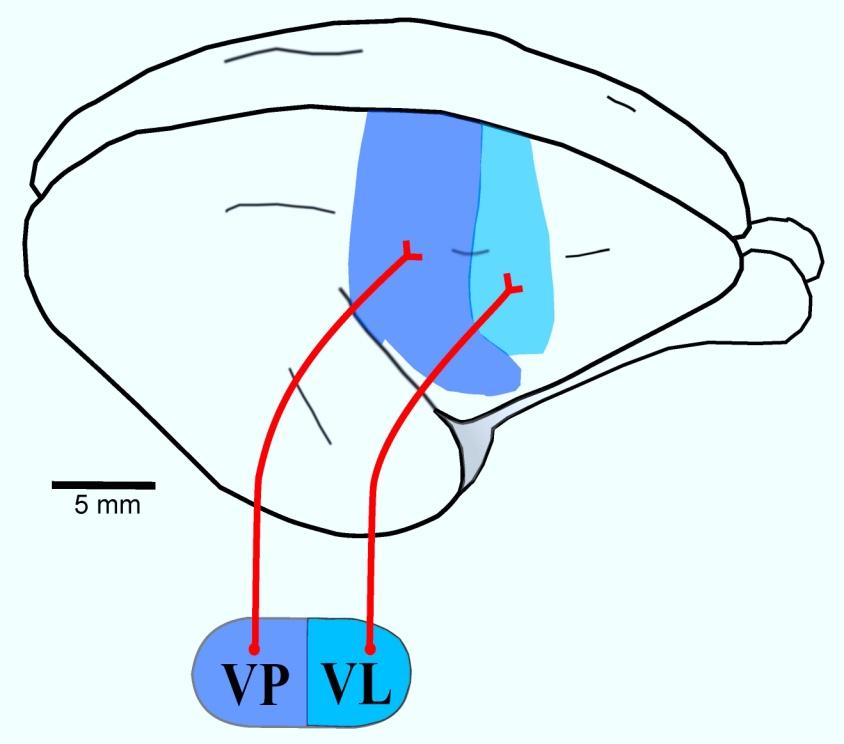
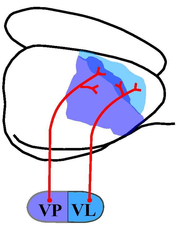
27 From entorhinal cortex to dentate gyrus to CA3 (via mossy fibers) to CA1 (via Schaffer collaterals of CA3 cell axons) to subiculum Fig 28 10a Hippocampus: input through the perforant path (axon 1), then through 3 synapses to the subiculum 27
28 Local circuits in the hippocampus LTP can occur at each synapse in this circuit. 28
29 Evolution of corpus striatum and rest of endbrain: speculations [from class 26] 1. Beginnings: a link between olfactory inputs and motor control: The link becomes Ventral striatum. It was a modifiable link (capable of experience-induced change). Other inputs reached the striatum 2. Non-olfactory inputs invade the striatal integrating mechanisms (via paleothalamic structures). 3. Early expansions of endbrain: striatal and pallial. Non-olfactory inputs to pallium [Note the two pathways going caudally from the olfactory system.] areas 4. Pre-mammalian & then mammalian expansions of cortex and striatum: For the striatum, the earlier outputs and inputs remain as connections with neocortex expand. 29
30 Mesulam, fig. 1-6 In red, the connections from neocortex that most directly influence the autonomic nervous system. The amygdala is a major player in this kind of connection. We focus on the amygdala next. Extrapersonal Space Primary Sensory and Motor Areas IDIOTYPIC CORTEX Modality-Specific (unimodal) Association Areas HOMOTYPICAL ISOCORTEX High-Order (heteromodal) Association Areas temporal pole - caudal orbitofrontal anterior insula-cingulate-parahippocampal PARALIMBIC AREAS septum - s. innominata-amygdala -piriform c.-hippocampus LIMBIC AREAS (CORTICOID + ALLOCORTEX) Hypothalamus Internal Milieu Image by MIT OpenCourseWare. 30

31 Fig

32 Frontal sections: the limbic system of rodent Find the Amygdala, the Stria Terminalis, and the Bed Nucleus of the Stria Terminalis Can you also identify the positions of the fornix fibers from the hippocampal formation? 32
33 Eyes, optic nerves, optic tracts; split optic chiasm Dorsal View Ventral View Fig
34 Major afferents of dorsal striatum: DA axons from the substantia nigra Sensory inputs via the paleothalamus Inputs via the neocortex Globus pallidus Fig 30-2 Image by MIT OpenCourseWare. 34
35 Cortex + Caudate Nucleus More ancient outputs have been added (in blue) Pathways in red are inhibitory, except where indicated by +. Note the "Double Inhibition" Putamen Globus Pallidus + Thalamus (VL,VA) Outputs to Subthalamus & Caudal Midbrain Locomotor Region Substantia Nigra Pars Reticulata Superior Colliculus (for orienting movements) Omits the dopamine reward pathways and the ventral striatum Corticobulbar & Corticospinal Tracts Fig 30-8 Image by MIT OpenCourseWare. 35
36 Dominant inputs to the dorsal striatum in mammals come from the neocortex Supplementary Motor Area Motor Cortex Somatosensory Cortex Topography of cortico striatal projections in primates: Sensorimotor areas to Putamen; Prefrontal areas to Head of Caudate; Posterior areas to Caudate (tail & medial head) Caudate Nucleus (head) Putamen Caudate Nucleus (tail) Fig 30 10a 36 Image by MIT OpenCourseWare.

37 Fig
38 REVIEW: Archetypal embryonic stage Dorsal dc Lateral Medial cx Ventral s s st st dc am cx = neocortex dc = dorsal cortex (pallium) dvr = dorsal ventricular ridge h = hyperpallium s = septum st = striatum Fig 12-2 Frog dvr st Homeobox gene expression: Emx-1 Dlx-1 Evolution of telencephalon based on expression patterns of regulatory genes during development Turtle s dvr h st Chick Mouse Image by MIT OpenCourseWare.
39 Human neocortex: 3 cytoarchitectural methods: The dominant cell type is the pyramidal cell. Is there a consistent pattern of connections of these neocortical cells? Fig 32-1a 39
40 Fig 32-2 Sketch of a column of cells in the neocortex. Note the different types of axons: afferent, efferent, association 40
41 Connections of neurons in the different cortical layers (We described this once before, but less schematically: see fig 32.2.) Layer 1 Layer 2 Layer 3 Layer 4 Layer 5 Layer 6 Omits all lateral interconnections and long-distance projections Why does layer 1 have no outputs? 41
42 The girdle of paralimbic areas: olfactocentric & hippocampocentric (from Mesulam) Figure removed due to copyright restrictions. 42

43 Virginia opossum Brush-tailed possum Rat Galago Fig
44 Hedgehog neocortex, salamander dorsal pallium, turtle dorsal cortex (Striedter p 270) Note the differences in trajectories of axons from the thalamus (in red). Figure removed due to copyright restrictions. 44
45 Fig

46 Fig


47 Hemisphere Lateral view Hemisphere Medial view Brainstem, schematic lateral view Major functional modules of the CNS Fig Sensory- Perceptual Motor Behavior Motivation 47

48 Fig
49 MIT OpenCourseWare Brain Structure and Its Origins Spring 2014 For information about citing these materials or our Terms of Use, visit:
9.14 Class 32 Review. Limbic system
 9.14 Class 32 Review Limbic system 1 Lateral view Medial view Brainstem, sagittal section Sensory- Perceptual Motor Behavior Major functional modules of the CNS Motivation Courtesy of MIT Press. Used with
Visual system invades the endbrain: pathways to striatum and cortex (continued) Why this happened in evolution
 Why this happened in evolution") Visual system invades the endbrain: pathways to striatum and cortex (continued) Why this happened in evolution What were the adaptive advantages? Visual information reaching the striatum directly: Advantages
Visual system invades the endbrain: pathways to striatum and cortex (continued) Why this happened in evolution What were the adaptive advantages? Visual information reaching the striatum directly: Advantages
A sketch of the central nervous system and its origins. MIT 9.14 Classes 31
 A sketch of the central nervous system and its origins G. E. Schneider 2014 Part 9: Hypothalamus & Limbic System MIT 9.14 Classes 31 The vertebrate medial pallium; in mammals: the hippocampal formation
9.14 Qs on post-midterm class sessions and Schneider chapters 18-34
 9.14 Qs on post-midterm class sessions and Schneider chapters 18-34 (Bold font indicates terms you should be able to define.) Ch 18 Ch 19 Ch 20 1. In comparative neuroanatomical studies, the taste system
CNS pathways. topics. The auditory nerve, and the cochlear nuclei of the hindbrain
 CNS pathways topics The auditory nerve, and the cochlear nuclei of the hindbrain Sensory channels of information flow in CNS Pathways to medial geniculate body of thalamus Functional categorization of
The basal forebrain: Questions, chapter 29:
 The basal forebrain: Questions, chapter 29: 7) What is the "basal forebrain", and what is its involvement in Alzheimer' s Disease? The acetylcholine-containing neurons of the nucleus basalis of Meynart
9.14 Classes #21-23: Visual systems
 9.14 Classes #21-23: Visual systems Questions based on Schneider chapter 20 and classes: 1) What was in all likelihood the first functional role of the visual sense? Describe the nature of the most primitive
Thus, there evolved the major types of functions of the neocortical mantle:
 Thus, there evolved the major types of functions of the neocortical mantle: Location sense This set the stage for more and more anticipation of inputs Object sense This also set the stage for more and
Prof. Saeed Abuel Makarem & Dr.Sanaa Alshaarawy
 Prof. Saeed Abuel Makarem & Dr.Sanaa Alshaarawy 1 Objectives By the end of the lecture, you should be able to: Describe the anatomy and main functions of the thalamus. Name and identify different nuclei
From Brodal, 2 nd ed. fig 20.1; 3 rd ed. Fig 21.1
 From Brodal, 2 nd ed. fig 20.1; 3 rd ed. Fig 21.1 Note the names of the layers-- named for cell types rather than for axonal stratification. Figure removed due to copyright restrictions. Please see figure
Psyc 311A, fall 2008 Conference week 3 TA: Jürgen Germann
 Psyc 311A, fall 2008 Conference week 3 TA: Jürgen Germann e-mail: jurgen.germann@mcgill.ca Overview: 1. Meninges 2. Cerebral cortex-cytoarchitecture 3. Diencephalon (thalamus/hypothalamus) (this replaces
LIMBIC SYSTEM. Dr. Amani A. Elfaki Associate Professor Department of Anatomy
 LIMBIC SYSTEM Dr. Amani A. Elfaki Associate Professor Department of Anatomy Learning Objectives Define the limbic system Identify the parts of the limbic system Describe the circulation of the limbic system
MIT 9.14 Class 30 Hormonal and other influences on brain development and plasticity (Limbic system 3) Book chapter 27
 Book chapter 27") A sketch of the central nervous system and its origins G. E. Schneider 2014 Part 9: Hypothalamus & Limbic System MIT 9.14 Class 30 Hormonal and other influences on brain development and plasticity (Limbic
A sketch of the central nervous system and its origins G. E. Schneider 2014 Part 9: Hypothalamus & Limbic System MIT 9.14 Class 30 Hormonal and other influences on brain development and plasticity (Limbic
Orientation, Development, Gross Anatomy, Blood Supply and Meninges References... 3
 Section I Orientation, Development, Gross Anatomy, Blood Supply and Meninges... 1 1 Orientation... 3 References... 3 2 Development... 7 Early Morphogenesis... 7 FormationoftheBrainRegions... 9 Histogenesis...
Outline of the next three lectures
 Outline of the next three lectures Lecture 35 Anatomy of the human cerebral cortex gross and microscopic cell types connections Vascular supply of the cerebral cortex Disorders involving the cerebral cortex
Introduction to the Central Nervous System: Internal Structure
 Introduction to the Central Nervous System: Internal Structure Objective To understand, in general terms, the internal organization of the brain and spinal cord. To understand the 3-dimensional organization
Systems Neuroscience Dan Kiper. Today: Wolfger von der Behrens
 Systems Neuroscience Dan Kiper Today: Wolfger von der Behrens wolfger@ini.ethz.ch 18.9.2018 Neurons Pyramidal neuron by Santiago Ramón y Cajal (1852-1934, Nobel prize with Camillo Golgi in 1906) Neurons
Classes #5-6: Specializations in CNS evolution
 Classes #5-6: Specializations in CNS evolution Questions based on Schneider chapter 5: 1. Does ontogeny really recapitulate phylogeny? What is a phylotypic stage? Explain the terms and discuss the concepts
CEREBRUM & CEREBRAL CORTEX
 CEREBRUM & CEREBRAL CORTEX Seonghan Kim Dept. of Anatomy Inje University, College of Medicine THE BRAIN ANATOMICAL REGIONS A. Cerebrum B. Diencephalon Thalamus Hypothalamus C. Brain Stem Midbrain Pons
P. Hitchcock, Ph.D. Department of Cell and Developmental Biology Kellogg Eye Center. Wednesday, 16 March 2009, 1:00p.m. 2:00p.m.
 Normal CNS, Special Senses, Head and Neck TOPIC: CEREBRAL HEMISPHERES FACULTY: LECTURE: READING: P. Hitchcock, Ph.D. Department of Cell and Developmental Biology Kellogg Eye Center Wednesday, 16 March
Thalamus: VA VM, MD S N. GPi Superior colliculus. compacta reticulata
 . Putamen & Caudate Putamen & Caudate GPe Neocortex Thalamus: VA VM, MD S N GPi Superior colliculus compacta reticulata Substantia Nigra Pedunculopontine nuc. of midbrain ret.form. Satellites of the corpus
The neurvous system senses, interprets, and responds to changes in the environment. Two types of cells makes this possible:
 NERVOUS SYSTEM The neurvous system senses, interprets, and responds to changes in the environment. Two types of cells makes this possible: the neuron and the supporting cells ("glial cells"). Neuron Neurons
Chapter 3. Structure and Function of the Nervous System. Copyright (c) Allyn and Bacon 2004
 Allyn and Bacon 2004") Chapter 3 Structure and Function of the Nervous System 1 Basic Features of the Nervous System Neuraxis: An imaginary line drawn through the center of the length of the central nervous system, from the
Chapter 3 Structure and Function of the Nervous System 1 Basic Features of the Nervous System Neuraxis: An imaginary line drawn through the center of the length of the central nervous system, from the
Brain anatomy and artificial intelligence. L. Andrew Coward Australian National University, Canberra, ACT 0200, Australia
 Brain anatomy and artificial intelligence L. Andrew Coward Australian National University, Canberra, ACT 0200, Australia The Fourth Conference on Artificial General Intelligence August 2011 Architectures
PSY 302: CHAPTER 3 NOTES THE BRAIN (PART II) - 9/5/17. By: Joseline
 - 9/5/17. By: Joseline") PSY 302: CHAPTER 3 NOTES THE BRAIN (PART II) - 9/5/17 By: Joseline Left 3 MAJOR FISSURES : 2HEMISPHERES Right Lateral Ventricle Central Fissure Third Ventricle Sulcus Lateral Fissure Gyros Fissure- Fissures
PSY 302: CHAPTER 3 NOTES THE BRAIN (PART II) - 9/5/17 By: Joseline Left 3 MAJOR FISSURES : 2HEMISPHERES Right Lateral Ventricle Central Fissure Third Ventricle Sulcus Lateral Fissure Gyros Fissure- Fissures
Telencephalon (Cerebral Hemisphere)
") Telencephalon (Cerebral Hemisphere) OUTLINE The Cortex - Lobes, Sulci & Gyri - Functional Subdivisions - Limbic Lobe & Limbic System The Subcortex - Basal Ganglia - White Matter (Internal Capsule) - Relations
Telencephalon (Cerebral Hemisphere) OUTLINE The Cortex - Lobes, Sulci & Gyri - Functional Subdivisions - Limbic Lobe & Limbic System The Subcortex - Basal Ganglia - White Matter (Internal Capsule) - Relations
Chapter 2: Studies of Human Learning and Memory. From Mechanisms of Memory, second edition By J. David Sweatt, Ph.D.
 Chapter 2: Studies of Human Learning and Memory From Mechanisms of Memory, second edition By J. David Sweatt, Ph.D. Medium Spiny Neuron A Current Conception of the major memory systems in the brain Figure
Nsci 2100: Human Neuroanatomy 2017 Examination 3
 Name KEY Lab Section Nsci 2100: Human Neuroanatomy 2017 Examination 3 On this page, write your name and lab section. On your bubble answer sheet, enter your name (last name, space, first name), internet
For more information about how to cite these materials visit
 Author(s): Peter Hitchcock, PH.D., 2009 License: Unless otherwise noted, this material is made available under the terms of the Creative Commons Attribution Non-commercial Share Alike 3.0 License: http://creativecommons.org/licenses/by-nc-sa/3.0/
PROPERTY OF ELSEVIER SAMPLE CONTENT - NOT FINAL. Gross Anatomy and General Organization of the Central Nervous System
 3 Gross Anatomy and General Organization of the Central Nervous System C h a p t e r O u t l i n e The Long Axis of the CNS Bends at the Cephalic Flexure Hemisecting a Brain Reveals Parts of the Diencephalon,
Stanley Pruisinger 1980's
 Neuroanatomy Prion disease cerebellum chapter b/c cerebellar ataxia here as a warning for obvious reasons. Creutzfeldt - Jakob Disease (CJD) "Spongiform" (brain turns to sponge) Jews in Lybia who ate
BASAL GANGLIA. Dr JAMILA EL MEDANY
 BASAL GANGLIA Dr JAMILA EL MEDANY OBJECTIVES At the end of the lecture, the student should be able to: Define basal ganglia and enumerate its components. Enumerate parts of Corpus Striatum and their important
Biological Bases of Behavior. 3: Structure of the Nervous System
 Biological Bases of Behavior 3: Structure of the Nervous System Neuroanatomy Terms The neuraxis is an imaginary line drawn through the spinal cord up to the front of the brain Anatomical directions are
I: To describe the pyramidal and extrapyramidal tracts. II: To discuss the functions of the descending tracts.
 Descending Tracts I: To describe the pyramidal and extrapyramidal tracts. II: To discuss the functions of the descending tracts. III: To define the upper and the lower motor neurons. 1. The corticonuclear
Developmental sequence of brain
 Cerebellum Developmental sequence of brain Fourth week Fifth week Location of cerebellum Lies above and behind the medullar and pons and occupies posterior cranial fossa Location of cerebellum External
Limbic system. Dr Devendra Save
 Limbic system Dr Devendra Save Named by Paul Broca. Introduction Limbic = border (Greek word) It is structure forming border between hypothalamus and cerebral cortex Is functional anatomic system of interconnected
Anatomy of the basal ganglia. Dana Cohen Gonda Brain Research Center, room 410
 Anatomy of the basal ganglia Dana Cohen Gonda Brain Research Center, room 410 danacoh@gmail.com The basal ganglia The nuclei form a small minority of the brain s neuronal population. Little is known about
M555 Medical Neuroscience Lab 1: Gross Anatomy of Brain, Crainal Nerves and Cerebral Blood Vessels
 M555 Medical Neuroscience Lab 1: Gross Anatomy of Brain, Crainal Nerves and Cerebral Blood Vessels Anatomical Directions Terms like dorsal, ventral, and posterior provide a means of locating structures
MITOCW MIT9_14S14_lec35.mp3
 MITOCW MIT9_14S14_lec35.mp3 The following content is provided under a Creative Commons license. Your support will help MIT OpenCourseWare continue to offer high quality educational resources for free.
Cerebral Cortex 1. Sarah Heilbronner
 Cerebral Cortex 1 Sarah Heilbronner heilb028@umn.edu Want to meet? Coffee hour 10-11am Tuesday 11/27 Surdyk s Overview and organization of the cerebral cortex What is the cerebral cortex? Where is each
Vision II. Steven McLoon Department of Neuroscience University of Minnesota
 Vision II Steven McLoon Department of Neuroscience University of Minnesota 1 Ganglion Cells The axons of the retinal ganglion cells form the optic nerve and carry visual information into the brain. 2 Optic
Basal Ganglia. Today s lecture is about Basal Ganglia and it covers:
 Basal Ganglia Motor system is complex interaction between Lower motor neurons (spinal cord and brainstem circuits) and Upper motor neurons (pyramidal and extrapyramidal tracts) plus two main regulators
nucleus accumbens septi hier-259 Nucleus+Accumbens birnlex_727
 Nucleus accumbens From Wikipedia, the free encyclopedia Brain: Nucleus accumbens Nucleus accumbens visible in red. Latin NeuroNames MeSH NeuroLex ID nucleus accumbens septi hier-259 Nucleus+Accumbens birnlex_727
CEREBRUM Dr. Jamila Elmedany Dr. Essam Eldin Salama
 CEREBRUM Dr. Jamila Elmedany Dr. Essam Eldin Salama Objectives At the end of the lecture, the student should be able to: List the parts of the cerebral hemisphere (cortex, medulla, basal nuclei, lateral
Leah Militello, class of 2018
 Leah Militello, class of 2018 Objectives 1. Describe the general organization of cerebral hemispheres. 2. Describe the locations and features of the different functional areas of cortex. 3. Understand
Medical Neuroscience Tutorial
 Pain Pathways Medical Neuroscience Tutorial Pain Pathways MAP TO NEUROSCIENCE CORE CONCEPTS 1 NCC1. The brain is the body's most complex organ. NCC3. Genetically determined circuits are the foundation
Exam 2 PSYC Fall (2 points) Match a brain structure that is located closest to the following portions of the ventricular system
 Match a brain structure that is located closest to the following portions of the ventricular system") Exam 2 PSYC 2022 Fall 1998 (2 points) What 2 nuclei are collectively called the striatum? (2 points) Match a brain structure that is located closest to the following portions of the ventricular system
Exam 2 PSYC 2022 Fall 1998 (2 points) What 2 nuclei are collectively called the striatum? (2 points) Match a brain structure that is located closest to the following portions of the ventricular system
Introduction to Systems Neuroscience. Nov. 28, The limbic system. Daniel C. Kiper
 Introduction to Systems Neuroscience Nov. 28, 2017 The limbic system Daniel C. Kiper kiper@ini.phys.ethz.ch http: www.ini.unizh.ch/~kiper/system_neurosci.html LIMBIC SYSTEM The term limbic system mean
MITOCW MIT9_14S14_lec23.mp3
 MITOCW MIT9_14S14_lec23.mp3 The following content is provided under a Creative Commons license. Your support will help MIT OpenCourseWare continue to offer high quality educational resources for free.
Gross Morphology of the Brain
 Gross Morphology of the Brain Done by : Marah Marahleh & Razan Krishan *slides in bold Principal Parts of the Brain Cerebrum : largest part of the brain Diencephalon Thalamus & hypothalamus Cerebellum
Lawrence & Kuypers, 1968:
 Lawrence & Kuypers, 1968: Lesion #2: destruction of medial brainstem pathways (added to pyramidotomy) Defective axial control: Righting: only after 10-40 days Falling: failure to elicit the usual corrective
Anatomy of the Hippocampus
 Anatomy of the Hippocampus Lecture 3.2 David S. Touretzky September, 2015 Human Hippocampus 2 Human Hippocampus 3 Hippocampus Means Seahorse Dissected human hippocampus next to a specimen of hippocampus
10/3/2016. T1 Anatomical structures are clearly identified, white matter (which has a high fat content) appears bright.
 appears bright.") H2O -2 atoms of Hydrogen, 1 of Oxygen Hydrogen just has one single proton and orbited by one single electron Proton has a magnetic moment similar to the earths magnetic pole Also similar to earth in that
H2O -2 atoms of Hydrogen, 1 of Oxygen Hydrogen just has one single proton and orbited by one single electron Proton has a magnetic moment similar to the earths magnetic pole Also similar to earth in that
Cephalization. Nervous Systems Chapter 49 11/10/2013. Nervous systems consist of circuits of neurons and supporting cells
 Nervous Systems Chapter 49 Cephalization Nervous systems consist of circuits of neurons and supporting cells Nervous system organization usually correlates with lifestyle Organization of the vertebrate
For more information about how to cite these materials visit
 Author(s): Peter Hitchcock, PH.D., 2009 License: Unless otherwise noted, this material is made available under the terms of the Creative Commons Attribution Non-commercial Share Alike 3.0 License: http://creativecommons.org/licenses/by-nc-sa3.0/
Human Anatomy. Brain and Cranial Nerves
 Human Anatomy Brain and Cranial Nerves 1 Brain and Cranial Nerves An adult brain weighs between 1.35 and 1.4 kilograms (kg) (around 3 pounds) and has a volume of about 1200 cubic centimeters (cc). Brain
Anatomy and Physiology (Bio 220) The Brain Chapter 14 and select portions of Chapter 16
 The Brain Chapter 14 and select portions of Chapter 16") Anatomy and Physiology (Bio 220) The Brain Chapter 14 and select portions of Chapter 16 I. Introduction A. Appearance 1. physical 2. weight 3. relative weight B. Major parts of the brain 1. cerebrum 2.
Anatomy and Physiology (Bio 220) The Brain Chapter 14 and select portions of Chapter 16 I. Introduction A. Appearance 1. physical 2. weight 3. relative weight B. Major parts of the brain 1. cerebrum 2.
神經解剖學 NEUROANATOMY BASAL NUCLEI 盧家鋒助理教授臺北醫學大學醫學系解剖學暨細胞生物學科臺北醫學大學醫學院轉譯影像研究中心.
 神經解剖學 NEUROANATOMY BASAL NUCLEI 盧家鋒助理教授臺北醫學大學醫學系解剖學暨細胞生物學科臺北醫學大學醫學院轉譯影像研究中心 http://www.ym.edu.tw/~cflu OUTLINE Components and Pathways of the Basal Nuclei Functions and Related Disorders of the Basal Nuclei
Overview of Brain Structures
 First Overview of Brain Structures Psychology 470 Introduction to Chemical Additions Steven E. Meier, Ph.D. All parts are interrelated. You need all parts to function normally. Neurons = Nerve cells Listen
Auditory and Vestibular Systems
 Auditory and Vestibular Systems Objective To learn the functional organization of the auditory and vestibular systems To understand how one can use changes in auditory function following injury to localize
Human Brain and Senses October 13, 2008 Page 1. Examination of the Human Brain
 Human Brain and Senses October 13, 2008 Page 1 Examination of the Human Brain With only a few hours today we can only begin to scratch the surface of a complex subject like neuroanatomy. The purpose of
Limbic system. Lecture 29, November 10, 2017
 Limbic system Lecture 29, November 10, 2017 Circadian rhythms (Latin, approximately a day ) Regulation of our daily rhythm Eating Sleeping Defecating Periods of activity Suprachiasmatic n. http://slideplayer.com/slide/6351731/
A. General features of the basal ganglia, one of our 3 major motor control centers:
 Reading: Waxman pp. 141-146 are not very helpful! Computer Resources: HyperBrain, Chapter 12 Dental Neuroanatomy Suzanne S. Stensaas, Ph.D. March 1, 2012 THE BASAL GANGLIA Objectives: 1. What are the main
The Central Nervous System I. Chapter 12
 The Central Nervous System I Chapter 12 The Central Nervous System The Brain and Spinal Cord Contained within the Axial Skeleton Brain Regions and Organization Medical Scheme (4 regions) 1. Cerebral Hemispheres
Brainstem: Midbrain. 1. Midbrain gross external anatomy 2. Internal structure of the midbrain:
 Brainstem: Midbrain 1. Midbrain gross external anatomy 2. Internal structure of the midbrain: cerebral peduncles tegmentum tectum (guadrigeminal plate) Midbrain Midbrain general features location between
THE CENTRAL NERVOUS SYSTEM. The Brain & Spinal Cord
 THE CENTRAL NERVOUS SYSTEM The Brain & Spinal Cord Review: Nervous System Parallel Distributed Processing Composition of the CNS Nuclei: Clusters of neurons in the CNS ( neighborhoods ) Fiber Tracts/Pathways:
A. General features of the basal ganglia, one of our 3 major motor control centers:
 Reading: Waxman pp. 141-146 are not very helpful! Computer Resources: HyperBrain, Chapter 12 Dental Neuroanatomy Suzanne S. Stensaas, Ph.D. April 22, 2010 THE BASAL GANGLIA Objectives: 1. What are the
Course Booklet. We have felt the pain that Neuroscience is giving you.
 Exams Stressing You Out? Take Action! Course Booklet NEUR 1202 Carleton University* *TranscendFinals is not affiliated with the university We have felt the pain that Neuroscience is giving you. Our mission
Brainstem. Steven McLoon Department of Neuroscience University of Minnesota
 Brainstem Steven McLoon Department of Neuroscience University of Minnesota 1 Course News Change in Lab Sequence Week of Oct 2 Lab 5 Week of Oct 9 Lab 4 2 Goal Today Know the regions of the brainstem. Know
Group D: Central nervous system yellow
 Group D: Central nervous system yellow Central nervous system 1. General structure of nervous system (neuron, glia, synapsis, mediators, receptors) Main points: types of neurons and glial cells, synapses,
Neuroanatomy. Dr. Maha ELBeltagy. Assistant Professor of Anatomy Faculty of Medicine The University of Jordan
 Neuroanatomy Dr. Maha ELBeltagy Assistant Professor of Anatomy Faculty of Medicine The University of Jordan 2018 Prof Yousry 10/15/17 Types of brain fibers THE WHITE MATTER OF THE BRAIN The white matter
Motor Functions of Cerebral Cortex
 Motor Functions of Cerebral Cortex I: To list the functions of different cortical laminae II: To describe the four motor areas of the cerebral cortex. III: To discuss the functions and dysfunctions of
Sensory systems. Taste/gustatory
 Sensory systems Taste/gustatory Sensory systems basic concepts Modality of Sensation Receptor Sensory Tract primary neuron secondary neuron tertiary neuron termination Receptors of sensory systems - primary
Parts of the Brain. Hindbrain. Controls autonomic functions Breathing, Heartbeat, Blood pressure, Swallowing, Vomiting, etc. Upper part of hindbrain
 Parts of the Brain The human brain is made up of three main parts: 1) Hindbrain (or brainstem) Which is made up of: Myelencephalon Metencephalon 2) Midbrain Which is made up of: Mesencephalon 3) Forebrain
Biological Bases of Behavior. 8: Control of Movement
 Biological Bases of Behavior 8: Control of Movement m d Skeletal Muscle Movements of our body are accomplished by contraction of the skeletal muscles Flexion: contraction of a flexor muscle draws in a
Neural Communication. Central Nervous System Peripheral Nervous System. Communication in the Nervous System. 4 Common Components of a Neuron
 Neural Communication Overview of CNS / PNS Electrical Signaling Chemical Signaling Central Nervous System Peripheral Nervous System Somatic = sensory & motor Autonomic = arousal state Parasympathetic =
Study Guide Unit 2 Psych 2022, Fall 2003
 Study Guide Unit 2 Psych 2022, Fall 2003 Subcortical Anatomy 1. Be able to locate the following structures and be able to indicate whether they are located in the forebrain, diencephalon, midbrain, pons,
Department of Human Anatomy GUIDELINES. nuclei. The lateral ventricles. White substance of cerebral hemispheres. course 1
 Department of Human Anatomy GUIDELINES Academic discipline Human Anatomy Module 2 Content module 11 Study subject The olfactory brain. Basal nuclei. The lateral ventricles. White substance of cerebral
The Nervous system is divided into 2 major divisions: 1) Central Nervous System (CNS): found within bones & consists of:
 Central Nervous System (CNS): found within bones & consists of:") The Nervous system is divided into 2 major divisions: 1) Central Nervous System (CNS): found within bones & consists of: - The Brain: within the skull, composed of cerebrum, cerebellum and brain stem.
The Nervous system is divided into 2 major divisions: 1) Central Nervous System (CNS): found within bones & consists of: - The Brain: within the skull, composed of cerebrum, cerebellum and brain stem.
Internal Organisation of the Brainstem
 Internal Organisation of the Brainstem Major tracts and nuclei of the brainstem (Notes) The brainstem is the major pathway for tracts and houses major nuclei, that contain sensory, motor and autonomics
Brain Mechanisms of Emotion 1 of 6
 Brain Mechanisms of Emotion 1 of 6 I. WHAT IS AN EMOTION? A. Three components (Oately & Jenkins, 1996) 1. caused by conscious or unconscious evaluation of an event as relevant to a goal that is important
Overview of the Nervous System (some basic concepts) Steven McLoon Department of Neuroscience University of Minnesota
 Steven McLoon Department of Neuroscience University of Minnesota") Overview of the Nervous System (some basic concepts) Steven McLoon Department of Neuroscience University of Minnesota 1 Coffee Hour Tuesday (Sept 11) 10:00-11:00am Friday (Sept 14) 8:30-9:30am Surdyk s
Overview of the Nervous System (some basic concepts) Steven McLoon Department of Neuroscience University of Minnesota 1 Coffee Hour Tuesday (Sept 11) 10:00-11:00am Friday (Sept 14) 8:30-9:30am Surdyk s
Property of Chonghao Zhao, MD, Ph.D.
 UCLA Orofacial Pain Lecture Series # 2 March 17 th 2011 Overview of Basal Ganglion, Thalamus, Hypothalamus, Brainstem, and Spinal Cord Neuroanatomy and MR Anatomy Chong-hao Zhao, MD, PhD American Board
Nervous System, Neuroanatomy, Neurotransmitters
 Nervous System, Neuroanatomy, Neurotransmitters Neurons Structure of neurons Soma Dendrites Spines Axon Myelin Nodes of Ranvier Neurons Structure of neurons Axon collaterals 1 Neurons Structure of neurons
PSYCH 260 Exam 2. March 2, Answer the questions using the Scantron form. Name:
 PSYCH 260 Exam 2 March 2, 2017 Answer the questions using the Scantron form. Name: 1 1 Main Please put in their proper order the steps that lead to synaptic communication between neurons. Begin with the
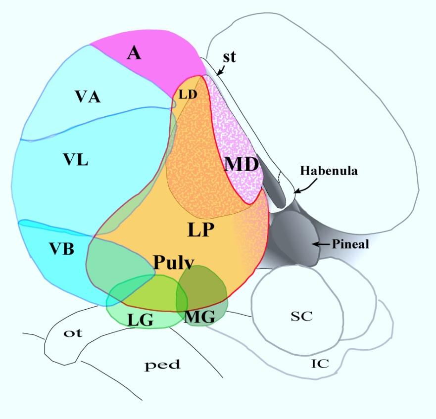
Thalamic nuclei. Each thalamus has several well defined borders: Introduction. Thalamus
 Thalamic nuclei Introduction For the successful completion of any task, some sort of recognition, identification and organisation is needed. Imagine what would happen if employees in a team would just
Basal Ganglia. Introduction. Basal Ganglia at a Glance. Role of the BG
 Basal Ganglia Shepherd (2004) Chapter 9 Charles J. Wilson Instructor: Yoonsuck Choe; CPSC 644 Cortical Networks Introduction A set of nuclei in the forebrain and midbrain area in mammals, birds, and reptiles.
Laboratory Manual for Comparative Anatomy and Physiology Figure 15.1 Transparency Master 114
 Neuron Capillary Astrocyte Microglial cell Neuron Fluid-filled cavity Process of oligodendrocyte Ependymal cells Brain or spinal cord tissue Myelin sheath Nerve fibers Figure 15.1 Transparency Master 114
SOMATIC SENSATION PART I: ALS ANTEROLATERAL SYSTEM (or SPINOTHALAMIC SYSTEM) FOR PAIN AND TEMPERATURE
 FOR PAIN AND TEMPERATURE") Dental Neuroanatomy Thursday, February 3, 2011 Suzanne S. Stensaas, PhD SOMATIC SENSATION PART I: ALS ANTEROLATERAL SYSTEM (or SPINOTHALAMIC SYSTEM) FOR PAIN AND TEMPERATURE Reading: Waxman 26 th ed, :
Dental Neuroanatomy Thursday, February 3, 2011 Suzanne S. Stensaas, PhD SOMATIC SENSATION PART I: ALS ANTEROLATERAL SYSTEM (or SPINOTHALAMIC SYSTEM) FOR PAIN AND TEMPERATURE Reading: Waxman 26 th ed, :
THE CENTRAL NERVOUS SYSTE M
 THE CENTRAL NERVOUS SYSTE M Structure and Functio n THIRD EDITIO N PER BRODAL A Brief Survey, x i Studying the Structures and Function of the Nervous System, xii i Animal Experiments Crucial for Progress,
b. The groove between the two crests is called 2. The neural folds move toward each other & the fuse to create a
 Chapter 13: Brain and Cranial Nerves I. Development of the CNS A. The CNS begins as a flat plate called the B. The process proceeds as: 1. The lateral sides of the become elevated as waves called a. The
biological psychology, p. 40 The study of the nervous system, especially the brain. neuroscience, p. 40
 biological psychology, p. 40 The specialized branch of psychology that studies the relationship between behavior and bodily processes and system; also called biopsychology or psychobiology. neuroscience,
Chemical Control of Behavior and Brain 1 of 9
 Chemical Control of Behavior and Brain 1 of 9 I) INTRO A) Nervous system discussed so far 1) Specific 2) Fast B) Other systems extended in space and time 1) Nonspecific 2) Slow C) Three components that
Page 1 L 58. The University of Connecticut Schools of Medicine and Dental Medicine Humans Systems: Organ Systems /2013 RETICULAR FORMATION
 Page 1 L 58 Douglas L. Oliver, Ph.D. The University of Connecticut Schools of Medicine and Dental Medicine Humans Systems: Organ Systems 1 2012/2013 RETICULAR FORMATION Lecture Lecture: Douglas Oliver
The Nervous System: Sensory and Motor Tracts of the Spinal Cord
 15 The Nervous System: Sensory and Motor Tracts of the Spinal Cord PowerPoint Lecture Presentations prepared by Steven Bassett Southeast Community College Lincoln, Nebraska Introduction Millions of sensory
The Neuroscience of Music in Therapy
 Course Objectives The Neuroscience of Music in Therapy Unit I. Learn Basic Brain Information Unit II. Music in the Brain; Why Music Works Unit III. Considerations for Populations a. Rehabilitation b. Habilitation
Detailed protocol Only dissected human brain samples are stored. The microdissection is performed on frozen brains and the samples are kept on -70 C.
 2008 Detailed protocol Only dissected human brain samples are stored. The microdissection is performed on frozen brains and the samples are kept on -70 C. BrainNet Europe II Project Co-ordinator: Prof.
Department of Cognitive Science UCSD
 Department of Cognitive Science UCSD Verse 1: Neocortex, frontal lobe, Brain stem, brain stem, Hippocampus, neural node, Right hemisphere, Pons and cortex visual, Brain stem, brain stem, Sylvian fissure,
Chapter 8. Control of movement
 Chapter 8 Control of movement 1st Type: Skeletal Muscle Skeletal Muscle: Ones that moves us Muscles contract, limb flex Flexion: a movement of a limb that tends to bend its joints, contraction of a flexor
CEREBRUM. Dr. Jamila EL Medany
 CEREBRUM Dr. Jamila EL Medany Objectives At the end of the lecture, the student should be able to: List the parts of the cerebral hemisphere (cortex, medulla, basal nuclei, lateral ventricle). Describe