Lateralization of Function. Human Brain
|
|
|
- Eugene Ward
- 5 years ago
- Views:
Transcription


1 Lateralization of Function Dr. Coulson Cognitive Science Department UCSD Human Brain An extension of the spinal cord 1


2 Cerebral Hemispheres Corpus Callosum 2


3 Cartoon View of Brain Cerebral Lobes 3


4 Neurons Brain composed of neurons 100 billion Neurons both send and receive signals to other cells in form of pulses Important parts Cell body Axon Synapse Connectivity Each neuron connected to 10,000 other neurons Point of contact is the synapse Computing power of brain comes from connections 4

5 Cortex Two millimeters thick and has area of 1.5 square meters Cartoon View: Frontal Lobe In front of central sulcus Decisions, judgments, emotions 5
6 Cartoon View: Parietal Lobe Behind central sulcus Perception of stimuli related to touch, pressure, temperature, pain Cartoon View: Temporal Lobe Below lateral fissure Perception, recognition, auditory processing 6


7 Cartoon View: Occipital Lobe Located at back of brain, behind the parietal lobe and temporal lobe Vision Lateralization of Function One side of the brain is more crucial for a given function and/or more efficient at the underlying computational tasks Typically a matter of degree Strongly vs. Weakly Lateralized Motor control a good example of a lateralized function 7


8 Sensorimotor Cortex 8

9 Motor Control What about language? Language is a paradigmatic example of a lateralized cognitive phenomenon 9


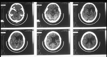
10 Wada Test Lateralization of Function Most evidence of lateralized brain function comes from observing how brain damage affects behavior on various sorts of cognitive tasks 10
 Only utterance was tan Broca s Discovery")


11 Paul Broca 19 th century French neurologist Star patient: Leborgne Understood most of what was said to him Able to eat, drink (move mouth and tongue) Only utterance was tan Broca s Discovery Leborgne s brain had damage to the lower rear portion of frontal lobe, lower front portion of parietal lobe, and upper part of the temporal lobe Broca deemed frontal lobe damage most important Aphasia partial or total loss of ability to articulate ideas due to brain damage Broca s Area lower rear portion of frontal lobe, adjacent to motor cortex Inferior frontal gyrus Brodmann s Areas 44/45 11
 Broca s Aphasia M.E. Cinderella...poor...um 'dopted her...scrubbed floor, um, tidy...poor, um...'dopted...si-sisters and mother.")
12 Brodmann s Areas Korbinian Brodmann examined brain cells with various stains designed to detect chemical differences between areas Brain areas defined by cytoarchitectonic characteristics known as Brodmann s Areas 52 areas in the human brain (though some subdivided into a, b, etc) Broca s Aphasia M.E. Cinderella...poor...um 'dopted her...scrubbed floor, um, tidy...poor, um...'dopted...si-sisters and mother...ball. Ball, prince um, shoe... Examiner Keep going. M.E. Scrubbed and uh washed and un...tidy, uh, sisters and mother, prince, no, prince, yes. Cinderella hooked prince. (Laughs.) Um, um, shoes, um, twelve o'clock ball, finished. Examiner So what happened in the end? M.E. Married. Examiner How does he find her? M.E. Um, Prince, um, happen to, um...prince, and Cinderalla meet, um met um met. Examiner What happened at the ball? They didn't get married at the ball. M.E. No, um, no...i don't know. Shoe, um found shoe... 12

13 Wernicke s Aphasia 1871 Karl Wernicke reported a different sort of language disorder Symptoms Talk fluently, excessively Use made up words Don t understand, in spite of intact hearing Wernicke s Area 13
14 Wernicke s Area Wernicke s Aphasic C.B. Uh, well this is the... the /dodu/ of this. This and this and this and this. These things going in there like that. This is /sen/ things here. This one here, these two things here. And the other one here, back in this one, this one /gesh/ look at this one. Examiner: Yeah, what's happening there? C.B. I can't tell you what that is, but I know what it is, but I don't know where it is. But I don't know what's under. I know it's you couldn't say it's... I couldn't say what it is. I couldn't say what that is. This shu-- that should be right in here. That's very bad in there. Anyway, this one here, and that, and that's it. This is the getting in here and that's the getting around here, and that, and that's it. This is getting in here and that's the getting around here, this one and one with this one. And this one, and that's it, isn't it? I don't know what else you'd want. Describing a picture of a child taking a cookie 14

15 Pop Quiz Pop Quiz 15


16 Sex Differences Women more vulnerable to aphasia after damage to frontal lobe Men more vulnerable to aphasia after damage to parietal and temporal lobe areas Similar sex differences in apraxia, impairment in voluntary motions Wernicke-Geschwind Model Broca s Area stores motor representation of speech Wernicke s Area stores auditory representation of speech sounds Connected by fiber tract known as arcuate fasiculus Considered an oversimplified model 16


17 Wernicke-Geschwind Model: Repeating a Spoken Word Reading a Written Word 17
18 Concepts Ventral prefrontal X cortex Motor word Comprehension Arcuate Fasciculus Association Cortex Posterior Temporal Auditory word Cortex Comprehension Speech motor output Auditory input Broca s Aphasia psychology.rutgers.edu/~rypma/ Ventral prefrontal cortex Motor word Comprehension Concepts Arcuate Fasciculus Association Cortex XPosterior Temporal Auditory word Cortex Comprehension Speech motor output Auditory input Wernicke s Aphasia psychology.rutgers.edu/~rypma/ 18
19 Ventral prefrontal cortex Motor word Comprehension Concepts XArcuate Fasciculus Association Cortex Posterior Temporal Auditory word Cortex Comprehension Speech motor output Auditory input Conduction Aphasia psychology.rutgers.edu/~rypma/ Reprise Wada Test Broca s Aphasia Wernicke s Aphasia Conduction Aphasia But remember, these models are cartoons 19


20 Electrocortical Stimulation Cheesy Demo 20


21 Where does stimulation interrupt naming? Representation of Language in Bilinguals 21


22 If language is left, what is right? President Woodrow Wilson Suffered RH stroke during Versailles Peace Conference after WWI Participants noticed nothing wrong with him BUT his personality seemed to change overnight From friendly and conciliatory to unpleasant and vindictive Woodrow Wilson Weeks later he suffered another stroke that resulted in paralysis of the left side of his body Which side did this stroke affect? He denied there was anything wrong with him Issued press release saying he hurt his left arm in a fall Anosagnosia Wife & close advisors hid his medical problems from public and ran shadow government 22

23 Right Hemisphere Damage Anosagnosia Why can t you move your hand? Somebody has ahold of it. I think it s the weather. I could warm it up and it would be alright. I have a shirt on. Why can t you walk? I could walk at home, but not here. It s slippery here. 23
24 Abnormal Body Image Patients may deny that their left hand is their own hand and wonder why someone else in in bed with them Hemineglect Inability to attend to objects (even one s own body) on one side of space Typically left side of space after right parietal damage 24


25 Unilateral (left) Neglect Right parietal lesion Neglect = Failure to report, respond, or even orient to stimuli on the contralateral side of the body 25

26 Draw the face of a clock, put in all of the numbers and set the hands for 10 after 11 Neglect 26

 Preserved visual")

27 Anton Radershceidt Neglect & Mental Imagery Asked to imagine the Piazza del Duomo in Milan from two different vantage points, a neglect patient describes different parts of the square -- Bisiach & Luzzatti (1978) Preserved visual knowledge and ability to visualize from different perspectives, but mental image lacks detail about the left half of space in each case! 27


28 Functional imaging: Right parietal neglect occurs because the left parietal lobe does not have a map of left visual field Only R side active Posner and Raichle,


29 Dressing Apraxia Patients w/large RH stroke have trouble getting arms into sleeves and legs into pants Know what they re supposed to do, but unable to do it due to defective body image What about sign language? Language, but body image and spatial relationships very important for understanding Worse with LH or RH damage? LH damage results in aphasia in signers, while RH damage leads to visuo-spatial deficits but largely intact language 29

30 Visuospatial Ability in Aphasic & Non-Aphasic Signers fmri: Spoken vs. Signed Language 30
31 Summary LH damage Communicative disorders Frontal damage leads to expressive disorders, trouble with grammatical complexity Posterior damage leads to receptive disorders, trouble with meaning RH damage Anosagnosia Body image disorder Hemineglect Electrocortical Stimulation Naming disruption w/stimulation in LH, not typically RH Exact locale varies widely from individual to individual Different languages disrupted at slightly different sites in cortex Thanks To contact Prof. Coulson: 31
Human Brain. Lateralization of Function. An extension of the spinal cord. Dr. Coulson Cognitive Science Department UCSD
 Lateralization of Function Human Brain An extension of the spinal cord Dr. Coulson Cognitive Science Department UCSD Cerebral Hemispheres Corpus Callosum Cerebral Lobes Neurons Brain composed of neurons
Human Brain. Lateralization of Function. Cortex. Cerebral Hemispheres. An extension of the spinal cord. Dr. Coulson Cognitive Science Department UCSD
 Lateralization of Function Human Brain An extension of the spinal cord Dr. Coulson Cognitive Science Department UCSD Cerebral Hemispheres Two millimeters thick and has area of 1.5 square meters Corpus
Lateralization of Function. Dr. Coulson Cognitive Science Department UCSD
 Lateralization of Function Dr. Coulson Cognitive Science Department UCSD Human Brain An extension of the spinal cord Cortex Two millimeters thick and has area of 1.5 square meters Cerebral Hemispheres
Higher Cortical Function
 Emilie O Neill, class of 2016 Higher Cortical Function Objectives Describe the association cortical areas processing sensory, motor, executive, language, and emotion/memory information (know general location
Learning Objectives.
 Emilie O Neill, class of 2016 Learning Objectives 1. Describe the types of deficits that occur with lesions in association areas including: prosopagnosia, neglect, aphasias, agnosia, apraxia 2. Discuss
Cognitive Neuroscience Cortical Hemispheres Attention Language
 Cognitive Neuroscience Cortical Hemispheres Attention Language Based on: Chapter 18 and 19, Breedlove, Watson, Rosenzweig, 6e/7e. Cerebral Cortex Brain s most complex area with billions of neurons and
PSY 215 Lecture 17 (3/28/2010) (Lateralization in the Brain) Dr. Achtman PSY 215
 (Lateralization in the Brain) Dr. Achtman PSY 215") PSY 215 Lecture 17 Topic: Lateralization in the Brain Chapter 14.1, pages 403-414 Corrections: Lecture 16 (page 4) Broca s Area: trouble producing language, comprehension is okay. Announcements: Review
PSY 215 Lecture 17 Topic: Lateralization in the Brain Chapter 14.1, pages 403-414 Corrections: Lecture 16 (page 4) Broca s Area: trouble producing language, comprehension is okay. Announcements: Review
Neocortex. Hemispheres 9/22/2010. Psychology 472 Pharmacology of Psychoactive Drugs. Structures are divided into several section or lobes.
 Neocortex Psychology 472 Pharmacology of Psychoactive Drugs 1 Is the most developed in Humans Has many folds and fissures The folds of tissue are called gyri or a gyrus (single) The fissures or valleys
Homework Week 2. PreLab 2 HW #2 Synapses (Page 1 in the HW Section)
") Homework Week 2 Due in Lab PreLab 2 HW #2 Synapses (Page 1 in the HW Section) Reminders No class next Monday Quiz 1 is @ 5:30pm on Tuesday, 1/22/13 Study guide posted under Study Aids section of website
Homework Week 2 Due in Lab PreLab 2 HW #2 Synapses (Page 1 in the HW Section) Reminders No class next Monday Quiz 1 is @ 5:30pm on Tuesday, 1/22/13 Study guide posted under Study Aids section of website
Exam 1 PSYC Fall 1998
 Exam 1 PSYC 2022 Fall 1998 (2 points) Briefly describe the difference between a dualistic and a materialistic explanation of brain-mind relationships. (1 point) True or False. George Berkely was a monist.
Introduction to Physiological Psychology Review
 Introduction to Physiological Psychology Review ksweeney@cogsci.ucsd.edu www.cogsci.ucsd.edu/~ksweeney/psy260.html n Learning and Memory n Human Communication n Emotion 1 What is memory? n Working Memory:
The Nervous System. Divisions of the Nervous System. Branches of the Autonomic Nervous System. Central versus Peripheral
 The Nervous System Divisions of the Nervous System Central versus Peripheral Central Brain and spinal cord Peripheral Everything else Somatic versus Autonomic Somatic Nerves serving conscious sensations
Brain anatomy tutorial. Dr. Michal Ben-Shachar 459 Neurolinguistics
 Brain anatomy tutorial Dr. Michal Ben-Shachar 459 Neurolinguistics The human brain Left hemisphere Right hemisphere http://www.brainmuseum.org/ Zoom out Zoom in Types of Brain Tissue Gray Matter: Cell
fmri (functional MRI)
") Lesion fmri (functional MRI) Electroencephalogram (EEG) Brainstem CT (computed tomography) Scan Medulla PET (positron emission tomography) Scan Reticular Formation MRI (magnetic resonance imaging) Thalamus
Lesion fmri (functional MRI) Electroencephalogram (EEG) Brainstem CT (computed tomography) Scan Medulla PET (positron emission tomography) Scan Reticular Formation MRI (magnetic resonance imaging) Thalamus
Announcement. Danny to schedule a time if you are interested.
 Announcement If you need more experiments to participate in, contact Danny Sanchez (dsanchez@ucsd.edu) make sure to tell him that you are from LIGN171, so he will let me know about your credit (1 point).
Myers Psychology for AP*
 Myers Psychology for AP* David G. Myers PowerPoint Presentation Slides by Kent Korek Germantown High School Worth Publishers, 2010 *AP is a trademark registered and/or owned by the College Board, which
Overview of Brain Structures
 First Overview of Brain Structures Psychology 470 Introduction to Chemical Additions Steven E. Meier, Ph.D. All parts are interrelated. You need all parts to function normally. Neurons = Nerve cells Listen
The human brain. of cognition need to make sense gives the structure of the brain (duh). ! What is the basic physiology of this organ?
. ! What is the basic physiology of this organ?") The human brain The human brain! What is the basic physiology of this organ?! Understanding the parts of this organ provides a hypothesis space for its function perhaps different parts perform different
The human brain The human brain! What is the basic physiology of this organ?! Understanding the parts of this organ provides a hypothesis space for its function perhaps different parts perform different
Lecture 35 Association Cortices and Hemispheric Asymmetries -- M. Goldberg
 Lecture 35 Association Cortices and Hemispheric Asymmetries -- M. Goldberg The concept that different parts of the brain did different things started with Spurzheim and Gall, whose phrenology became quite
Functional Neuroanatomy. IBRO ISN African Neuroscience School 4-13 th Dec 2014 Nairobi, Kenya
 Functional Neuroanatomy IBRO ISN African Neuroscience School 4-13 th Dec 2014 Nairobi, Kenya What is/are the function(s) of the nervous system? Sensation Perception Visceral activities (Homeostasis) Behavior
Hemispheric Specialization (lateralization) Each lobe of the brain has specialized functions (Have to be careful with this one.)
 Each lobe of the brain has specialized functions (Have to be careful with this one.)") Cerebral Cortex Principles contralaterality the right half of your brain controls the left half of your body and vice versa. (contralateral control.) Localization of function Specific mental processes
Cerebral Cortex Principles contralaterality the right half of your brain controls the left half of your body and vice versa. (contralateral control.) Localization of function Specific mental processes
The Nervous System and the Endocrine System
 The Nervous System and the Endocrine System Neurons: The Building Blocks of the Nervous System Nervous System The electrochemical communication system of the body Sends messages from the brain to the
Chapter 2 Test. 1. Evolutionary structures within the are the most primitive. *a. hindbrain b. thalamus c. forebrain d. midbrain e.
 Cognitive Psychology In and Out of the Laboratory 5th Edition Galotti TEST BANK Full clear download (no formatting errors) at: https://testbankreal.com/download/cognitive-psychology-laboratory-5thedition-galotti-test-bank/
P. Hitchcock, Ph.D. Department of Cell and Developmental Biology Kellogg Eye Center. Wednesday, 16 March 2009, 1:00p.m. 2:00p.m.
 Normal CNS, Special Senses, Head and Neck TOPIC: CEREBRAL HEMISPHERES FACULTY: LECTURE: READING: P. Hitchcock, Ph.D. Department of Cell and Developmental Biology Kellogg Eye Center Wednesday, 16 March
25/09/2012. Capgras Syndrome. Chapter 2. Capgras Syndrome - 2. The Neural Basis of Cognition
 Chapter 2 The Neural Basis of Cognition Capgras Syndrome Alzheimer s patients & others delusion that significant others are robots or impersonators - paranoia Two brain systems for facial recognition -
Review of Week 2. COGS1 Spring 2019
 Review of Week 2 COGS1 Spring 2019 Quiz B in section during week 3 Quiz B will be on week 2 reading and lecture material. Sign up on Piazza!!! Coulson Lateralization and Aphasia What does lateralization
Cerebrum-Cerebral Hemispheres. Cuneyt Mirzanli Istanbul Gelisim University
 Cerebrum-Cerebral Hemispheres Cuneyt Mirzanli Istanbul Gelisim University The largest part of the brain. Ovoid shape. Two incompletely separated cerebral hemispheres. The outer surface of the cerebral
2Lesson. Outline 1.5. Lesson Plan. The OVERVIEW. Lesson 1.5: How do the parts of my brain work together? LESSON. Unit1.2
 Outline 2Lesson Unit1.2 OVERVIEW Rationale: This lesson is intended to emphasize the need of the structures of the brain to work together to create and control behaviors, like the creation and comprehension
Chapter 14, Part 2! The Cerebrum and Cranial Nerves! pp !
 Chapter 14, Part 2! The Cerebrum and Cranial pp. 482 505! SECTION 14-9! The cerebrum, the largest region of the brain, contains motor, sensory, and association areas! 2! 1! ! Chapter 14 Part 2 Brain/Cranial
The origins of localization
 Association Cortex, Asymmetries, and Cortical Localization of Affective and Cognitive Functions Michael E. Goldberg, M.D. The origins of localization The concept that different parts of the brain did different
PsychoBrain. 31 st January Dr Christos Pliatsikas. Lecturer in Psycholinguistics in Bi-/Multilinguals University of Reading
 PsychoBrain 31 st January 2018 Dr Christos Pliatsikas Lecturer in Psycholinguistics in Bi-/Multilinguals University of Reading By the end of today s lecture you will understand Structure and function of
Introduction to the Nervous System. Code: HMP 100/ UPC 103/ VNP 100. Course: Medical Physiology. Level 1 MBChB/BDS/BPharm
 Introduction to the Nervous System. Code: HMP 100/ UPC 103/ VNP 100. Course: Medical Physiology Level 1 MBChB/BDS/BPharm Lecture 2. Functional Organisation of the Nervous System Lecture Outline 1.1 Introduction
-Zeina Assaf. -Omar Odeh. - Maha Beltagy
 -3 -Zeina Assaf -Omar Odeh - Maha Beltagy 1 P a g e The Inferior Surface Of The Brain The inferior surface of the brain is divide by the stem of the lateral fissure into 2 parts : The orbital surface and
Parts of the Brain. Hindbrain. Controls autonomic functions Breathing, Heartbeat, Blood pressure, Swallowing, Vomiting, etc. Upper part of hindbrain
 Parts of the Brain The human brain is made up of three main parts: 1) Hindbrain (or brainstem) Which is made up of: Myelencephalon Metencephalon 2) Midbrain Which is made up of: Mesencephalon 3) Forebrain
How We Grow & Change
 How We Grow & Change Neural Development What makes up nerves? Neurons! (single cells) Interesting Facts About Neurons: Average brain has approx 100 billion neurons and we only use 10% (10 billion neurons)!
Association Cortex, Asymmetries, and Cortical Localization of Affective and Cognitive Functions. Michael E. Goldberg, M.D.
 Association Cortex, Asymmetries, and Cortical Localization of Affective and Cognitive Functions Michael E. Goldberg, M.D. The origins of localization The concept that different parts of the brain did different
Biological Process 9/7/10. (a) Anatomy: Neurons have three basic parts. 1. The Nervous System: The communication system of your body and brain
 Anatomy: Neurons have three basic parts. 1. The Nervous System: The communication system of your body and brain") Biological Process Overview 1. The Nervous System: s (a) Anatomy, (b) Communication, (c) Networks 2. CNS/PNS 3. The Brain (a) Anatomy, (b) Localization of function 4. Methods to study the brain (Dr. Heidenreich)
Biological Process Overview 1. The Nervous System: s (a) Anatomy, (b) Communication, (c) Networks 2. CNS/PNS 3. The Brain (a) Anatomy, (b) Localization of function 4. Methods to study the brain (Dr. Heidenreich)
Chapter 14, Part 2! Chapter 14 Part 2 Brain/Cranial Nerves! The Cerebrum and Cranial Nerves! pp !
 Chapter 14, Part 2! The Cerebrum and Cranial pp. 482 505! SECTION 14-9! The cerebrum, the largest region of the brain, contains motor, sensory, and association areas! 2! White Matter of the Cerebrum! 1.
1. Processes nutrients and provides energy for the neuron to function; contains the cell's nucleus; also called the soma.
 1. Base of brainstem; controls heartbeat and breathing 2. tissue destruction; a brain lesion is a naturally or experimentally caused destruction of brain tissue 3. A thick band of axons that connects the
Specific Sulci/Fissures:
 Specific Sulci/Fissures: Central Sulcus Longitudinal Fissure Sylvian/Lateral Fissure Transverse Fissure http://www.bioon.com/book/biology/whole/image/1/1-8.tif.jpg http://www.dalbsoutss.eq.edu.au/sheepbrains_me/human_brain.gif
Cortical Organization. Functionally, cortex is classically divided into 3 general types: 1. Primary cortex:. - receptive field:.
 Cortical Organization Functionally, cortex is classically divided into 3 general types: 1. Primary cortex:. - receptive field:. 2. Secondary cortex: located immediately adjacent to primary cortical areas,
Sensorimotor Functioning. Sensory and Motor Systems. Functional Anatomy of Brain- Behavioral Relationships
 Sensorimotor Functioning Sensory and Motor Systems Understanding brain-behavior relationships requires knowledge of sensory and motor systems. Sensory System = Input Neural Processing Motor System = Output
Basic Brain Structure
 The Human Brain Basic Brain Structure Composed of 100 billion cells Makes up 2% of bodies weight Contains 15% of bodies blood supply Uses 20% of bodies oxygen and glucose Brain Protection Surrounded by
FRONTAL LOBE. Central Sulcus. Ascending ramus of the Cingulate Sulcus. Cingulate Sulcus. Lateral Sulcus
 FRONTAL LOBE Central Ascending ramus of the Cingulate Cingulate Lateral Lateral View Medial View Motor execution and higher cognitive functions (e.g., language production, impulse inhibition, reasoning
PSYC& 100: Biological Psychology (Lilienfeld Chap 3) 1
 1") PSYC& 100: Biological Psychology (Lilienfeld Chap 3) 1 1 What is a neuron? 2 Name and describe the functions of the three main parts of the neuron. 3 What do glial cells do? 4 Describe the three basic
PSYC& 100: Biological Psychology (Lilienfeld Chap 3) 1 1 What is a neuron? 2 Name and describe the functions of the three main parts of the neuron. 3 What do glial cells do? 4 Describe the three basic
Psych 56L/ Ling 51: Acquisition of Language
 Psych 56L/ Ling 51: Acquisition of Language Lecture 4 Biological Bases of Language II Announcements Be working on HW1 (due 1/26/12) Be working on bio bases review questions Check out the reference material
Ways we Study the Brain. Accidents Lesions CAT Scan PET Scan MRI Functional MRI
 The Brain Ways we Study the Brain Accidents Lesions CAT Scan PET Scan MRI Functional MRI Accidents Phineas Gage Story Personality changed after the accident. What this this tell us? That different part
Psy /16 Human Communication. By Joseline
 Psy-302 11/16 Human Communication By Joseline Lateralization Left Hemisphere dominance in speech production in 95% of right handed and 70% of left handed people Left -> Timing, Sequence of events Right
XIXth Century: Localization of Functions to Different Parts of the Brain
 XIXth Century: Localization of Functions to Different Parts of the Brain Studies by Bell and Magendie initiated an extremely important scientific procedure,, where a specific part of the nervous system
shows syntax in his language. has a large neocortex, which explains his language abilities. shows remarkable cognitive abilities. all of the above.
 Section: Chapter 14: Multiple Choice 1. Alex the parrot: pp.529-530 shows syntax in his language. has a large neocortex, which explains his language abilities. shows remarkable cognitive abilities. all
CISC 3250 Systems Neuroscience
 CISC 3250 Systems Neuroscience Levels of organization Central Nervous System 1m 10 11 neurons Neural systems and neuroanatomy Systems 10cm Networks 1mm Neurons 100μm 10 8 neurons Professor Daniel Leeds
LEC 1B ANATOMY OF THE NERVOUS SYSTEM. Cogs 17 * UCSD
 LEC 1B ANATOMY OF THE NERVOUS SYSTEM Cogs 17 * UCSD Cerebral Cortex A 6-layer sheet of cells, unfolded = < 1 m square X 3 mm thick Cortex 6 layers of cells Nissl Stain for Cell Bodies Info projected to
Cerebral Cortex 1. Sarah Heilbronner
 Cerebral Cortex 1 Sarah Heilbronner heilb028@umn.edu Want to meet? Coffee hour 10-11am Tuesday 11/27 Surdyk s Overview and organization of the cerebral cortex What is the cerebral cortex? Where is each
CEREBRUM. Dr. Jamila EL Medany
 CEREBRUM Dr. Jamila EL Medany Objectives At the end of the lecture, the student should be able to: List the parts of the cerebral hemisphere (cortex, medulla, basal nuclei, lateral ventricle). Describe
XIXth Century: Localization of Functions to Different Parts of the Brain
 XIXth Century: Localization of Functions to Different Parts of the Brain Studies by Bell and Magendie initiated an extremely important scientific procedure,, where a specific part of the nervous system
Inside Your Patient s Brain Michelle Peterson, APRN, CNP Centracare Stroke and Vascular Neurology
 Inside Your Patient s Brain Michelle Peterson, APRN, CNP Centracare Stroke and Vascular Neurology Activity Everyone stand up, raise your right hand, tell your neighbors your name 1 What part of the brain
Cerebral Cortex Structure, Function, Dysfunction Reading Ch 10 Waxman Dental Neuroanatomy Lecture. Suzanne Stensaas, Ph.D.
 Cerebral Cortex Structure, Function, Dysfunction Reading Ch 10 Waxman Dental Neuroanatomy Lecture Suzanne Stensaas, Ph.D. March 7, 2012 Anatomy Review Lobes and layers Brodmann s areas Vascular Supply
It Doesn t Take A Lot of Brains to Understand the Brain: Functional Neuroanatomy Made Ridiculously Simple
 It Doesn t Take A Lot of Brains to Understand the Brain: Functional Neuroanatomy Made Ridiculously Simple 6 th Annual Northern Kentucky TBI Conference March 23, 2012 www.bridgesnky.org James F. Phifer,
Long term effects of Acquired Brain Injury. Dr Alyson Norman
 Long term effects of Acquired Brain Injury Dr Alyson Norman Overview Consequences of Acquired Brain Injury (ABI): Cognitive (the way people think) Physical Affective (emotional effects) Behavioural Psychosocial
THE CENTRAL NERVOUS SYSTEM. The Brain & Spinal Cord
 THE CENTRAL NERVOUS SYSTEM The Brain & Spinal Cord Review: Nervous System Parallel Distributed Processing Composition of the CNS Nuclei: Clusters of neurons in the CNS ( neighborhoods ) Fiber Tracts/Pathways:
Brain Structures. Some scientists divide the brain up into three parts. Hindbrain Midbrain Forebrain
 The Brain Phineas Gage Play The Frontal Lobes and Behavior: The Story of Phineas Gage (12:03) Module #25 from The Brain: Teaching Modules (2 nd edition). http://www.learner.org/resources/series1 42.html
49a A&P: Nervous System -! Synaptic Transmission and Central Nervous System
 49a A&P: Nervous System -! Synaptic Transmission and Central Nervous System 49a A&P: Nervous System -! Synaptic Transmission and Central Nervous System! Class Outline" 5 minutes" "Attendance, Breath of
Nervous System: Part IV The Central Nervous System The Brain
 Nervous System: Part IV The Central Nervous System The Brain Can you survive when part of your brain is destroyed? 2 Essential Knowledge 3.D.2 2. Cells communicate with each other through direct contact
The Nervous System. Nerves, nerves everywhere!
 The Nervous System Nerves, nerves everywhere! Purpose of the Nervous System The information intake and response system of the body. Coordinates all body functions, voluntary and involuntary! Responds to
Brain and behaviour (Wk 6 + 7)
") Brain and behaviour (Wk 6 + 7) What is a neuron? What is the cell body? What is the axon? The basic building block of the nervous system, the individual nerve cell that receives, processes and transmits
Brain and behaviour (Wk 6 + 7) What is a neuron? What is the cell body? What is the axon? The basic building block of the nervous system, the individual nerve cell that receives, processes and transmits
Name: Period: Test Review: Chapter 2
 Name: Period: Test Review: Chapter 2 1. The function of dendrites is to A) receive incoming signals from other neurons. B) release neurotransmitters into the spatial junctions between neurons. C) coordinate
Does Wernicke's Aphasia necessitate pure word deafness? Or the other way around? Or can they be independent? Or is that completely uncertain yet?
 Does Wernicke's Aphasia necessitate pure word deafness? Or the other way around? Or can they be independent? Or is that completely uncertain yet? Two types of AVA: 1. Deficit at the prephonemic level and
Neuroanatomy. Cerebral Cortex: Movement and Speech
 Neuroanatomy Cerebral Cortex: Movement and Speech Functional Neuroanatomy Phrenology: Pseudoscience Functional neuroanatomy is the study of how different parts of the brain control different aspects of
Brain-Behavior Network. Central Nervous System. Cerebral Cortex Gyrus and Sulcus. Nervous System
 Brain-Behavior Network Nervous System Sensory information comes into and decisions come out of the central nervous system (CNS) Central Nervous System The nerves outside the CNS are called the peripheral
Disorders affecting region: depression anxiety
 Amygdala Involved in learning, and the processing of emotional memories. Measures sensory input for potential threat level, then hypothalamus Regulates volatile emotions like fear and anger. Disorders
Layered organization of cortex: Paleocortex 3 layers hippocampal formation / ventral & medial cortex closest to brainstem
 Layered organization of cortex: Paleocortex 3 layers hippocampal formation / ventral & medial cortex closest to brainstem Archicortex 3-4 layers hippocampal formation / amygdala Neocortex 6 layers more
synapse neurotransmitters Extension of a neuron, ending in branching terminal fibers, through which messages pass to other neurons, muscles, or glands
 neuron synapse The junction between the axon tip of a sending neuron and the dendrite of a receiving neuron Building block of the nervous system; nerve cell Chemical messengers that cross the synaptic
The Brain and Cranial Nerves Pg Three Main Regions of the Brain. Forebrain
 The Brain and Cranial Nerves Pg. 129 Three Main Regions of the Brain Forebrain Cerbral hemispheres Diencephalon Midbrain Brain stem Hindbrain Pons Cerebellum Medulla oblongata Interprets sensory inputs
CEREBRUM Dr. Jamila Elmedany Dr. Essam Eldin Salama
 CEREBRUM Dr. Jamila Elmedany Dr. Essam Eldin Salama Objectives At the end of the lecture, the student should be able to: List the parts of the cerebral hemisphere (cortex, medulla, basal nuclei, lateral
Motor Systems I Cortex. Reading: BCP Chapter 14
 Motor Systems I Cortex Reading: BCP Chapter 14 Principles of Sensorimotor Function Hierarchical Organization association cortex at the highest level, muscles at the lowest signals flow between levels over
The Brain and Cranial Nerves Pg. 129
 The Brain and Cranial Nerves Pg. 129 Three Main Regions of the Brain Forebrain Cerbral hemispheres Diencephalon Midbrain Brain stem Hindbrain Pons Cerebellum Medulla oblongata Forebrain Interprets sensory
Topic 11 - Parietal Association Cortex. 1. Sensory-to-motor transformations. 2. Activity in parietal association cortex and the effects of damage
 Topic 11 - Parietal Association Cortex 1. Sensory-to-motor transformations 2. Activity in parietal association cortex and the effects of damage Sensory to Motor Transformation Sensory information (visual,
Test Bank. Multiple Choice
 Chapter 2: The Brain: An Overview of Structure and Function Test Bank Multiple Choice 1. Evolutionary structures within the are the most primitive. a. hindbrain b. thalamus c. forebrain d. midbrain Answer
BRAIN DEVELOPMENT: HELPING ALL CHILDREN DO AND BE THEIR BEST. February 9, :30 8:00 p.m. (Delicious Dinner First!)
") BRAIN DEVELOPMENT: HELPING ALL CHILDREN DO AND BE THEIR BEST February 9, 2012 5:30 8:00 p.m. (Delicious Dinner First!) Most annoying thing about learning about the brainmom was right! Everything we talk
BRAIN DEVELOPMENT: HELPING ALL CHILDREN DO AND BE THEIR BEST February 9, 2012 5:30 8:00 p.m. (Delicious Dinner First!) Most annoying thing about learning about the brainmom was right! Everything we talk
CNS composed of: Grey matter Unmyelinated axons Dendrites and cell bodies White matter Myelinated axon tracts
 CNS composed of: Grey matter Unmyelinated axons Dendrites and cell bodies White matter Myelinated axon tracts The Brain: A Quick Tour Frontal Lobe Control of skeletal muscles Personality Concentration
WHAT ARE the COMPONENTS OF THE NERVOUS SYSTEM?
 The Nervous System WHAT ARE the COMPONENTS OF THE NERVOUS SYSTEM? The nervous system is made of: the brain & the spinal cord the nerves the senses There are lots of proteins and chemicals in your body
Modules 4 & 6. The Biology of Mind
 Modules 4 & 6 The Biology of Mind 1 Neuron - 100 Billion - Communication System Glial cells Cell body (nucleus) Dendrites Axon Axon Terminals (terminal buttons) Synaptic cleft 3 4 Communication Within
Psychology of Language
 PSYCH 150 / LIN 155 UCI COGNITIVE SCIENCES syn lab Psychology of Language Prof. Jon Sprouse 03.07.13: Extra slides about animal brains 1 Comparative primatology in search of the biological foundation of
Nervous system, integration: Overview, and peripheral nervous system:
 Nervous system, integration: Overview, and peripheral nervous system: Some review & misc. parts [Fig. 28.11B, p. 573]: - white matter --> looks white due to the myelinated sheaths, which are quite fatty.
Wetware: The Biological Basis of Intellectual Giftedness
 Wetware: The Biological Basis of Intellectual Giftedness Why is "giftedness" such a puzzle for parents? Why is there so much confusion? The most common plea heard on TAGFAM is "my child is different; please
To understand AD, it is important to
 To understand AD, it is important to know a bit about the brain. This part of Unraveling the Mystery gives an inside view of the normal brain, how it works, and what happens during aging. The brain is
AP Psychology Exam Review. The Brain. Brain Mnemonics by Michael Britt
 AP Psychology Exam Review The Brain Brain Mnemonics by Michael Britt www.thepsychfiles.com Figure 2.2, p. 67 Pons - regulates waking and relaxing. Put a d in pons and you have ponds. Ponds are relaxing
a) Central sulcus- shallow groove that runs across brain sagitally
 Central sulcus- shallow groove that runs across brain sagitally") KEY BRAIN Brain Gross Anatomy Terms 1) Explain each of the following in terms of structure of the brain a) Central sulcus- shallow groove that runs across brain sagitally b) Lateral fissure- deep groove
KEY BRAIN Brain Gross Anatomy Terms 1) Explain each of the following in terms of structure of the brain a) Central sulcus- shallow groove that runs across brain sagitally b) Lateral fissure- deep groove
The CNS and PNS: How is our Nervous System Organized?
 Honors Biology Guided Notes Chapter 28 Nervous System Name 28.10 28.19 The CNS and PNS: How is our Nervous System Organized? ANIMAL NERVOUS SYSTEMS Define Cephalization and Centralization. What type of
MULTI-CHANNEL COMMUNICATION
 INTRODUCTION Research on the Deaf Brain is beginning to provide a new evidence base for policy and practice in relation to intervention with deaf children. This talk outlines the multi-channel nature of
STRUCTURAL ORGANIZATION OF THE NERVOUS SYSTEM
 STRUCTURAL ORGANIZATION OF THE NERVOUS SYSTEM STRUCTURAL ORGANIZATION OF THE BRAIN The central nervous system (CNS), consisting of the brain and spinal cord, receives input from sensory neurons and directs
Cerebral Cortex: Association Areas and Memory Tutis Vilis
 97 Cerebral Cortex: Association Areas and Memory Tutis Vilis a) Name the 5 main subdivisions of the cerebral cortex. Frontal, temporal, occipital, parietal, and limbic (on the medial side) b) Locate the
Human Nervous System
 Human Nervous System A network of interconnected parts that controls behavior & connects us to the world Central Nervous System consists of the brain and spinal cord Peripheral Nervous System consists
1--One Brain...or Two?--2
 Página 1 de 5 1--One Brain...or Two?--2 Left How many brains do you have - one or two? Actually, this is quite easy to answer...you have only one brain. However, the cerebral hemispheres are divided right
Cognitive domain: Knowledge Answer location: Introduction: Knowledge from Cognitive Deficits Question type: MS Ans: C
 1 McBride and Cutting, Cognitive Psychology: Theory, Process, and Methodology Instructor Resources Chapter 2: Cognitive Neuroscience Multiple Choice 1. Neuroscientists have learned a great deal about which
Announcements. Exam 1. VII. Imaging techniques of the brain. Anatomical/Structural Scans. Structural Scans: CT. Structural Scans: CT 2/17/2014
 Exam 1 None at the moment! Announcements Mean 78.0% Median 80% Mode 86% Min 26% Max 98% Std Dev 12.6% VII. Imaging techniques of the brain A. CT: anatomical B. MRI: anatomical C. fmri: functional D. SPECT
Exam 1. Mean 78.0% Median 80% Mode 86% Min 26% Max 98% Std Dev 12.6%
 Exam 1 Mean 78.0% Median 80% Mode 86% Min 26% Max 98% Std Dev 12.6% None at the moment! Announcements VII. Imaging techniques of the brain A. CT: anatomical B. MRI: anatomical C. fmri: functional D. SPECT
Clinical Learning Exercise #1
 Clinical Learning Exercise #1 Exercise: We are going to assume nothing is wrong with the peripheral nervous system and attempt to identify the central nervous system anatomical location for the following
Bob Jacobs, Ph.D., Colorado College First Grade Lesson Plan Example. Introduction Who are we? Where are we from? What are we doing/ Why are we here?
 Introduction Who are we? Where are we from? What are we doing/ Why are we here? Brainstorming Where is your brain? What does your brain do? What does it look like? What do you want to learn?/ What questions
Psychology in Your Life
 Sarah Grison Todd Heatherton Michael Gazzaniga Psychology in Your Life SECOND EDITION Chapter 2 The Role of Biology in Psychology 1 2016 W. W. Norton & Company, Inc. 2.1 How Do Our Nervous Systems Affect