Incidental Radiology Findings
|
|
|
- Mark Burke
- 5 years ago
- Views:
Transcription


1 Incidental Radiology Findings D Arcy Little MD CCFP FRCPC Radiologist-in-Chief Orillia Soldiers Memorial Hospital 1
 15.8 31.3%).")
2 Incidental findings in imaging diagnostic tests: a systematic review Review Article British Journal of Radiology (2010) 83, B Lumbreras, PhD, L Donat, MSC and I Hernández-Aguado, PhD Conclusions: Overall, the mean frequency of incidental findings was 23.6% (95% confidence interval (CI) %). The frequency of incidental findings was higher in studies involving CT technology (mean 31.1%, 95% CI %), in patients with an unspecific initial diagnosis (mean 30.5, 95% CI ). Trapped by an Incidental Finding 2

3 Issue 1: Selecting the Right Test Radiology is complex and rapidly changing. Practice parameters based on an explicit, detailed, objective analysis of specific problems may aid physicians in the selection of tests. Consulting the radiologist can also help choose which tests to order. Point of ordering guidelines and reminders may be helpful in the future. Example: Lung Cancer Screening Do you screen for lung cancer? If so, what do you use? 3


4 CXR and Lung Cancer Chest X-Rays Can Detect Early Lung Cancer But Also Can Produce Many False-Positive Results Issue 2: Communication between Radiologist and Clinician: nearly impossible to provide a comprehensive word picture of every abnormality. Subtleties of appearance that are of differential diagnostic importance are difficult to capture in words. The significance of some lesions may depend on further knowledge of the patient history. Communication is important. Numerous studies have verified that more accurate interpretations are possible if pertinent clinical information is provided. 4


5 Radiology Language Radiologists often use their own semiquantitative words to convey probability. Phrases like conceivably, worrisome for, consistent with, suggestive of, and cannot exclude are used to varying degrees at different institutions. Not only do clinicians interpret the same phrase differently but different radiologists use the same phrase differently. Bayes Theorem Under the Bayesian interpretation of probability, probability, or uncertainty, measures confidence that something is true. BIRADS CATEGORIES: 5


6 The Problem with Probabilities In order to use Bayes theorem, the disease prevalence in similar patients (pretest probability) and the test result must be mathematically combined. Unfortunately, estimating disease prevalence and quantifying the test result in a way that can be used in Bayes theorem require considerable guesswork. Medicine is not a science. Guesswork and intuition will always be needed because we cannot do all (or even most) of the scientific experiments that would be required for unambiguous answers. Clinical judgment and the use of Bayes theorem both require intuition. The major difference is that Bayes theorem requires physicians to be explicit about their intuition. When we are guessing, we should explicitly state that we are guessing. Overview of Incidental Findings: 6


7 Incidental Findings on Brain MRI in the General Population n engl j med 357;18 Nov 1, 2007 White Matter Microangiopathic Changes 7


8 Significance of epidural fat on MRI 8


9 Thyroid and Neck Thyroid Nodules: Thyroid nodules are a common clinical problem. Epidemiologic studies have shown the prevalence of palpable thyroid nodules to be approximately 5% in women and 1% in men living in iodinesufficient parts of the world. In contrast, high-resolution ultrasound (US) can detect thyroid nodules in 19 67% of randomly selected individuals with higher frequencies in women and the elderly. 9
10 Revised American Thyroid Association Management Guidelines for Patients with Thyroid Nodules and Differentiated Thyroid Cancer THYROID Volume 19, Number 11, 2009 The American Thyroid Association (ATA) Guidelines Taskforce on Thyroid Nodules and Differentiated Thyroid Cancer, 2009 RECOMMENDATION 1 Measure serum TSH in the initial evaluation of a patient with a thyroid nodule. If the serum TSH is subnormal, a radionuclide thyroid scan should be performed using either technetium 99mTc pertechnetate or 123I. Recommendation rating: A 10
11 RECOMMENDATION 2 Thyroid sonography should be performed in all patients with known or suspected thyroid nodules. Recommendation rating: A Thyroid US can answer the following questions: Is there truly a nodule that corresponds to the palpable abnormality? How large is the nodule? Does the nodule have benign or suspicious features? Is suspicious cervical lymphadenopathy present? Is the nodule greater than 50% cystic? Is the nodule located posteriorly in the thyroid gland? These last two features might decrease the accuracy of FNA biopsy performed with palpation. Also, there may be other thyroid nodules present that require biopsy based on their size and appearance. RECOMMENDATION 12 (a) In the presence of two or more thyroid nodules >1 cm, those with a suspicious sonographic appearance should be aspirated preferentially. Recommendation rating: B (b) If none of the nodules has a suspicious sonographic appearance and multiple sonographically similar coalescent nodules with no intervening normal parenchyma are present, the likelihood of malignancy is low and it is reasonable to aspirate the largest nodules only and observe the others with serial US examinations. Recommendation rating: C 11
12 microcalcifications; hypoechoic; Suspicious features: increased nodular vascularity; infiltrative margins; taller than wide on transverse view. RECOMMENDATION 13 A low or low-normal serum TSH concentration may suggest the presence of autonomous nodule(s). A technetium 99mTc pertechnetate or 123I scan should be performed and directly compared to the US images to determine functionality of each nodule >1 1.5 cm. FNA should then be considered only for those isofunctioning or nonfunctioning nodules, among which those with suspicious sonographic features should be aspirated preferentially. Recommendation rating: B Follow up Thyroid nodules diagnosed as benign require follow-up because of a low, but not negligible, false-negative rate of up to 5% with FNA, which may be even higher with nodules >4 cm. Nodule growth is not in and of itself pathognomonic of malignancy, but growth is an indication for repeat biopsy. One reasonable definition of growth is a 20% increase in nodule diameter with a minimum increase in two or more dimensions of at least 2mm. 12

13 RECOMMENDATION 14 (a) It is recommended that all benign thyroid nodules be followed with serial US examinations 6 18 months after the initial FNA. If nodule size is stable (i.e., no more than a 50% change in volume or <20% increase in at least two nodule dimensions in solid nodules or in the solid portion of mixed cystic solid nodules), the interval before the next follow-up clinical examination or US may be longer, e.g., every 3 5 years. Recommendation rating: C (b) If there is evidence for nodule growth either by palpation or sonographically (more than a 50%change in volume or a 20% increase in at least two nodule dimensions with a minimal increase of 2mm in solid nodules or in the solid portion of mixed cystic solid nodules), the FNA should be repeated, preferably with US guidance. Recommendation rating: B 13
14 High-risk history: History of thyroid cancer in one or more first degree relatives; history of external beam radiation as a child; exposure to ionizing radiation in childhood or adolescence; prior hemithyroidectomy with discovery of thyroid cancer, 18FDG avidity on PET scanning; MEN2=FMTC-associated RET protooncogene mutation, calcitonin >100 pg=ml. MEN, multiple endocrine neoplasia; familial medullary thyroid cancer. 14


15 Solitary Lung Nodule diagnostic and therapeutic problem. range from a few millimeters up to 4 cm or more in size. larger than 2.5 cm and contain no calcium, it is highly probable that they are carcinomas. certain steps should be taken to ascertain the nature of the lesion. If the lesion was not present on films made within the preceding 1 or 2 years in a patient (particularly a smoker) older than 40 years of age, malignant tumor is the first consideration. If the nodule has undergone no increase in size for at least 2 years, it should be observed at intervals for several more years. CT should be used in the examination of patients with solitary pulmonary nodules to determine the presence or absence of calcification. When calcification is present the lesion is most likely benign, unless the calcification is focal and eccentric, which may indicate a calcified granuloma engulfed by tumor. 15


16 Lung Nodule Calcification Malignancy A or B? 16


17 Calcification and malignancy: A calculated likelihood ratio for malignancy with a benign pattern of calcification approaches zero. But only approximately 13% of malignant nodules have a non-benign pattern of calcification. Nodule Features: Fleischner Society Criteria: Heber MacMahon et al. November 2005 Radiology, 237,


18 PET Scans Ontario provides PET scans for patients with a solitary pulmonary nodule and potentially resectable nonsmall cell lung cancer POSITRON EMISSION TOMOGRAPHY (PET) PET is a coalition of physics, chemistry, physiology and medicine Measures physiologic parameters noninvasively COMPONENTS OF PET EXAM: 18


19 PET PRINCIPLES: PET imaging differs from conventional radionuclide imaging because it uses radionuclides that decay with positron emission. A positron has the same mass as an electron but has a positive charge. PET imaging consists of detection of these photons in coincidence. Recall, the energy equivalence of the mass of an electron is 511 kev. 18F-FDG is a structural analog of 2- deoxyglucose Enriched in tissues with high glucose consumption: inflammatory cells, tumour cells 19


20 Cyclotron A cyclotron is a machine used to accelerate charged particles to high energies. The first cyclotron was built by Ernest Orlando Lawrence and his graduate student, M. Stanley Livingston, at the University of California, Berkley, in the early 1930's. Cyclotron Physiological Localization 20


21 Time line for Production and Use of FDG: 21


22 22
 scans, with width and length used to assess severity.")

23 Coronary artery calcifications: significance of incidental detection on CT scans. Radiology Sep;172(3): A scoring system was devised for the assessment of coronary artery calcifications apparent on computed tomographic (CT) scans, with width and length used to assess severity. The degree of calcification was compared with the presence of stenoses of 70% or greater at cardiac catheterization in 46 patients who underwent both studies. Although many significantly stenosed vessels showed no calcification, heavy calcifications had a high positive predictive value for significant disease. In a separate branch of the study, the perioperative cardiac morbidity and mortality were compared in 30 age- and sex-matched pairs of patients undergoing thoracotomy who did and did not have coronary calcifications on CT scans obtained before surgery. Patients with calcifications had a higher frequency of cardiac complications, including arrhythmias, ischemia, hypotension, myocardial infarction, and death. 23
 and patients after cholecystectomy (8.7 ± 2.")
 proves not to be an additional factor for CBD widening in comparison with patients without stone disease.")

24 What is the normal calibre of the CBD after cholecystectomy? The difference between patients without cholecystolithiasis (6.2 ± 2.3 mm) and patients after cholecystectomy (8.7 ± 2.9 mm) was statistically significant (P < ). Cholecystolithiasis (6.0 ± 1.6 mm) proves not to be an additional factor for CBD widening in comparison with patients without stone disease. The CBD is larger in older people. An upper limit of 10 mm seems reasonable for patients with and without cholecystolithiasis. A significant increase in CBD width after cholecystectomy was found and measurements up to 14 mm may occur. Measurements have to be correlated with clinical and laboratory findings. European Radiology Volume 8, Number 8, 1998,

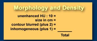
25 TSTC (too small to characterize lesions) We cannot diagnose them with certainty as: cysts: water-density, sharp demarcation hemangiomas: slowly progressive peripheral nodular enhancement of arterial density malignant lesions: inhomogeneous, irregular demarcation, peripheral enhancement less than arterial density For this type of lesions which, due to their small size and atypical imaging features, cannot be confidently categorized, the term TSTC (to small to characterize) lesions has been coined. c709d Incidental hypervascular lesions Incidental hypervascular lesions are also very common findings in liver imaging. It is important to differentiate between 'touch' and 'don't touch' lesions. Benign 'don't touch' hypervascular tumors include hemangioma, FNH and small adenomas. 'Touch' lesions include large adenomas (more then 5 cm) and malignant tumors like Hepatocelular carcinoma (HCC), Fibrolamellar carcinoma (FLHCC) and metastases. These enhancing, solid lesions should be differentiated from vascular lesions, like hepatic aneurysm, aortaportal shunt or pseudoaneurysm. 25


26 -Typical hemangioma. -Enhancement in arterial phase is almost isodense to the aorta. -as contrast diffuses toward the center of the lesion, the level of enhancement lowers slowly, -On the left a typical hemangioma. -in the late phase is still hyperdense compared to the vascular spaces. Enhancement in 'capillary blush' -'capillary blush' due to an abundant capillary network which characterizes FNH, adenoma, HCC and hypervascular metastases. -As capillaries are surrounded by tissue the overall enhancement will be less dense compared to the enhancement of the vascular spaces in hemangioma. -Hence, in capillary blush, the enhancement occurs slightly later compared to the aorta and is less dense than the aorta. Focal Nodular hyperplasia: -non-neoplastic, hyperplastic response to a congenital vascular malformation. -At late arterial phase, FNH typically presents with a bright homogeneous enhancement, but less intense than the aorta with a hypodense central scar. -Smaller (< 3cm) FNH, often lack a central scar. -The radiating hypodense fibrous bands or septa, arising from the scar, are not infrequent and quite characteristic. -At portal phase, FNH is often iso-attenuating to the normal liver and may be difficult to deliniate. -Delayed phase often shows hyperattenuation of the central scar and septa due to late opacification of the fibrotic components. -No calcifications, inhomogeneity or capsule should be seen in FNH. 26

, although multiple adenomas have been")


 -'Every hypervascular lesion in a cirrhotic liver is HCC until proven")
27 Hepatic adenomas : -are uncommon benign epithelial liver tumors that develop in an otherwise normal appearing liver. -They are seen predominantly in young women (20 to 44 years-old), are frequently located in the right hepatic lobe, and are typically solitary (70 to 80 percent), although multiple adenomas have been described in patients with prolonged contraceptive use, glycogen storage diseases (GSD), and hepatic adenomatosis. -Adenomas range in size from 1 to 30 cm. -Symptoms, such as abdominal pain, are more likely with larger lesions. -hypervascular lesions, somewhat less dense than we would expect in FNH. -Both lesions demonstrate a halo of a capsule, which should not be apparent in FNH. -Unlike in FNH, the enhancement is inhomogeneous and in the portovenous and equilibrium phase the lesions are not isodense to the liver. Hepatocellular carcinoma (HCC) -'Every hypervascular lesion in a cirrhotic liver is HCC until proven otherwise' '. -On the left we see a cirrhotic liver with irregular margins (arrows), suggesting that the hypervascular lesion is a HCC. -The inhomogeneous enhancement and the partial capsule are helpful for the diagnosis HCC, but even if these features were not present, our diagnosis still would be HCC. 27


28 Hypervascular metastases -A hypervascular primary tumor like endocrine tumors (thyroid, carcinoid), renal cell tumors and some breast carcinomas. -Often coexisting hypo- and hypervascular metastases. -Larger lesions are often inhomogeneous due to central necrosis. 28


29 Features of a cyst on Ultrasound: Features of a cyst on CT: 29
30 II F Bosniak renal cyst classification Bosniak 1 simple cyst, anechoic, imperceptable wall, rounded work up : nil % malignant : ~ 0 Bosniak 2 minimally complex, single thin (< 1mm) septations, thin Ca++; non-enhancing high-attenuation renal lesions of less than 3 cm are also included in this category; these lesions are generally well marginated. work up : nil % malignant : ~ 0 30


31 Bosniak renal cyst classification Bosniak 2F minimally complex but requiring follow up, thin septation, thick Ca++, hyperdense. for a hyperdense cyst on CT to be 2F needs to meet the following criteria: > 3 cm diameter, mostly intrarenal (less than 25% of wall visible); no enhancement work up : needs ultrasound / CT follow up % malignant : ~ 5 % Bosniak 3 indeterminate, thick or multiple septations, mural nodule, hyperdense on CT (see 2F) treatment / work up : partial nephrectomy or RF ablation in elderly / poor surgical risk % malignant : ~ 50% Bosniak 4 clearly malignant, solid mass with cystic spaces surgery treatment: partial / total nephrectomy % malignant : ~100% Bosniak Classification: Bosniak II F Require follow up CT yearly for 5 years to establish stability. 31


32 Guidelines on the management of renal cyst disease Can Urol Assoc J April; 4(2): Guidelines on the management of renal cyst disease There are no randomized controlled trials with regards to follow-up or management of cystic renal masses, as such, the recommendations are primarily expert opinion. At this time, category I and II renal cysts, do not require further imaging or follow-up. Patients in Category IIF, because of the approximate 5% malignant risk, do require periodic imaging. (There is no consensus or evidence based interval determined for follow-up imaging.) Combination of ultrasound and MRI should be considered as follow-up for Bosniak IIF and reduces the lifetime radiation dose (once the lesion has been characterized by triphasic CT scan) in patients younger than 50 years. For Category III (50% malignant risk) and category IV (75% to 90% malignant risk), surgical excision is recommended (Level 3 evidence, Grade B recommendation). Although MRI may add further information, it should be used as an adjunct to CT scans in difficult cases (Level 4 evidence, Grade C recommendation). 32
33 33


34 659fc 34


35 f0d8d 35


36 36


37 Basics In patients less than 30 years, a narrow zone of transition indicates benignancy. Mets and myeloma need to be included in the differential diagnosis of a well-defined bone lesion in a patient over 40 years. Infection and eosinophilic granuloma can mimic a malignant tumor with cortical destruction and aggressive periosteal reaction. Malignant bone tumors do not cause benign periosteal reaction. 37


38 38

39 Any Incidental lesions I have not covered that you want to ask about? Darcy.little@sympatico.ca 39
Sonographic Features of Thyroid Nodules & Guidelines for Management
 Sonographic Features of Thyroid Nodules & Guidelines for Management Mark A. Lupo, MD, FACE, ECNU Thyroid & Endocrine Center of Florida Assistant Clinical Professor of Medicine Florida State University,
Tania Gallant MD, FRCPC Internal Medicine Update April
 Tania Gallant MD, FRCPC Internal Medicine Update April 28 2017 Disclosures Honoraria/Ad board: Sanofi-Aventis, Janssen, Merck Frosst, Eli-Lilly, Astra Zeneca, Boehringer-Ingelheim Objectives By the end
Evaluation and Management of Thyroid Nodules. Nick Vernetti, MD, FACE Palm Medical Group Las Vegas, Nevada
 Evaluation and Management of Thyroid Nodules Nick Vernetti, MD, FACE Palm Medical Group Las Vegas, Nevada Disclosure Consulting Amgen Speaking Amgen Objectives Understand the significance of incidental
Evaluation of Thyroid Nodules
 Evaluation of Thyroid Nodules Stephan Kowalyk, MD January 25 28, 2018 1 Primary goal Exclude malignancy Incidental thyroid nodules If found on CT, MRI, PET scan, carotid Doppler ULTRASOUND!! January 25
Thyroid Nodule. Disclosure. Learning Objectives P A P A P A 3/18/2014. Nothing to disclose.
 Thyroid Nodule Evaluating the patient with a thyroid nodule and some management options. Miguel V. Valdez PA C Disclosure Nothing to disclose. Learning Objectives Examination of thyroid gland Options for
HEPATO-BILIARY IMAGING
 HEPATO-BILIARY IMAGING BY MAMDOUH MAHFOUZ MD PROF.OF RADIOLOGY CAIRO UNIVERSITY mamdouh.m5@gmail.com www.ssregypt.com CT ABDOMEN Indications Patient preparation Patient position Scanogram Fasting 4-6 hours
Approach to Thyroid Nodules
 Approach to Thyroid Nodules Alice Y.Y. Cheng, MD, FRCPC Twitter: @AliceYYCheng Copyright 2017 by Sea Courses Inc. All rights reserved. No part of this document may be reproduced, copied, stored, or transmitted
Financial Disclosure
 Benign Liver Masses Adil Abdalla, MBBS Creighton University-CHI Health August 25, 2018 Financial Disclosure Nothing to disclose Financial Disclosure 1 Objectives To assess patients with benign liver tumors
ACG Clinical Guideline: Diagnosis and Management of Focal Liver Lesions
 ACG Clinical Guideline: Diagnosis and Management of Focal Liver Lesions Jorge A. Marrero, MD, 1 Joseph Ahn, MD, FACG, 2 K. Rajender Reddy, MD, FACG 3 1 University of Texas at Southwestern, Dallas, Texas,
Thyroid Nodules. Family Medicine Refresher Course Geeta Lal MD, FACS April 2, No financial disclosures
 Thyroid Nodules Family Medicine Refresher Course Geeta Lal MD, FACS April 2, 2014 No financial disclosures Objectives Review epidemiology Work up of Thyroid nodules Indications for FNAB Evolving role of
Liver Tumors. Prof. Dr. Ahmed El - Samongy
 Liver Tumors Prof. Dr. Ahmed El - Samongy Objective 1. Identify the most important features of common benign liver tumors 2. Know the risk factors, diagnosis, and management of hepatocellular carcinoma
of Thyroid Lesions Comet Tail Crystals
 2 Ultrasound Features of Thyroid Lesions There are many different features indicating a certain benign or malignant tumor type, but many of these are overlapping signs. Combining several features is considered
Principal Site Investigator ENHANCE (Evaluation of Thyroid FNA Genomic Signature) study: An IRB approved study with funding to Rochester Regional
 study: An IRB approved study with funding to Rochester Regional") October 20 th 2018 Principal Site Investigator ENHANCE (Evaluation of Thyroid FNA Genomic Signature) study: An IRB approved study with funding to Rochester Regional Health from Veracyte Review ultrasound
October 20 th 2018 Principal Site Investigator ENHANCE (Evaluation of Thyroid FNA Genomic Signature) study: An IRB approved study with funding to Rochester Regional Health from Veracyte Review ultrasound
Renal masses - the role of diagnostic imaging
 Renal masses - the role of diagnostic imaging Poster No.: C-2471 Congress: ECR 2015 Type: Educational Exhibit Authors: V. Rai#; Bjelovar/HR Keywords: Cysts, Cancer, Structured reporting, Ultrasound, MR,
CT Screening for Lung Cancer for High Risk Patients
 CT Screening for Lung Cancer for High Risk Patients The recently published National Lung Cancer Screening Trial (NLST) showed that low-dose CT screening for lung cancer reduces mortality in high-risk patients
CTA/MRA of Pediatric Hepatic Masses Radiology-Pathology Correlation
 Acta Radiológica Portuguesa, Vol.XVIII, nº70, pág. 41-50, Abr.-Jun., 2006 CTA/MRA of Pediatric Hepatic Masses Radiology-Pathology Correlation Marilyn J. Siegel Mallinckrodt Institute of Radiology, Washington
11/1/2014. Radiologic incidentalomas Ordering pitfalls Newer technology and applications
 Bilal Tahir, MD Gitasree Borthakur, MD Indiana University School of Medicine Department of Radiology & Imaging Sciences October 31, 2014 ACP 2014 Dr. V. Aaron Nuclear (vaaron@iupui.edu) Dr. S. Westphal
Radiology of hepatobiliary diseases
 GI cycle - Lecture 14 436 Teams Radiology of hepatobiliary diseases Objectives 1. To Interpret plan x-ray radiograph of abdomen with common pathologies. 2. To know the common pathologies presentation.
Objectives. 1)To recall thyroid nodule ultrasound characteristics that increase the risk of malignancy
To recall thyroid nodule ultrasound characteristics that increase the risk of malignancy") Evaluation and Management of Thyroid Nodules in Primary Care Chris Sadler, MA, PA C, CDE, DFAAPA Medical Science Outcomes Liaison Intarcia Diabetes and Endocrine Associates La Jolla, CA Past President
Evaluation and Management of Thyroid Nodules in Primary Care Chris Sadler, MA, PA C, CDE, DFAAPA Medical Science Outcomes Liaison Intarcia Diabetes and Endocrine Associates La Jolla, CA Past President
objectives Pitfalls and Pearls in PET/CT imaging Kevin Robinson, DO Assistant Professor Department of Radiology Michigan State University
 objectives Pitfalls and Pearls in PET/CT imaging Kevin Robinson, DO Assistant Professor Department of Radiology Michigan State University To determine the regions of physiologic activity To understand
THYROID NODULES: THE ROLE OF ULTRASOUND
 THYROID NODULES: THE ROLE OF ULTRASOUND NOVEMBER 2017 DR. DEAN DURANT DEFINITION Thyroid nodule: Focal area within the thyroid gland with echogenicity different from surrounding parenchyma. THYROID NODULES
CT & MRI of Benign Liver Neoplasms Srinivasa R Prasad
 CT & MRI of Benign Liver Neoplasms Srinivasa R Prasad No financial disclosures Acknowledgements Many thanks to Drs. Heiken, Narra & Menias (MIR) Dr. Sahani (MGH) for sharing images Benign Liver Tumors:
Newcastle HPB MDM updated radiology imaging protocol recommendations. Author Dr John Scott. Consultant Radiologist Freeman Hospital
 Newcastle HPB MDM updated radiology imaging protocol recommendations Author Dr John Scott. Consultant Radiologist Freeman Hospital This document is intended as a guide to aid radiologists and clinicians
Objectives. How to Investigate Thyroid Nodules like A Pro
 How to Investigate Thyroid Nodules like A Pro Chris Sadler, MA, PA C, CDE, DFAAPA Medical Science Outcomes Liaison Intarcia Diabetes and Endocrine Associates La Jolla, CA Past President ASEPA Disclosures
Imaging of liver and pancreas
 Imaging of liver and pancreas.. Disease of the liver Focal liver disease Diffusion liver disease Focal liver disease Benign Cyst Abscess Hemangioma FNH Hepatic adenoma HCC Malignant Fibrolamellar carcinoma
Dr Claire Smith, Consultant Radiologist St James University Hospital Leeds
 Dr Claire Smith, Consultant Radiologist St James University Hospital Leeds Imaging in jaundice and 2ww pathway Image protocol Staging Limitations Pancreatic cancer 1.2.4 Refer people using a suspected
INTERDISCIPLINARY DISCUSSIONS IN LOCALISED RCC DIAGNOSIS AND SURGICAL STRATEGIES FOR ATYPICAL RENAL CYSTIC LESIONS. Maria Cova
 INTERDISCIPLINARY DISCUSSIONS IN LOCALISED RCC DIAGNOSIS AND SURGICAL STRATEGIES FOR ATYPICAL RENAL CYSTIC LESIONS Maria Cova Radiology Department University of Trieste (IT) Eleventh European International
A rare case of solitary toxic nodule in a 3yr old female child a case report
 Volume 3 Issue 1 2013 ISSN: 2250-0359 A rare case of solitary toxic nodule in a 3yr old female child a case report *Chandrasekaran Maharajan * Poongkodi Karunakaran *Madras Medical College ABSTRACT A three
Imaging in Pediatric Thyroid disorders: US and Radionuclide imaging. Deepa R Biyyam, MD Attending Pediatric Radiologist
 Imaging in Pediatric Thyroid disorders: US and Radionuclide imaging Deepa R Biyyam, MD Attending Pediatric Radiologist Imaging in Pediatric Thyroid disorders: Imaging modalities Outline ACR-SNM-SPR guidelines
Risk of Thyroid Cancer Based on Thyroid Ultrasound Imaging Characteristics
 Risk of Thyroid Cancer Based on Thyroid Ultrasound Imaging Characteristics Diabetes Update and Advances in Endocrinology & Metabolism Vickie A Feldstein MD Rebecca Smith- Bindman MD Department of Radiology
Thyroid in a Nutshell Dublin Catherine Kirkpatrick Consultant Sonographer ULHT
 Thyroid in a Nutshell Dublin 2017 Catherine Kirkpatrick Consultant Sonographer ULHT Acknowledgements Dr. Steve Colley Dr. Rhodri Evans Dr. Rhian Rhys Dr. Andrew McQueen Aims Anatomy & Physiology Incidence
Adina Alazraki, MD, FAAP Assistant Professor Radiology and Pediatrics Emory University and Children s Healthcare of Atlanta
 Adina Alazraki, MD, FAAP Assistant Professor Radiology and Pediatrics Emory University and Children s Healthcare of Atlanta Review recently published pediatric guidelines for management of thyroid nodules
Workup of a Solid Liver Lesion
 Workup of a Solid Liver Lesion Joseph B. Cofer MD FACS Chief Quality Officer Erlanger Health System Affiliate Professor of Surgery UTHSC-Chattanooga I have no financial or other relationships with any
Rodney C Richie MD FACP FCCP DBIM Texas Life and EMSI
 Rodney C Richie MD FACP FCCP DBIM Texas Life and EMSI Pulmonary Nodules Well-circumscribed, radiographic opacities measuring 3 cm in diameter Surrounded by aerated lung Not associated with atelectesis
Role of imaging in RCC. Ultrasonography. Solid lesion. Cystic RCC. Solid RCC 31/08/60. From Diagnosis to Treatment: the Radiologist Perspective
 Role of imaging in RCC From Diagnosis to Treatment: the Radiologist Perspective Diagnosis Staging Follow up Imaging modalities Limitations and pitfalls Duangkamon Prapruttam, MD Department of Therapeutic
Can Color Doppler Sonography Aid in the Prediction of Malignancy of Thyroid Nodules?
 Article Can Color Doppler Sonography Aid in the Prediction of Malignancy of Thyroid Nodules? Mary C. Frates, MD, Carol B. Benson, MD, Peter M. Doubilet, MD, PhD, Edmund S. Cibas, MD, Ellen Marqusee, MD
Jesse Civan, M.D. Medical Director, Jefferson Liver Tumor Center
 Liver Tumors Jesse Civan, M.D. Medical Director, Jefferson Liver Tumor Center Differential Diagnosis Malignant Metastatic from non-hepatic primary Hepatocellular carcinoma Cholangiocarcinoma Biliary cystcarcinoma
PULMONARY NODULES AND MASSES : DIAGNOSTIC APPROACH AND NEW MANAGEMENT GUIDELINES. https://tinyurl.com/hmpn2018
 PULMONARY NODULES AND MASSES : DIAGNOSTIC APPROACH AND NEW MANAGEMENT GUIDELINES Heber MacMahon MB, BCh Department of Radiology The University of Chicago https://tinyurl.com/hmpn2018 Disclosures Consultant
Interesting Cases from Liver Tumor Board. Jeffrey C. Weinreb, M.D.,FACR Yale University School of Medicine
 Interesting Cases from Liver Tumor Board Jeffrey C. Weinreb, M.D.,FACR Yale University School of Medicine jeffrey.weinreb@yale.edu Common Liver Diseases Hemangioma Cyst FNH Focal Fat/Sparing THID Non-Cirrhotic
GUIDELINES FOR PULMONARY NODULE MANAGEMENT : RECENT CHANGES AND UPDATES
 Venice 2017 GUIDELINES FOR PULMONARY NODULE MANAGEMENT : RECENT CHANGES AND UPDATES Heber MacMahon MB, BCh Department of Radiology The University of Chicago Disclosures Consultant for Riverain Medical
MANAGEMENT RECOMMENDATIONS
 1 MANAGEMENT RECOMMENDATIONS 1. Adrenal masses!!!!!!! page 2 2. Liver Masses!!!!!!! page 3 3. Obstetric US Soft Markers for Aneuploidy!! pages 4-6 4. Ovarian and Adnexal Cysts!!!!! pages 7-10 5. Pancreatic
The Incidental Renal lesion
 The Incidental Renal lesion BACKGROUND Increase in abdominal CT/US in last 15 years Resulted in detection of many (small) renal lesions 50% > 50yrs has at least 1 lesion majority simple cysts Renal lesions
Imaging in breast cancer. Mammography and Ultrasound Donya Farrokh.MD Radiologist Mashhad University of Medical Since
 Imaging in breast cancer Mammography and Ultrasound Donya Farrokh.MD Radiologist Mashhad University of Medical Since A mammogram report is a key component of the breast cancer diagnostic process. A mammogram
Sonographic imaging of pediatric thyroid disorders in childhood. Experiences and report in 150 cases
 Sonographic imaging of pediatric thyroid disorders in childhood. Experiences and report in 150 cases M. Mearadji International Foundation for Pediatric Imaging Aid Sonographic technique. Use of high frequency
Learning Objectives. 1. Identify which patients meet criteria for annual lung cancer screening
 Disclosure I, Taylor Rowlett, DO NOT have a financial interest /arrangement or affiliation with one or more organizations that could be perceived as a real or apparent conflict of interest in the context
International Journal of Current Medical Sciences- Vol. 6, Issue,, pp , June, 2016 A B S T R A C T
 ISSN: 2320-8147 International Journal of Current Medical Sciences- Vol. 6, Issue,, pp. 122-126, June, 2016 COMPUTED TOMOGRAPHY IN HEPATIC METASTASES Ananthakumar P and Adaikkappan M., Available online
PET IMAGING (POSITRON EMISSION TOMOGRAPY) FACT SHEET
 FACT SHEET") Positron Emission Tomography (PET) When calling Anthem (1-800-533-1120) or using the Point of Care authorization system for a Health Service Review, the following clinical information may be needed to
Positron Emission Tomography (PET) When calling Anthem (1-800-533-1120) or using the Point of Care authorization system for a Health Service Review, the following clinical information may be needed to
LUNG NODULES: MODERN MANAGEMENT STRATEGIES
 Department of Radiology LUNG NODULES: MODERN MANAGEMENT STRATEGIES Christian J. Herold M.D. Department of Biomedical Imaging and Image-guided Therapy Medical University of Vienna Vienna, Austria Pulmonary
Evaluation of Liver Mass Lesions. American College of Gastroenterology 2013 Regional Postgraduate Course
 Evaluation of Liver Mass Lesions American College of Gastroenterology 2013 Regional Postgraduate Course Lewis R. Roberts, MB ChB, PhD Division of Gastroenterology and Hepatology Mayo Clinic College of
PRACTICE GUIDELINES: Thyroid Nodules and Cancer 2017 ESEO Alexandria
 PRACTICE GUIDELINES: Thyroid Nodules and Cancer 2017 ESEO Alexandria James V. Hennessey MD Associate Professor of Medicine Harvard Medical School Case 1 28 year old woman sees OB for routine visit ROS:
Oh, I get it, the TSH goes up and down
 Evaluation and Management of the Thyroid Nodule Oh, I get it, the TSH goes up and down UCSF Head and Neck Conference October 24, 2008 Peter A. Singer, M.D. Professor and Chief Clinical Endocrinology University
Chief Complain. Liver lesion found in routine health check 41 days ago
 Chief Complain Liver lesion found in routine health check 41 days ago Present Illness On 2005-7-26 at 台北署立醫院 he underwent a health check for the first time. Abdominal US showed suspicious of a 6*5 cm hepatoma,
Pediatric Retroperitoneal Masses Radiologic-Pathologic Correlation
 Acta Radiológica Portuguesa, Vol.XVIII, nº 70, pág. 61-70, Abr.-Jun., 2006 Pediatric Retroperitoneal Masses Radiologic-Pathologic Correlation Marilyn J. Siegel Mallinckrodt Institute of Radiology, Washington
ADRENAL MEDULLARY DISORDERS: PHAEOCHROMOCYTOMAS AND MORE
 ADRENAL MEDULLARY DISORDERS: PHAEOCHROMOCYTOMAS AND MORE DR ANJU SAHDEV READER AND CONSULTANT RADIOLOGIST QUEEN MARY UNIVERSITY AND ST BARTHOLOMEW S HOSPITAL BARTS HEALTH, LONDON, UK DISCLOSURE OF CONFLICT
Hepatobiliary and Pancreatic Malignancies
 Hepatobiliary and Pancreatic Malignancies Gareth Eeson MD MSc FRCSC Surgical Oncologist and General Surgeon Kelowna General Hospital Interior Health Consultant, Surgical Oncology BC Cancer Agency Centre
Practical Approach to Thyroid Nodules:Ultrasound Criteria for Performing FNA Revisited
 Practical Approach to Thyroid Nodules:Ultrasound Criteria for Performing FNA Revisited Poster No.: C-0100 Congress: ECR 2013 Type: Educational Exhibit Authors: S. Kuzmich, S. Sritharan, S. MUKUNDHAN, M.
Normal Sonographic Anatomy
 hapter 2:The Liver DUNSTAN ABRAHAM Normal Sonographic Anatomy Homogeneous, echogenic texture (Figure 2-1) Measures approximately 15 cm in length and 10 12.5 cm anterior to posterior; measurement taken
Thyroid Nodules. Dr. HAKIMI, SpAK Dr. MELDA DELIANA, SpAK Dr. SISKA MAYASARI LUBIS, SpA
 Thyroid Nodules ENDOCRINOLOGY DIVISION ENDOCRINOLOGY DIVISION Dr. HAKIMI, SpAK Dr. MELDA DELIANA, SpAK Dr. SISKA MAYASARI LUBIS, SpA Anatomical Considerations The Thyroid Nodule Congenital anomalies Thyroglossal
Thyroid Nodules: US Risk Stratification. Alex Tessnow, MD, FACE, ECNU University of Texas Southwestern Associate Professor of Medicine Dallas, Texas
 Thyroid Nodules: US Risk Stratification Alex Tessnow, MD, FACE, ECNU University of Texas Southwestern Associate Professor of Medicine Dallas, Texas Which of the following is true? A. All echogenic foci
MTP: Thyroid Nodules
 Canadian Endocrine Update MTP: Thyroid Nodules Deric Morrison MD, FRCP, ECNU Assistant Professor, Division of Endocrinology and Metabolism, Western University April 2014 Faculty/Presenter Disclosure Faculty:
Malignant Focal Liver Lesions
 Malignant Focal Liver Lesions Other Than HCC Pablo R. Ros, MD, MPH, PhD Departments of Radiology and Pathology University Hospitals Cleveland Medical Center Case Western Reserve University Pablo.Ros@UHhospitals.org
Case-based discussion:
 Case-based discussion: Pailin Kongmebhol, M.D. Department of Radiology Faculty of Medicine Chiang Mai University There are many guidelines for managing thyroid nodules Two important guidelines: 2015 American
Women s Health in General Practice Symposium 2015 Thyroid & Parathyroid Cases
 Women s Health in General Practice Symposium 2015 Thyroid & Parathyroid Cases Bill Fleming Epworth Freemasons Hospital 1 Common Endocrine Presentations anatomical problems thyroid nodule / goitre embryological
Thyroid Ultrasonography: clinical and radiological correlations
 Thyroid Ultrasonography: clinical and radiological correlations Dr.M.Thijs Radiology Anatomy Inflammatory Thyroid Disease Benign lesions Thyroid tumors Thyroglossal duct cyst Anatomy Transverse Longitudinal
NEOPLASMS OF THE THYROID PATHOLOGY OF PARATHYROID GLANDS. BY: Shifaa Qa qa
 NEOPLASMS OF THE THYROID PATHOLOGY OF PARATHYROID GLANDS BY: Shifaa Qa qa Neoplasmas of the thyroid thyroid nodules Neoplastic ---- benign, malignant Non neoplastic Solitary nodules ----- neoplastic Nodules
4/22/2010. Hakan Korkmaz, MD Assoc. Prof. of Otolaryngology Ankara Dıșkapı Training Hospital-Turkey.
 Management of Differentiated Thyroid Cancer: Head Neck Surgeon Perspective Hakan Korkmaz, MD Assoc. Prof. of Otolaryngology Ankara Dıșkapı Training Hospital-Turkey Thyroid gland Small endocrine gland:
Diagnostic Dilemmas of Breast Imaging
 Diagnostic Dilemmas of Breast Imaging Common Causes of Error in Breast Cancer Detection By: Jason Cord, M.D. Mammography: Initial Imaging The standard for detection of breast cancer Screening mammography
The Focal Hepatic Lesion: Radiologic Assessment
 The Focal Hepatic Lesion: Radiologic Assessment Kevin Kuo, Harvard Medical School Year III Our Patient: PS 67 y/o female w/ long history of alcohol use Drinking since age 18, up to one bottle of wine/day
Essentials of Clinical MR, 2 nd edition. 65. Benign Hepatic Masses
 65. Benign Hepatic Masses Pulse sequences acquired for abdominal MRI typically consist of fast acquisition schemes such as single-shot turbo spin echo (i.e. HASTE) and gradient echo schemes such as FLASH
Endocrinology and Metabolic Disorder Unit Regina Apostolorum Hospital
 Enrico Papini Endocrinology and Metabolic Disorder Unit Regina Apostolorum Hospital Albano Laziale, Italy The Following Faculty have provide no information regarding significant relationship with commercial
Index. radiologic.theclinics.com. Note: Page numbers of article titles are in boldface type.
 Index Note: Page numbers of article titles are in boldface type. A ACC. See Adrenal cortical carcinoma. Acromegaly and the pituitary gland, 551 Acute suppurative thyroiditis, 405, 406 Addison, Thomas and
MEASUREMENT OF EFFECT SOLID TUMOR EXAMPLES
 MEASUREMENT OF EFFECT SOLID TUMOR EXAMPLES Although response is not the primary endpoint of this trial, subjects with measurable disease will be assessed by standard criteria. For the purposes of this
Complete Summary GUIDELINE TITLE. Liver lesion characterization. BIBLIOGRAPHIC SOURCE(S)
") Complete Summary GUIDELINE TITLE Liver lesion characterization. BIBLIOGRAPHIC SOURCE(S) Foley WD, Bree RL, Gay SB, Glick SN, Heiken JP, Huprich JE, Levine MS, Ros PR, Rosen MP, Shuman WP, Greene FL, Rockey
Complete Summary GUIDELINE TITLE Liver lesion characterization. BIBLIOGRAPHIC SOURCE(S) Foley WD, Bree RL, Gay SB, Glick SN, Heiken JP, Huprich JE, Levine MS, Ros PR, Rosen MP, Shuman WP, Greene FL, Rockey
USGFNA of thyroid nodules
 US Guided FNA (USGFNA) of neck masses INTERVENTIONAL HEAD & NECK ULTRASOUND Brendan C. Stack, Jr., MD., FACS, FACE Professor Otolaryngology-Head and Neck Surgery Indications Technique Interpretation Results
Ultrasound of the Breast BASICS FOR THE ORDERING CLINICIAN
 Ultrasound of the Breast BASICS FOR THE ORDERING CLINICIAN Breast Ultrasound Anatomy Skin Breast Parenchyma Pectoralis Fascia Pectoralis Breast Ultrasound Anatomy Indications for Breast Ultrasound Palpable
How To Approach Renal Masses? - Differential Diagnosis On Image
 How To Approach Renal Masses? - Differential Diagnosis On Image Poster No.: C-1646 Congress: ECR 2015 Type: Educational Exhibit Authors: A. E. A. G. Costa, A. Gomes, A. Duarte, I. Távora; Lisbon/PT Keywords:
Criteria of Malignancy. Evaluation Score
 30 5 Diagnostic Criteria Criteria of Malignancy Table 5.2 lists criteria in contrast-enhancing MR mammography that strongly indicate the presence of malignancy or are unspecific. Unifactorial evaluation
CLINICAL PRESENTATION AND RADIOLOGY QUIZ QUESTION
 Donald L. Renfrew, MD Radiology Associates of the Fox Valley, 333 N. Commercial Street, Suite 100, Neenah, WI 54956 4/30/2011 Radiology Quiz of the Week # 18 Page 1 CLINICAL PRESENTATION AND RADIOLOGY
Imaging Guided Biopsy. Edited & Presented by ; Hussien A.B ALI DINAR. Msc Lecturer,Reporting Sonographer
 Imaging Guided Biopsy Edited & Presented by ; Hussien A.B ALI DINAR. Msc Lecturer,Reporting Sonographer Objective By the End of this lessons you should : Define what biopsy Justify Aim to perform biopsy
The radiological spectrum of thyroid malignancy
 The radiological spectrum of thyroid malignancy Poster No.: C-2575 Congress: ECR 2012 Type: Educational Exhibit Authors: K. Cortis, W. Scicluna, A. Mizzi ; Rabat/MT, Birkirkara/MT Keywords: Ultrasound-Colour
Incidentalomas in Radiology GRAND ROUNDS 5/23/2017. AJ Lewis, MD MBA Boca Radiology Group
 Incidentalomas in Radiology GRAND ROUNDS 5/23/2017 AJ Lewis, MD MBA Boca Radiology Group Disclosures NONE Incidental Findings EVERYWHERE INCIDENTAL FINDINGS The Latest Management Recommendations CT or
Study of validity of ultrasonographic diagnosis in relation to Fine Needle Aspiration Cytology (FNAC) diagnosis
 diagnosis") Original article: Study of validity of ultrasonographic diagnosis in relation to Fine Needle Aspiration Cytology (FNAC) diagnosis *Dr Rajvi Matalia, ** Dr Y.P.Sachdev, ***Dr D.S.Kulkarni *Junior Resident,
Original article: Study of validity of ultrasonographic diagnosis in relation to Fine Needle Aspiration Cytology (FNAC) diagnosis *Dr Rajvi Matalia, ** Dr Y.P.Sachdev, ***Dr D.S.Kulkarni *Junior Resident,
PEDIATRIC Ariel Katz MD
 PEDIATRIC Ariel Katz MD Dept. Otolaryngology Head &Neck Surgery Wolfson Medical Center Holon, Israel OBJECTIVES Overview/Background Epidemiology/Etiology Intro to Guidelines Workup Treatment Follow-Up
CME Article Clinics in diagnostic imaging (135)
") Medical Education Singapore Med J 2011; 52(5) : 384 CME Article Clinics in diagnostic imaging (135) Pojchamarnwiputh S, Muttarak M, Sriplakich S H 1a 1b 1c 1d Fig. 1 (a) Axial unenhanced; (b & c) delayed
Medical Education Singapore Med J 2011; 52(5) : 384 CME Article Clinics in diagnostic imaging (135) Pojchamarnwiputh S, Muttarak M, Sriplakich S H 1a 1b 1c 1d Fig. 1 (a) Axial unenhanced; (b & c) delayed
SCBT-MR 2015 Incidentaloma on Chest CT
 SCBT-MR 2015 Incidentaloma on Chest CT Reginald F. Munden MD, DMD, MBA I have no conflicts of interest to report Incidentaloma Pulmonary Nodule Mediastinal Lymph Node Coronary Artery Calcium Incidental
Recently role of non-invasive diagnostics methods
 CERTAIN ASPECTS OF NLS-DIAGNOSTICS OF LIVER FOCAL PATHOLOGY A.Y. Shvack, V.I. Nesterov, N.L. Ogluzdina This article contains information about NLS-graphy application in diagnostics of liver focal affections:
PAPILLARY THYROID CARCINOMA PRESENTING AS A LATERAL NECK MASS MASS. Dr. Pamela Hanson DO PGY3
 PAPILLARY THYROID CARCINOMA PRESENTING AS A LATERAL NECK MASS MASS Dr. Pamela Hanson DO PGY3 MK CASE PRESENTATION 28 yo Female presented to the ENT Clinic in October 2016, with the complaint of chronic
Thyroid nodules. Most thyroid nodules are benign
 Thyroid nodules Postgraduate Course in General Surgery Jessica E. Gosnell MD Assistant Professor March 22, 2011 Most thyroid nodules are benign thyroid nodules occur in 77% of the world s population palpable
RING ENCHANCING LESION BY M.S. HEMHNATH
 RING ENCHANCING LESION BY M.S. HEMHNATH A 21 YRS FEMALE CAME WITH H/O HEADACHE AND SEIZURE FOR THE PAST ONE MONTH. NO OTHER FOCAL NEUROLOGICAL DEFICIT. DIFFERENTIAL DIAGNOSIS For this case are Neurocysticerosis
Neckmasses in infancy and childhood: Clinical and radiological classification and imaging approaches M. Mearadji
 Neckmasses in infancy and childhood: Clinical and radiological classification and imaging approaches M. Mearadji International Foundation for Pediatric Imaging Aid Introduction Neck masses are a frequent
Pulmonary Nodules. Michael Morris, MD
 Pulmonary Nodules Michael Morris, MD Case 45 year old healthy male Smokes socially Normal physical exam Pre-employment screening remote +PPD screening CXR nodular opacity Case 45 year old healthy male
Endocrine University, 2016 AACE-ACE-MAYO CLINIC
 Endocrine University, 2016 AACE-ACE-MAYO CLINIC Dev Abraham MD, MRCP (UK), ECNU Professor of Medicine (clinical), Division of Endocrinology Adjunct Professor of Surgery and Pathology Medical Director,
Thyroid Nodules: US Risk Stratification and FNA Guidelines
 Thyroid Nodules: US Risk Stratification and FNA Guidelines Mark A. Lupo, MD, FACE, ECNU Thyroid & Endocrine Center of Florida Assistant Clinical Professor of Medicine Florida State University, College
ACRIN 6666 IM Additional Evaluation: Additional Views/Targeted US
 Additional Evaluation: Additional Views/Targeted US For revised or corrected form check box and fax to 215-717-0936. Instructions: The form is completed based on recommendations (from ID form) for additional
Cardiac Imaging Tests
 Cardiac Imaging Tests http://www.medpagetoday.com/upload/2010/11/15/23347.jpg Standard imaging tests include echocardiography, chest x-ray, CT, MRI, and various radionuclide techniques. Standard CT and
Thyroid US. Background: Thyroid/Neck US. Use of Office Ultrasound in the Thyroid Surgery Practice
 2010 UCSF Head and Neck Endocrine Surgery Course Use of Office Ultrasound in the Thyroid Surgery Practice Lisa A. Orloff, MD FACS Dept of Otolaryngology-Head and Neck Surgery University of California,
Pulmonary Nodules & Masses
 Pulmonary Nodules & Masses A Diagnostic Approach Heber MacMahon The University of Chicago Department of Radiology Disclosure Information Consultant for Riverain Technology Minor equity in Hologic Royalties
Thyroid and Parathyroid Ultrasound Protocol
 Thyroid and Parathyroid Ultrasound Protocol Reviewed By: Anna Ellermeier, MD Last Reviewed: December 2017 Contact: (866) 761-4200, Option 1 **NOTE for all examinations: 1. If documenting possible flow
THYROID CANCER IN CHILDREN. Humberto Lugo-Vicente MD FACS FAAP Professor Pediatric Surgery UPR School of Medicine
 THYROID CANCER IN CHILDREN Humberto Lugo-Vicente MD FACS FAAP Professor Pediatric Surgery UPR School of Medicine Thyroid nodules Rare Female predominance 4-fold as likely to be malignant Hx Radiation exposure?
Case Scenario 1: Thyroid
 Case Scenario 1: Thyroid History and Physical Patient is an otherwise healthy 80 year old female with the complaint of a neck mass first noticed two weeks ago. The mass has increased in size and is palpable.