An Image Repository for Chest CT
|
|
|
- Martina Gregory
- 6 years ago
- Views:
Transcription
1 An Image Repository for Chest CT Francesco Frajoli for the Chest CT in Antibody Deficiency Group

.")
2 An Image Repository for Chest CT he Chest CT in Antibody Deficiency Group is an international and interdisciplinary group that works together to improve pulmonary diagnostics in patients with antibody deficiency syndrome. available for download at ne of the projects of the Group is to uniformely document chest CT scans. A common list of pulmonary pathologies is rated according to the same criteria. The list of findings is available for download at the website of the group. Findings may be documented in the ESID registry, a national registry, or sent by fax to Ulrich Baumann (for details see docutmentation sheet). The following collection of images and comments aims to standardise the rating of pulmonary CT findings and thus improve the quality of the data. e are greatly indebted to Francesco Frajoli, MD, radiologist at Sapienza University, Rome, Italy, EU, who prepared and commented the following collection of CT images for this purpose. This collection is available free of charge for educational use. Reproduction is allowed only with reference to the author and the Chest CT in ADS Group. Hanover, July 2012 Ulrich Baumann
3
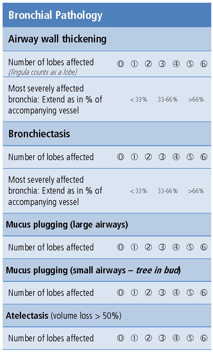
4 Air Wall Thickening It s a morphological abnormality observed in chronic airway disease. On HRTC scans is only assessed subjectively. It may render airways visible in more distal areas of the lung than normally expected. The severity of peribronchial thickening was designated as < 33 % if the thickness of the bronchial wall was equal to the diameter of the adjacent vessel; was designated as 33-66% if the thickness was greater than and up to twice the diameter of an adjacent vessel; and was designated > 66% if the thickness was greater than twice the diameter of an adjacent vessel. Normal airway artery dimensions: AWT is < 33% of AOD (<20% total airway diameter in normal airway) AWT BLD AOD

5 Most severely affected bronchi: Extend in % of accompany vessel AWT: AOD < 33%


6 Most severely affected bronchi: Extend in % of accompany vessel AWT: AOD 33-66%

7 Most severely affected bronchia: Extend in % of accompanying vessel AWT : AOD > 66%


8 Considerations Peripheral air wall thickness Most of the peripheral air wall thickening should be considered as >66 %

9 BRONCHIECTASIS Pathology. Bronchiectasis is irreversible localized or diffuse bronchial dilatation, usually resulting from chronic infection, proximal airway obstruction, or congenital bronchial abnormality. CT scans. Morphologic criteria on thin-section CT scans include bronchial dilatation with respect to the accompanying pulmonary artery (signet ring sign), lack of tapering of bronchi, and identification of bronchi within 1 cm of the pleural surface. Bronchiectasis may be classified as cylindric, varicose, or cystic, depending on the appearance of the affected bronchi. It is often accompanied by bronchial wall thickening, mucoid impaction, and smallairways abnormalities. Transverse CT scan shows varicose Bronchiectasis. Normal airway lumen to outer arterial diameter BLD AOD


10 Most severely affected bronchia: Extend in % of accompany vessel Vessel : bronchia < 33%


11 Most severely affected bronchia: Extend in % of accompany vessel Vessel : bronchia %

12 Most severely affected bronchia: Extend in % of accompany vassel Vessel : bronchia > 66%


13 Considerations Peripheral bronchiectasis Most of the peripheral bronchiectasis should be considered as >66 %


14 Mucus plugging Central mucous plugging was defined as an opacity filling a defined bronchus, and peripheral mucous plugging was defined as the presence of either dilated bronchi or peripheral thin branching structures or centrilobular nodules in the peripheral lung. Large airways mucus plugging

15 Small airways mucus plugging


16 Tree in bud Small airways mucus plugging It consists of small centrilobular nodules of soft tissue attenuation connected to multiple branching linear structures of similar caliber that originate from as single stalk


17 Atelectasis Pathophysiology. Atelectasis is reduced inflation of all or part of the lung. One of the commonest mechanisms is resorption of air distal to airway obstruction (eg, an endobronchial neoplasm). The synonym collapse is often used interchangeably with atelectasis, particularly when it is severe or accompanied by obvious increase in lung opacity. CT scans. Reduced volume is seen, accompanied by increased attenuation in the affected part of the lung. Atelectasis is often associated with abnormal displacement of fissures, bronchi, vessels, diaphragm, heart, or mediastinum. The distribution can be lobar, segmental, or subsegmental. Atelectasis is often qualified by descriptors such as linear, discoid, or platelike.
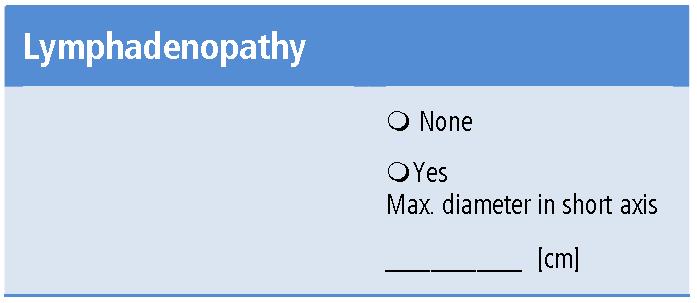
 and adenopathy. CT scans. There is a wide range in the size of normal lymph nodes.")

18 Lymphadenopathy Pathology. By common usage, the term lymphadenopathy is usually restricted to enlargement, due to any cause, of the lymph nodes. Synonyms include lymph node enlargement (preferred) and adenopathy. CT scans. There is a wide range in the size of normal lymph nodes. Mediastinal and hilar lymph nodes range in size from sub-ct resolution to 12 mm. Somewhat arbitrary thresholds for the upper limit of normal of 1 cm in short axis diameter for mediastinal nodes and 3 mm for most hilar nodes have been reported, but size criteria do not allow reliable differentiation between healthy and diseased lymph nodes.
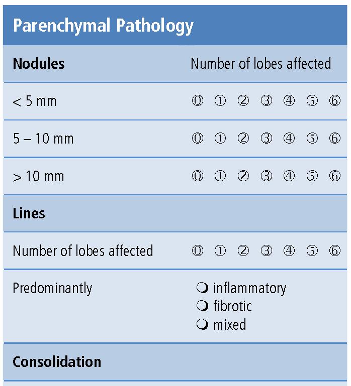
 Centrilobular nodules appear separated by several millimeters from the pleural surfaces, fissures, and interlobular septa. They may be of soft-tissue or ground-glass attenuation.")
 A ground-glass nodule (synonym, nonsolid nodule) manifests as hazy increased attenuation in the lung that does not obliterate the bronchial and vascular margins.")
19 Nodule On CT scans a nodule appears as a rounded or irregular opacity, well or poorly defined, measuring up to 3 cm in diameter. (a) Centrilobular nodules appear separated by several millimeters from the pleural surfaces, fissures, and interlobular septa. They may be of soft-tissue or ground-glass attenuation. Ranging in size from a few millimeters to a centimeter, centrilobular nodules are usually ill-defined. (b) A micronodule is less than 3 mm in diameter. (c) A ground-glass nodule (synonym, nonsolid nodule) manifests as hazy increased attenuation in the lung that does not obliterate the bronchial and vascular margins. (d) A solid nodule has homogenous soft-tissue attenuation. (e) A part-solid nodule (synonym, semisolid nodule) consists of both groundglass and solid soft-tissue attenuation components. Transverse CT scan shows irregular nodule in left lower lobe.

20 Nodule CT scan shows a nodule < 5 mm

21 Nodule CT scan shows a Nodule 5-10 mm


22 Nodule Tc scan shows a nodule > 10 mm
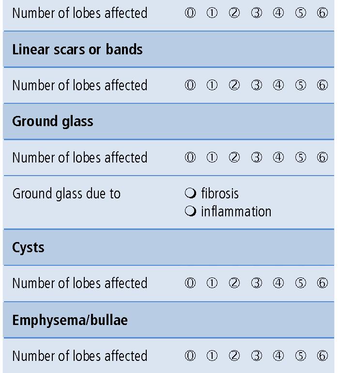
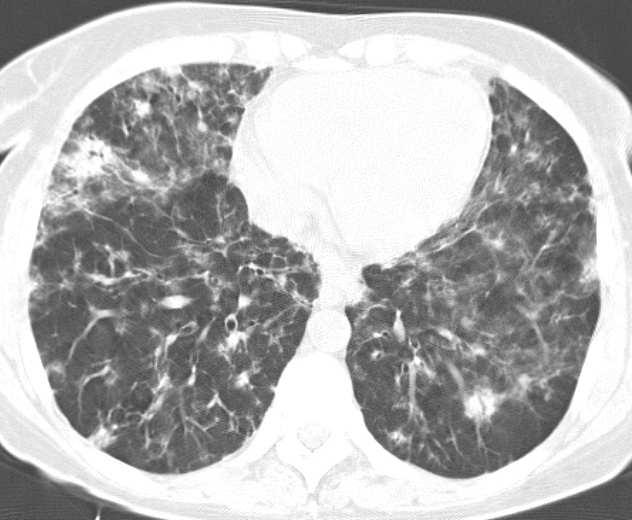
23 Intralobular lines CT scans. Intralobular lines are visible as fine linear opacities in a lobule when the intralobular interstitial tissue is abnormally thickened. When numerous, they may appear as a fine reticular pattern. Intralobular lines may be seen in various conditions, including interstitial fibrosis and alveolar proteinosis
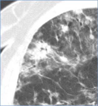
24 Inflammatory Lines

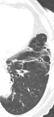
25 Fibrotic lines Mixed lines

26 Consolidation Pathology. Consolidation refers to an exudate or other product of disease that replaces alveolar air, rendering the lung solid (as in infective pneumonia). CT scans. Consolidation appears as a homogeneous increase in pulmonary parenchymal attenuation that obscures the margins of vessels and airway walls. An air bronchogram may be present. The attenuation characteristics of consolidated lung are only rarely helpful in differential diagnosis (eg, decreased attenuation in lipoid pneumonia and increased in amiodarone toxicity). Transverse CT scan shows a consolidation in the middle lobe
, partial collapse of alveoli, increased capillary blood volume, or a combination of these,")
27 Ground-glass opacity On CT scans, it appears as hazy increased opacity of lung, with preservation of bronchial and vascular margins. It is caused by partial filling of airspaces, interstitial thickening (due to fluid, cells, and/or fibrosis), partial collapse of alveoli, increased capillary blood volume, or a combination of these, the common factor being the partial displacement of air. Ground-glass opacity is less opaque than consolidation, in which bronchovascular margins are obscured. Transverse CT scan shows ground glass opacity

 and occur without associated pulmonary emphysema.")
28 Cysts Pathology. A cyst is any round circumscribed space that is surrounded by an epithelial or fibrous wall of variable thickness A cyst appears as a round parenchymal low-attenuating area with a well-defined interface with normal lung. Cysts have variable wall thickness but are usually thinwalled (<2 mm) and occur without associated pulmonary emphysema. Cysts in the lung usually contain air but occasionally contain fluid or solid material. The term is often used to describe enlarged thin walled airspaces in patients with lymphangioleiomyomatosis or Langerhans cell histiocytosis; thicker walled honeycomb cysts are seen in patients with end-stage fibrosis.

29 Emphysema Pathology. Emphysema is characterized by permanently enlarged airspaces distal to the terminal bronchiole with destruction of alveolar walls. Absence of obvious fibrosis was historically regarded as an additional criterion, but the validity of that criterion has been questioned because some interstitial fibrosis may be present in emphysema secondary to cigarette smoking. Emphysema is usually classified in terms of the part of the acinus predominantly affected: proximal (centriacinar, more commonly termed centrilobular, emphysema), distal (paraseptal emphysema), or whole acinus (panacinar or, less commonly, panlobular emphysema). The CT appearance of emphysema consists of focal areas or regions of low attenuation, usually without visible walls. In the case of panacinar emphysema, decreased attenuation is more diffuse. Transverse CT scan shows centrilobular emphysema.

30 Bulla Pathology. An airspace measuring more than 1 cm usually several centimeters in diameter, sharply demarcated by a thin wall that is no greater than 1 mm in thickness. A bulla is usually accompanied by emphysematous changes in the adjacent lung. CT scans. A bulla appears as a rounded focal area of decreased attenuation, 1 cm or more in diameter, bounded by a thin wall. Multiple bullae are often present and are associated with other signs of pulmonary emphysema (centrilobular and paraseptal). Axial CT scan shows large bulla in left lower lung zone.

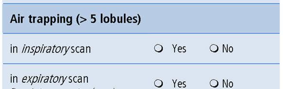
31 Air trapping Pathophysiology. Air trapping is retention of air in the lung distal to an obstruction (usually partial). CT scans. Air trapping is seen on end-expiration CT scans as parenchymal areas with less than normal increase in attenuation and lack of volume reduction. Comparison between inspiratory and expiratory CT scans can be helpful when air trapping is subtle or diffuse. Differentiation from areas of decreased attenuation resulting from hypoperfusion as a consequence of an occlusive vascular disorder (eg, chronic thromboembolism) may be problematic, but other findings of airways versus vascular disease are usually present. Transverse CT scans at end inspiration and end expiration show air trapping.
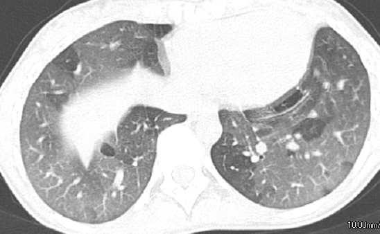
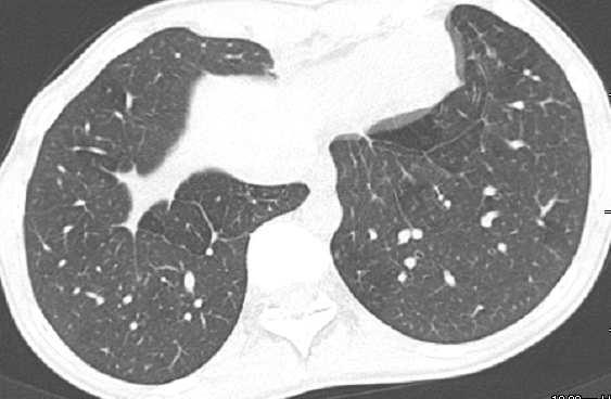
32 INSPIRATION EXPIRATION
Acute and Chronic Lung Disease
 KATHOLIEKE UNIVERSITEIT LEUVEN Faculty of Medicine Acute and Chronic Lung Disease W De Wever, JA Verschakelen Department of Radiology, University Hospitals Leuven, Belgium Clinical utility of HRCT To detect
HRCT in Diffuse Interstitial Lung Disease Steps in High Resolution CT Diagnosis. Where are the lymphatics? Anatomic distribution
 Steps in High Resolution CT Diagnosis Pattern of abnormality Distribution of disease Associated findings Clinical history Tomás Franquet MD What is the diagnosis? Hospital de Sant Pau. Barcelona Secondary
DISEASES OF THE RESPIRATORY SYSTEM 2018 DR HEYAM AWAD LECTURE 2: ATELECTASIS AND EMPHYSEMA
 DISEASES OF THE RESPIRATORY SYSTEM 2018 DR HEYAM AWAD LECTURE 2: ATELECTASIS AND EMPHYSEMA INTRODUCTION In this lecture we will discuss atelectasis which is a complication of several medical and surgical
Case 1: Question. 1.1 What is the main pattern of this HRCT? 1. Intralobular line 2. Groundglass opacity 3. Perilymphatic nodule
 HRCT WORK SHOP Case 1 Case 1: Question 1.1 What is the main pattern of this HRCT? 1. Intralobular line 2. Groundglass opacity 3. Perilymphatic nodule Case 1: Question 1.2 What is the diagnosis? 1. Hypersensitivity
Radiologic-pathologic correlation of pulmonary diseases
 The 1578 th Chest Conference/ 3 rd Biennial Clinical- Radiologic-Pathologic Correlation Radiologic-pathologic correlation of pulmonary diseases Harumi Itoh, M.D. University of Fukui, Japan Centriacinar
Case 1 : Question. 1.1 What is the intralobular distribution? 1. Centrilobular 2. Perilymphatic 3. Random
 Interesting case Case 1 Case 1 : Question 1.1 What is the intralobular distribution? 1. Centrilobular 2. Perilymphatic 3. Random Case 1: Answer 1.1 What is the intralobular distribution? 1. Centrilobular
PULMONARY TUBERCULOSIS RADIOLOGY
 PULMONARY TUBERCULOSIS RADIOLOGY RADIOLOGICAL MODALITIES Medical radiophotography Radiography Fluoroscopy Linear (conventional) tomography Computed tomography Pulmonary angiography, bronchography Ultrasonography,
Glossary of terms in thoracic imaging and clinicalradiological correlation. What every radiologist should
 Glossary of terms in thoracic imaging and clinicalradiological correlation. What every radiologist should know. Poster No.: C-1565 Congress: ECR 2012 Type: Educational Exhibit Authors: A. Moreno Pastor,
Workshop Cyst & Lucency. How to Approach
 Workshop Cyst & Lucency How to Approach To Approach Cystic Lung Disease True cysts? Cavitary disease Cystic bronchiectasis Mosaic attenuation Subpleural cysts Bullae Paraseptal emphysema Honeycombing Birt
10/17/2016. Nuts and Bolts of Thoracic Radiology. Objectives. Techniques
 Nuts and Bolts of Thoracic Radiology October 20, 2016 Carleen Risaliti Objectives Understand the basics of chest radiograph Develop a system for interpreting chest radiographs Correctly identify thoracic
TB Radiology for Nurses Garold O. Minns, MD
 TB Nurse Case Management Salina, Kansas March 31-April 1, 2010 TB Radiology for Nurses Garold O. Minns, MD April 1, 2010 TB Radiology for Nurses Highway Patrol Training Center Salina, KS April 1, 2010
Interesting Cases. Pulmonary
 Interesting Cases Pulmonary 54M with prior history of COPD, hep B/C, and possible history of TB presented with acute on chronic dyspnea, and productive cough Hazy opacity overlying the left hemithorax
Cystic Lung Disease: a Comparison of Cystic Size, as Seen on Expiratory and Inspiratory HRCT Scans
 Cystic Lung Disease: a Comparison of Cystic Size, as Seen on Expiratory and Inspiratory HRCT Scans Ki-Nam Lee, MD 1 Seong-Kuk Yoon, MD 1 Seok Jin Choi, MD 2 Jin Mo Goo, MD 3 Kyung-Jin Nam, MD 1 Index words:
Usual Interstitial pneumonia and Nonspecific Interstitial Pneumonia. Nitra and the Gangs.
 Usual Interstitial pneumonia and Nonspecific Interstitial Pneumonia Nitra and the Gangs. บทน ำและบทท ๓, ๑๐, ๑๒, ๑๓, ๑๔, ๑๕, ๑๗ Usual Interstitial Pneumonia (UIP) Most common & basic pathologic pattern
Financial disclosure COMMON DIAGNOSES IN HRCT. High Res Chest HRCT. HRCT Pre test. I have no financial relationships to disclose. Anatomy Nomenclature
 Financial disclosure I have no financial relationships to disclose. Douglas Johnson D.O. Cardiothoracic Imaging Gaston Radiology COMMON DIAGNOSES IN HRCT High Res Chest Anatomy Nomenclature HRCT Sampling
Diseases of the Lung and Respiratory Tract, Part I. William Bligh-Glover M.D. Department of Anatomy, CWRU
 Diseases of the Lung and Respiratory Tract, Part I William Bligh-Glover M.D. Department of Anatomy, CWRU Educational objectives: Distinguish the types of atelectasis and their etiologies Distinguish the
Cryptogenic Organizing Pneumonia Diagnosis Approach Based on a Clinical-Radiologic-Pathologic Consensus
 Cryptogenic Organizing Pneumonia Diagnosis Approach Based on a Clinical-Radiologic-Pathologic Consensus Poster No.: C-1622 Congress: ECR 2012 Type: Scientific Exhibit Authors: C. Cordero Lares, E. Zorita
Low-dose CT Lung Cancer Screening Guidelines for Pulmonary Nodules Management Version 2
 Low-dose CT Lung Cancer Screening Guidelines for Pulmonary Nodules Management Version 2 The Committee for Management of CT-screening-detected Pulmonary Nodules 2009-2011 The Japanese Society of CT Screening
An Introduction to Radiology for TB Nurses
 An Introduction to Radiology for TB Nurses Garold O. Minns, MD September 14, 2017 TB Nurse Case Management September 12 14, 2017 EXCELLENCE EXPERTISE INNOVATION Garold O. Minns, MD has the following disclosures
Manish Powari Regional Training Day 10/12/2014
 Manish Powari Regional Training Day 10/12/2014 Large number of different types of Interstitial Lung Disease (ILD). Most are very rare Most patients present with one of a smaller number of commoner diseases
Eun-Young Kang, M.D., Jae Wook Lee, M.D., Ji Yung Choo, M.D., Hwan Seok Yong, M.D., Ki Yeol Lee, M.D., Yu-Whan Oh, M.D.
 Eun-Young Kang, M.D., Jae Wook Lee, M.D., Ji Yung Choo, M.D., Hwan Seok Yong, M.D., Ki Yeol Lee, M.D., Yu-Whan Oh, M.D. Department of Radiology, Korea University Guro Hospital, College of Medicine, Korea
8/14/2017. Objective: correlate radiographic findings of common lung diseases to actual lung pathologic features
 What is that lung disease? Pulmonary Patterns & Correlated Pathology Dr. Russell Tucker, DACVR Objective: correlate radiographic findings of common lung diseases to actual lung pathologic features Improved
Diagnosis of TB: Radiology David Finlay, MD
 TB Intensive Tyler, Texas June 2-4, 2010 Diagnosis of TB: Radiology David Finlay, MD June 3, 2010 2stages stages- Tuberculosis 1. primary infection 2. reactivation, or post primary disease 2 1 Primary
Typical and atypical findings of pulmonary sarcoidosis at high resolution CT
 Typical and atypical findings of pulmonary sarcoidosis at high resolution CT Poster No.: C-0169 Congress: ECR 2013 Type: Educational Exhibit Authors: L. Raposo Rodríguez, C. Mejía, B. Escobar Mallada,
ARDS - a must know. Page 1 of 14
 ARDS - a must know Poster No.: C-1683 Congress: ECR 2016 Type: Authors: Keywords: DOI: Educational Exhibit M. Cristian; Turda/RO Education and training, Edema, Acute, Localisation, Education, Digital radiography,
Pediatric High-Resolution Chest CT
 Pediatric High-Resolution Chest CT Alan S. Brody, MD Professor of Radiology and Pediatrics Chief, Thoracic Imaging Cincinnati Children s s Hospital Cincinnati, Ohio, USA Pediatric High-Resolution CT Short
Mimics in chest disease: interstitial opacities
 Insights Imaging (2013) 4:9 27 DOI 10.1007/s13244-012-0207-7 PICTORIAL REVIEW Mimics in chest disease: interstitial opacities Anastasia Oikonomou & Panos Prassopoulos Received: 19 June 2012 / Revised:
How does COPD really work?
 How does COPD really work? by Alex Goodell View online Where does COPD fit in the mix of respiratory diseases? I ve made a map of the major pathologies outlined in Robbins and First Aid (obviously these
The Imaging Analysis of Pulmonary Sarcodiosis
 www.cancercellresearch.org ISSN: 2161-2609 Article The Imaging Analysis of Pulmonary Sarcodiosis Xin He, Chuanyu Zhang* Department of Radiology, Affiliated Hospital of Qingdao University, Qingdao, China
The Respiratory System. Dr. Ali Ebneshahidi
 The Respiratory System Dr. Ali Ebneshahidi Functions of The Respiratory System To allow gases from the environment to enter the bronchial tree through inspiration by expanding the thoracic volume. To allow
Chest X-ray Interpretation
 Chest X-ray Interpretation Introduction Routinely obtained Pulmonary specialist consultation Inherent physical exam limitations Chest x-ray limitations Physical exam and chest x-ray provide compliment
Lung Allograft Dysfunction
 Lung Allograft Dysfunction Carlos S. Restrepo M.D. Ameya Baxi M.D. Department of Radiology University of Texas Health San Antonio Disclaimer: We do not have any conflict of interest or financial gain to
Bronchial syndrome. Atelectasis Draining bronchus Bronchiectasis
 Bronchial syndrome Atelectasis Draining bronchus Bronchiectasis Etienne Leroy Terquem Pierre L Her SPI / ISP Soutien Pneumologique International / International Support for Pulmonology Atelectasis Consequence
CT findings in multifocal or diffuse non-mucinous bronchioloalveolar carcinoma (BAC)
") CT findings in multifocal or diffuse non-mucinous bronchioloalveolar carcinoma (BAC) Poster No.: C-2192 Congress: ECR 2014 Type: Educational Exhibit Authors: I. Sandu, A. R. Popita, I.-A. Brumboiu; Cluj-Napoca/RO
CT findings in multifocal or diffuse non-mucinous bronchioloalveolar carcinoma (BAC) Poster No.: C-2192 Congress: ECR 2014 Type: Educational Exhibit Authors: I. Sandu, A. R. Popita, I.-A. Brumboiu; Cluj-Napoca/RO
I9 COMPLETION INSTRUCTIONS
 The I9 Form is completed for each screening exam at T0, T1, and T2. At T0 (baseline), the I9 documents comparison review of the baseline screen (C2 Form) with any historical images available. At T1 and
Pulmonary Manifestations of Systemic Lupus Erythematosus 1
 Pulmonary Manifestations of Systemic Lupus Erythematosus 1 Kee Hyuk Yang, M.D., Yo Won Choi, M.D., Seok Chol Jeon, M.D., Choong Ki Park, M.D., Kyung in Joo, M.D., Chang Kok Hahm, M.D., Seung Ro Lee, M.D.
Imaging of the Lung. István Battyány
 Imaging of the Lung István Battyány Anatomy of airways: (asszimetric dichotomy) trachea Main bronchus Lobar bronchus }lung, lobe 1. segmental bronchus subsegmental bronchus bronchus lobularis bronchiolus
ACUTE PULMNARY INFECTIONS: UNDERSTANDING THE CHEST RADIOGRAPH. Leonard E. Swischuk, M.D. University of Texas Medical Branch
 ACUTE PULMNARY INFECTIONS: UNDERSTANDING THE CHEST RADIOGRAPH Leonard E. Swischuk, M.D. University of Texas Medical Branch AUTHOR HAS NOTHING TO DECLARE LEARNING OBJETIVES Understand the pathophysiology
Congenital Lung Malformations: Radiologic-Pathologic Correlation
 Acta Radiológica Portuguesa, Vol.XVIII, nº 70, pág. 51-60, Abr.-Jun., 2006 Congenital Lung Malformations: Radiologic-Pathologic Correlation Marilyn J. Siegel Mallinckrodt Institute of Radiology, Washington
PULMONARY IMAGING: GETTING THE MOST INFORMATION FROM THORACIC RADIOGRAPHS
 PULMONARY IMAGING: GETTING THE MOST INFORMATION FROM THORACIC RADIOGRAPHS Peter Scrivani, DVM, DACVR Cornell University College of Veterinary Medicine, Ithaca, NY Outline Pulmonary Imaging Pulmonary anatomy
Chief Complain. For chemotherapy
 Chief Complain For chemotherapy Present Illness 93.12 Progressive weakness of R t arm for 1 year X-ray: peneative lesion over right proximal humorous Bone scan: multiple increased intake Biopsy of distal
The Thorax The Ever Challenging Pulmonary Patterns
 The Thorax The Ever Challenging Pulmonary Patterns Lisa G. Britt, DVM, MS, Diplomate American College of Veterinary Radiology, Clinical Assistant Professor @ University of Missouri s College of Veterinary
Role of High Resolution Computed Tomography in Evaluation of Pulmonary Diseases
 International J. of Healthcare & Biomedical Research, Volume:, Issue:, April, Pages 9-96 Role of High Resolution Computed Tomography in Evaluation of Pulmonary Diseases Dr. Abhijeet D. Nagapurkar*, Dr.
Imaging Small Airways Diseases: Not Just Air trapping. Eric J. Stern MD University of Washington
 Imaging Small Airways Diseases: Not Just Air trapping Eric J. Stern MD University of Washington What we are discussing SAD classification SAD imaging with MDCT emphasis What is a small airway? Airway with
Bronchiectasis: An Imaging Approach
 Bronchiectasis: An Imaging Approach Travis S Henry, MD Associate Professor of Clinical Radiology Cardiac and Pulmonary Imaging Section University of California, San Francisco Large Middle Small 1 Bronchiectasis
Daria Manos RSNA 2016 RC 401. https://medicine.dal.ca/departments/depar tment-sites/radiology/contact/faculty/dariamanos.html
 Daria Manos RSNA 2016 RC 401 https://medicine.dal.ca/departments/depar tment-sites/radiology/contact/faculty/dariamanos.html STEP1: Is this fibrotic lung disease? STEP 2: Is this a UIP pattern? If yes:
Pulmonary Patterns & Correlated Pathology
 Pulmonary Patterns & Correlated Pathology Russell Tucker, DVM, DACVR Washington State University College of Veterinary Medicine Objective: correlate radiographic findings of common lung diseases to actual
Tuberculosis: The Essentials
 Tuberculosis: The Essentials Kendra L. Fisher, MD, PhD THORACIC TUBERCULOSIS: THE BARE ESSENTIALS Kendra Fisher MD, FRCP (C) Department of Radiology Loma Linda University Medical Center TUBERCULOSIS ()
How to Analyse Difficult Chest CT
 How to Analyse Difficult Chest CT Complex diseases are:- - Large lesion - Unusual or atypical pattern - Multiple discordant findings Diffuse diseases are:- - Numerous findings in both sides 3 basic steps
C2 COMPLETION INSTRUCTIONS
 The C2 Form is completed for each screening exam at T0, T1, and T2. The C2 Form is to be completed by each of the following ACRIN-NLST study staff: the research associate (study coordinator), CT technologist,
Radiology of the respiratory disease
 Radiology of the respiratory disease [ Color index: Important Notes Extra ] [ Editing file Feedback Share your notes Shared notes ] Resources: - 435 Slides - 434 Team - 435 Notes Done by: - Mai Alageel
Chest XRay interpretation INTERPRETATIONS Identifications: Name & Date Technical evaluation Basic Interpretations
 Chest XRay interpretation INTERPRETATIONS Identifications: Name & Date Technical evaluation Basic Interpretations TECHNICAL EVALUATION 1. Projection: AP/PA view To differentiate between AP & PA films,
Downloaded from by on 01/23/18 from IP address Copyright ARRS. For personal use only; all rights reserved
 Downloaded from www.ajronline.org by 46.3.194.46 on 01/23/18 from IP address 46.3.194.46. Copyright RRS. For personal use only; all rights reserved sthma is an inflammatory disease of the lungs characterized
Thoracic CT pattern in lung cancer: correlation of CT and pathologic diagnosis
 19 th Congress of APSR PG of Lung Cancer (ESAP): Update of Lung Cancer Thoracic CT pattern in lung cancer: correlation of CT and pathologic diagnosis Kazuma Kishi, M.D. Department of Respiratory Medicine,
Chest Radiology Interpretation: Findings of Tuberculosis
 Chest Radiology Interpretation: Findings of Tuberculosis Get out your laptops, smart phones or other devices pollev.com/chestradiology Case #1 1 Plombage Pneumonia Cancer 2 Reading the TB CXR Be systematic!
TB Intensive Houston, Texas
 TB Intensive Houston, Texas October 15-17, 17 2013 Diagnosis of TB: Radiology Rosa M Estrada-Y-Martin, MD MSc FCCP October 16, 2013 Rosa M Estrada-Y-Martin, MD MSc FCCP, has the following disclosures to
COPD in Radiology, with a Focus on Bronchiectasis and Emphysema
 November, 2002 COPD in Radiology, with a Focus on Bronchiectasis and Emphysema Evan Lyon, Harvard Medical School, Year IV Course Director Why is COPD important? Its common: 30 million Americans living
Spectrum of Cystic Lung Disease and its Mimics. Kathleen Jacobs MD and Elizabeth Weihe MD UC San Diego Medical Center, Department of Radiology
 Spectrum of Cystic Lung Disease and its Mimics Kathleen Jacobs MD and Elizabeth Weihe MD UC San Diego Medical Center, Department of Radiology No Financial Disclosures Learning Objectives 1. Review the
Web Chapter 3. Image Gallery: Lesion detection on low dose chest CT
 Web Chapter 3 Image Gallery: Lesion detection on low dose chest CT Sarabjeet Singh, MD Mannudeep K. Kalra, MD *Eugene J. Mark, MD *James Stone, MD James H. Thrall, MD Department of Radiology and *Department
Visual Assessment of CT Findings in Smokers With Nonobstructed Spirometric Abnormalities in The COPDGene Study
 88 Chronic Obstructive Pulmonary Diseases: Journal of the COPD Foundation Original Research. Visual Assessment of CT Findings in Smokers With Nonobstructed Spirometric Abnormalities in The COPDGene Study
Collapse, Crowding, Consolidation, and Contrast: Imaging Findings of Atelectasis on Computed Tomography
 Collapse, Crowding, Consolidation, and Contrast: Imaging Findings of Atelectasis on Computed Tomography Garrana SH 1,2, Desouches SL 1,2, Rosado-de-Christenson ML 1,2, Henry TS 3, Kunin JR 1,2, Walker
Post treatment contribution in semiological analysis of thoracic helical CT
 Post treatment contribution in semiological analysis of thoracic helical CT Poster No.: C-0996 Congress: ECR 2010 Type: Educational Exhibit Topic: Chest - Miscellaneous Authors: M. Gaha, N. Mama, N. Arifa,
Radiological conference. Left upper lobe collapse. Citation Hong Kong Practitioner, 1998, v. 20 n. 9, p
 Title Radiological conference. Left upper lobe collapse Author(s) Wong, LLS; Peh, WCG Citation Hong Kong Practitioner, 1998, v. 20 n. 9, p. 513-517 Issued Date 1998 URL http://hdl.handle.net/10722/44672
Thin-Section CT Findings of Arc-Welders Pneumoconiosis
 Thin-Section CT Findings of Arc-Welders Pneumoconiosis Daehee Han, MD 1 Jin Mo Goo, MD 1 Jung-Gi Im, MD 1 Kyung Soo Lee, MD 2 Do Myung Paek, MD 3 Seong Ho Park, MD 1 Index words: Lungs, diseases Lungs,
11/10/2014. Multi-disciplinary Approach to Diffuse Lung Disease: The Imager s Perspective. Radiology
 Multi-disciplinary Approach to Diffuse Lung Disease: The Imager s Perspective Radiology Pathology Clinical 1 Role of HRCT Diagnosis Fibrosis vs. inflammation Next step in management Response to treatment
The crazy-paving pattern: A radiological-pathological correlated and illustrated overview
 The crazy-paving pattern: A radiological-pathological correlated and illustrated overview Poster No.: C-0827 Congress: ECR 2010 Type: Educational Exhibit Topic: Chest Authors: W. F. M. De Wever, J. Coolen,
Pulmonary Alveolar Microlithiasis: CT and pathologic findings in 10 patients
 Monaldi Arch Chest Dis 2005; 63: 1, 59-64 CASE REPORT Pulmonary Alveolar Microlithiasis: CT and pathologic findings in 10 patients H. Sumikawa 1,T. Johkoh 1, 2, N. Tomiyama 1, S. Hamada 1, M. Koyama 1,
HRCT Versus Volume Rendering (Three Colors, Three Densities Lung Images) in Diagnosis of Small Airway Disease: A Comparative Study
 in Diagnosis of Small Airway Disease: A Comparative Study") Med. J. Cairo Univ., Vol. 84, No. 1, March: 359-364, 2016 www.medicaljournalofcairouniversity.net HRCT Versus Volume Rendering (Three Colors, Three Densities Lung Images) in Diagnosis of Small Airway Disease:
Med. J. Cairo Univ., Vol. 84, No. 1, March: 359-364, 2016 www.medicaljournalofcairouniversity.net HRCT Versus Volume Rendering (Three Colors, Three Densities Lung Images) in Diagnosis of Small Airway Disease:
ARTICLE IN PRESS. Ahuva Grubstein a, Daniele Bendayan b, Ithak Schactman c, Maya Cohen a, David Shitrit b, Mordechai R. Kramer b,
 Respiratory Medicine (2005) 99, 948 954 Concomitant upper-lobe bullous emphysema, lower-lobe interstitial fibrosis and pulmonary hypertension in heavy smokers: report of eight cases and review of the literature
Thoracic sarcoidosis: Pictoral review of typical and atypical findings
 Thoracic sarcoidosis: Pictoral review of typical and atypical findings Poster No.: C-0804 Congress: ECR 2010 Type: Educational Exhibit Topic: Chest Authors: A. Ferreira, J. Calha; Lisbon/PT Keywords: Sarcoidosis,
September 2014 Imaging Case of the Month. Michael B. Gotway, MD. Department of Radiology Mayo Clinic Arizona Scottsdale, AZ
 September 2014 Imaging Case of the Month Michael B. Gotway, MD Department of Radiology Mayo Clinic Arizona Scottsdale, AZ Clinical History: A 57-year-old non-smoking woman presented to her physician as
Radiologists toolbox to differentiate alveolar versus interstitial lung diseases
 Radiologists toolbox to differentiate alveolar versus interstitial lung diseases Dr Sumer Shikhare, Dr Trishna Shimpi, Dr Ashish Chawla Khoo Teck Puat Hospital Singapore. Relevant financial disclosures
Radiologic Approach to Smoking Related Interstitial Lung Disease
 Radiologic Approach to Smoking Related Interstitial Lung Disease Poster No.: C-1854 Congress: ECR 2013 Type: Educational Exhibit Authors: K.-N. Lee, J.-Y. Han, E.-J. Kang, J. Kang; Busan/KR Keywords: Toxicity,
UERMMMC Department of Radiology. Basic Chest Radiology
 UERMMMC Department of Radiology Basic Chest Radiology PHYSICS DENSITIES BONE SOFT TISSUES WATER FAT AIR TELEROENTGENOGRAM Criteria for an Ideal Chest Radiograph 1. Upright 2. Posteroanterior View 3. Full
I8 COMPLETION INSTRUCTIONS
 The I8 Form is completed for each screening exam at T0, T1, and T2. At T0 (baseline), the I8 Form documents comparison review of the baseline screen (DR Form) with any historical images available. At T1
Lecture 3. Inflammatory Processes
 Lecture 3 Inflammatory Processes Process: Increased vascular permeability Water and cellular infiltrations Results: Abscess, ulceration, cavitation Penetration, perforation and fistula formation Scarring,
Respiratory System. Organization of the Respiratory System
 Respiratory System In addition to the provision of oxygen and elimination of carbon dioxide, the respiratory system serves other functions, as listed in (Table 15 1). Respiration has two quite different
Diagnostic Imaging of Diffuse Infiltrative Disease of the Lung
 Thematic Review Series Respiration 2004;71:4 19 DOI: 10.1159/000075642 Diagnostic Imaging of Diffuse Infiltrative Disease of the Lung Maurizio Zompatori a Claudio Bnà a Venerino Poletti c Enrica Spaggiari
Bronchioloalveolar Carcinoma Mimicking DILD:
 Bronchioloalveolar Carcinoma Mimicking DILD: A Case Report 1 Ju Young Lee, M.D., In Jae Lee, M.D., Dong Gyu Kim, M.D. 2, Soo Kee Min, M.D. 3, Min-Jeong Kim, M.D., Sung Il Hwang, M.D., Yul Lee, M.D., Sang
Thoracic Manifestations of Sarcoidosis Using Multi-Slice CT
 IOSR Journal of Dental and Medical Sciences (IOSR-JDMS) e-issn: 2279-0853, p-issn: 2279-0861.Volume 14, Issue 9 Ver. II (Sep. 2015), PP 63-68 www.iosrjournals.org Thoracic Manifestations of Sarcoidosis
4/16/2017. Learning Objectives. Interpretation of the Chest Radiograph. Components. Production of the Radiograph. Density & Appearance
 Interpretation of the Arthur Jones, EdD, RRT Learning Objectives Identify technical defects in chest radiographs Identify common radiographic abnormalities This Presentation is Approved for 1 CRCE Credit
Pulmonary CT Findings of Visceral Larva Migrans due to Ascaris suum
 Pulmonary CT Findings of Visceral Larva Migrans due to Ascaris suum Poster No.: E-0038 Congress: ESTI 2012 Type: Scientific Exhibit Authors: K. Honda, F. Okada, Y. Ando, A. Ono, S. Matsumoto, H. Mori;
CHAPTER 7.1 STRUCTURES OF THE RESPIRATORY SYSTEM
 CHAPTER 7.1 STRUCTURES OF THE RESPIRATORY SYSTEM Pages 244-247 DO NOW What structures, do you think, are active participating in the breathing process? 2 WHAT ARE WE DOING IN TODAY S CLASS Finishing Digestion
and localized ground glass opacities, or bronchiolar focal or multifocal micronodules;
 E1 Chest CT scan and Pneumoniae_YE Claessens et al- Supplementary methods Level of CAP probability according to CT scan - definite CAP: systematic alveolar condensation, or alveolar condensation with peripheral
MY High Resolution CT in Obstructive and Airways Lung Disease CH2-CHEST
 CH2-CHEST High Resolution CT in Obstructive and Airways Lung Disease W. Richard Webb Dept. of Radiology, University of California of San Francisco, San Francisco The recent development of high-resolution
HRCT FINDINGS IN EARLY CASES OF COPD- AN EXPERIENCE
 International Journal of Basic and Applied Medical Sciences ISSN: 2277-213 (Online) 213 Vol. 3 (3) September-December, pp.12-131/nazia et al. HRCT FINDINGS IN EARLY CASES OF COPD- AN EXPERIENCE *Nazia
Thoracic Sarcoidosis Imaging Updated: Jul 19, 2013
 Thoracic Sarcoidosis Imaging Updated: Jul 19, 2013 Overview Radiography Computed Tomography Magnetic Resonance Imaging Nuclear Imaging Show All Multimedia Library References Overview For patients with
Signs in Chest Radiology
 Signs in Chest Radiology Jonathan H. Chung, MD Disclosures No pertinent disclosures Jonathan H. Chung, MD Assistant Professor Institute t of fadvanced d Biomedical Imaging National Jewish Health Denver,
INTERSTITIAL LUNG DISEASE. Radhika Reddy MD Pulmonary/Critical Care Long Beach VA Medical Center January 5, 2018
 INTERSTITIAL LUNG DISEASE Radhika Reddy MD Pulmonary/Critical Care Long Beach VA Medical Center January 5, 2018 Interstitial Lung Disease Interstitial Lung Disease Prevalence by Diagnosis: Idiopathic Interstitial
Histopathology: pulmonary pathology
 Histopathology: pulmonary pathology These presentations are to help you identify basic histopathological features. They do not contain the additional factual information that you need to learn about these
New Horizons in the Imaging of the Lung
 New Horizons in the Imaging of the Lung Postprocessing. How to do it and when do we need it? Peter M.A. van Ooijen, MSc, PhD Principal Investigator, Radiology, UMCG Discipline Leader Medical Imaging Informatics
CHEST & ABDOMINAL X-RAYS MALIKA IBRAHIM CORE MEDICAL TRAINEE BLACKPOOL VICTORIA HOSPITAL DATA INTERPRETATION COURSE FEB 20, 2017
 CHEST & ABDOMINAL X-RAYS MALIKA IBRAHIM CORE MEDICAL TRAINEE BLACKPOOL VICTORIA HOSPITAL DATA INTERPRETATION COURSE FEB 20, 2017 1. Sample x-rays 2. Basic chest x-ray interpretation skills 3. Chest x-ray
I have no relevant conflicts of interest to disclose
 I have no relevant conflicts of interest to disclose Diffuse parenchymal lung disease (DPLD) and its associations Secondary lobular anatomy DPLD History, clinical findings, temporal evolution, and exposures
Typical and Atypical Manifestations of Intrathoracic Sarcoidosis
 Typical and typical Manifestations of Intrathoracic Sarcoidosis Hyun Jin Park, MD 1 Jung Im Jung, MD 1 Myung Hee Chung, MD 1 Sun Wha Song, MD 1 Hyo Lim Kim, MD 1 Jun Hyun aik, MD 1 Dae Hee Han, MD 1 Ki
Airways Disease MDT - 6th May 2014
 Airways Disease MDT - 6th May 2014 The inaugural AD-MDT was held on 6/5/14. The AIM of the meeting is to develop the skills and knowledge to be able to run an AD-MDT - the time frame from the start to
Imaging of the Lung in Children
 Imaging of the Lung in Children Imaging methods X-Ray of the Lung (Anteroposterior, ) CT, HRCT MRI USG Congenital developmental defects of the lungs Agenesis, aplasia, hypoplasia Tension pulmonary anomalies
Role of Computed Tomography in Diagnosis of Diffuse Lung Diseases Chauhan Jayant 1*, Panchal Pankaj 2, Faruqui Tehzeeb 3
 ORIGINAL ARTICLE Role of Computed Tomography in Diagnosis of Diffuse Lung Diseases Chauhan Jayant 1*, Panchal Pankaj 2, Faruqui Tehzeeb 3 1 MD,DTCD,Additional Professor& HOD, 2,3 MBBS, 3 rd year resident