Case Discussion Splenic Abscess
|
|
|
- Prosper Pearson
- 5 years ago
- Views:
Transcription
1 Case Discussion Splenic Abscess
2 Personal Data Gender: male Birth Date: 1928/Mar/06th Allergy: Mefenamic Smoking: 0.5 PPD for 55 years Alcohol: negative (?)
3 4 Months Ago Abdominal pain: epigastric area and LUQ of the abdomen Denied trauma Frequency: once every 3 to 4 days Abdominal sonography in 國泰 H.: Gall stone Renal cyst Hydronephrosis Deformed spleen r/o tumor
4 End of This July Hospitalization in 嘉義長庚 Impression: gall stone Laparoscopic cholecystectomy
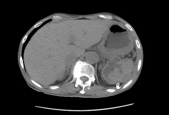
5 End of September Sudden persisted epigastralgia Elevated amylase and lipase levels CT scan of the abdomen: Blurred outline of the spleen with several hypodense areas in it Close contact of the pancreatic tail with the spleen Left pleural effusion, bil. Pleural thickening, inflammation in left lower lung
6 Mid-October Transferred to our ED Abdominal pain: Epigastric, LUQ and periumbilical areas Radiation to the left shoulder Frequency: once a day Cold sweating, pale face Body weight loss: Kg in 3 months Knee-chest position Trauma history: denied
7 Laboratory Studies AST: 43 IU/L Amylase/Lipase: 746/422 U/L CRP: 1.40 mg/dl White count, CEA, CA 19-9: WNL

8 CXR Pleural thickening

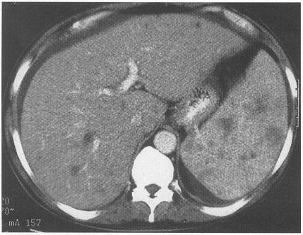
9 CXR, Lateral Pleural effusion

10 CT Scan Hypodense lesion with ill-defined border Inflammatory process Pleural effusion
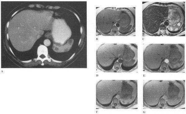
11 Pre-CM

12 Post-CM Pleural effusion
13 Stomach Multiple hypodense lesions with ill-defined borders and heterogenous contents Spleen

14 Hypodense lesion without enhancement
15 Lesion without enhancement Inflammatory process

16 Enlarged pancreas with relatively decreased density
17 Differential Diagnosis Cystic lesion With or without cell debris? Solitary or multiple? With or without enhancement? Other associated findings? Correlating patient s data and history
18 Hematoma or Abscess Heterogenous content with relatively higher density than pure fluid Inflammatory process of the peripheral tissues Infective vs. trauma: denied histories of trauma
19 Hemangioma Mostly similar to hemangiomas of the liver Delayed enhancement Mostly detected incidentally MRI T1: low signal intensity or iso-intensity T2: high signal intensity
20
21 Lymphangioma Usually: Cystic in spleen Asymptomatic Multiple Thin-walled Well-marginated Subcapsular in location Attenuation: between HU
22 Lymphoma Most common splenic malignancy Diffusely affected Splenomegaly Necrosis cystic lesion
23
24 Metastasis Most common: breast, lung, melanoma Cystic lesion: ovary, breast, endometrium, melanoma Melanoma: usually hypervascular Breast cancer
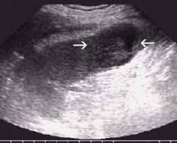
25 Diagnosis & Management Splenic abscess, suspecting secondary to chronic pancreatitis Cefamezine + Gentamicin Splenectomy Pus culture: no growth
26 Discussion Splenic Abscess
27 Clinical Presentation Fever: 95% Abdominal pain: 60% LUQ pain: 38% Left chest pain: 17% Left shoulder pain: 10% RUQ pain: 6% Weakness: 22% Chills: 22% Nausea/Vomiting: 16% Anorexia: 15% Diaphoresis: 12% Weight loss: 11% Change in bowel habits: 9%
28 Abdominal tenderness: 59% Splenomegaly: 54% Hepatomegaly: 16% Abdominal distention: 13% Ascites: 7% Costovertebral angle tenderness: 5% Friction rub: 3%
29 Dullness at left thoracic base: 33% Left basilar rales: 21% Elevated left diaphragm: 18% Left pleural friction rub: 5%
30 Lab Studies Leukocytosis: 70% Immunocompromised patients with fungal abscesses Elevated alkaline phosphatase level Positive blood culture: 60%
31 Image Studies CXR Abnormal in 80% of patients Elevated left hemidiaphragm: 33% Pleural effusion: 28% Abdominal radiograph Abnormal soft tissue density or gas pattern: 35% Radioisotope scanning: of little value
32 Ultrasound Repeatable for interval change Nonspecific Highly variable and not easy to interpret CT Test of choice Sensitivity: ~100% Low-density lesions without enhancement CT-guided aspiration
33 Diagnostic procedure CT-guided or ultrasound-guided aspiration Proper antibiotic treatment
34 Indication Once identified, always treated!
35

36 Grainger & Allison's Diagnostic Radiology: A Textbook of Medical Imaging, 4th ed.
37 plenic_fig1.aspx
38 Treatment Medical treatment Interventional treatment involving antimicrobials Primary medical management: controversial Surgical treatment Percutaneous drainage or splenectomy + antimicrobial therapy
39 Percutaneous drainage Uniloculate solitary abscess Not contraindicated splenic flexure of the colon and the pleural space risk of injury Patients with cavities that have calcified walls or patients with a history of travel to endemic areas Echinococcus
40 Percutaneous drainage Complications: Hemorrhage Pleural empyema Pneumothorax Fistula formation Contraindications: Contiguous process A phlegmonous or poorly differentiated lesion on CT scan Multiloculated or debris-filled abscess Uncontrollable coagulopathy
41 Splenectomy Splenotomy: Reserved for only the sickest patients who have contraindications to both splenectomy and percutaneous drainage
42 Complications Mortality rate: 100% if left untreated Rupture: Most common peritoneal cavity: 6.6% Rupture into the bowel, bronchus, or pleural space Colon obstruction Splenocutaneous fistula
43 Complications of Treatment Atelectasis Left-sided pleural effusion Pneumonia Subphrenic abscess Pancreatic injury with fistula or pseudocyst Thrombocytosis Overwhelming postsplenectomy sepsis
Imaging of liver and pancreas
 Imaging of liver and pancreas.. Disease of the liver Focal liver disease Diffusion liver disease Focal liver disease Benign Cyst Abscess Hemangioma FNH Hepatic adenoma HCC Malignant Fibrolamellar carcinoma
Resident Teaching Conference 10/16/09 Rondi Kauffmann Resident presenter William Nealon Faculty presenter
 Resident Teaching Conference 10/16/09 Rondi Kauffmann Resident presenter William Nealon Faculty presenter KC 59 year old male Referred to Surgery clinic for incidentally discovered 5cm x 3cm pancreatic
PANCREATIC PSEUDOCYSTS. Madhuri Rao MD PGY-5 Kings County Hospital Center
 PANCREATIC PSEUDOCYSTS Madhuri Rao MD PGY-5 Kings County Hospital Center 34 yo M Case Presentation PMH: Chronic pancreatitis (ETOH related) PSH: Nil Meds: Nil NKDA www.downstatesurgery.org Symptoms o Chronic
Chief complaint. A mass at right chest
 Chief complaint A mass at right chest Present illness This 1-year-5-month-old girl had a mass at right side chest since one month ago. flat and not tender at first In the recent 2 days, the mass enlarged
Morning Report. Allison Haden, MD October 1, 2002
 Morning Report Allison Haden, MD October 1, 2002 68 yo WM with N/V and abdominal pain Became ill about 2 weeks ago, per pt. intermittent nausea: worse w/eating vomiting within 30min of eating, nonbilious,
Cystic liver lesion and eosinophilia
 November, 2005 Cystic liver lesion and eosinophilia Jakob Begun, Harvard medical School Year III Patient Presentation 55 year old Cape Verde female presented to her PCP with 6 month history of variable
objectives Pitfalls and Pearls in PET/CT imaging Kevin Robinson, DO Assistant Professor Department of Radiology Michigan State University
 objectives Pitfalls and Pearls in PET/CT imaging Kevin Robinson, DO Assistant Professor Department of Radiology Michigan State University To determine the regions of physiologic activity To understand
Plain abdomen The standard films are supine & erect AP views (alternative to erect, lateral decubitus film is used in ill patients).
.") Plain abdomen The standard films are supine & erect AP views (alternative to erect, lateral decubitus film is used in ill patients). The stomach can be readily identified by its location, gastric rugae
Plain abdomen The standard films are supine & erect AP views (alternative to erect, lateral decubitus film is used in ill patients). The stomach can be readily identified by its location, gastric rugae
Appendix 5. EFSUMB Newsletter. Gastroenterological Ultrasound
 EFSUMB Newsletter 87 Examinations should encompass the full range of pathological conditions listed below A log book listing the types of examinations undertaken should be kept Training should usually
Chief Complaint. Retroperitoneal cystic mass incidentally found at health examination center.
 Personal Information Age: 34 y/o Sex: female Past history: major systemic medical history(-) surgical history(-), family history(-) Denied food or drug allergy Chief Complaint Retroperitoneal cystic mass
CLINICAL MANIFESTATIONS AND DIAGNOSIS OF ACUTE PANCREATITIS. Raed Abu Sham a, M.D
 CLINICAL MANIFESTATIONS AND DIAGNOSIS OF ACUTE PANCREATITIS Raed Abu Sham a, M.D ACUTE PANCREATITIS Acute inflammatory process of the pancreas that resolves both clinically and histologically. It is usually
Sex: 女 Age: 51 Occupation: 無 Admission date:92/07/22
 Sex: 女 Age: 51 Occupation: 無 Admission date:92/07/22 Chief complaint Unknown fever for one month Hand tremor and left huge renal tumor was noted Present illness Suffered from fever for one month, hand
Afternoon Session Cases
 Afternoon Session Cases Case 1 19 year old woman Presented with abdominal pain to community hospital Mild incr WBC a14, 000, Hg normal, lipase 100 (normal to 75) US 5.2 x 3.7 x 4 cm mass in porta hepatis
Patient Information. Age: 8 y/o Sex: Female. Date of Admission: Date of Discharge:
 Patient Information Age: 8 y/o Sex: Female Date of Admission: 92-10-08 Date of Discharge: 92-10-18 Chief Complaint Severe admominal pain and vomiting with dysuria since last afternoon Present Illness Lower
All Those Liver Masses are not Necessarily from the Liver: A Case of a Giant Adrenal Pseudocyst Mimicking a Hepatic Cyst
 ISSN 1941-5923 DOI: 10.12659/AJCR.893798 Received: 2014.02.06 Accepted: 2015.02.18 Published: 2015.02.06 All Those Liver Masses are not Necessarily from the Liver: A Case of a Giant Adrenal Pseudocyst
Imaging of common diseases of hepatobiliary and GI system
 Imaging of common diseases of hepatobiliary and GI system Natthaporn Tanpowpong, M.D. Diagnostic radiology Faculty of Medicine, Chulalongkorn University Normal plain radiograph A = Common bile duct
Perforation of a Duodenal Diverticulum. Elective Student S. C.
 Perforation of a Duodenal Diverticulum 2008 4 Elective Student S. C. Case History An elderly male presented to the Emergency Department with abdominal pain. Chief Complaint: Worsening, diffuse abdominal
Documentation Dissection
 History of Present Illness: Documentation Dissection The patient is a 50-year-old male c/o symptoms for past 4 months 1, severe 2 bloating and stomach cramps, some nausea, vomiting, diarrhea. In last 3
A CASE REPORT OF SPONTANEOUS BILOMA - AN ENIGMATIC SURGICAL PROBLEM
 A CASE REPORT OF SPONTANEOUS BILOMA - AN ENIGMATIC SURGICAL PROBLEM *Sumanta Kumar Ghosh and Biswajit Mukherjee ESIC Medical College, Joka, Kolkata, India *Author for Correspondence ABSTRACT Occurrence
General history. Basic Data : Age :62y/o Date of admitted: Married status : Married
 General history Basic Data : Age :62y/o Date of admitted:940510 Married status : Married General history Chief Complain : bilateral ovarian cyst incidentally being found out during pap smear. Present Illness
Case Study: #3: Gallbladder Carcinoma?
 Case Study: #3: Gallbladder Carcinoma? By: Megan Wyatt K. SON Wyatt 225 2B1 RDMS, RVT Patient: Male 85 YOA Caucasian Indication: Elevated Alkaline Phosphatase History Annual physical showed elevated alkaline
Back to Basics: What Imaging Test should I order? Jeanne G. Hill, M.D. Pediatric Radiology Medical University of South Carolina
 Back to Basics: What Imaging Test should I order? Jeanne G. Hill, M.D. Pediatric Radiology Medical University of South Carolina Disclosure Neither I nor any member of my immediate family has a relevant
Clinical Radiological Pathological Conference
 Clinical Radiological Pathological Conference CASE 1: A 59-year-old female Housekeeper Live in Phuket, Thailand Progressive dyspnea for 1 year Present illness 1 year PTA : She developed dyspnea on exertion
간암의조직검사 : 언제, 어떻게? 계명대학교의과대학내과학교실 정우진
 간암의조직검사 : 언제, 어떻게? 계명대학교의과대학내과학교실 정우진 간생검한다 vs 안한다? M/81 Alcoholic LC, albumin 4.0, bil 0.6, Cr 1.06, glucose 141, afp 2.2, CA19-9 12.41 CT: R/O HCC in S8, R/O CC M/69 HBV(-), HCV(-), social alcoholics
GASTROENTEROLOGY ESSENTIALS
 GASTROENTEROLOGY ESSENTIALS Practical Gastroenterology 8/25/2018 Jahnavi Koppala, MBBS Abdullah Abdussalam, MD A 48-year-old male was evaluated for noncardiac chest pain. Treatment with PPI twice daily
Name : 黃 XX Age : 52 Sex : 女 Occupation : 廚房阿姨 Marital status : 已婚
 Name : 黃 XX Age : 52 Sex : 女 Occupation : 廚房阿姨 Marital status : 已婚 Chief Complaint Mild postprandial fullness for 2 months Present Illness This 52 year-old female suffered from intermittent post-prandial
performed to help sway the clinician in what the appropriate diagnosis is, which can substantially alter the treatment of management.
 Hello, I am Maura Polansky at the University of Texas MD Anderson Cancer Center. I am a Physician Assistant in the Department of Gastrointestinal Medical Oncology and the Program Director for Physician
Radiology of hepatobiliary diseases
 GI cycle - Lecture 14 436 Teams Radiology of hepatobiliary diseases Objectives 1. To Interpret plan x-ray radiograph of abdomen with common pathologies. 2. To know the common pathologies presentation.
GASTRO-INTESTINAL TRACT INFECTIONS - ANTIMICROBIAL MANAGEMENT
 GASTRO-INTESTINAL TRACT INFECTIONS - ANTIMICROBIAL MANAGEMENT Name & Title Of Author: Dr Linda Jewes, Consultant Microbiologist Date Amended: December 2016 Approved by Committee/Group: Drugs & Therapeutics
Acute Abdomen. Nirav Patel MD, FACS Banner University Medical Center - Phoenix
 Acute Abdomen Nirav Patel MD, FACS Banner University Medical Center - Phoenix ? Diffuse periumbilical with localization to RLQ + Nausea, anorexia, fevers - Diarrhea, emesis Exacerbated by movement, bumps
Bacterial pneumonia with associated pleural empyema pleural effusion
 EMPYEMA Synonyms : - Parapneumonic effusion - Empyema thoracis - Bacterial pneumonia - Pleural empyema, pleural effusion - Lung abscess - Complicated parapneumonic effusions (CPE) 1 Bacterial pneumonia
Chief Complain. Liver lesion found in routine health check 41 days ago
 Chief Complain Liver lesion found in routine health check 41 days ago Present Illness On 2005-7-26 at 台北署立醫院 he underwent a health check for the first time. Abdominal US showed suspicious of a 6*5 cm hepatoma,
Pediatric TB Intensive Houston, Texas October 14, 2013
 Pediatric TB Intensive Houston, Texas October 14, 2013 Radiologic Presentation of Childhood TB Susan D. John, MD, FACR October 14, 2013 Disclosures I have no disclosures or conflicts of interest to report
Malignant Focal Liver Lesions
 Malignant Focal Liver Lesions Other Than HCC Pablo R. Ros, MD, MPH, PhD Departments of Radiology and Pathology University Hospitals Cleveland Medical Center Case Western Reserve University Pablo.Ros@UHhospitals.org
Cystic Disease of the Liver Work Up and Management. Louis Ferrari MD, PGY 3 6/9/16 SUNY Downstate Medical Center
 Cystic Disease of the Liver Work Up and Management Louis Ferrari MD, PGY 3 6/9/16 SUNY Downstate Medical Center The Case 73F presents to clinic after diagnostic laparoscopy at OSH. Known liver mass for
Case Scenario 1. Discharge Summary
 Case Scenario 1 Discharge Summary A 69-year-old woman was on vacation and noted that she was becoming jaundiced. Two months prior to leaving on that trip, she had had a workup that included an abdominal
Appropriate Imaging Tests Lead to Meaningful Results. Dr. Richard Wasley May 2011
 Appropriate Imaging Tests Lead to Meaningful Results Dr. Richard Wasley May 2011 Summarize the advantages and limitations of specific imaging tests and why clinical information is so important to radiologists
Cholecystitis is defined as nonspecific inflammation of the gallbladder with or without cholelithiasis. Types: calculous and acalculous.
 Cholecystitis is defined as nonspecific inflammation of the gallbladder with or without cholelithiasis. Types: calculous and acalculous. Anatomy of the gallbladder The gallbladder, a pear-shaped reservoir
Correspondence should be addressed to Justin Cochrane;
 Case Reports in Gastrointestinal Medicine Volume 2015, Article ID 794282, 4 pages http://dx.doi.org/10.1155/2015/794282 Case Report Acute on Chronic Pancreatitis Causing a Highway to the Colon with Subsequent
Pediatric TB Intensive Houston, Texas
 Pediatric TB Intensive Houston, Texas November 13, 2009 Radiographic Manifestations of Pediatric TB Susan D. John, MD, FACR November 13, 2009 Radiologic Presentation of Childhood TB Susan D. John, MD,
Jaundice. Agnieszka Dobrowolska- Zachwieja, MD, PhD
 Jaundice Agnieszka Dobrowolska- Zachwieja, MD, PhD Jaundice definition Jaundice, as in the French jaune, refers to the yellow discoloration of the skin. It arises from the abnormal accumulation of bilirubin
Pleural Diseases. Dr Matthew J Knight Consultant Respiratory Physician
 Pleural Diseases Dr Matthew J Knight Consultant Respiratory Physician What do you need to know? What do you need to know? Pleura- normal anatomy and physiology Pleural effusions Causes and investigations
Splenic Cystic Lesions - Differential Diagnosis
 Splenic Cystic Lesions - Differential Diagnosis Poster No.: C-1343 Congress: ECR 2013 Type: Educational Exhibit Authors: N. Neto, P. G. M. G. Ferreira, A. Vasconcelos ; Lisboa/PT, 1 2 2 3 1 3 Amadora/PT,
Radiology. Undergraduate Radiology Sample Questions
 Radiology Undergraduate Radiology Sample Questions April 2012 The following examples are offered of questions that might be used to assess undergraduate radiology. There are 3 different styles: An OSCE
Case Study #2. Case Study #1 cont 9/28/2011. CAPA 2011 Christy Wilson PA C. LH is 78 yowf with PMHx of metz breast CA presents
 Case Study #1 CAPA 2011 Christy Wilson PA C 46 yo female presents with community acquired PNA (CAP). Her condition worsened and she was transferred to the ICU and placed on mechanical ventilation. Describe
INVESTIGATIONS OF GASTROINTESTINAL DISEAS
 INVESTIGATIONS OF GASTROINTESTINAL DISEAS Lecture 1 and 2 دز اسماعيل داود فرع الطب كلية طب الموصل Radiological tests of structure (imaging) Plain X-ray: May shows soft tissue outlines like liver, spleen,
Extraosseous myeloma: imaging features
 Extraosseous myeloma: imaging features C. Santos Montón, R. Corrales, J. M. Bastida Bermejo, M. Villanueva Delgado, R. E. Correa Soto, J. M. Alonso Sánchez; Salamanca/ES Learning objectives -To review
Chest XRay interpretation INTERPRETATIONS Identifications: Name & Date Technical evaluation Basic Interpretations
 Chest XRay interpretation INTERPRETATIONS Identifications: Name & Date Technical evaluation Basic Interpretations TECHNICAL EVALUATION 1. Projection: AP/PA view To differentiate between AP & PA films,
Liver Cancer (Hepatocellular Carcinoma or HCC) Overview
 Overview") Liver Cancer (Hepatocellular Carcinoma or HCC) Overview Recent advances in liver cancer care seek to address the rising incidence of liver cancer, which has steadily increased over the past three decades.
Liver Cancer (Hepatocellular Carcinoma or HCC) Overview Recent advances in liver cancer care seek to address the rising incidence of liver cancer, which has steadily increased over the past three decades.
Causes of abdominal pain Doctors in the ED spend lots of time and money diagnosing abdominal pain. They still often do not know the exact cause
 1 2 3 What's Going On in There? EMS and Abdominal Pain Kevin McFarlane BSN,RN,CEN,CPEN,EMT Southwest Emergency Education and Consulting What is going on in there Acute Abdomen Sudden onset of pain within
CT 101 :Pancreas and Spleen
 CT 101 :Pancreas and Spleen Shikha Khullar,, MD, MPH Division of Radiology University of South Alabama The Pancreas Normal Pancreas 3 Phase Pancreatic CT Non contrast Arterial phase : 30-35 35 second
Case 1. Intro to Gallbladder & Pancreas Pathology. Case 1 DIAGNOSIS??? Acute Cholecystitis. Acute Cholecystitis. Helen Remotti M.D.
 Cholecystitis acute chronic Gallbladder tumors Adenomyoma (benign) Adenocarcinoma Pancreatitis acute chronic Pancreatic tumors Intro to Gallbladder & Pancreas Pathology Helen Remotti M.D. Case 1 70 year
Dr Claire Smith, Consultant Radiologist St James University Hospital Leeds
 Dr Claire Smith, Consultant Radiologist St James University Hospital Leeds Imaging in jaundice and 2ww pathway Image protocol Staging Limitations Pancreatic cancer 1.2.4 Refer people using a suspected
FIRST COAST SERVICE OPTIONS FLORIDA MEDICARE PART B LOCAL COVERAGE DETERMINATION
 FIRST COAST SERVICE OPTIONS FLORIDA MEDICARE PART B LOCAL COVERAGE DETERMINATION CPT/HCPCS Codes 72192 Computed tomography, pelvis; without contrast material 72193 with contrast material(s) 72194 without
In any operation. Indications. Anaesthesia. Position of the patient. Incision. Steps of the operation. Complications.
 In any operation Indications. Anaesthesia. Position of the patient. Incision. Steps of the operation. Complications. Abdominal operation I position for operation Supine Abdominal operation I position for
Safe Answers For The American Board of Surgery Certifying Exam & Recertifying Exam
 Safe Answers For The American Board of Surgery Certifying Exam & Recertifying Exam By Sarmad Aji, MD., FACS. A comprehensive review of the most commonly asked questions on the American Board of Surgery
Management of Pleural Effusion
 Management of Pleural Effusion Development of Pleural Effusion pulmonary capillary pressure (CHF) capillary permeability (Pneumonia) intrapleural pressure (atelectasis) plasma oncotic pressure (hypoalbuminemia)
Ultrasound ICD-10-CM
 Ultrasound ICD-10-CM Clinical Documentation Guides Brought to you by www.codingstrategies.com The Resource for Physician and Outpatient Coding, Compliance & ICD-10-CM OTHER CLINICAL DOCUMENTATION GUIDES
SPECIAL DIAGNOSTIC STUDIES IN BLUNT TRAUMA OLEH : Prof.DR.Dr Abdul Rasyid SpRad (K),Ph.D Dr.Evo Elidar Sp.Rad
,Ph.D Dr.Evo Elidar Sp.Rad") SPECIAL DIAGNOSTIC STUDIES IN BLUNT TRAUMA OLEH : Prof.DR.Dr Abdul Rasyid SpRad (K),Ph.D Dr.Evo Elidar Sp.Rad Trauma Emergency Room layout Ideally the trauma emergency room is centrally located to provide
SPECIAL DIAGNOSTIC STUDIES IN BLUNT TRAUMA OLEH : Prof.DR.Dr Abdul Rasyid SpRad (K),Ph.D Dr.Evo Elidar Sp.Rad Trauma Emergency Room layout Ideally the trauma emergency room is centrally located to provide
Diagnostic Approach to Pleural Effusion
 Diagnostic Approach to Pleural Effusion Objectives Define the leading causes of pleural effusion Classify the type of effusion Identify procedures and tests associated with diagnosis 2 Agenda Basic anatomy
Radiological Investigations of Abdominal Trauma
 76 77 Investigations of Abdominal Trauma Introduction: Trauma to abdominal organs is a common cause of patient morbidity and mortality among trauma patients. Causes of abdominal trauma include blunt injuries,
Undergraduate Teaching
 Prof. James F Meaney Undergraduate Teaching Chest X-Ray Understanding the normal anatomical by reference to cross sectional imaging Radiology? It s FUN! Cryptic puzzle Sudoku (Minecraft?) It s completely
Name that disease. C.J. Wolf, MD, CPC, CPC-H. University of Texas System Case #1
 Name that Disease C.J. Wolf, MD, CPC, CPC-H Asst. Systemwide Compliance Officer University of Texas System cwolf@utsystem.edu Case #1 17 year old girl who has lost 20 lbs in the last six months Thin and
Cardiac Mass in a 15-Year-Old Boy
 Cardiac Mass in a 15-Year-Old Boy Echocardiographic Case Report Hortensia Vuçini Department of Cardiology and Cardiac Surgery UHC Mother Theresa Tirana, Albania October 20, 2007 Case Presentation 15 year-old
GENERAL ABDOMINAL IMAGING PERITONEAL SPACE, PANCREAS, & SPLEEN. VMB 960 March 25, 2013
 GENERAL ABDOMINAL IMAGING PERITONEAL SPACE, PANCREAS, & SPLEEN VMB 960 March 25, 2013 REFERENCE Chapters 35-36 Pages 650-678 Chapter 37 Pages 694-701 Chapter 3 Pages 38-49 OBJECTIVES Radiography and Ultrasound
Emergency Surgery Course Graz, March ACUTE PANCREATITIS. Carlos Mesquita Coimbra
 ACUTE PANCREATITIS Carlos Mesquita Coimbra ESSENTIALS (1) AP occurs when digestive enzymes become activated while still in the pancreas, causing inflammation repeated bouts of AP can lead to chronic pancreatitis
Diagnosis of TB: Radiology David Finlay, MD
 TB Intensive Tyler, Texas June 2-4, 2010 Diagnosis of TB: Radiology David Finlay, MD June 3, 2010 2stages stages- Tuberculosis 1. primary infection 2. reactivation, or post primary disease 2 1 Primary
Radiology of the abdomen Lecture -1-
 Radiology of the abdomen Lecture -1- Objectives To know radiology modalities used in abdomen imaging mainly GI tract. To know advantages and disadvantages of each modality. To know indications and contraindications
Abdomen Sonography Examination Content Outline
 Abdomen Sonography Examination Content Outline (Outline Summary) # Domain Subdomain Percentage 1 2 3 Anatomy, Perfusion, and Function Pathology, Vascular Abnormalities, Trauma, and Postoperative Anatomy
Systemic lupus erythematosus (SLE): Pleuropulmonary Manifestations
: Pleuropulmonary Manifestations") 08/30/10 09/26/10 Systemic lupus erythematosus (SLE): Pleuropulmonary Manifestations Camila Downey S. Universidad de Chile, School of Medicine, Year VII Harvard University, School of Medicine Sept 17,
08/30/10 09/26/10 Systemic lupus erythematosus (SLE): Pleuropulmonary Manifestations Camila Downey S. Universidad de Chile, School of Medicine, Year VII Harvard University, School of Medicine Sept 17,
MANAGEMENT RECOMMENDATIONS
 1 MANAGEMENT RECOMMENDATIONS 1. Adrenal masses!!!!!!! page 2 2. Liver Masses!!!!!!! page 3 3. Obstetric US Soft Markers for Aneuploidy!! pages 4-6 4. Ovarian and Adnexal Cysts!!!!! pages 7-10 5. Pancreatic
CT EVALUATION OF GASTRIC LESIONS:
 CT EVALUATION OF GASTRIC LESIONS: Pictural essay Hasni Bouraoui I, Kahloun A, Jemni H, Elouni F, Moulahi H, Daadoucha A, Ben Ali A, Sriha B, Tlili Graies K Departments of Radiology, Gastro enterology,
Management of Gallbladder Disease
 Management of Gallbladder Disease Steven B. Johnson, MD, FACS, FCCM Professor and Chairman, Department of Surgery Program Director, Phoenix Integrated Surgical Residency University of Arizona College of
Splenectomy Procedure Information
 Where is the spleen located? Splenectomy Procedure Information Your spleen is located in the upper left side of the abdomen, partly protected by your lower ribs. It lies beneath the diaphragm; near the
Birthday: 1952/07/31 Date of admission:1999/12/30 Age:48 y/o Past medication:esrd under regular HD for 5+ years; denied DM and HTN
 Birthday: 1952/07/31 Date of admission:1999/12/30 Age:48 y/o Past medication:esrd under regular HD for 5+ years; denied DM and HTN Chief Complaint : 1)intermittent LLQ cramping pain for 2 months 2) LGI
Esophageal injuries. 新光急診張志華醫師 Facebook.com/jack119
 Esophageal injuries 新光急診張志華醫師 Facebook.com/jack119 Pre-test 1 What is the most common cause of esophageal injuries? A. Traffic accidents B. Gunshot wounds C. Iatrogenic Pre-test 2 Which contrast agent
Lung Cancer - Suspected
 Lung Cancer - Suspected Shared Decision Making Lung Cancer: http://www.enhertsccg.nhs.uk/ Patient presents with abnormal CXR Lung cancer - clinical presentation History and Examination Incidental finding
Personal data. Age : 63 Gender : male
 Personal data Age : 63 Gender : male Chief complain No specific symptom or discomfort A hepatic mass, found by abdominal sonography of routine health exam on 88-12-08 Past history 1984-3-3 Old CVA with
Case 1307 Mesothelial cysts
 Case 1307 Mesothelial cysts Vinhais S, Monteiro M, Cunha TM INSTITUTO PORTUGUÊS DE ONCOLOGIA de Francisco Gentil de LISBOA Section: Gastro-Intestinal Imaging Published: 2001, Nov. 23 Patient: 44 year(s),
Spontaneous Regression of Pancreatic. Pseudocyst Mimicking a Submucosal. Tumor of the Stomach with Upper. Gastrointestinal Bleeding.
 2006 17 128-132 Spontaneous Regression of Pancreatic Pseudocyst Mimicking a Submucosal Tumor of the Stomach with Upper Gastrointestinal Bleeding Report of a Case Kuo-Chih Tseng, Yu-Hsi Hsieh, Chang-An
Abdominal Examination Benchmarks
 Abdominal Examination Benchmarks Preparation and Positioning: Stand on the right side of the patient. The patient should be supine and double draped so only the abdomen is exposed o To relax the abdominal
12 Blueprints Q&A Step 2 Surgery
 12 Blueprints Q&A Step 2 Surgery 34. A 40-year-old female has been referred to you for a recent ER and hospital admission, from which she was given a diagnosis of acute diverticulitis. Treatment at that
Science & Technologies
 A GIANT LIVER HYDATIDE CYST SIMULTANEOUSLY PERFORATED TO PERITONEAL AND PLEURAL CAVITIES A RARE CASE REPORT. Ivan P. Novakov Department of Special Surgery; Medical University - Plovdiv Abstract. Background.
Case 1. A 35-year-old male presented with fever, cough, and purulent sputum for one week. This was his CXR (Fig. 1.1). What is the diagnosis?
. What is the diagnosis?") 1 Interpreting Chest X-Rays CASE 1 Fig. 1.1 Case 1. A 35-year-old male presented with fever, cough, and purulent sputum for one week. This was his CXR (Fig. 1.1). What is the diagnosis? CASE 1 Interpreting
1 Interpreting Chest X-Rays CASE 1 Fig. 1.1 Case 1. A 35-year-old male presented with fever, cough, and purulent sputum for one week. This was his CXR (Fig. 1.1). What is the diagnosis? CASE 1 Interpreting
What s your diagnosis?
 What s your diagnosis? Signalment: 9 year old MC 2.7 kg Papillion Presenting Complaint: Presented for work up of anorexia and vomiting History: He had presented to cardiology for work up of a grad IV/VI
Chest Radiology Interpretation: Findings of Tuberculosis
 Chest Radiology Interpretation: Findings of Tuberculosis Get out your laptops, smart phones or other devices pollev.com/chestradiology Case #1 1 Plombage Pneumonia Cancer 2 Reading the TB CXR Be systematic!
ACUTE ABDOMEN IN OLDER CHILDREN. Carlos J. Sivit M.D.
 ACUTE ABDOMEN IN OLDER CHILDREN Carlos J. Sivit M.D. ACUTE ABDOMEN Clinical condition characterized by severe abdominal pain developing over several hours ACUTE ABDOMINAL PAIN Common childhood complaint
U Nordic Forum - Trauma & Emergency Radiology. Lecture Objectives. MDCT in Acute Pancreatitis. Acute Pancreatitis: Etiologies
 Nordic Forum - Trauma & Emergency Radiology Lecture Objectives MDCT in Acute Pancreatitis Borut Marincek Institute of Diagnostic Radiology niversity Hospital Zurich, Switzerland To describe the role of
is time consuming and expensive. An intra-operative assessment is not going to be helpful if there is no more tissue that can be taken to improve the
 My name is Barry Feig. I am a Professor of Surgical Oncology at The University of Texas MD Anderson Cancer Center in Houston, Texas. I am going to talk to you today about the role for surgery in the treatment
Respiratory Diseases and Disorders
 Chapter 9 Respiratory Diseases and Disorders Anatomy and Physiology Chest, lungs, and conducting airways Two parts: Upper respiratory system consists of nose, mouth, sinuses, pharynx, and larynx Lower
Guidelines, Policies and Statements D5 Statement on Abdominal Scanning
 Guidelines, Policies and Statements D5 Statement on Abdominal Scanning Disclaimer and Copyright The ASUM Standards of Practice Board have made every effort to ensure that this Guideline/Policy/Statement
Characterization of adrenal lesions on CT and MRI: all that a radiologist must know
 Characterization of adrenal lesions on CT and MRI: all that a radiologist must know Poster No.: C-2476 Congress: ECR 2013 Type: Educational Exhibit Authors: N. Benzina, S. MAJDOUB, C. H. ZARRAD, H. Zaghouani,
BIOE221. Session 6. Abdominal Examination. Bioscience Department. Endeavour College of Natural Health endeavour.edu.au
 BIOE221 Session 6 Abdominal Examination Bioscience Department Examination of the Abdomen Session objectives Understand the organs / structures that are present in the abdominal cavity Understand the importance
Disorders of the Liver, Gallbladder and Pancreas
 Disorders of the Liver, Gallbladder and Pancreas Objectives: Disorders of the liver Disorders of the gall bladder Disorders of the pancreas Part 1: Disorders of the Liver 1 Jaundice: is a manifestation
Bronchogenic Carcinoma
 A 55-year-old construction worker has smoked 2 packs of ciggarettes daily for the past 25 years. He notes swelling in his upper extremity & face, along with dilated veins in this region. What is the most
Ventriculoperitoneal Shunt with Communicating Peritoneal & Subcutaneous Pseudocysts Formation
 International Journal of Health Sciences, Qassim University, Vol. 8, No. 1 (January-March 2014) Case Report Ventriculoperitoneal Shunt with Communicating Peritoneal & Subcutaneous Pseudocysts Formation
Ultrasound Of The Acute Abdomen a survival guide. Alison McGuinness Consultant sonographer Mid Yorks Hospitals NHS Trust
 Ultrasound Of The Acute Abdomen a survival guide Alison McGuinness Consultant sonographer Mid Yorks Hospitals NHS Trust Definition Rapid onset of severe abdominal pain usually requiring admission to hospital
ROLE OF COMPUTED TOMOGRAPHY IN EVALUATION OF ASCITES Ramesh Chander 1, Kamlesh Gupta 2, Arvinder Singh 3, V. K. Rampal 4, Mohit Khandelwal 5
 ROLE OF COMPUTED TOMOGRAPHY IN EVALUATION OF ASCITES Ramesh Chander 1, Kamlesh Gupta 2, Arvinder Singh 3, V. K. Rampal 4, Mohit Khandelwal 5 HOW TO CITE THIS ARTICLE: Ramesh Chander, Kamlesh Gupta, Arvinder
IT 의료융합 1 차임상세미나 복부질환초음파 이재영
 IT 의료융합 1 차임상세미나 2013-4-3 복부질환초음파 이재영 나는오늘누구를위하여 종을울리나? 전통적의료 의사 공학설계자 의사 최첨단진단장비들 USG, CT, MRI 환자 환자 현대의료 사용자중심의사고 US in the Abdomen Detection DDx Look Behavior Response by external stimuli Guiding Tool
X-ray Corner. Imaging of The Pancreas. Pantongrag-Brown L
 X-ray Corner 125 Imaging of The Pancreas Modern imaging modalities commonly used in pancreas include ultrasound (US), CT, and MRI. Pancreas is a retroperitoneal organ which makes it difficult to visualize