Developmental sequence of brain
|
|
|
- Anabel O’Neal’
- 5 years ago
- Views:
Transcription
1 Cerebellum

2 Developmental sequence of brain Fourth week Fifth week

3 Location of cerebellum Lies above and behind the medullar and pons and occupies posterior cranial fossa

4 Location of cerebellum

5 External structures Consists of two cerebellar hemispheres united in the midline by the vermis Primary fissure

6 External structures Lobules of cerebellum: Anterior lobe, posterior lobe, flocculonodular lobe. Posterolateral fissure


7 External structures Tonsil of cerebellum two elevated masses on inferior surface of hemispheric portion, just nearby foramen magnum Cerebellar tonsilar hernia

8 Internal structures Cerebellar cortex gray matter Granular layer, piriform (Purkinje) cell layer, molecular layer Cerebellar medulla white matter Superior / Middle / Inferior cerebellar peduncles Cerebellar nuclei gray matter within white matter Fastigial, globose, emboliform, dentate nuclei Interposed nucleus

9 Cerebellar cortex Molecular layer Piriform cell layer Granular layer White matter

10 Cerebellar nuclei Fastigial, globose, emboliform, dentate nuclei
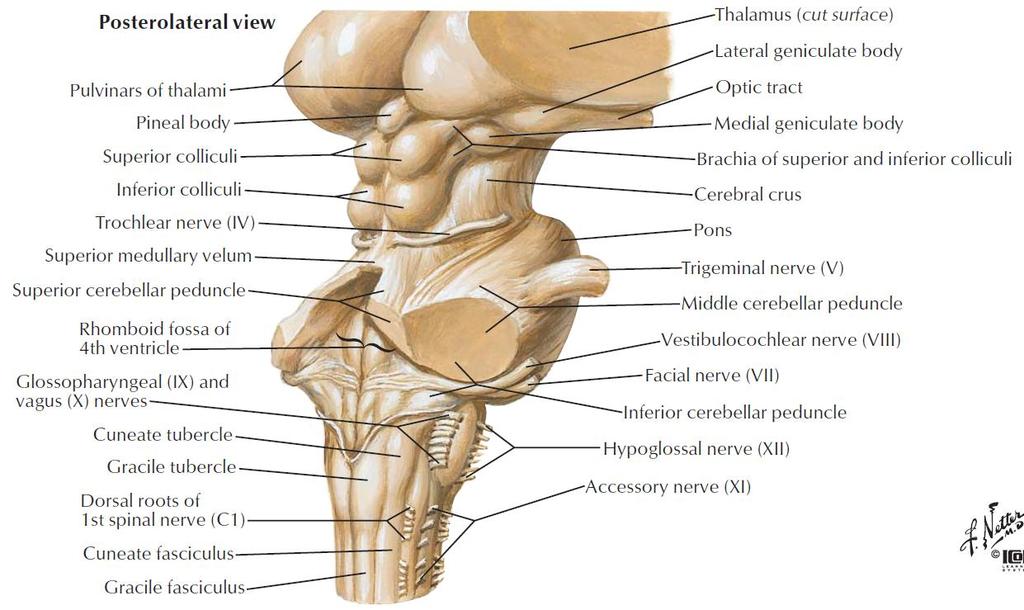
11 Cerebellar peduncles
")
 connects with medulla and with spinal cord, contain both afferent and efferent")
12 Cerebellar peduncles superior cerebellar peduncle (brachium conjunctivum) connects with midbrain, contain mostly efferent fibers middle cerebellar peduncle (brachium pontis) connects with pons, contain afferent fibers inferior cerebellar peduncle (restiform body) connects with medulla and with spinal cord, contain both afferent and efferent fibers


13 Functional divisions of cerebellum Vestibulocerebellum Archicerebellum Flocculonodular lobe Spinocerebellum Paleocerebellum Vermis and intermediate zone Cerebrocerebellum Neocerebellum Lateral zone Flocculonodular lobe Intermediate zone Vermis Lateral zone
14 Connections and functions of Vestibulocerebellum Connections cerebellum Afferents: receives inputs from vestibular nuclei and vestibular ganglion Efferents: projects to the vestibular nucleus vestibulospinal tract and medial longitudinal fasciculus motor neurons of anterior horn and cranial nuclei innervating extraocular muscles
15 Function: regulation of eyeball movements and maintenance of body equilibrium.
16 Spinocerebellum Connections Afferents: receives somatic sensory information via spinocerebellar tracts Efferents: Vermis projects to the fastigial nucleus vestibular nuclei and reticular formation vestibulospinal tract and reticulospinal tract motor neurons of anterior horn Intermediate zone projects to the interposed nuclei Contralateral red nucleus rubrospinal tract motor neurons of anterior horn Contralateral VL of thalamus cerebral cortex coticospinal tract motor neurons of anterior horn
17 Function: modulate the muscular tension.
18 Cerebrocerebellum Connections Afferents: receives input from the cerebral cortex via a relay in pontine nuclei Efferents: projects to dentate nucleus VL of thalamus primary motor cortex corticospinal tract motor neurons of anterior horn
19 Function: modulate the coordination of voluntary and fineness movement, including determination of movement strength, direction and amplitude.
20 Cerebellar functions Cerebellum has functions of 1. maintaining body posture, 2. controlling muscular tension, 3. coordinating of voluntary movement, but NOT initiating and performing voluntary movement.

21 Cerebellar injury symptoms Typical signs of cerebellar injury: a. Ataxia: incoordination of voluntary movement b. Nystagmus: tremor of eyeball c. Intention tremor Nose-to-finger test
22 Diencephalon
23 Location

24 Location Located between midbrain and telencephalon, mostly surrounded by cerebral hemispheres

25 Divisions of diencephalon (dorsal) thalamus metathalamus epithalamus subthalamus hypothalamus

26 External features A large egg-shaped nucleus mass, Right and left portion of thalamus are joined by interthalamic adhesion Anterior end called anterior thalamic tubercle, Posterior end called pulvinar Terminal stria Hypothalamic sulcus Dorsal thalamus

27 Dorsal thalamus
28 Subdivision Principal Nuclei Common abbreviation Ant. nuclear group Anterior Med. nuclear group dorsomedial Lat. nuclear group Dorsal layer Lateral dorsal LD Lateral posterior Pulvinar Ventral layer Ventral anterior VA Ventral lateral (intermediate) Ventral posterior Ventral posterolateral Ventral posteromedial LP VL (VI) VP VPL VPM
29 Functional subdivision Non-specific projecting nuclei ---- receive afferents from olfactory brain and reticular formation of brain stem (ascending reticular activating system), project to hypothalamus, corpus striatum and diffusely to cerebral cortex. Midline nuclei Intralaminar nuclei Thalamic reticular nuclei Internuncial nuclei ---- receive a wide variety of afferent fibers and in turn project widely to association areas of cerebral cortex, involved in higher nervous activity of brain. Anterior nuclear group Medial nuclear group Dorsal layer of lateral nuclear group
30 Functional subdivision Specific relaying nuclei Ventral anterior nucleus (VA) Ventral lateral nucleus (VL) Receive inputs from corpus striatum, dentate nucleus, and substantia nigra, project to motor cortex to regulate body movement. Ventral posteromedial nucleus (VPM) -receives trigeminal lemniscus and taste fibers Ventral posterolateral nucleus (VPL) -receives medial lemniscus and spinal lemniscus Projects to primary somatic sensory area via central thalamic radiations

 Medial")

31 Metathalamus Lateral geniculate body (LGN) Medial geniculate body (MGN) Specific relaying nuclei
 inferior colliculus Projects to auditory area via acoustic")
 Projects to visual area via optic radiation")
32 Medial geniculate body (MGN) Relay station of audition Receive fibers from (brachium of) inferior colliculus Projects to auditory area via acoustic radiation Lateral geniculate body (LGN) Relay station of vision Receive fibers from retina (optic tract) Projects to visual area via optic radiation Metathalamus

33 Epithalamus Includes Thalamic medullary stria Habenular trigone Habenular commissure Pineal body: Melatonin posterior commissure Dorsal view


34 Subthalamus Ventrally to the dorsal thalamus Contains subthalamic nucleus involved in movement regulation. subthalamic nucleus
35 Hypothalamus Position-lies ventral to thalamus Boundaries Superior: hypothalamic sulcus Inferior: optic chiasma tuber cinereum Infundibulum (hypophysis) mamillary body Anterior: interventricular foramen Posterior: continues with midbrain tegmentum
36 Hypothalamus


37 Hypothalamic divisions Anterior to posterior Preoptic region Supraoptic region Tuberal region Mamillary region A P
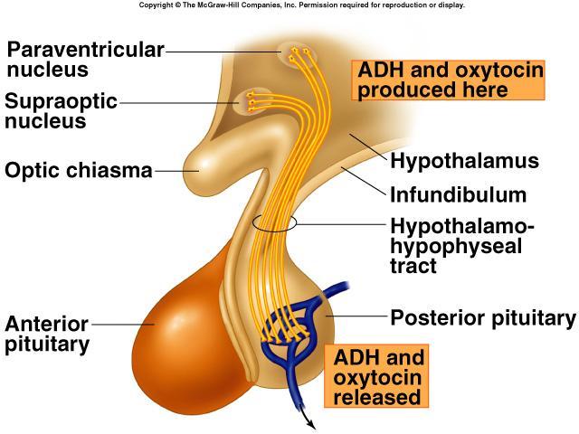
38 Principal nuclei in hypothalamus Preoptic region: preoptic nucleus Supraoptic region Supraoptic nucleus Paraventricular nucleus produce antidiuretic hormone (ADH or vasopressin) and oxytocin Tuberal region Infundibular (Arcuate) nucleus Ventromedial nucleus Dorsomedial nucleus Mamillary region Mamillary nucleus Posterior hypothalamic nucleus

39 Anterior commissure Preoptic nucleus Anterior hypothalamic nucleus Supraoptic nucleus Suprachiasmatic nucleus Optic chiasma Infundibulum (stalk of the pituitary gland) Paraventricular nucleus Fornix Arcuate nucleus Pituitary gland Dorsomedial nucleus Posterior hypothalamic nucleus Lateral hypothalamic area Ventromedial nucleus Mammillary body The main hypothalamic nuclei

40 Hypothalamus -- connection Connects with limbic system, brainstem, spinal cord and dorsal thalamus Connects with hypophysis
41 Hypothalamus -- connection Supraoptic nucleus supraopticohypophysial tract posterior lobe of hypophysis (neurohypophysis) Paraventricular nucleus paraventriculohypophysial tract posterior lobe of hypophysis (neurohypophysis)
42

43 Hypothalamus -- connection Parvicellular neurons in the arcuate nucleus and nearby region secrete releasing and inhibiting hormones tuberoinfundibular tract portal vein of hypophysis anterior lobe of hypophysis

44 Thyroidstimulating hormone Adrenocorticot ropic hormone
45 Functions of hypothalamus Neuroendocrine center Subcortical higher regulatory center of the visceral activity, such as temperature regulation food ingestion reproduction fluid and electrolyte balance Regulate emotional responses and biological rhythms


46 Third ventricle Position: a narrow ventriclar cleft lies within diencephalon Lateral wall: dorsal thalamus and hypothalamus Communication Lateral ventricles third ventricle midbrain aqueduct fourth ventricle
Copy Right- Hongqi ZHANG-Department of Anatomy-Fudan University. Systematic Anatomy. Nervous system Cerebellum. Dr.Hongqi Zhang ( 张红旗 )
") Systematic Anatomy Nervous system Cerebellum Dr.Hongqi Zhang ( 张红旗 ) Email: zhanghq58@126.com 1 The Cerebellum Cerebellum evolved and developed with the complication of animal movement. Key points about
Systematic Anatomy Nervous system Cerebellum Dr.Hongqi Zhang ( 张红旗 ) Email: zhanghq58@126.com 1 The Cerebellum Cerebellum evolved and developed with the complication of animal movement. Key points about
Located below tentorium cerebelli within posterior cranial fossa. Formed of 2 hemispheres connected by the vermis in midline.
 The Cerebellum Cerebellum Located below tentorium cerebelli within posterior cranial fossa. Formed of 2 hemispheres connected by the vermis in midline. Gray matter is external. White matter is internal,
Connection of the cerebellum
 CEREBELLUM Connection of the cerebellum The cerebellum has external layer of gray matter (cerebellar cortex ), & inner white matter In the white matter, there are 3 deep nuclei : (a) dentate nucleus laterally
Cerebellum John T. Povlishock, Ph.D.
 Cerebellum John T. Povlishock, Ph.D. OBJECTIVES 1. To identify the major sources of afferent inputs to the cerebellum 2. To define the pre-cerebellar nuclei from which the mossy and climbing fiber systems
Ch 13: Central Nervous System Part 1: The Brain p 374
 Ch 13: Central Nervous System Part 1: The Brain p 374 Discuss the organization of the brain, including the major structures and how they relate to one another! Review the meninges of the spinal cord and
Abdullah AlZibdeh. Dr. Maha ElBeltagy. Maha ElBeltagy
 19 Abdullah AlZibdeh Dr. Maha ElBeltagy Maha ElBeltagy Introduction In this sheet, we discuss the cerebellum; its lobes, fissures and deep nuclei. We also go into the tracts and connections in which the
Unit VIII Problem 5 Physiology: Cerebellum
 Unit VIII Problem 5 Physiology: Cerebellum - The word cerebellum means: the small brain. Note that the cerebellum is not completely separated into 2 hemispheres (they are not clearly demarcated) the vermis
Cerebellum. Steven McLoon Department of Neuroscience University of Minnesota
 Cerebellum Steven McLoon Department of Neuroscience University of Minnesota 1 Anatomy of the Cerebellum The cerebellum has approximately half of all the neurons in the central nervous system. The cerebellum
b. The groove between the two crests is called 2. The neural folds move toward each other & the fuse to create a
 Chapter 13: Brain and Cranial Nerves I. Development of the CNS A. The CNS begins as a flat plate called the B. The process proceeds as: 1. The lateral sides of the become elevated as waves called a. The
Brainstem. Amadi O. Ihunwo, PhD School of Anatomical Sciences
 Brainstem Amadi O. Ihunwo, PhD School of Anatomical Sciences Lecture Outline Constituents Basic general internal features of brainstem External and Internal features of Midbrain Pons Medulla Constituents
Prof. Saeed Abuel Makarem & Dr.Sanaa Alshaarawy
 Prof. Saeed Abuel Makarem & Dr.Sanaa Alshaarawy 1 Objectives By the end of the lecture, you should be able to: Describe the anatomy and main functions of the thalamus. Name and identify different nuclei
 Spinal Cord Tracts DESCENDING SPINAL TRACTS: Are concerned with somatic motor function, modification of ms. tone, visceral innervation, segmental reflexes. Main tracts arise form cerebral cortex and others
Introduction to the Central Nervous System: Internal Structure
 Introduction to the Central Nervous System: Internal Structure Objective To understand, in general terms, the internal organization of the brain and spinal cord. To understand the 3-dimensional organization
Brainstem. By Dr. Bhushan R. Kavimandan
 Brainstem By Dr. Bhushan R. Kavimandan Development Ventricles in brainstem Mesencephalon cerebral aqueduct Metencephalon 4 th ventricle Mylencephalon 4 th ventricle Corpus callosum Posterior commissure
Diencephalon ( interbrain )
") Diencephalon ( interbrain ) 1. Diencephalon regional organization and internal structure: thalamus topographic and nuclear organization metathalamus the geniculate bodies epithalamus pineal gland and habenula
Diencephalon ( interbrain ) 1. Diencephalon regional organization and internal structure: thalamus topographic and nuclear organization metathalamus the geniculate bodies epithalamus pineal gland and habenula
Anatomy and Physiology (Bio 220) The Brain Chapter 14 and select portions of Chapter 16
 The Brain Chapter 14 and select portions of Chapter 16") Anatomy and Physiology (Bio 220) The Brain Chapter 14 and select portions of Chapter 16 I. Introduction A. Appearance 1. physical 2. weight 3. relative weight B. Major parts of the brain 1. cerebrum 2.
Anatomy and Physiology (Bio 220) The Brain Chapter 14 and select portions of Chapter 16 I. Introduction A. Appearance 1. physical 2. weight 3. relative weight B. Major parts of the brain 1. cerebrum 2.
Motor System Hierarchy
 Motor Pathways Lectures Objectives Define the terms upper and lower motor neurons with examples. Describe the corticospinal (pyramidal) tract and the direct motor pathways from the cortex to the trunk
Voluntary Movement. Ch. 14: Supplemental Images
 Voluntary Movement Ch. 14: Supplemental Images Skeletal Motor Unit: The basics Upper motor neuron: Neurons that supply input to lower motor neurons. Lower motor neuron: neuron that innervates muscles,
Hypothalamus. To learn how the brain regulates neuroendocrine secretions NTA Ch 14, pgs Key Figs: 14-3; 14-4,
 Hypothalamus Objectives To learn the general organization of the hypothalamus and the functions of the major nuclei NTA Ch 14, pgs. 419-422 Key Figs: 14-2, 14-3 To learn how the brain regulates neuroendocrine
BIO 210 CHAPTER 13. The Central Nervous System SUPPLEMENT 2. PowerPoint by John McGill Supplemental Notes by Beth Wyatt CEREBELLUM
 BIO 210 CHAPTER 13 The Central Nervous System SUPPLEMENT 2 PowerPoint by John McGill Supplemental Notes by Beth Wyatt CEREBELLUM Second Largest Division of the Brain Lies Below the Posterior Portion of
Gross Morphology of the Brain
 Gross Morphology of the Brain Done by : Marah Marahleh & Razan Krishan *slides in bold Principal Parts of the Brain Cerebrum : largest part of the brain Diencephalon Thalamus & hypothalamus Cerebellum
The neurvous system senses, interprets, and responds to changes in the environment. Two types of cells makes this possible:
 NERVOUS SYSTEM The neurvous system senses, interprets, and responds to changes in the environment. Two types of cells makes this possible: the neuron and the supporting cells ("glial cells"). Neuron Neurons
Medial View of Cerebellum
 Meds 5371 System Neuroscience D. L. Oliver CEREBELLUM Anterior lobe (spinal) Posterior lobe (cerebral) Flocculonodular lobe (vestibular) Medial View of Cerebellum 1 Ventral View of Cerebellum Flocculus
Auditory and Vestibular Systems
 Auditory and Vestibular Systems Objective To learn the functional organization of the auditory and vestibular systems To understand how one can use changes in auditory function following injury to localize
Lecture 4 The BRAINSTEM Medulla Oblongata
 Lecture 4 The BRAINSTEM Medulla Oblongata Introduction to brainstem 1- Medulla oblongata 2- Pons 3- Midbrain - - - occupies the posterior cranial fossa of the skull. connects the narrow spinal cord
Brainstem. Steven McLoon Department of Neuroscience University of Minnesota
 Brainstem Steven McLoon Department of Neuroscience University of Minnesota 1 Course News Change in Lab Sequence Week of Oct 2 Lab 5 Week of Oct 9 Lab 4 2 Goal Today Know the regions of the brainstem. Know
By Dr. Saeed Vohra & Dr. Sanaa Alshaarawy
 By Dr. Saeed Vohra & Dr. Sanaa Alshaarawy 1 By the end of the lecture, students will be able to : Distinguish the internal structure of the components of the brain stem in different levels and the specific
Chapter 13 Brain and Cranial Nerves
 Chapter 13 Brain and Cranial Nerves 13-1 Brain and Cranial Nerves Brain Part of CNS contained in cranial cavity Control center for many of body s functions Much like a complex computer but more Parts of
Medical Neuroscience Tutorial
 Pain Pathways Medical Neuroscience Tutorial Pain Pathways MAP TO NEUROSCIENCE CORE CONCEPTS 1 NCC1. The brain is the body's most complex organ. NCC3. Genetically determined circuits are the foundation
DEVELOPMENT OF BRAIN
 Ahmed Fathalla OBJECTIVES At the end of the lecture, students should: List the components of brain stem. Describe the site of brain stem. Describe the relations between components of brain stem & their
The Cerebellum. Little Brain. Neuroscience Lecture. Dr. Laura Georgescu
 The Cerebellum Little Brain Neuroscience Lecture Dr. Laura Georgescu Learning Objectives 1. Describe functional anatomy of the cerebellum- its lobes, their input and output connections and their functions.
Internal Organisation of the Brainstem
 Internal Organisation of the Brainstem Major tracts and nuclei of the brainstem (Notes) The brainstem is the major pathway for tracts and houses major nuclei, that contain sensory, motor and autonomics
For more information about how to cite these materials visit
 Author(s): Peter Hitchcock, PH.D., 2009 License: Unless otherwise noted, this material is made available under the terms of the Creative Commons Attribution Non-commercial Share Alike 3.0 License: http://creativecommons.org/licenses/by-nc-sa/3.0/
Development of Brain Stem, Cerebellum and Cerebrum
 Development of Brain Stem, Cerebellum and Cerebrum The neural tube cranial to the 4th pair of somites develop into the brain. 3 dilatations and 2 flexures form at the cephalic end of the neural tube during
FUNCTION: It COORDINATES movement HOW IT WORKS
 CEREBELLUM Chris Cohan, Ph.D. Dept. of Pathology/Anat Sci University at Buffalo Objectives: Describe the anatomy of the cerebellum, its 3 functions and associated regions Describe how the cerebellum influences
Neuroanatomy. Dr. Maha ELBeltagy. Assistant Professor of Anatomy Faculty of Medicine The University of Jordan
 Neuroanatomy Dr. Maha ELBeltagy Assistant Professor of Anatomy Faculty of Medicine The University of Jordan 2018 THE BRAIN STEM It includes: Midbrain Pons Medulla oblongata Anterior view Anterior view
Omar Sami. Aseel Abdeen. Muhammad Al-Salem. 1 P a g e
 Omar Sami Aseel Abdeen Muhammad Al-Salem 1 P a g e Using only section 2 record, I wrote this sheet; as the video is not ready yet. Despite pointing the structures, I ve tried to include all the scientific
The Cerebellum. The Little Brain. Neuroscience Lecture. PhD Candidate Dr. Laura Georgescu
 The Cerebellum The Little Brain Neuroscience Lecture PhD Candidate Dr. Laura Georgescu Learning Objectives 1. Describe functional anatomy of the cerebellum - its lobes, their input and output connections
Nsci 2100: Human Neuroanatomy 2017 Examination 3
 Name KEY Lab Section Nsci 2100: Human Neuroanatomy 2017 Examination 3 On this page, write your name and lab section. On your bubble answer sheet, enter your name (last name, space, first name), internet
THE CEREBELLUM SUDIVISIONS, STRUCTURE AND CONNECTIONS
 THE CEREBELLUM Damage to the cerebellum produces characteristic symptoms primarily with respect to the coordination of voluntary movements. The cerebellum receives information from the skin, joints, muscles,
Unit VIII Problem 3 Neuroanatomy: Brain Stem, Cranial Nerves and Scalp
 Unit VIII Problem 3 Neuroanatomy: Brain Stem, Cranial Nerves and Scalp - Brain stem: It is connected to the cerebellum and cerebral hemispheres. Rostral end of brain stem: diencephalon is the area which
PSY 302: CHAPTER 3 NOTES THE BRAIN (PART II) - 9/5/17. By: Joseline
 - 9/5/17. By: Joseline") PSY 302: CHAPTER 3 NOTES THE BRAIN (PART II) - 9/5/17 By: Joseline Left 3 MAJOR FISSURES : 2HEMISPHERES Right Lateral Ventricle Central Fissure Third Ventricle Sulcus Lateral Fissure Gyros Fissure- Fissures
PSY 302: CHAPTER 3 NOTES THE BRAIN (PART II) - 9/5/17 By: Joseline Left 3 MAJOR FISSURES : 2HEMISPHERES Right Lateral Ventricle Central Fissure Third Ventricle Sulcus Lateral Fissure Gyros Fissure- Fissures
Neuroanatomy Dr. Maha ELBeltagy Assistant Professor of Anatomy Faculty of Medicine The University of Jordan 2018
 Neuroanatomy Dr. Maha ELBeltagy Assistant Professor of Anatomy Faculty of Medicine The University of Jordan 2018 Dr Maha ELbeltagy THE BRAIN STEM Dr Maha ELbeltagy It includes: Midbrain - Pons - Medulla
Chapter 13 Lecture Outline *
 Anatomy and Physiology, Seventh Edition Rod R. Seeley Idaho State University Trent D. Stephens Idaho State University Philip Tate Phoenix College Chapter 13 Lecture Outline * *See PowerPoint Image Slides
Blood supply to the brain Blood brain barrier isolates neural tissue from general circulation
 The Brain and Cranial Nerves Objectives Name the major regions of the brain and describe their functions. Discuss the formation, circulation, and functions of the CSF. List the main components of the medulla
Hypothalamus. lies below the hypothalamic sulcus. includes the following ventral surface structures:
 Hypothalamus I. Overview The Hypothalamus is a division of the diencephalon. lies within the floor and ventral part of the walls of the third ventricle, functions primarily in the maintenance of homeostasis.
Principles of Anatomy and Physiology
 Principles of Anatomy and Physiology 14 th Edition CHAPTER 14 The Brain and Cranial Nerves Introduction The purpose of the chapter is to: 1. Understand how the brain is organized, protected, and supplied
Property of Chonghao Zhao, MD, Ph.D.
 UCLA Orofacial Pain Lecture Series # 2 March 17 th 2011 Overview of Basal Ganglion, Thalamus, Hypothalamus, Brainstem, and Spinal Cord Neuroanatomy and MR Anatomy Chong-hao Zhao, MD, PhD American Board
For more information about how to cite these materials visit
 Author(s): Peter Hitchcock, PH.D., 2009 License: Unless otherwise noted, this material is made available under the terms of the Creative Commons Attribution Non-commercial Share Alike 3.0 License: http://creativecommons.org/licenses/by-nc-sa3.0/
Fig. 1.4 (A B). The brain stem. Anterior view. Bar: 5 mm.
. The brain stem. Anterior view. Bar: 5 mm.") Section I A Fig. 1.4 (A B). The brain stem. Anterior view. Bar: 5 mm. Medulla The boundaries between the spinal cord and medulla are noted on Fig. 1.30. The pontomedullary sulcus (1) separates the medulla
Section I A Fig. 1.4 (A B). The brain stem. Anterior view. Bar: 5 mm. Medulla The boundaries between the spinal cord and medulla are noted on Fig. 1.30. The pontomedullary sulcus (1) separates the medulla
M555 Medical Neuroscience Lab 1: Gross Anatomy of Brain, Crainal Nerves and Cerebral Blood Vessels
 M555 Medical Neuroscience Lab 1: Gross Anatomy of Brain, Crainal Nerves and Cerebral Blood Vessels Anatomical Directions Terms like dorsal, ventral, and posterior provide a means of locating structures
CEREBRUM & CEREBRAL CORTEX
 CEREBRUM & CEREBRAL CORTEX Seonghan Kim Dept. of Anatomy Inje University, College of Medicine THE BRAIN ANATOMICAL REGIONS A. Cerebrum B. Diencephalon Thalamus Hypothalamus C. Brain Stem Midbrain Pons
Brain and Cranial Nerves (Ch. 15) Human Anatomy lecture. caudal = toward the spinal cord)
 Human Anatomy lecture. caudal = toward the spinal cord)") Insight: Some cranial nerve disorders Brain and Cranial Nerves (Ch. 15) Human Anatomy lecture I. Overview (Directional terms: rostral = toward the forehead caudal = toward the spinal cord) A. 3 Major parts
Insight: Some cranial nerve disorders Brain and Cranial Nerves (Ch. 15) Human Anatomy lecture I. Overview (Directional terms: rostral = toward the forehead caudal = toward the spinal cord) A. 3 Major parts
Basal Nuclei (Ganglia)
") Doctor said he will not go deep within these slides because we will take them in physiology, so he will explain the anatomical structures, and he will go faster in the functions sheet in yellow Basal Nuclei
Doctor said he will not go deep within these slides because we will take them in physiology, so he will explain the anatomical structures, and he will go faster in the functions sheet in yellow Basal Nuclei
Diencephalon ( interbrain )
") Diencephalon ( interbrain ) 1. Diencephalon regional organization and internal structure: thalamus topographic and nuclear organization metathalamus the geniculate bodies epithalamus pineal gland and habenula
Diencephalon ( interbrain ) 1. Diencephalon regional organization and internal structure: thalamus topographic and nuclear organization metathalamus the geniculate bodies epithalamus pineal gland and habenula
I: To describe the pyramidal and extrapyramidal tracts. II: To discuss the functions of the descending tracts.
 Descending Tracts I: To describe the pyramidal and extrapyramidal tracts. II: To discuss the functions of the descending tracts. III: To define the upper and the lower motor neurons. 1. The corticonuclear
Non-cranial nerve nuclei
 Brainstem Non-cranial nerve nuclei Nucleus Gracile nucleus Cuneate nucleus Infeiro olivary nucleus Pontine nucleus inferior colliculus superior colliculus Red nucleus Substantia nigra Pretectal area Site
Faculty of Dental Medicine and Surgery. Sem 4 Cerebellum Dr. Abbas
 Faculty of Dental Medicine and Surgery Sem 4 Cerebellum Dr. Abbas Anatomy of the cerebellum Cerebellum Configurations External - located in posterior cranial fossa - communicate with other structure via
Organization of The Nervous System PROF. SAEED ABUEL MAKAREM
 Organization of The Nervous System PROF. SAEED ABUEL MAKAREM Objectives By the end of the lecture, you should be able to: List the parts of the nervous system. List the function of the nervous system.
The Nervous System: Sensory and Motor Tracts of the Spinal Cord
 15 The Nervous System: Sensory and Motor Tracts of the Spinal Cord PowerPoint Lecture Presentations prepared by Steven Bassett Southeast Community College Lincoln, Nebraska Introduction Millions of sensory
Cranial Nerve VIII (The Vestibulo-Cochlear Nerve)
") Cranial Nerve VIII (The Vestibulo-Cochlear Nerve) Please view our Editing File before studying this lecture to check for any changes. Color Code Important Doctors Notes Notes/Extra explanation Objectives
Cranial Nerve VIII (The Vestibulo-Cochlear Nerve) Please view our Editing File before studying this lecture to check for any changes. Color Code Important Doctors Notes Notes/Extra explanation Objectives
Thalamus and the Internal Capsule
 This power point is made available as an educational resource or study aid for your use only. This presentation may not be duplicated for others and should not be redistributed or posted anywhere on the
The Nervous System PART B
 7 The Nervous System PART B PowerPoint Lecture Slide Presentation by Jerry L. Cook, Sam Houston University ESSENTIALS OF HUMAN ANATOMY & PHYSIOLOGY EIGHTH EDITION ELAINE N. MARIEB The Reflex Arc Reflex
PROPERTY OF ELSEVIER SAMPLE CONTENT - NOT FINAL. Gross Anatomy and General Organization of the Central Nervous System
 3 Gross Anatomy and General Organization of the Central Nervous System C h a p t e r O u t l i n e The Long Axis of the CNS Bends at the Cephalic Flexure Hemisecting a Brain Reveals Parts of the Diencephalon,
DIENCEPHALON. ..Central core of the forebrain..consists of three paired structures
 DIENCEPHALON..Central core of the forebrain..consists of three paired structures ---------- --thalamus, --------hypothalamus, -------epithalamus..encloses the third ventricle Diencephalon between cerebral
Telencephalon (Cerebral Hemisphere)
") Telencephalon (Cerebral Hemisphere) OUTLINE The Cortex - Lobes, Sulci & Gyri - Functional Subdivisions - Limbic Lobe & Limbic System The Subcortex - Basal Ganglia - White Matter (Internal Capsule) - Relations
Telencephalon (Cerebral Hemisphere) OUTLINE The Cortex - Lobes, Sulci & Gyri - Functional Subdivisions - Limbic Lobe & Limbic System The Subcortex - Basal Ganglia - White Matter (Internal Capsule) - Relations
Done by : Areej Al-Hadidi
 Brainstem &diencephalon Done by : Areej Al-Hadidi Brainstem Functions Ascending and descending tracts Reflex centers Cardiovascular and respiratory centers Coughing, sneezing, swallowing Nuclei of the
SENSORY (ASCENDING) SPINAL TRACTS
 SPINAL TRACTS") SENSORY (ASCENDING) SPINAL TRACTS Dr. Jamila El-Medany Dr. Essam Eldin Salama OBJECTIVES By the end of the lecture, the student will be able to: Define the meaning of a tract. Distinguish between the different
SENSORY (ASCENDING) SPINAL TRACTS Dr. Jamila El-Medany Dr. Essam Eldin Salama OBJECTIVES By the end of the lecture, the student will be able to: Define the meaning of a tract. Distinguish between the different
For more information about how to cite these materials visit
 Attribution: Department of Neurology, 2009 License: Unless otherwise noted, this material is made available under the terms of the Creative Commons Attribution Non-commercial Share Alike 3.0 License: http://creativecommons.org/licenses/by-nc-sa/3.0/
Chapter 3. Structure and Function of the Nervous System. Copyright (c) Allyn and Bacon 2004
 Allyn and Bacon 2004") Chapter 3 Structure and Function of the Nervous System 1 Basic Features of the Nervous System Neuraxis: An imaginary line drawn through the center of the length of the central nervous system, from the
Chapter 3 Structure and Function of the Nervous System 1 Basic Features of the Nervous System Neuraxis: An imaginary line drawn through the center of the length of the central nervous system, from the
If I Only Had a Brain
 If I Only Had a Brain A Heart. (The Nerve!) Regions of the Brain Cerebral hemisphere Diencephalon Cerebellum (b) Adult brain Brain stem Regions of the Brain: Cerebrum Precentral gyrus Frontal lobe Central
Lecturer. Prof. Dr. Ali K. Al-Shalchy MBChB/ FIBMS/ MRCS/ FRCS 2014
 Lecturer Prof. Dr. Ali K. Al-Shalchy MBChB/ FIBMS/ MRCS/ FRCS 2014 Dorsal root: The dorsal root carries both myelinated and unmyelinated afferent fibers to the spinal cord. Posterior gray column: Long
Basal nuclei, cerebellum and movement
 Basal nuclei, cerebellum and movement MSTN121 - Neurophysiology Session 9 Department of Myotherapy Basal Nuclei (Ganglia) Basal Nuclei (Ganglia) Role: Predict the effects of various actions, then make
Embryonic Brain Development
 Chapter 14 The Brain and Cranial Nerves Largest organ in the body? Brain functions in sensations, memory, emotions, decision making, behavior 19-1 19-2 Embryonic Brain Development Principal Parts of the
Thalamus and Sensory Functions of Cerebral Cortex
 Thalamus and Sensory Functions of Cerebral Cortex I: To describe the functional divisions of thalamus. II: To state the functions of thalamus and the thalamic syndrome. III: To define the somatic sensory
Thalamic nuclei. Each thalamus has several well defined borders: Introduction. Thalamus
 Thalamic nuclei Introduction For the successful completion of any task, some sort of recognition, identification and organisation is needed. Imagine what would happen if employees in a team would just
Copy Right- Hongqi ZHANG-Department of Anatomy-Fudan University. Systematic Anatomy. Nervous system Telencephalon. Dr.Hongqi Zhang ( 张红旗 )
") Systematic Anatomy Nervous system Telencephalon Dr.Hongqi Zhang ( 张红旗 ) Email: zhanghq58@126.com 1 The Telencephalon Gray matter Cortex Basilar nuclei White matter-medulla Lateral ventricles General introduction
Systematic Anatomy Nervous system Telencephalon Dr.Hongqi Zhang ( 张红旗 ) Email: zhanghq58@126.com 1 The Telencephalon Gray matter Cortex Basilar nuclei White matter-medulla Lateral ventricles General introduction
Lecture - Chapter 13: Central Nervous System
 Lecture - Chapter 13: Central Nervous System 1. Describe the following structures of the brain, what is the general function of each: a. Cerebrum b. Diencephalon c. Brain Stem d. Cerebellum 2. What structures
14 - Central Nervous System. The Brain Taft College Human Physiology
 14 - Central Nervous System The Brain Taft College Human Physiology Development of the Brain The brain begins as a simple tube, a neural tube. The tube or chamber (ventricle) is filled with cerebrospinal
Organization of The Nervous System PROF. MOUSAED ALFAYEZ & DR. SANAA ALSHAARAWY
 Organization of The Nervous System PROF. MOUSAED ALFAYEZ & DR. SANAA ALSHAARAWY Objectives At the end of the lecture, the students should be able to: List the parts of the nervous system. List the function
The Nervous System 7PART B. PowerPoint Lecture Slide Presentation by Patty Bostwick-Taylor, Florence-Darlington Technical College
 PowerPoint Lecture Slide Presentation by Patty Bostwick-Taylor, Florence-Darlington Technical College The Nervous System 7PART B What is a reflex? What is a reflex? What is meant by the statement that
CASE 48. What part of the cerebellum is responsible for planning and initiation of movement?
 CASE 48 A 34-year-old woman with a long-standing history of seizure disorder presents to her neurologist with difficulty walking and coordination. She has been on phenytoin for several days after having
Color Atlas of Human Anatomy, Vol. 3
 Color Atlas of Human Anatomy, Vol. 3 Nervous System and Sensory Organs von Werner Kahle, Michael Frotscher 1. Auflage Thieme 2010 Verlag C.H. Beck im Internet: www.beck.de ISBN 978 3 13 533506 3 Zu Leseprobe
Stanley Pruisinger 1980's
 Neuroanatomy Prion disease cerebellum chapter b/c cerebellar ataxia here as a warning for obvious reasons. Creutzfeldt - Jakob Disease (CJD) "Spongiform" (brain turns to sponge) Jews in Lybia who ate
The Cerebellum. Outline. Lu Chen, Ph.D. MCB, UC Berkeley. Overview Structure Micro-circuitry of the cerebellum The cerebellum and motor learning
 The Cerebellum Lu Chen, Ph.D. MCB, UC Berkeley 1 Outline Overview Structure Micro-circuitry of the cerebellum The cerebellum and motor learning 2 Overview Little brain 10% of the total volume of the brain,
I. Anatomy of the Brain A. Cranial Meninges and Ventricles of the Brain 1. Meninges a. Dura mater 1) Endosteal/Periosteal Layer - Outer 2) Meningeal
 Endosteal/Periosteal Layer - Outer 2) Meningeal") I. Anatomy of the Brain A. Cranial Meninges and Ventricles of the Brain 1. Meninges a. Dura mater 1) Endosteal/Periosteal Layer - Outer 2) Meningeal Layer - Inner 3) Falx cerebri a) Superior sagittal sinus
I. Anatomy of the Brain A. Cranial Meninges and Ventricles of the Brain 1. Meninges a. Dura mater 1) Endosteal/Periosteal Layer - Outer 2) Meningeal Layer - Inner 3) Falx cerebri a) Superior sagittal sinus
Chapter 14: The Brain and Cranial Nerves. Copyright 2009, John Wiley & Sons, Inc.
 Chapter 14: The Brain and Cranial Nerves Development of the Brain Three to four-week embryo: prosencephalon, mesencephalon and rhombencephalon. Five-week embryo: telencephalon (cerebrum), diencephalon
Biological Bases of Behavior. 3: Structure of the Nervous System
 Biological Bases of Behavior 3: Structure of the Nervous System Neuroanatomy Terms The neuraxis is an imaginary line drawn through the spinal cord up to the front of the brain Anatomical directions are
Model 3-50B or 3-88 III VIII. Olfactory Nerve. Optic Nerve. Oculomotor Nerve. Trochlear Nerve. Trigeminal Nerve. Abducens Nerve.
 Model 3-50B or 3-88 I Olfactory Nerve II Optic Nerve Oculomotor Nerve III IV Trochlear Nerve Trigeminal Nerve V VI Abducens Nerve Glossopharyngeal Nerve IX VII Facial Nerve VIII Vestibocochlear Nerve or
PETER PAZMANY CATHOLIC UNIVERSITY Consortium members SEMMELWEIS UNIVERSITY, DIALOG CAMPUS PUBLISHER
 PETER PAZMANY CATHOLIC UNIVERSITY SEMMELWEIS UNIVERSITY Development of Complex Curricula for Molecular Bionics and Infobionics Programs within a consortial* framework** Consortium leader PETER PAZMANY
Human Brain and Senses October 13, 2008 Page 1. Examination of the Human Brain
 Human Brain and Senses October 13, 2008 Page 1 Examination of the Human Brain With only a few hours today we can only begin to scratch the surface of a complex subject like neuroanatomy. The purpose of
Brainstem. Telencephalon Diencephalon Cerebellum Brain stem
 Brainstem Brainstem 脑 脊髓 Brainstem Telencephalon Diencephalon Cerebellum Brain stem Ventral view Lateral view 10 pairs of the cranial nerves are attached to the brain stem The brainstem Midbrain Pons Medulla
The Central Nervous System I. Chapter 12
 The Central Nervous System I Chapter 12 The Central Nervous System The Brain and Spinal Cord Contained within the Axial Skeleton Brain Regions and Organization Medical Scheme (4 regions) 1. Cerebral Hemispheres
Sheep Brain Dissection
 Sheep Brain Dissection Mammalian brains have many features in common. Human brains may not be available, so sheep brains often are dissected as an aid to understanding the mammalian brain since he general
PHYSIOLOHY OF BRAIN STEM
 PHYSIOLOHY OF BRAIN STEM Learning Objectives The brain stem is the lower part of the brain. It is adjoining and structurally continuous with the spinal cord. 1 Mid Brain 2 Pons 3 Medulla Oblongata The
Subcortical Motor Systems: cerebellum
 Outline Subcortical Motor Systems: cerebellum 陽明大學醫學院腦科所陳昌明副教授 Anatomy Cerebellar cortex Neuronal circuitry Cerebellar connections Vestibulocerebellum Spinocerebellum Neocerebellum Other cerebellar functions
The Nervous system is divided into 2 major divisions: 1) Central Nervous System (CNS): found within bones & consists of:
 Central Nervous System (CNS): found within bones & consists of:") The Nervous system is divided into 2 major divisions: 1) Central Nervous System (CNS): found within bones & consists of: - The Brain: within the skull, composed of cerebrum, cerebellum and brain stem.
The Nervous system is divided into 2 major divisions: 1) Central Nervous System (CNS): found within bones & consists of: - The Brain: within the skull, composed of cerebrum, cerebellum and brain stem.
1. The cerebellum coordinates fine movement through interactions with the following motor-associated areas:
 DENT/OBHS 131 2009 Take-home test 4 Week 6: Take-home test (2/11/09 close 2/18/09) 1. The cerebellum coordinates fine movement through interactions with the following motor-associated areas: Hypothalamus
Chapter 18: The Brain & Cranial Nerves. Origin of the Brain
 Chapter 18: The Brain & Cranial Nerves BIO 218 Fall 2015 Origin of the Brain The brain originates from a structure called the neural tube, which arises during a developmental stage called neurulation.