Comment je réponds au clinicien qui me demande de classer (selon ISSVA) une tache de naissance?
|
|
|
- Linette Montgomery
- 5 years ago
- Views:
Transcription
1 Comment je réponds au clinicien qui me demande de classer (selon ISSVA) une tache de naissance? Ph. Clapuyt, L. Boon Cliniques universitaires saint Luc Université catholique de Louvain 1200 Bruxelles

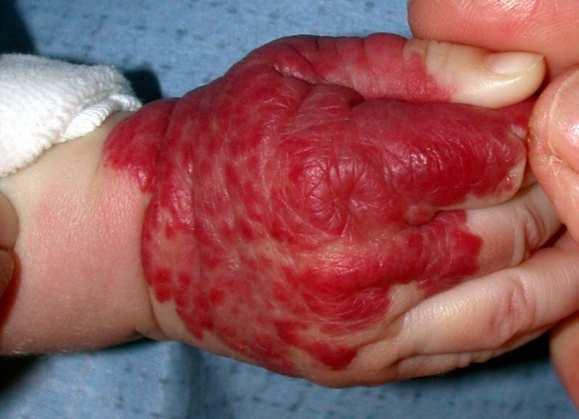


2 Les angiomes
3
4 are imaging studies helpful for diagnosis of vascular malformations? in the vast majority : NO


5 in the vast majority : NO VM
6 caveats : differential diagnosis venous malformation? lymphatic malformation? the radiologist is not always able to answer the question!

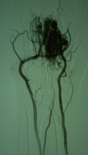
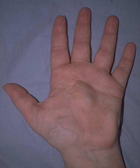
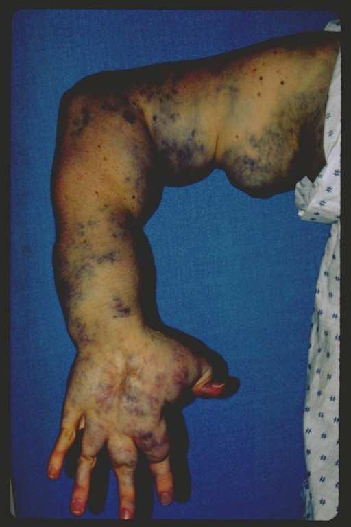
7 LM? - VM? VM (finger)
")
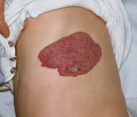
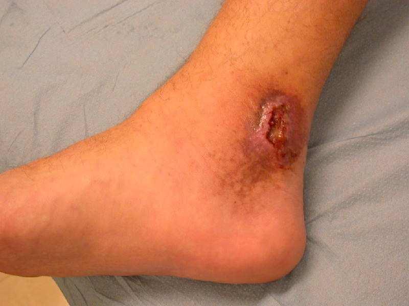
8 LM? - VM? LM (leg)

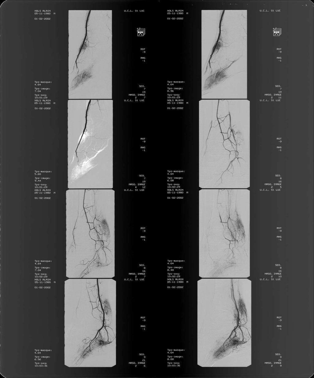
9 LM? - VM? VM

10 LM? - VM? LM
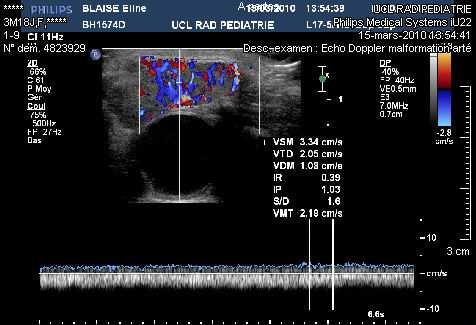
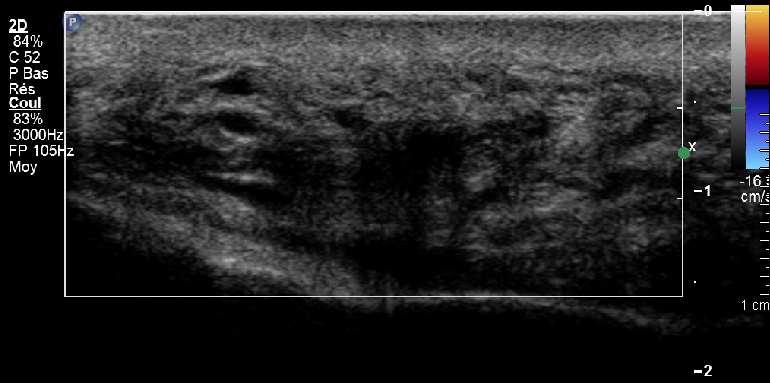
11 VM / LM: differential diagnosis some clues cyst-like channels VM draining vein(s) phlebolith(s) very slow flow filling during compression/ decompression
12 VM / LM: differential diagnosis some clues cyst-like channels VM draining vein(s) phlebolith(s) very slow flow filling during compression/ decompression LM variable sized cyst(s) frequent fluid/fluid (debris) level (hemorrhage) no flow into cyst vessels in septa not compressible

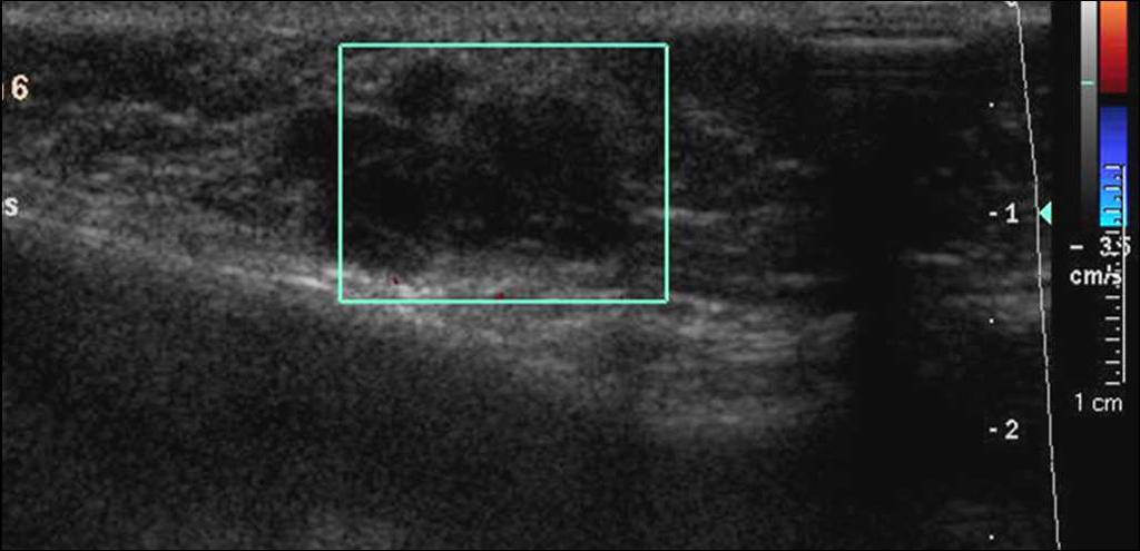
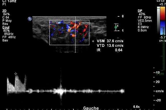
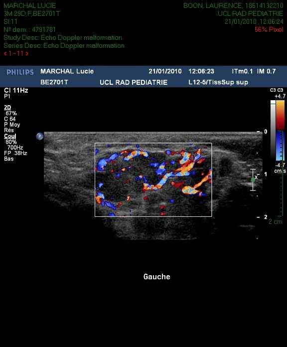
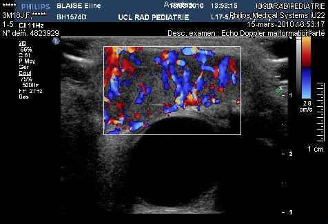
13 doubtful cases : Doppler US at rest compression venous malformation

14 phlebolith venous malformation

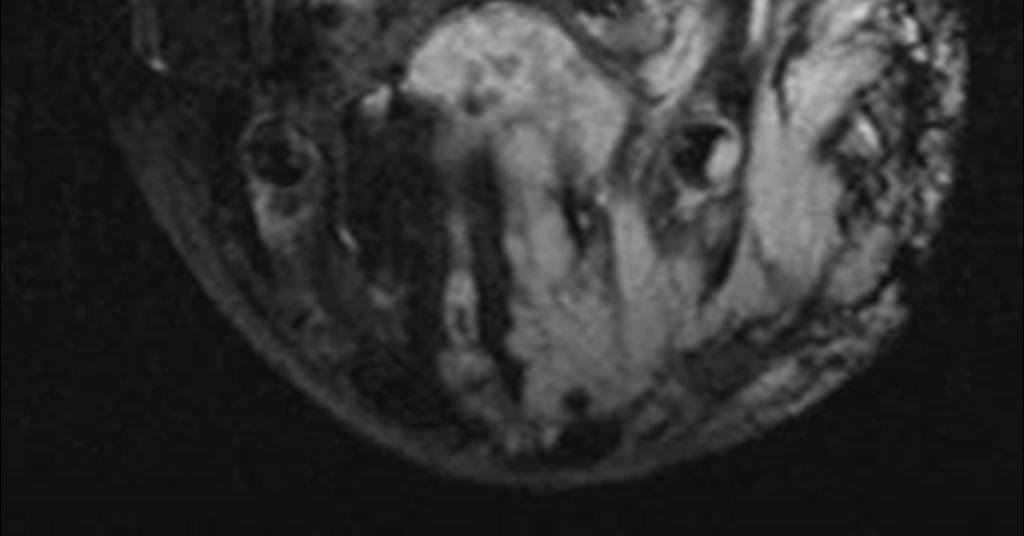
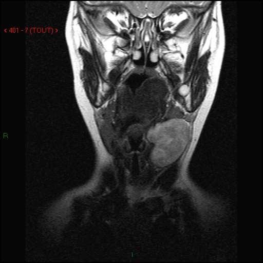
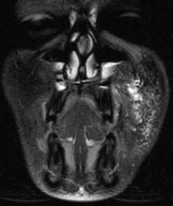
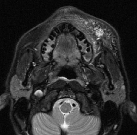
15 fluid-fluid levels a b T1 T2 lymphatic malformation

16 in the vast majority : NO CM


17 doubtful cases : Doppler US hemangioma?


18 hemangioma soft tissue mass vascular network
19

20
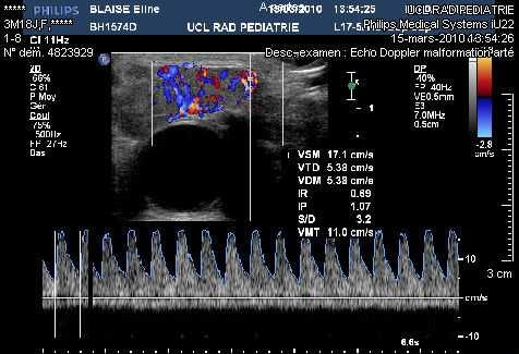
21 hemangioma soft tissue mass vascular network ++ low RI (often < 0.50) fast flow in veins large feeding vessels and draining veins
22 hemangioma?
23 hemangioma
24 caveats : differential diagnosis soft tissue mass or swelling! mandatory for precise characterization and/or diagnosis
25 caveats : differential diagnosis

26 caveats : differential diagnosis rhabdomyosarcoma


27 caveats : differential diagnosis
28 are imaging studies helpful for diagnosis of vascular malformations? in the vast majority : NO in particular cases : NO but additional information for follow-up and treatment
29 in particular cases : NO but additional information for follow-up and treatment

30 Sturge - Weber syndrome MRI T1 + injection


31 in particular cases : NO but additional information for follow-up and treatment AVM
32 are imaging studies helpful for diagnosis of vascular malformations? in the vast majority : NO in particular cases : NO but additional information for follow-up and treatment in some cases : mandatory for precise characterization and/or diagnosis

33 in some cases : mandatory for precise characterization and/or diagnosis CM?


34 CM? no mass! nidus


35 arterio-venous malformation feeding artery fast flow low resistance RI pulsatility in vein


36 arterio-venous malformation feeding artery affected side feeding artery normal side
37 pre-therapeutic work-up sclerotherapy LM VM embolisation surgery
38 LM macrocysts
39 a b c LM microcysts d
40 pre-therapeutic work-up sclerotherapy embolisation surgery : anatomical landmarks
41 LM
42 imaging tools ultrasound + Doppler screening first modality morphology flow hemodynamic pattern MRI (or CT) cross-sectional anatomy depth extension arteriography (pre-)therapeutic approach
43

44 venous malformation T 1 p r e T 1 p o s t
45 vascular birthmarks tumor - mass YES NO
46 vascular birthmarks tumor - mass YES NO typical vascular architecture atypical - no vessels
47 vascular birthmarks tumor - mass YES NO typical vascular architecture atypical - no vessels hemangioma NICH RICH
48 vascular birthmarks tumor - mass YES NO typical vascular architecture atypical - no vessels hemangioma NICH RICH malignant tumor benign tumor LM!
49 vascular birthmarks tumor - mass YES NO typical vascular architecture atypical - no vessels vascular malformation hemangioma NICH RICH malignant tumor benign tumor LM!
50 vascular birthmarks tumor - mass YES NO typical vascular architecture atypical - no vessels vascular malformation hemangioma NICH malignant tumor fast flow slow (no) flow RICH benign tumor LM!
51 vascular birthmarks tumor - mass YES NO typical vascular architecture atypical - no vessels vascular malformation hemangioma NICH malignant tumor fast flow slow (no) flow RICH benign tumor LM! arterio-venous malformation arterio-venous fistula
52 vascular birthmarks tumor - mass YES NO typical vascular architecture Doppler US atypical - no vessels Doppler US vascular malformation hemangioma NICH RICH malignant tumor benign tumor LM! fast flow Doppler US slow (no) flow Doppler US arterio-venous malformation arterio-venous fistula capillary malformation venous malformation lymphatic malformation
Sonography of soft-tissue vascular lesions
 Sonography of soft-tissue vascular lesions Oscar M. Navarro Associate Professor, University of Toronto Dept. of Diagnostic Imaging, The Hospital for Sick Children Toronto, Canada Declaration of Disclosure
Ultrasound of soft-tissue vascular anomalies
 Ultrasound of soft-tissue vascular anomalies Oscar M. Navarro Associate Professor, University of Toronto Dept. of Diagnostic Imaging, The Hospital for Sick Children Toronto, Canada Declaration of Disclosure
Ultrasound imaging of vascular anomalies: pearls and pitfalls
 Ultrasound imaging of vascular anomalies: pearls and pitfalls Oscar Navarro, MD Dept. of Medical Imaging, University of Toronto Dept. of Diagnostic Imaging, The Hospital for Sick Children Declaration of
Vessel malformation vascular malformation vascular malformation vascular malformation Vascular malformation vascular malformations
 A vascular malformation is another type of birthmark, or congenital (present at birth) growth, made up of arteries, veins, capillaries, or lymphatic vessels. There are several different types of malformations
Classification des Malformations vasculaires
 Classification des Malformations vasculaires Gilles Soulez, MD, MSc, FSIR Professeur Titulaire et Chairman Dpt Radiologie, Radio-Oncologie et Medecine Nucléaire Université de Montréal Basic principles
Vascular Tumors in Children and Adults. Thuy Phung, MD, PhD Houston Methodist Hospital Texas Children s Hospital Baylor College of Medicine
 Vascular Tumors in Children and Adults Thuy Phung, MD, PhD Houston Methodist Hospital Texas Children s Hospital Baylor College of Medicine What are these lesions? (Marcelo Hochman, MD) What are these lesions?
Diagnostic imaging of cervical vascular malformations
 Diagnostic imaging of cervical vascular malformations Poster No.: C-0781 Congress: ECR 2013 Type: Educational Exhibit Authors: A. Llanes Rivada, D. Dualde-Beltrán, L. Ariño Montaner, J. 1 2 1 1 1 1 1 Joudanin
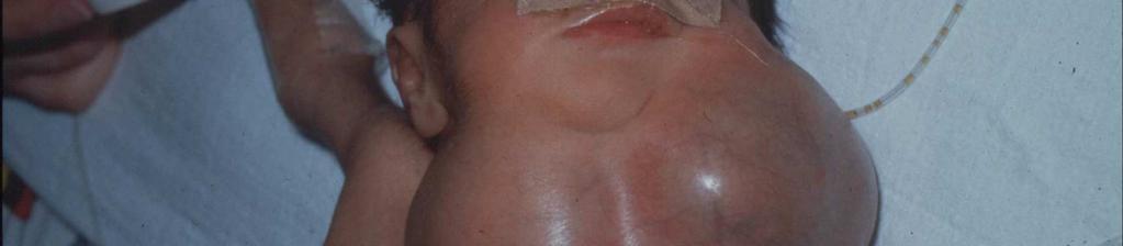
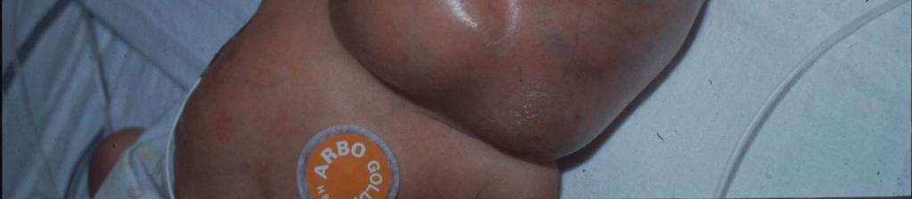
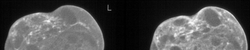
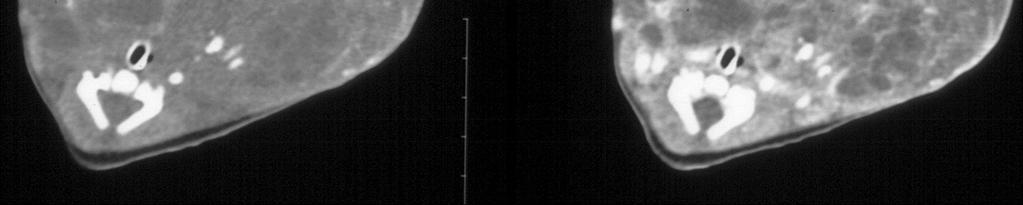
Young patient with swelling of mandible
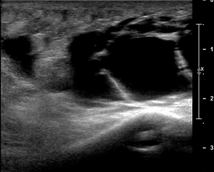
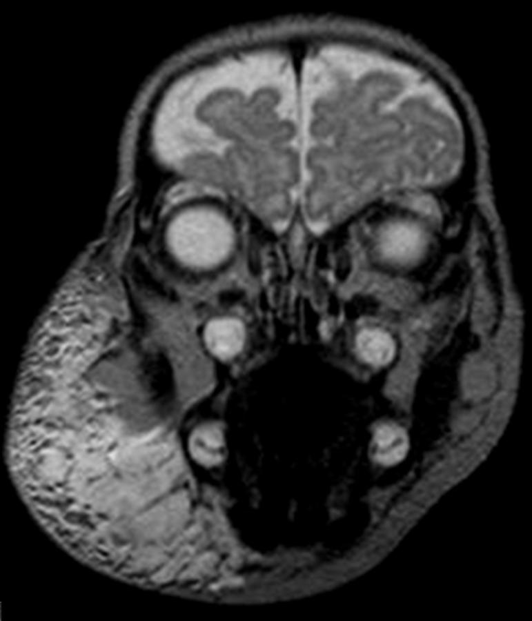
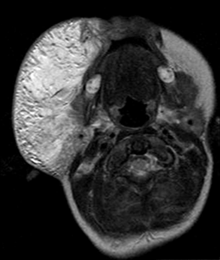
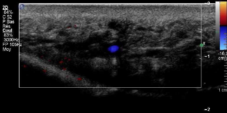
 HR J Young patient with swelling of mandible, p. 66-71 Clinical Case - Test Yourself Interventional Young patient with swelling of mandible Nikolaos Galanakis 1, Maria Papadaki 2, Dimitrios Tsetis 1 1
Vascular Malformations of the Head & Neck
 Vascular Malformations of the Head & Neck Jared Steinklein, MD Lenox Hill Hospital The New York Head & Neck Institute Vascular Birthmark Institute of New York Disclosures The presenter has no financial
Angiographic features of rapidly involuting congenital hemangioma (RICH)
") Pediatr Radiol (2003) 33: 15 19 DOI 10.1007/s00247-002-0726-3 CASE REPORT Orhan Konez Patricia E. Burrows John B. Mulliken Steven J. Fishman Harry P.W. Kozakewich Angiographic features of rapidly involuting
Pediatr Radiol (2003) 33: 15 19 DOI 10.1007/s00247-002-0726-3 CASE REPORT Orhan Konez Patricia E. Burrows John B. Mulliken Steven J. Fishman Harry P.W. Kozakewich Angiographic features of rapidly involuting
2/2/14 INTRODUCTION. Vascular Anomalies: An Introduction to Classification and Coordination of Care. VASCULAR ANOMALIES: History and Classification
 INTRODUCTION Vascular Anomalies: An Introduction to Classification and Coordination of Care Cindy Kerr,RN,MSN,CPNP Erin Spera,RN,MSN,CPNP Mary Beth Sylvia,RN,MS,FNP-BC 1in 3 newborns has a vascular birthmark
HIGH-FLOW ARTERIOVENOUS MALFORMATION WİTHİN ENLARGED FETAL LEG (Congenital Hemangioma vs Parkes Weber Syndrome)
") HIGH-FLOW ARTERIOVENOUS MALFORMATION WİTHİN ENLARGED FETAL LEG (Congenital Hemangioma vs Parkes Weber Syndrome) DORUK CEVDI KATLAN, MD Department of Obstetrics and Gynecology / Perinatology Suleymaniye
HIGH-FLOW ARTERIOVENOUS MALFORMATION WİTHİN ENLARGED FETAL LEG (Congenital Hemangioma vs Parkes Weber Syndrome) DORUK CEVDI KATLAN, MD Department of Obstetrics and Gynecology / Perinatology Suleymaniye
Rare case of a Congenital Arteriovenous malformation (Park Weber angiodysplasia) around the elbow with median nerve compression
 around the elbow with median nerve compression") ISPUB.COM The Internet Journal of Orthopedic Surgery Volume 14 Number 1 Rare case of a Congenital Arteriovenous malformation (Park Weber angiodysplasia) around the elbow with median nerve compression S
ISPUB.COM The Internet Journal of Orthopedic Surgery Volume 14 Number 1 Rare case of a Congenital Arteriovenous malformation (Park Weber angiodysplasia) around the elbow with median nerve compression S
Imaging features of pediatric vascular malformations of the skin and subcutaneous tissue.
 Imaging features of pediatric vascular malformations of the skin and subcutaneous tissue. Poster No.: C-1962 Congress: ECR 2012 Type: Educational Exhibit Authors: R. F. Ocete Pérez, M. Fajardo Cascos,
Explaining All of the Options for AVM: Cerebral Arteriovenous Malformation
 Explaining All of the Options for AVM: Cerebral Arteriovenous Malformation Recorded on: November 19, 2012 Bernard Bendok, M.D. Director of the Neurointerventional Program Northwestern Memorial Hospital
Imaging Work-Up of a Neck Mass - Adults & Children
 Disclosures Imaging Work-Up of a Neck Mass - Adults & Children I have nothing to disclose Christine M Glastonbury MBBS Professor of Radiology & Biomedical Imaging Otolaryngology-Head & Neck Surgery and
Original Article Study on new classification and treatment of vascular malformations in the extremities
 Int J Clin Exp Med 2018;11(4):4021-4029 www.ijcem.com /ISSN:1940-5901/IJCEM0072003 Original Article Study on new classification and treatment of vascular malformations in the extremities Xuejian Liu, Hailin
Attending Physician Statement- Stroke
 Instruction to doctor: This patient is insured with us against the happening of certain contingent events associated with his health A claim has been submitted in connection with Stroke / Intracranial
What effects will proximal or distal disease have on a waveform?
 Spectral Doppler Interpretation Director of Ultrasound Education & Quality Assurance Baylor College of Medicine Division of Maternal-Fetal Medicine Maternal Fetal Center Imaging Manager Texas Children
What effects will proximal or distal disease have on an waveform?
 Spectral Doppler Interpretation Director Director of of Ultrasound Ultrasound Education Education & & Quality Quality Assurance Assurance Baylor Baylor College College of of Medicine Medicine Division
PEDIATRICS WK 3 HEAD AND NECK ALISON WALLACE MD, PHD
 PEDIATRICS WK 3 HEAD AND NECK ALISON WALLACE MD, PHD Topics 1. Cervical lymphadenopathy 2. Lymphatic malformation 3. Thyroglossal duct cysts 4. Branchial cleft cysts 5. Thyroid masses CASE 1 Case 1 A 2
Hamburg Classification: Vascular Malformation
 1 2 Hamburg Classification: Vascular Malformation 9 3 Dirk A. Loose and Raul E. Mattassi 4 5 6 7 8 9 10 11 12 13 14 15 16 17 18 19 20 21 22 23 24 25 26 27 28 29 30 31 32 AU1 Vascular malformations occur
Hemangioma of Tongue with Phlebolith: A Rare presentation
 Journal of Government Dental College and Hospital, October 2017, Vol.-04, Issue- 01, P. 20-25 Original article: Hemangioma of Tongue with Phlebolith: A Rare presentation 1 Dr. Jigna S Shah (MDS) 1, 2 Dr.
Vascular malformations: an update on imaging and management
 Review Arch Argent Pediatr 2016;114(2):167-176 / 167 Vascular malformations: an update on imaging and management Sergio Sierre, M.D. a, DaríoTeplisky, M.D. a and José Lipsich, M.D. a a. Department of Interventional
When is Limb Edema Not Heart Failure
 When is Limb Edema Not Heart Failure An Approach to the Swollen Leg Greg Harding M.D. Vascular Surgeon Faculty/Presenter Disclosure Faculty: Greg Harding M.D. Relationships with commercial interests: None
Vascular malformations and their management
 Information for patients Vascular malformations and their management Introduction This leaflet tells you about the condition known as vascular malformation, including venous malformation, arteriovenous
Features compression after open and endovascular operation in vascular malformation
 Features compression after open and endovascular operation in vascular malformation Sapelkin Sergey Institute of Surgery named A.V. Vishnevsky, Moscow, Russia 21.10.2017 CIRC Meeting, Grassau AV-malformations:
INFECTION. HIV Infection DWI
 HIV Infection INFECTION DWI Fig Axial CT and MRI images show multiple enlarged lymph nodes in the neck as well as in the parotid gland bilaterally. These nodes were suppurative with positive diffusion.
Swelling. Size: measure exact size in cm using a tape measure (measure longitudinal and transverse axis and if possible the depth)
") Swelling Inspection Site: exact anatomic position Number: single or multiple Shape: spherical, oval, kidney-shaped or irregular Size: measure exact size in cm using a tape measure (measure longitudinal
Swelling Inspection Site: exact anatomic position Number: single or multiple Shape: spherical, oval, kidney-shaped or irregular Size: measure exact size in cm using a tape measure (measure longitudinal
Radiographic and statistical analysis of Brain Arteriovenous Malformations.
 Radiographic and statistical analysis of Brain Arteriovenous Malformations. Poster No.: C-0996 Congress: ECR 2017 Type: Educational Exhibit Authors: C. E. Rodriguez 1, A. Lopez Moreno 1, D. Sánchez Paré
Vascular Imaging in the Pediatric Abdomen. Jonathan Swanson, MD
 Vascular Imaging in the Pediatric Abdomen Jonathan Swanson, MD Goals and Objectives To understand the imaging approach, appearance, and clinical manifestations of the common pediatric abdominal vascular
PDF hosted at the Radboud Repository of the Radboud University Nijmegen
 PDF hosted at the Radboud Repository of the Radboud University Nijmegen The following full text is a publisher's version. For additional information about this publication click this link. http://hdl.handle.net/066/5
Traumatic Arteriovenous Malformation of Cheek: A Case Report and Review of Literature
 10.5005/jp-journals-10003-1138 CASE REPORT Traumatic Arteriovenous Malformation of Cheek: A Case Report and Review of Literature Vadisha Srinivas Bhat, Rajeshwary Aroor, B Satheesh Kumar Bhandary, Shama
Neckmasses in infancy and childhood: Clinical and radiological classification and imaging approaches M. Mearadji
 Neckmasses in infancy and childhood: Clinical and radiological classification and imaging approaches M. Mearadji International Foundation for Pediatric Imaging Aid Introduction Neck masses are a frequent
Vascular Malformations: A Review
 58 Vascular Malformations: A Review Joshua A. Cox, MD 1 Erica Bartlett, MD 1 Edward I. Lee, MD 1 1 Division of Plastic Surgery, Baylor College of Medicine, Houston, Texas Semin Plast Surg 2014;28:58 63.
Ultrasound diagnostics of a spontaneous arteriovenous fistula of the head and neck
 Case report Cite as: Zakharkina MV, Chechetkin O, Krotenkova MV, Konovalov RN: Ultrasound diagnostics of a spontaneous arteriovenous fistula of the head and neck.. Submitted: 29.03.2017 ccepted: 24.05.2017
Soft Tissue Hemangiomas: High-resolution Grayscale and Color Doppler Ultrasonographic Features in 43 Patients
 O R I G I N A L A R T I C L E Soft Tissue Hemangiomas: High-resolution Grayscale and Color Doppler Ultrasonographic Features in 43 Patients Chia-Yu Keng 1,2, Howard Haw-Chang Lan 1,2,3 *, Clayton Chi-Chang
Brain AVM with Accompanying Venous Aneurysm with Intracerebral and Intraventricular Hemorrhage
 Cronicon OPEN ACCESS EC PAEDIATRICS Case Report Brain AVM with Accompanying Venous Aneurysm with Intracerebral and Intraventricular Hemorrhage Dimitrios Panagopoulos* Neurosurgical Department, University
Alcohol injections for management of vascular malformations. Clinical case report. Hendro Sudjono Yuwono
 Alcohol injections for management of vascular malformations Clinical case report Hendro Sudjono Yuwono Department of Surgery, School of Medicine, Padjadjaran University Bandung, Indonesia Abstract Vascular
Vascular Malformations of the Brain: A Review of Imaging Features and Risks
 Vascular Malformations of the Brain: A Review of Imaging Features and Risks Comprehensive Neuroradiology: Best Practices October 27-30, 2016 Sudhakar R. Satti, MD Associate Director Neurointerventional
High-Flow Vascular Malformation of Ear: A Case Report
 256 Ear high-flow vascular malformation Case Report High-Flow Vascular Malformation of Ear: A Case Report Ankit Gupta 1*, Shyam Gupta 1, Akhil Kumar 2, Sameek Bhattacharaya 1, Manoj Jha 1, Vinay Tiwari
Role of imaging in the diagnosis of vascular malformations (VM)
") Review Article Role of imaging in the diagnosis of vascular malformations (VM) Katayoun Samadi, Gloria Maria Salazar Division of Interventional Radiology, Department of Radiology, Massachusetts General
Review Article Role of imaging in the diagnosis of vascular malformations (VM) Katayoun Samadi, Gloria Maria Salazar Division of Interventional Radiology, Department of Radiology, Massachusetts General
CHARACTERIZATION OF CONGENITAL VASCULAR MALFORMATION IN THE EXTREMITIES USING WHOLE BODY BLOOD POOL SCINTIGRAPHY AND LYMPHSCINTIGRAPHY
 77 Lymphology 42 (2009) 77-84 CHARACTERIZATION OF CONGENITAL VASCULAR MALFORMATION IN THE EXTREMITIES USING WHOLE BODY BLOOD POOL SCINTIGRAPHY AND LYMPHSCINTIGRAPHY Y.H. Kim, J.Y. Choi, Y.W. Kim, D.I.
Controversies & updates in Vascular Surgery
 Controversies & updates in Vascular Surgery Paris - january 24 2018 Venous session VENOUS ODDITIES DUPLEX IMAGE Philippe LEMASLE Le Chesnay - France I have no financial relationship to disclose Case n
Role of imaging in RCC. Ultrasonography. Solid lesion. Cystic RCC. Solid RCC 31/08/60. From Diagnosis to Treatment: the Radiologist Perspective
 Role of imaging in RCC From Diagnosis to Treatment: the Radiologist Perspective Diagnosis Staging Follow up Imaging modalities Limitations and pitfalls Duangkamon Prapruttam, MD Department of Therapeutic
Alice Fung, MD Oregon Health and Science University
 Alice Fung, MD Oregon Health and Science University Disclosure Comments The speaker Alice Fung, MD Has relevant financial relationships to disclose. Received honorarium from (Guerbet). This individual
Vascular malformations: classification, imaging and treatment
 Vascular malformations: classification, imaging and treatment Poster No.: C-2166 Congress: ECR 2015 Type: Educational Exhibit Authors: C. De Angelis, A. Goracci, G. Mauri, D. Poretti, V. Pedicini, 1 2
An Arteriovenous Malformation in the Suprapatellar Fat Pad of the Knee associated with Klippel-Trenaunay- Weber Syndrome: A Case Report 1
 n rteriovenous Malformation in the Suprapatellar Fat Pad of the Knee associated with Klippel-Trenaunay- Weber Syndrome: Case Report 1 Mi Hyun Park, M.D., Soon Tae Kwon, M.D., yung Seok Shin, M.D., Young
MR Imaging of Soft-Tissue Vascular Malformations: Diagnosis, Classification, and Therapy Follow-up 1
 Note: This copy is for your personal non-commercial use only. To order presentation-ready copies for distribution to your colleagues or clients, contact us at www.rsna.org/rsnarights. VASCULAR/INTERVENTIONAL
Case Report Testicular Arteriovenous Malformation: Gray-Scale and Color Doppler Ultrasonography Features
 Volume 2011, Article ID 876206, 4 pages doi:10.11/2011/876206 Case Report Testicular Arteriovenous Malformation: Gray-Scale and Color Doppler Ultrasonography Features Fatih Gulsen, 1 Ismail Mihmanli, 1
Imaging of liver and pancreas
 Imaging of liver and pancreas.. Disease of the liver Focal liver disease Diffusion liver disease Focal liver disease Benign Cyst Abscess Hemangioma FNH Hepatic adenoma HCC Malignant Fibrolamellar carcinoma
Clinical Perspective: Vascular Anomalies with revised 2014 ISSVA Classification
 Clinical Perspective: Vascular Anomalies with revised 2014 ISSVA Classification Joao uilherme Amaral Pediatric Interventional Radiologist Division Chief Image uided Therapy Centre - The Hospital for Sick
Cardiovascular Imaging
 Cardiovascular Imaging Cardiovascular Imaging Cardio and Vascular Imaging Vascularization / Angiogenesis Cardiovascular Imaging metabolic imaging of the heart myocardial perfusion imaging Cardiac CT Vascularization
Endovascular Therapy for Arteriovenous Malformation
 Endovascular Therapy for Arteriovenous Malformation Wayne F. Yakes, MD Vascular malformations involving the spinal cord are technically challenging clinical entities to diagnosis and ultimately treat.
Love your legs again Varicose Veins
 Love your legs again Varicose Veins Veins are the vessels that return blood to the heart once it has circulated through the body (as opposed to arteries, which carry oxygen-rich blood from the heart to
OHTAC Recommendation. Endovascular Laser Treatment for Varicose Veins. Presented to the Ontario Health Technology Advisory Committee in November 2009
 OHTAC Recommendation Endovascular Laser Treatment for Varicose Veins Presented to the Ontario Health Technology Advisory Committee in November 2009 April 2010 Issue Background The Ontario Health Technology
2014 CPT Codes: What Your Practice Needs to Know. December 12, 2013
 2014 CPT Codes: What Your Practice Needs to Know December 12, 2013 2014 CPT Changes 335 changes, 175 new codes, 107 revisions, 47 deletions Changes to upper and lower GI codes, breast biopsies, peripheral
Sclerotherapy for Venous Vascular and Lymphatic Malformations
 Service: Imaging Sclerotherapy for Venous Vascular and Lymphatic Malformations Exceptional healthcare, personally delivered Your doctor has requested that you have sclerotherapy. We hope that the following
Orbital facia. Periororbital facia Orbital septum Bulbar facia Muscular facia
 Anatomy Orbital facia Periororbital facia Orbital septum Bulbar facia Muscular facia Physiology of symptoms 1) Proptosis ( exophthalmos) Pseudoproptosis Axial Non axial Pulsating Positional Intermittent
External Jugular Vein Vascular Malformation: Sonographic and MR Imaging Appearances
 AJNR Am J Neuroradiol 25:338 342, February 2004 Case Report External Jugular Vein Vascular Malformation: Sonographic and MR Imaging Appearances Anil T. Ahuja, Hok-Yuen Yuen, Ka-Tak Wong, Ann D. King, Victor
Vascular Surgery Rotation Objectives for Junior Residents (PGY-1 and 2)
") Vascular Surgery Rotation Objectives for Junior Residents (PGY-1 and 2) Definition Vascular surgery is the specialty concerned with the diagnosis and management of congenital and acquired diseases of the
Vascular Surgery Rotation Objectives for Junior Residents (PGY-1 and 2) Definition Vascular surgery is the specialty concerned with the diagnosis and management of congenital and acquired diseases of the
MINERVA MEDICA COPYRIGHT
 ISVI-IUA Consensus Document Diagnostic Guidelines of Vascular Anomalies: Vascular Malformations and Hemangiomas B. B. LEE, P. L. ANTIGNANI, V. BARALDINI, I. BAUMGARTNER, P. BERLIEN, F. BLEI, G. P. CARRAFIELLO,
Marc Norman, Ph.D. - Do Not Use without Permission 1. Cerebrovascular Accidents. Marc Norman, Ph.D. Department of Psychiatry
 Cerebrovascular Accidents Marc Norman, Ph.D. Department of Psychiatry Neuropsychiatry and Behavioral Medicine Neuropsychology Clinical Training Seminar 1 5 http://www.nlm.nih.gov/medlineplus/ency/images/ency/fullsize/18009.jpg
MASSIVE EPISTAXIS IN A NEONATE: A SYMPTOM OF VEIN OF GALEN MALFORMATION!
 CASE REPORT MASSIVE EPISTAXIS IN A NEONATE: A SYMPTOM OF VEIN OF GALEN MALFORMATION! Shagufta Wahab 1, Rizwan Ahmad Khan 2, Manjari Thapa Manger 3 1. Radiodiagnosis, Aligarh Muslim University, Aligarh,
Vascular Technology Examination Content Outline
 Vascular Technology Examination Content Outline (Outline Summary) # Domain Subdomain Percentage 1 Normal Anatomy, Perfusion, and Function Evaluate normal anatomy, perfusion, function 2 Pathology, Perfusion,
ED Diagnosis of DVT or tools to rule out DVT in your ED
 ED Diagnosis of DVT or tools to rule out DVT in your ED Ralph Wang UCSF Department of Emergency Medicine 53 yo f c/o left leg swelling recent cholecystectomy its midnight how do you manage this patient?
Liver Cancer (Hepatocellular Carcinoma or HCC) Overview
 Overview") Liver Cancer (Hepatocellular Carcinoma or HCC) Overview Recent advances in liver cancer care seek to address the rising incidence of liver cancer, which has steadily increased over the past three decades.
Liver Cancer (Hepatocellular Carcinoma or HCC) Overview Recent advances in liver cancer care seek to address the rising incidence of liver cancer, which has steadily increased over the past three decades.
Abdomen Sonography Examination Content Outline
 Abdomen Sonography Examination Content Outline (Outline Summary) # Domain Subdomain Percentage 1 2 3 Anatomy, Perfusion, and Function Pathology, Vascular Abnormalities, Trauma, and Postoperative Anatomy
Physician s Vascular Interpretation Examination Content Outline
 Physician s Vascular Interpretation Examination Content Outline (Outline Summary) # Domain Subdomain Percentage 1 2 3 4 5 6 Cerebrovascular Abdominal Peripheral Arterial - Duplex Imaging Peripheral Arterial
Update on Pediatric Vascular Tumors
 Update on Pediatric Vascular Tumors Jim Treat, MD Associate Professor of Clinical Pediatrics and Dermatology No Relevant Conflicts of Interest PHOTOGRAPHY & VIDEOTAPING ARE STRICTLY PROHIBITED IN ALL EDUCATIONAL
GLUT-1: an extra diagnostic tool to differentiate between haemangiomas and vascular malformations q
 The British Association of Plastic Surgeons (2005) 58, 348 352 GLUT-1: an extra diagnostic tool to differentiate between haemangiomas and vascular malformations q J. Leon-Villapalos a, K. Wolfe b, L. Kangesu
Imaging features of orbital neoplasm developed in pediatrics
 Imaging features of orbital neoplasm developed in pediatrics Poster No.: C-1119 Congress: ECR 2015 Type: Educational Exhibit Authors: J. H. Yoo; Seoul/KR Keywords: Eyes, Head and neck, Paediatric, CT,
Vascular Anomalies. Dr Kurosh Parsi MBBS, Msc (MED), FACD, FACP Vascular Birthmark Clinic Sydney Children s Hospital
, FACD, FACP Vascular Birthmark Clinic Sydney Children s Hospital") Vascular Anomalies Dr Kurosh Parsi MBBS, Msc (MED), FACD, FACP Vascular Birthmark Clinic Sydney Children s Hospital Vascular Anomalies Can be divided into two types: Tumours Malformations Vascular Tumours
Vascular Anomalies Dr Kurosh Parsi MBBS, Msc (MED), FACD, FACP Vascular Birthmark Clinic Sydney Children s Hospital Vascular Anomalies Can be divided into two types: Tumours Malformations Vascular Tumours
Evaluation of Neck Mass. Disclosure. Learning Objectives 3/24/2014. Karen T. Pitman MD, FACS Banner MDACC, Gilbert AZ. Nothing to disclose
 Evaluation of Neck Mass Karen T. Pitman MD, FACS Banner MDACC, Gilbert AZ Nothing to disclose Disclosure Learning Objectives 1. Describe a systematic method to evaluate a patient with a neck mass 2. Select
Spontaneous occlusion of a cerebral arteriovenous malformation after subtotal endovascular embolisation
 206 Chiriac et al Spontaneous occlusion of a cerebral arteriovenous malformation Spontaneous occlusion of a cerebral arteriovenous malformation after subtotal endovascular embolisation A. Chiriac, N. Dobrin*,
Supratentorial cerebral arteriovenous malformations : a clinical analysis
 Original article: Supratentorial cerebral arteriovenous malformations : a clinical analysis Dr. Rajneesh Gour 1, Dr. S. N. Ghosh 2, Dr. Sumit Deb 3 1Dept.Of Surgery,Chirayu Medical College & Research Centre,
Chief Complaint. Retroperitoneal cystic mass incidentally found at health examination center.
 Personal Information Age: 34 y/o Sex: female Past history: major systemic medical history(-) surgical history(-), family history(-) Denied food or drug allergy Chief Complaint Retroperitoneal cystic mass
Historical perspective
 SPINAL AVM Introduction Vascular malformations of spinal cord are a rare clinical entity, representing 5% of all primary spinal cord lesions, with arteriovenous malformations(avm) & cavernous malformations
ADDITIONS. The following codes have been added.
 ADDITIONS The following codes have been added. 99446 Interprofessional telephone/internet assessment and management service provided by treating/requesting physician or other qualified health care professional;
Conflict of Interest: none. Neonatal Airway Masses. Neonatal Respiratory Papillomatosis. Paul J. Samuels, MD
 Paul J. Samuels, MD Professor of Anesthesiology and Pediatrics Director of Education Cincinnati Children s Hospital Cincinnati, Ohio Conflict of Interest: none Neonatal Respiratory Papillomatosis Caused
UPSTATE Comprehensive Stroke Center. Neurosurgical Interventions Satish Krishnamurthy MD, MCh
 UPSTATE Comprehensive Stroke Center Neurosurgical Interventions Satish Krishnamurthy MD, MCh Regional cerebral blood flow is important Some essential facts Neurons are obligatory glucose users Under anerobic
Looking Outside the Box: Incidental Extracardiac Finding in Echo
 Looking Outside the Box: Incidental Extracardiac Finding in Echo Dr. Aijaz Shah Head of Division, Adult Echocardiography Laboratory Prince Sultan Cardiac Centre Riyadh Case 1 17 year old boy presented
Diagnostic imaging of lymphatic malformations
 Diagnostic imaging of lymphatic malformations Poster No.: C-1440 Congress: ECR 2016 Type: Educational Exhibit Authors: M. M. Coman 1, M. T. A. Buzan 2, S. Manole 3, S. M. Dudea 3 ; 1 2 3 Campia Turzii/RO,
Parathyroid Imaging: Current Concepts. Maria Gule-Monroe, M.D. Nancy Perrier, M.D.
 Parathyroid Imaging: Current Concepts Maria Gule-Monroe, M.D. Nancy Perrier, M.D. Disclosures None Objectives Ultrasound characteristics of parathyroid adenomas vs. lymph nodes 4D-CT evaluation of hyperparathyroidism
Dural Arteriovenous Malformations and Fistulae (DAVM S DAVF S)
") Jorge Guedes Campos NEUROIMAGING DEPARTMENT HOSPITAL SANTA MARIA UNIVERSITY OF LISBON PORTUGAL DEFINITION region of arteriovenous shunting confined to a leaflet of packymeninges often adjacent to a major
Jorge Guedes Campos NEUROIMAGING DEPARTMENT HOSPITAL SANTA MARIA UNIVERSITY OF LISBON PORTUGAL DEFINITION region of arteriovenous shunting confined to a leaflet of packymeninges often adjacent to a major
CTA/MRA of Pediatric Hepatic Masses Radiology-Pathology Correlation
 Acta Radiológica Portuguesa, Vol.XVIII, nº70, pág. 41-50, Abr.-Jun., 2006 CTA/MRA of Pediatric Hepatic Masses Radiology-Pathology Correlation Marilyn J. Siegel Mallinckrodt Institute of Radiology, Washington
Head and Neck Vascular Lesions: Characterization of the Flow Pattern by the Use of Three-Phase CT
 Head and Neck Vascular Lesions: Characterization of the Flow Pattern by the Use of Three-Phase CT Chang-Woo Ryu, MD 1 Jae Kyun Kim, MD 2 Sang Joon Kim, MD 3 Jeong Hyun Lee, MD 3 Jeoung Hyun Kim, MD 3 Hong
Fluid responsiveness Monitoring in Surgical and Critically Ill Patients
 Fluid responsiveness Monitoring in Surgical and Critically Ill Patients Impact clinique de la Goal-directed-therapy Patrice FORGET, M.D Cliniques universitaires Saint Luc Université catholique de Louvain,
Vascular surgery is a specialty that deals with diseases of the vascular system (i.e. arteries, veins
 Vascular Surgery Vascular surgery is a specialty that deals with diseases of the vascular system (i.e. arteries, veins and lymphatics). These are managed by medical therapy, interventional procedures,
ASDIN 7th Annual Scientific Meeting DISCLOSURES TECHNICAL CONSIDERATIONS TECHNICAL CONSIDERATIONS UTILITY OF ULTRASOUND IN EVALUATING ACCESS
 DISCLOSURES UTILITY OF ULTRASOUND IN EVALUATING ACCESS DYSFUNCTION None Vandana Dua Niyyar, MD Assistant Professor of Medicine, Division of Nephrology, Emory University UTILITY OF ULTRASOUND IN ACCESS
Deep Vein Thrombosis
 Deep Vein Thrombosis Introduction Deep vein thrombosis (DVT) is a blood clot in a vein. This condition can affect men and women of any age and race. DVT is a potentially serious condition. If not treated,
Synovial hemangioma of the suprapatellar bursa
 Synovial hemangioma of the suprapatellar bursa Poster No.: P-0040 Congress: ESSR 2013 Type: Authors: Keywords: DOI: Scientific Exhibit A. YESILDAG, S. Keskin, H. Kalkan, S. Kucuksen, U. Kerimoglu; Konya/TR
Insuffisance mitrale
 Imagerie Multimodalité dans les Valvulopathies: De quoi avons nous besoin? Insuffisance mitrale Agnès Pasquet, MD, PhD Pôle de Recherche Cardiovasculaire Institut de Recherche Expérimentale et Clinique
LOWER EXTREMITY VENOUS COMPRESSION ULTRASOUND. CPT Stacey Good, DO Emergency Medicine Ultrasound Fellow Madigan Army Medical Center
 LOWER EXTREMITY VENOUS COMPRESSION ULTRASOUND CPT Stacey Good, DO Emergency Medicine Ultrasound Fellow Madigan Army Medical Center Learning Objectives Setup and patient positioning for optimizing success
Klippel - Trenaunay Syndrome (KTS) When and What to do? Dr. Ayhan ŞENOL SBU.Gazi Yasargil ETH. Diyarbakır /TURKEY
 When and What to do? Dr. Ayhan ŞENOL SBU.Gazi Yasargil ETH. Diyarbakır /TURKEY") Klippel - Trenaunay Syndrome (KTS) When and What to do? Dr. Ayhan ŞENOL SBU.Gazi Yasargil ETH. Diyarbakır /TURKEY Disclosure Speaker name: Ayhan ŞENOL I have the following potential conflicts of interest
Klippel - Trenaunay Syndrome (KTS) When and What to do? Dr. Ayhan ŞENOL SBU.Gazi Yasargil ETH. Diyarbakır /TURKEY Disclosure Speaker name: Ayhan ŞENOL I have the following potential conflicts of interest
Lung sequestration and Scimitar syndrome
 Lung sequestration and Scimitar syndrome Imaging approaches M. Mearadji International Foundation for Pediatric Imaging Aid Rotterdam, The Netherlands Pulmonary sequestration Pulmonary sequestration (PS)
Imaging of Cerebrovascular Disease
 Imaging of Cerebrovascular Disease A Practical Guide Val M. Runge, MD Editor-in-Chief of Investigative Radiology Institute for Diagnostic, Interventional, and Pediatric Radiology Inselspital, University
Name : 黃 XX Age : 52 Sex : 女 Occupation : 廚房阿姨 Marital status : 已婚
 Name : 黃 XX Age : 52 Sex : 女 Occupation : 廚房阿姨 Marital status : 已婚 Chief Complaint Mild postprandial fullness for 2 months Present Illness This 52 year-old female suffered from intermittent post-prandial
For exam: VL DUPLEX EXTREMITY VEINS UNILAT LT
 For exam: VL DUPLEX EXTREMITY VEINS UNILAT LT - 8870390 METHOD/TECHNIQUE: The veins of the left upper extremity were studied at multiple For exam: VL DUPLEX EXTREMITY VEINS UNILAT RT - 8870400 METHOD/TECHNIQUE: