EUS-guided FNAB. Differential Diagnosis 5/1/2017. EUS-FNA of Solid and Cystic Lesions:
|
|
|
- Arthur Hicks
- 6 years ago
- Views:
Transcription

 Cystic degeneration of")
1 EUS-FNA of Solid and Cystic Lesions: Part 1: Solid Masses Martha Bishop Pitman, M.D. Director, Cytopathology Massachusetts General Hospital Professor of Pathology Harvard Medical School Boston, MA EUS-guided FNAB Transgastric: body and tail Transduodenal: head Linear e-scope Color Doppler Aspiration needle Transgastric or transduodenal aspiration Cytology specimen Differential Diagnosis Solid Chronic pancreatitis Ductal adenocarcinoma Acinar cell carcinoma Pancreatic endocrine neoplasm Solid psuedopapillary tumor Pancreatoblastoma Metastasis Cystic Pseudocyst Serous cyst Mucinous cyst (MCN and IPMN) Cystic degeneration of typically solid tumors PEN SPN other Other more rare cysts Simple cysts Lymphoepithelial cyst Peripancreatic cysts 1


2 Cytology Interpretation Multimodal Approach Clinical Information Patient age and gender Symptoms Past medical history Radiological Information Location of mass in the pancreas (and thus organ traversed for EUS) Mass characteristics Solid or cystic» Size, contours, invasion» Cyst structure: uni- or multilocular; thick/thin wall, Ca++, nodule/mass in the wall» Gross cyst contents: thick, viscous, thin, water, clear, brown Ancillary tests: CEA, amylase, molecular analysis Quality FNA Quality specimen High cellularity Cells representative of the lesion Quality preparations Quality interpretation Training of interpreter Experience of interpreter Team approach to diagnosis EUS-FNAB Smaller transducers Sharper needles Disposable needles Needle ridges Sharp stylet Beveled needle 2
 Alcohol fixed Air dried CllBl Cell Block")
 or")


3 Optimal Preparation of EUS- FNAB of Solid Masses Direct Smears (ROSE) Alcohol fixed Air dried CllBl Cell Block kpreparations Rinsings and dedicated pass into RPMI or formalin Enrich material with large bore needle (19g) or pro-core If cellularity appears too scant for cell block, process fluid as cytospin, ThinPrep or SurePath Dedicated pass for flow cytometry if lymphoma is suspected or lymphoid dominant lesion noted on rapid interpretation Newer Needles Acquire- Boston Scientific 3


4 Cytohistology Autoimmune pancreatitis Poorly-differentiated PDAC Normal Pancreas (bivalved pancreatic head) Courtesy of 4 th Series AFIP Fascicle on Tumors of the Pancreas Acini and intercalated cells Normal Pancreas 4


5 Normal Pancreas Ducts and Islets Normal Pancreas Acinar Cells Normal Pancreas 5


6 6


7 Normal Pancreas Ductal Cells 7


8 Duodenal Contamination Duodenal Contamination 8


9 Duodenal Contamination Gastric Contamination Gastric Contamination 9

10 Differential Diagnosis of Solid Pancreatic Masses Solid Chronic pancreatitis Ductal adenocarcinoma Metastasis Pancreatic neuroendocrine tumor CP Acinar cell carcinoma Pancreatoblastoma Solid-pseudopapillary neoplasm Images: AFIP Pancreas fascicle 2007 Differential Diagnosis of Solid Pancreatic Masses Solid Chronic pancreatitis Ductal adenocarcinoma Metastasis Pancreatic neuroendocrine tumor Acinar cell carcinoma Solid-pseudopapillary neoplasm Images: AFIP Pancreas fascicle 2007 Differential Diagnostic Approach to Evaluating the Slide Solid Glandular smear pattern versus solid cellular smear pattern Glandular smear pattern» Malignant or not Solid cellular smear pattern» Endocrine or not Cyst Extracellular mucin Yes: thick or thin No: CEA elevated? KRAS positive? Epithelial cells Yes: mucinous?» Yes: low grade or atypical or malignant Multimodal parameters: gender, symptoms, location, imaging 10


11 Glandular Smear Pattern Solid-Cellular smear pattern Solid Cellular Neoplasm- Smear Pattern Clinical and Radiological Features of PDAC y.o. M>F Radiating epigastric pain with wt. loss Jaundice Migratory thrombophlebitis Sudden onset DM Double duct sign on CT Hypodense mass in panc head with irregular borders; atrophy elsewhere Cigarette smoking Long term DM Family history 31 st degree relatives: 32x 21 st degree relatives: 6x 11 st degree relatives: 2.3x Germline mutations PJS [STK11/LKB1]: 132x FAMMM [p16/cdkn2a] FANC BRCA2 Familial CP [PRSS1/SPINK1] 11


12 High Grade Adenocarcinoma Marked nuclear atypia hyperchromasia pleomorphism overlapping Prominent nucleoli Single atypical cells Mitoses Coagulative Necrosis 12


13 Variants of PDAC: Adenosquamous Carcinoma p63 mucicarmine Variants of PDAC: Undifferentiated Carcinoma with Osteoclast-type Giant Cells CD68 Variants of PDAC Colloid Carcinoma Undifferentiated Carcinoma Signet Ring Cell Carcinoma Foamy Gland Carcinoma 13

 Anisonucleosis 4:1 in a group")
14 Well-Differentiated PDAC Image courtesy of Dr. Volkan Adsay Limitations of Cytology compared to Histology Lack of architecture Cannot see distribution of ductal structures Cannot see abnormal localization Cannot see if ducts are adjacent to medium sized vessels, wrapping around nerves or isolated in fat Cannot see contours or angulation of ducts Cannot see luminal contents Cannot see stromal reaction Criteria for Well-differentiated Adenocarcinoma Irregular cellular distribution in a sheet (drunken honeycomb) Anisonucleosis 4:1 in a group Anisonucleosis 4:1 in a group Parachromatin clearing Irregular nuclear membranes, often subtle Abundant cytoplasm, often visibly mucinous 14


15 Drunken Honeycomb Anisonucleosis Parachromatin clearing Drunken Honeycomb Anisonucleosis Cytoplasmic mucin Parachromatin clearing Parachromatin clearing Nuclear crowding Irregular nuclear membranes Visible cytoplasmic mucin 15


16 Drunken honeycomb Exaggerated vacuolated cytoplasm Well-differentiated Adenocarcinoma Cell block preparation of needle rinsings Chronic Pancreatitis mostly ductal cells scantily cellular some islet cells monolayered sheets cohesive, few single cells maintained polarity minimal nuclear overlap mild anisonucleosis smooth nuclear membranes rare/normal mitoses no coagulative necrosis CP CA 16


")


 2010 Nov 9;")
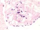
17 Chronic Pancreatitis Key Cytologic Features: -Fragments of acinar tissue with acini splayed apart by fibrosis -Stromal fragments -Inflammatory cells (lymphocytes, plasma cells, macrophages and siderophages) -Background debris and calcification -Ductal epithelium with only mild cytologic atypia -No definite features of neoplasia Autoimmune Pancreatitis JOP. J Pancreas (Online) 2010 Nov 9; 11(6): Autoimmune Pancreatitis: Mass Forming Lesion 17


18 Type 1 vs Type 2 AIP Type 1 AIP Type 2 AIP Age Elderly, 7 th decade Middle age, 5 th decade Sex Male M:F 1:1 Presentation Jaundice (75%) Acute Pancreatitis (15%) Pain Jaundice Systemic Disease Yes No Elevated serum IgG4 80% Uncommon IBD No association Present in 16-30% of cases Histology Periductal inflammation and one of the following: Storiform fibrosis Obliterative phlebitis Long-term outcomes Frequent relapses No relapses Periductal inflammation and one of the following: Ductal/lobular abscesses Ductal ulceration with neutrophils Autoimmune Pancreatitis Deshpande V, Mino-Kenudson M, Brugge WR, Pitman MB, Castill CF, Warshaw AL Lauwers GY. Am J Surg Pathol. 2005; 29; AIP: Core Biopsy Type 1 Type 2 18
 Immunohistochemical Staining")
19 IgG 4 Staining IgG 4 positive plasma cells relative to generic IgG positive plasma cells No defined criteria Heterogeneity of plasma cell distribution in the tissue Cellblock and core biopsy samples are small leading to sampling error Epithelial Atypia in AIP Ancillary Tests: Benign vs Malignant Ductal Epithelium (AFIP Pancreas Fascicle 2007, Chapter 17, Table 17-4) Immunohistochemical Staining in the Diagnosis of Ductal Adenocarcinoma Benign Glands (chronic pancreatitis, benign bile duct lesions) (% Abnormal Expression) Ductal Adenocarinoma CEA (monoclonal)* B CA p53** 0 60 Smad4 (dpc4)*** 0 55 Mesothelin 5 95 *abnormal expression is cytoplasmic reactivity **abnormal expression is reactivity in >20% of nuclei ***abnormal expression is loss of cytoplasmic and nuclear activity 19


20 The Immunohistochemical Expression Pattern of SMAD4, p53, and CDX2 is Helpful in Diagnosing Pancreatic Ductal Adenocarcinoma in Endoscopic Ultrasound-Guided Fine Needle Aspirations Jian Shen M.D., Ph.D., Edmund S. Cibas M.D., and Xiaohua Qian M.D., Ph.D. Brigham and Women s Hospital, Harvard Medical School, Boston, Massachusetts Modern Path 2007; 20(Suppl 2): 82A CDX2 p53 SMAD4 SMAD4 Loss Present in ~50% of PDAC; supports a malignant interpretation Metastases Rare compared to primary malignancy Any tumor can metastasize to the pancreas Renal cell carcinoma common metastasis that mimics primary PDAC Solitary mass Decades after nephrectomy (avg. 10 yrs) 20


21 Differential Diagnosis Foamy Gland Adenocarcinoma FGA CK19+ CA19-9+ SMAD4 - RCC EMA+ PAX8+ Anti-RCC+/- 21


22 Differential Diagnosis Lipid Rich Neuroendocrine Tumor PanNET Synapto+ Chromo + CK7+ RCC EMA+ PAX8+ Anti-RCC+/- Solid Cellular Neoplasms PanNET ACC SPN SPN PanNET ACC Solid Cellular Neoplasms 22


 Classic")

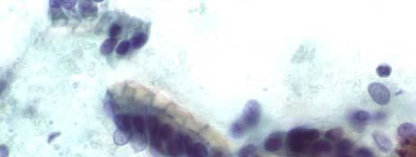
23 Pancreatic Neuroendocrine Tumor [PanNet, aka PEN, PET] Clinical Any age; y.o. M=F MEN, VHL syndromes Hormone effects in functional PanNet: insulin, glucagon for example Radiological Pancreatic tail>>head/body Round, well-circumscribed Sometimes cystic, CA++ Octreotide scan+ Histology Cellular monomorphic population of polygonal cells with various organoid patterns with scan stroma (occasionally Images: AFIP Pancreas fascicle 2007 hylanized or amyloid stroma) Classic Morphology: PanNET Single cells mostly Plasmacytoid Coarse, stippled chromatin -/+ nucleoli Variant Morphology: PanNET Lipid laden Prominent nucleoli Oncocytic 23


 GRADE")

24 IHC: PanNET Synaptophysin+ Chromogranin+ Trypsin Grading of NETs Best performed on resected specimen Requires evaluation of hottest area If resectable, grading on FNA material not necessary, wastes resources If unresectable, grading helps to separate G1/G2 from G3 and direct treatment Usually apparent on HE Grading GEP NETs (WHO, ENETS) GRADE MITOSES KI-67 1 < 2 AND <3% OR 3-20% 3 >20 OR >20% Tumor Tumor Carcinoma Korean Journal of Pathology 2013; 47(3):


25 CIN= chromosomal instability Poorly-Differentiated Neuroendocrine Carcinoma Small cell type Large cell type Secondarily Cystic Solid Neoplasms: Cystic PanNET 25


 +/- nucleoli Benign")
26 Acinar Cell Carcinoma Clinical Adults>>> children Mean age ~ 59 years Males>females ~4:1 Lipase hypersecretion syndrome= polyarthralgia + multi-focal subcutaneous fat necrosis Radiological Solid, circumscribed Large and bulky Rarely cystic Image: AFIP Pancreas fascicle 2007 Classic Morphology: Acinar Cohesive groups and single cells Many stripped naked nuclei Granular cytoplasm (and background) +/- nucleoli Benign versus Malignant Acinar Cell Population Benign Malignant 26


27 IHC:ACCa Trypsin+ Endocrine markers- Classic Morphology: PBL Clinical Most common malignant pancreatic neoplasm of children 2/3 s occur in children and 1/3 in adults Half in Asians Often identical to acinar cell carcinoma on FNA Diagnosis depends on identifying squamoid corpuscle on smears or cell block Solid-Pseudopapillary Neoplasm Clinical Rare but may represent up to 6% of all pancreatic neoplasms and 24% of resected cysts 89% in young women, mean age ~ 28 years 1/3 in head, 1/3 in body and 1/3 in tail Radiology shows large solid and cystic neoplasm Image: AFIP Pancreas fascicle


28 Classic Morphology: SPN Papillary branching Myxoid stroma Clinging cells and single cells Euchromatin Oval, indented, grooved nuclei Perinuclear vacuoles/globules l l IHC: SPN Beta-catenin CD10 CD56 Spenule/accessory spleen Lymphoid tissue Histiocytes Blood vessels CD8+ cells indicating splenic endothelial cells 28


29 Standardized Terminology and Nomenclature for Pancreaticobiliary Cytopathology from the Papanicolaou Society of Cytopathology I. Nondiagnostic II. Negative: Normal pancreatic tissue, splenule, LEC, pancreatitis (AIP) III. Atypical: Suggestive but not diagnostic of NET or SPN; indeterminate bile duct lesions IV. Neoplastic -Benign: SCA, NET microadenoma -Other: IPMN, MCN, PanNET, SPN V. Suspicious: Suggestive but not diagnostic of PDAC, Acinar Cell Ca., PanNEC VI. Positive/Malignant: PDAC, Acinar Cell Ca., PanNEC 29
EUS-guided FNAB. Differential Diagnosis 3/14/2018. EUS-FNA of Solid and Cystic Lesions: Part 1: Solid Masses
 EUS-FNA of Solid and Cystic Lesions: Part 1: Solid Masses Martha Bishop Pitman, M.D. Director, Cytopathology Massachusetts General Hospital Professor of Pathology Harvard Medical School Boston, MA EUS-guided
40th European Congress of Cytology Liverpool, UK, 2-5 th October 2016
 40th European Congress of Cytology Liverpool, UK, 2-5 th October 2016 EUS FNA of abdominal organs: An approach to reporting and triage for ancillary testing Date and time: Sunday 2 nd October 2016 15.00-16.30
Pancreatitis: A Potential Pitfall in Endoscopic Ultrasound Guided Pancreatic FNA
 Pancreatitis: A Potential Pitfall in Endoscopic Ultrasound Guided Pancreatic FNA Jack Yang, MD Department of Pathology, Medical University of South Carolina Objectives Understand the indication of EUS
Standardized Terminology in Pancreatobiliary Cytology: The Papanicolaou Society Guidelines
 Standardized Terminology in Pancreatobiliary Cytology: The Papanicolaou Society Guidelines Barbara Ann Centeno. M.D. Vice-Chair, Clinical Services, Anatomic Pathology Assistant Chief, Pathology Service
Pancreatic Cytopathology: A pragmatic approach. By Dr Miguel Perez-Machado MD. PhD. MRCPath Royal Free Hospital
 Pancreatic Cytopathology: A pragmatic approach. By Dr Miguel Perez-Machado MD. PhD. MRCPath Royal Free Hospital Indications of Cytology Sampling To document malignancy in patients with malignant appearing
Patient History. A 58 year old man presents with a 16 mm cyst in the pancreatic tail. The cyst is unilocular with a thick wall and no mural nodule.
 Case 1 Martha Bishop Pitman, MD Director of Cytopathology Massachusetts General Hospital Associate Professor of Pathology Harvard Medical School Boston, MA Patient History A 58 year old man presents with
Case 1. Case 1: EUS Report 5/1/2017. Interesting Cases of Pancreatic Masses
 Interesting Cases of Pancreatic Masses Martha Bishop Pitman, MD Professor of Pathology Harvard Medical School Director of Cytopathology Massachusetts General Hospital Boston, MA MASSACHUSETTS GENERAL PHYSICIANS
DIAGNOSTIC CHALLENGES Pancreas FNAB. Dr. M. Weir Oct 2017
 DIAGNOSTIC CHALLENGES Pancreas FNAB Dr. M. Weir Oct 2017 CONFLICT OF INTEREST DISCLOSURE I have not had in the past 3 years, a financial interest, arrangement or affiliation with one or more organizations
Advances in Pancreatic. Cytology. Martha B. Pitman, MD. Disclosure of Relevant Financial Relationships
 Advances in Pancreatic Cytology Martha B. Pitman, MD Disclosure of Relevant Financial Relationships USCAP requires that all planners (Education Committee) in a position to influence or control the content
Outline 11/2/2017. Pancreatic EUS-FNA general aspects. Cytomorphologic features of solid neoplasms/lesions of the pancreas
 ENDOSCOPIC ULTRASOUND GUIDED-FINE NEEDLE ASPIRATION CYTOLOGY OF PANCREAS Khalid Amin M.D. Assistant Professor Department of Laboratory Medicine and Pathology University of Minnesota Outline Pancreatic
Pancreatico-biliary cytology: a practical approach to diagnosis. Corina Cotoi
 Pancreatico-biliary cytology: a practical approach to diagnosis Corina Cotoi Pancreatico-biliary lesions Solid: Ductal adenocarcinoma Cholangiocarcinoma Acinar cell carcinoma Neuroendocrine tumour / carcinoma
Pancreatic Cytopathology: The Solid Neoplasms
 Pancreatic Cytopathology: The Solid Neoplasms Syed Z. Ali, M.D. Professor of Pathology and Radiology Director of Cytopathology The Johns Hopkins Hospital Baltimore, Maryland Pancreatic Cytopathology: Past,
Standardization of Nomenclature
 Standardized Terminology and Nomenclature for Pancreaticobiliary Cytopathology from the Papanicolaou Society of Cytopathology Lester J. Layfield, M.D. Professor and Chair University of Missouri Pathology
CYTOLOGY OF EUS- GUIDED FNA OF THE PANCREAS AND THE UPPER GI TRACT
 CYTOLOGY OF EUS- GUIDED FNA OF THE PANCREAS AND THE UPPER GI TRACT Barbara A. Centeno, M.D. Vice-Chair, Clinical Services Assistant Chief of Pathology Director of Cytopathology Department of Anatomic Pathology/Moffitt
Conflicts of Interest
 Standardized Terminology and Nomenclature for Pancreaticobiliary Cytopathology from the Papanicolaou Society of Cytopathology Lester J. Layfield, M.D. Professor and Chair University of Missouri Pathology
EUS-FNAB of GI Tract: Specimen Handling and Triage
 EUS-FNAB of GI Tract: Specimen Handling and Triage Amy Ly, M.D. Director, Fine Needle Aspiration Service Massachusetts General Hospital Assistant Professor of Pathology Harvard Medical School Boston, MA
Video Microscopy Tutorial 19
 Video Microscopy Tutorial 19 EUS FNA of Pancreatic Cysts Martha Pitman, MD There are no disclosures necessary. EUS-FNA of Pancreatic Cysts Martha Bishop Pitman, M.D. Massachusetts General Hospital Harvard
CHRONIC PANCREATITIS OR DUCTAL ADENOCARCINOMA? N. Volkan Adsay, \ MD
 CHRONIC PANCREATITIS OR DUCTAL ADENOCARCINOMA? N. Volkan Adsay, \ MD Case for discussion 67 y/o male Back pain and weight loss CT: 4.5 cm ill-defined, solid lesion in the head FNA/Core bx: Inconclusive
Evaluation and Management of Cystic Lesions of the Pancreas: When to Resect, When to Follow and When to Forget
 Evaluation and Management of Cystic Lesions of the Pancreas: When to Resect, When to Follow and When to Forget Randall Brand, MD Professor of Medicine Division of Gastroenterology, Hepatology and Nutrition
Pancreatic Cystic Lesions 원자력병원
 Pancreatic Cystic Lesions 원자력병원 박선 후 Lines of cellular differentiation Ductal Acinar Undetermined Ductal adenocarcinoma Serous/ mucinous tumor Intraductal papillary mucinous neoplasm Acinar cell carcinoma
Select problems in cystic pancreatic lesions
 Disclosure Select problems in cystic pancreatic lesions Five Prime Therapeutics shareholder Adicet Bio shareholder Bristol-Meyer Squibb advisory board grace.kim@ucsf.edu Pancreatic cystic lesions Intraductal
Neoplasias Quisticas del Páncreas
 SEAP -Aproximación Práctica a la Patología Gastrointestinal- Madrid, 26 de mayo, 2006 Neoplasias Quisticas del Páncreas Gregory Y. Lauwers, M.D. Director, Service Massachusetts General Hospital Harvard
Respiratory Tract Cytology
 Respiratory Tract Cytology 40 th European Congress of Cytology Liverpool, UK Momin T. Siddiqui M.D. Professor of Pathology and Laboratory Medicine Director of Cytopathology Emory University Hospital, Atlanta,
FNA of Thyroid. Toward a Uniform Terminology With Management Guidelines. NCI NCI Thyroid FNA State of the Science Conference
 FNA of Thyroid NCI NCI Thyroid FNA State of the Science Conference Toward a Uniform Terminology With Management Guidelines Thyroid Thyroid FNA Cytomorphology NCI Thyroid FNA State of the Science Conference
Salivary Glands 3/7/2017
 Salivary Glands 3/7/2017 Goals and objectives Focus on the entities unique to H&N Common board type facts Information for your future practice Salivary Glands Salivary Glands Major gland. Paratid. Submandibular.
Objectives. Pancreatic Cysts. Benefits and Limitations of the Cytologic Assessment of Cystic Pancreatic Lesions and Masses
 Benefits and Limitations of the Cytologic Assessment of Cystic Pancreatic Lesions and Masses Michelle D. Reid, MD, MSc Associate 1/24/15 Professor Director of Cytology - EUHM Michelle D Reid MD, MS Emory
Salivary Gland Cytology
 Salivary Gland Cytology Diagnostic challenges and potential pitfalls Tarik M. Elsheikh, MD Professor and Medical Director Anatomic Pathology Cleveland Clinic FNA Salivary Gland Lesions Indications Distinguish
Thyroid follicular neoplasms in cytology. Ulrika Klopčič Institute of Oncology, Department of Cytopathology, Ljubljana, Slovenia
 Thyroid follicular neoplasms in cytology Ulrika Klopčič Institute of Oncology, Department of Cytopathology, Ljubljana, Slovenia Lecture overview importance of FNAB in assessing thyroid lesions follicular
Endoscopic Ultrasound-guided FNA Cytology of the Pancreas
 Endoscopic Ultrasound-guided FNA Cytology of the Pancreas 67 th Annual California Society of Pathologists 2014 Laura Tabatabai, MD Professor of Pathology University of California, San Francisco Overview
Cytopathology Study Day 16 April RCPath - BAC. Digital cytology: EUS FNA pancreas and head and neck
 Cytopathology Study Day 16 April 2017 Guy s Hospital London RCPath - BAC Digital cytology: EUS FNA pancreas and head and neck R. Dina MD, FIAC, FRCPath Consultant Cyto/Histopathologist Hon Sen Lecturer
Biliary Tract Neoplasia: A Cyto-histologic Review. Michelle Reid, MD, MSc Professor of Pathology Director of Cytopathology Emory University Hospital
 Biliary Tract Neoplasia: A Cyto-histologic Review Michelle Reid, MD, MSc Professor of Pathology Director of Cytopathology Emory University Hospital Bile Duct Brushings (BDB) BDBs are the initial diagnostic
An Approach to Pancreatic Cysts. Introduction
 An Approach to Pancreatic Cysts Nalini M. Guda, MD Aurora St. Luke s Medical Center, Milwaukee Clinical Adjunct Professor of Medicine, University of Wisconsin School of Medicine and Public Health Introduction
Oncocytic-Appearing Salivary Gland Tumors. Oncocytic, Cystic, Mucinous, and High Grade Salivary Gland Tumors SALIVARY GLAND FNA: PART II
 William C. Faquin, MD, PhD Professor of Pathology Harvard Medical School Director of Head and Neck Pathology Massachusetts Eye and Ear Massachusetts General Hospital SALIVARY GLAND FNA: PART II Oncocytic,
Predictors of Malignancy in Thyroid Fine-Needle Aspirates Cyst Fluid Only Cases
 Predictors of Malignancy in Thyroid Fine-Needle Aspirates Cyst Fluid Only Cases Can Potential Clues of Malignancy Be Identified? Mohammad Jaragh, MD 1 ; V. Bessie Carydis, MMedSci (Cytol) 1 ; Christina
ENDOSCOPIC ULTRA SOUND GUIDED FNA OF GI TRACT AND PANCREAS
 ENDOSCOPIC ULTRA SOUND GUIDED FNA OF GI TRACT AND PANCREAS Prof. Fernando Schmitt Medical Faculty of Porto University, Porto, Portugal IPATIMUP General Secretary of the International Academy of Cytology
Salivary gland tumor cytologic and histologic correlation: Algorithmic and risk stratification based approaches
 Salivary gland tumor cytologic and histologic correlation: Algorithmic and risk stratification based approaches Christopher C. Griffith, MD, PhD Raja R. Seethala, MD 1. Salivary gland tumor cytology: A
FNA OF SALIVARY GLANDS: A PRACTICAL APPROACH
 FNA OF SALIVARY GLANDS: A PRACTICAL APPROACH FNA of Salivary Glands: Challenges Wide range of neoplastic and non-neoplastic lesions Cytological overlap between the different benign and malignant tumors
Cytyc Corporation - Case Presentation Archive - March 2002
 FirstCyte Ductal Lavage History: 68 Year Old Female Gail Index: Unknown Clinical History: Negative Mammogram in 1995 6 yrs. later presents with bloody nipple discharge Subsequent suspicious mammogram Suspicious
Presentation material is for education purposes only. All rights reserved URMC Radiology Page 1 of 98
 Presentation material is for education purposes only. All rights reserved. 2011 URMC Radiology Page 1 of 98 Radiology / Pathology Conference February 2011 Brooke Koltz, Cytopathology Resident Presentation
Objectives. Salivary Gland FNA: The Milan System. Role of Salivary Gland FNA 04/26/2018
 Salivary Gland FNA: The Milan System Dr. Jennifer Brainard Section Head Cytopathology Cleveland Clinic Objectives Introduce the Milan System for reporting salivary gland cytopathology Define cytologic
ROSE (Rapid on site evaluation) in EUS-FNA in solid and cystic pancreatic lesions: possibilities and limits
 in EUS-FNA in solid and cystic pancreatic lesions: possibilities and limits") ROSE (Rapid on site evaluation) in EUS-FNA in solid and cystic pancreatic lesions: possibilities and limits Charitini Salla, MD Head of Cytopathology Department, Hygeia Hospital, Athens, Greece There is
ROSE (Rapid on site evaluation) in EUS-FNA in solid and cystic pancreatic lesions: possibilities and limits Charitini Salla, MD Head of Cytopathology Department, Hygeia Hospital, Athens, Greece There is
The role of endoscopy in the diagnosis and treatment of cystic pancreatic neoplasms
 The role of endoscopy in the diagnosis and treatment of cystic pancreatic neoplasms CYSTIC LESIONS AND FLUID COLLECTIONS OF THE PANCREAS Their pathology ranges from pseudocysts and pancreatic necrosis
Biliary cytolgy and pancreatic endoscopic ultrasound-guided FNA. Leena Krogerus Helsinki, FINLAND
 Biliary cytolgy and pancreatic endoscopic ultrasound-guided FNA Leena Krogerus Helsinki, FINLAND Reasons for biliary cytology PSC- is a pre-neoplastc condition in youg individulas, the cure of which is
Background to the Thyroid Nodule
 William C. Faquin, MD, PhD Professor of Pathology Harvard Medical School Director of Head and Neck Pathology Massachusetts Eye and Ear Massachusetts General Hospital THYROID FNA: PART I Background to the
Cytological Sub-classification of Lung Cancer: Morphologic and Molecular Characteristics. Mercè Jordà, University of Miami
 Cytological Sub-classification of Lung Cancer: Morphologic and Molecular Characteristics Mercè Jordà, University of Miami Mortality Lung cancer is the most frequent cause of cancer incidence and mortality
Salivary Gland Cytology: A Clinical Approach to Diagnosis and Management of Atypical and Suspicious Lesions
 Salivary Gland Cytology: A Clinical Approach to Diagnosis and Management of Atypical and Suspicious Lesions W.C. Faquin, M.D., Ph.D. Massachusetts General Hospital Harvard Medical School, USA Marc Pusztaszeri,
Thyroid master class. Thyroid Fine needle aspiration cytology and liquid-based techniques: Hologic and Becton Dickinson
 Thyroid master class Thyroid Fine needle aspiration cytology and liquid-based techniques: Hologic and Becton Dickinson Principle of LBC Collection of cells in liquid medium Immediate fixation Processor-prepared
Thyroid Nodules: Understanding FNA Cytology (The Bethesda System for Reporting of Thyroid Cytopathology) Shamlal Mangray, MB, BS
 Shamlal Mangray, MB, BS") Thyroid Nodules: Understanding FNA Cytology (The Bethesda System for Reporting of Thyroid Cytopathology) Shamlal Mangray, MB, BS Attending Pathologist Rhode Island Hospital, Providence, RI DISCLOSURE:
Thyroid Nodules: Understanding FNA Cytology (The Bethesda System for Reporting of Thyroid Cytopathology) Shamlal Mangray, MB, BS Attending Pathologist Rhode Island Hospital, Providence, RI DISCLOSURE:
CASE 01 LA Path Slide Seminar 13 March, 08. Deepti Dhall, MD Department of Pathology and Laboratory Medicine Cedars-Sinai Medical Center
 CASE 01 LA Path Slide Seminar 13 March, 08 Deepti Dhall, MD Department of Pathology and Laboratory Medicine Cedars-Sinai Medical Center Clinical History 60 year old male presented with obstructive jaundice
Anatomy of the biliary tract
 Harvard-MIT Division of Health Sciences and Technology HST.121: Gastroenterology, Fall 2005 Instructors: Dr. Jonathan Glickman Anatomy of the biliary tract Figure removed due to copyright reasons. Biliary
Objectives. Atypical Glandular Cells. Atypical Endocervical Cells. Reactive Endocervical Cells
 2013 California Society of Pathologists 66 th Annual Meeting San Francisco, CA Atypical Glandular Cells to Early Invasive Adenocarcinoma: Cervical Cytology and Histology Christina S. Kong, MD Associate
Update on Thyroid FNA The Bethesda System. Shikha Bose M.D. Associate Professor Cedars Sinai Medical Center
 Update on Thyroid FNA The Bethesda System Shikha Bose M.D. Associate Professor Cedars Sinai Medical Center Thyroid Nodules Frequent occurrence Palpable: 4-7% of adults Ultrasound: 10-31% Majority benign
Gynecologic Cytopathology: Glandular lesions
 Gynecologic Cytopathology: Glandular lesions Lin Wai Fung (MSc, MPH, CMIAC) 17/4/2014 Glandular lesions of the uterus Endocervix Endometrium Normal endocervical cells Sheets, strips well-preserved architecture:
Biliary cytolgy and pancreatic endoscopic ultrasound-guided. Helsinki, FINLAND
 Biliary cytolgy and pancreatic endoscopic ultrasound-guided d FNA Leena Krogerus Helsinki, FINLAND Reasons for biliary cytology Obstructions PSC, a premalignant condition, the cure of which is transplantation
DIAGNOSTIC DILEMMA. Case Reports Clinical history. Materials and Methods
 DIAGNOSTIC DILEMMA A Metastatic Renal Carcinoid Tumor Presenting as Breast Mass: A Diagnostic Dilemma Farnaz Hasteh, M.D., 1 Robert Pu, M.D., Ph.D., 2 and Claire W. Michael, M.D. 2 * We present clinicopathological
Matthew McCollough, M.D. April 9, 2009 University of Louisville
 Matthew McCollough, M.D. April 9, 2009 University of Louisville List the differential diagnosis for pancreatic cysts Review the epidemiology Illustrate the types of cysts through case discussions Discuss
Pancreatic Cancer: The ABCs of the AJCC and WHO
 Pancreatic Cancer: The ABCs of the AJCC and WHO Aatur D. Singhi, MD PhD Assistant Professor University of Pittsburgh Medical Center Department of Pathology singhiad@upmc.edu Case presentation Objectives
DOWNLOAD ENTIRE DOCUMENT FROM
 PREVIEW ONLY 1 Atlas on Bethesda system for reporting Thyroid Cytology PREVIEW ONLY 2 OVERVIEW 1. Indications and goal of thyroid FNA 2. Contraindications 3. Procurement of cell sample 4. Staining methods
Pancreatic Cysts. Darius C. Desai, MD FACS St. Luke s University Health Network
 Pancreatic Cysts Darius C. Desai, MD FACS St. Luke s University Health Network None Disclosures Incidence Widespread use of cross sectional imaging Seen in over 2% of patients having abdominal imaging
Prepared By Jocelyn Palao and Layla Faqih
 Prepared By Jocelyn Palao and Layla Faqih The structure of the suspected atypical cell should always be compared to the structure of other similar, benign, cells which are present in the smears. The diagnosis
Mody. AIS vs. Invasive Adenocarcinoma of the Cervix
 Common Problems in Gynecologic Pathology Michael T. Deavers, M.D. Houston Methodist Hospital, Houston, Texas Common Problems in Gynecologic Pathology Adenocarcinoma in-situ (AIS) of the Cervix vs. Invasive
CLINICAL SIGNIFICANCE OF BENIGN EPITHELIAL CHANGES
 Papillomas. Papillomas are composed of multiple branching fibrovascular cores, each having a connective tissue axis lined by luminal and myoepithelial cells ( Fig. 23-11 ). Growth occurs within a dilated
Suspicious Cytologic Diagnostic Category in Endoscopic Ultrasound-Guided FNA of the Pancreas: Follow-Up and Outcomes
 Suspicious Cytologic Diagnostic Category in Endoscopic Ultrasound-Guided FNA of the Pancreas: Follow-Up and Outcomes Evan A. Alston, MD 1 ; Sejong Bae, PhD 2 ; and Isam A. Eltoum, MD, MBA 1 BACKGROUND:
Workshop 9. Cytopathology of Solid and Cystic Neoplasms of the Pancreas: Everything You Need To Know For Daily Practice.
 Workshop 9 Cytopathology of Solid and Cystic Neoplasms of the Pancreas: Everything You Need To Know For Daily Practice Michelle Reid, MD Momin Siddiqui, MD,FIAC There are no disclosures necessary. Cytopathology
Pancreatic malignant tumors are the fifth leading cause of cancerrelated
 44 CANCER CYTOPATHOLOGY Cytologic Criteria for Well Differentiated Adenocarcinoma of the Pancreas in Fine-Needle Aspiration Biopsy Specimens Fan Lin, M.D., Ph.D. 1 Gregg Staerkel, M.D. 2 1 Department of
EUS-FNA OF PANCREATIC EXOCRINE TUMORS COMPARISON OF EXPERIENCES WITH PATHOLOGICAL DIAGNOSIS
 EUS-FNA OF PANCREATIC EXOCRINE TUMORS COMPARISON OF EXPERIENCES WITH PATHOLOGICAL DIAGNOSIS Vincenzo Canzonieri, M.D. CRO - Aviano National Cancer Institute Dept of Pathology EUS European Cyto-Pathologist
Team approach is essential incorporating: radiology, gastroenterology, surgery and pathology Successful performance is operator dependent
 Committee V Post Brushing and FNA Testing Managment Options Daniel Kurtycz, M.D. Chair Committee V Committee V - Post brushing and FNA management options Team approach is essential incorporating: radiology,
Biliary tract tumors
 Short Course 2010 Annual Fall Meeting of the Korean Society for Pathologists Biliary tract tumors Joon Hyuk Choi, M.D., Ph.D. Professor, Department of Pathology, Yeungnam Univ. College of Medicine, Daegu,
Disclosure. Relevant Financial Relationship(s) None. Off Label Usage None MFMER slide-1
 None. Off Label Usage None MFMER slide-1") Disclosure Relevant Financial Relationship(s) None Off Label Usage None 2013 MFMER slide-1 Case Presentation A 43 year old male, with partial nephrectomy for a right kidney mass 2013 MFMER slide-2 2013
Disclosure Relevant Financial Relationship(s) None Off Label Usage None 2013 MFMER slide-1 Case Presentation A 43 year old male, with partial nephrectomy for a right kidney mass 2013 MFMER slide-2 2013
Pancreatic Cystic Neoplasms: Guidelines and beyond
 Pancreatic Cystic Neoplasms: Guidelines and beyond Kenneth J. Chang, MD, FACG, FASGE Executive Director, Comprehensive Digestive Disease Center Professor and Chief, Gastroenterology Vincent & Anna Kong
Pancreatic Cysts. Pancreatic Cysts. Multidisciplinary and Multimodal Approach To the Pre-Operative Diagnosis of Pancreatic Cysts
 Multidisciplinary and Multimodal Approach To the Pre-Operative Diagnosis of Pancreatic Cysts Martha Bishop Pitman, M.D. Director of Cytopathology Massachusetts General Hospital Professor of Pathology Harvard
Disclosures. Parathyroid Pathology. Objectives. The normal parathyroid 11/10/2012
 Disclosures Parathyroid Pathology I have nothing to disclose Annemieke van Zante MD/PhD Assistant Professor of Clinical Pathology Associate Chief of Cytopathology Objectives 1. Review the pathologic features
Radiology Pathology Conference
 Radiology Pathology Conference Nadia F. Yusaf, M.D. PGY-3 1/29/2010 Presentation material is for education purposes only. All rights reserved. 2010 URMC Radiology Page 1 of 90 Case 1 60 year- old man presents
Intraductal carcinoma of the prostate on needle biopsy: histologic features and clinical significance
 & 2006 USCAP, Inc All rights reserved 0893-3952/06 $30.00 www.modernpathology.org Intraductal carcinoma of the prostate on needle biopsy: histologic features and clinical significance Charles C Guo 1 and
Solid pseudopapillary tumour of the pancreas: Report of five cases
 ISPUB.COM The Internet Journal of Pathology Volume 8 Number 2 Solid pseudopapillary tumour of the pancreas: Report of five cases P Srilatha, V Manna, P Kanthilatha Citation P Srilatha, V Manna, P Kanthilatha..
Medullary Thyroid Carcinoma. This case was provided by Treant Hospital, Bethesda, Hoogeveen, The Netherlands
 Medullary Thyroid Carcinoma This case was provided by Treant Hospital, Bethesda, Hoogeveen, The Netherlands ADS-01504 Rev. 001 2016 Hologic, Inc. All rights reserved. Overview Medullary Thyroid Carcinoma
PAAF vs Core Biopsy en Lesiones Mamarias Case #1
 5/19/2014 PAAF vs Core Biopsy en Lesiones Mamarias Case #1 Fine Needle Aspiration Cytology of Breast: Correlation with Needle Core Biopsy 64-year-old woman Mass in breast Syed Hoda, MD CD31 Post-Radiation
Intro to Gallbladder & Pancreas Pathology
 Cholecystitis acute chronic Gallbladder tumors Adenomyoma (benign) Adenocarcinoma Pancreatitis acute chronic Pancreatic tumors Intro to Gallbladder & Pancreas Pathology Helen Remotti M.D. Gallstones (Cholelithiasis)
ACG Clinical Guideline: Diagnosis and Management of Pancreatic Cysts
 ACG Clinical Guideline: Diagnosis and Management of Pancreatic Cysts Grace H. Elta, MD, FACG 1, Brintha K. Enestvedt, MD, MBA 2, Bryan G. Sauer, MD, MSc, FACG (GRADE Methodologist) 3 and Anne Marie Lennon,
The Frozen Section: Diagnostic Challenges and Pitfalls
 The Frozen Section: Diagnostic Challenges and Pitfalls William C. Faquin, M.D., Ph.D. Director, Head and Neck Pathology Massachusetts General Hospital & Massachusetts Eye and Ear Infirmary Harvard Medical
"Atypical": Criteria and
 "Atypical": Criteria and Controversies Esther Rossi MD PhD MIAC Division of Anatomic Pathology and Cytology Catholic University of Sacred Heart Rome, Italy CASE HISTORY In 2015, 45 y/o woman underwent
PANCREATIC CYTOPATHOLOGY: PRACTICAL POINTS TO AVOID COMMON PITFALLS
 PANCREATIC CYTOPATHOLOGY: PRACTICAL POINTS TO AVOID COMMON PITFALLS Barbara A. Centeno, M.D. Senior Member and Director of Cytopathology/H. Lee Moffitt Cancer Center and Research Institute Professor/ Departments
Evaluation of AGA and Fukuoka Guidelines for EUS and surgical resection of incidental pancreatic cysts
 Evaluation of AGA and Fukuoka Guidelines for EUS and surgical resection of incidental pancreatic cysts Authors Alexander Lee 1, Vivek Kadiyala 2,LindaS.Lee 3 Institutions 1 Texas Digestive Disease Consultants,
Cytology for the Endocrinologist. Nicole Massoll M.D
 Cytology for the Endocrinologist Nicole Massoll M.D Objectives Discuss slide preperation Definitions of adequacy ROSE (Rapid On-Site Evaluation) Thyroid Cytology Adequacy Nicole Massoll M.D. University
Pancreatobiliary Frozen Section Nightmares
 Pancreatobiliary Frozen Section Nightmares Aatur D. Singhi, MD PhD Assistant Professor University of Pittsburgh Medical Center Department of Pathology singhiad@upmc.edu Objectives Briefly give an overview
5/21/2018. An Update on Pancreas Neoplasms. Arief Suriawinata, M.D. Lines of Differentiation in Pancreatic Neoplasms
 An Update on Pancreas Neoplasms Arief Suriawinata, M.D. Professor of Pathology and Laboratory Medicine Geisel School of Medicine at Dartmouth Department of Pathology and Laboratory Medicine Dartmouth-Hitchcock
DIAGNOSTIC SLIDE SEMINAR: PART 1 RENAL TUMOUR BIOPSY CASES
 DIAGNOSTIC SLIDE SEMINAR: PART 1 RENAL TUMOUR BIOPSY CASES Dr. Andrew J. Evans MD, PhD, FACP, FRCPC Consultant in Genitourinary Pathology University Health Network, Toronto, ON Case 1 43 year-old female,
Hyperchromatic Crowded Groups: What is Your Diagnosis? Session 3000
 Hyperchromatic Crowded Groups: What is Your Diagnosis? Session 3000 Thomas A. Bonfiglio, M.D. Professor Emeritus, Pathology and Laboratory Medicine University of Rochester Disclosures In the past 12 months,
INTRODUCTION TO PATHOLOGICAL TECHNIQUES. 1. Types of routine biopsy procedures 2. Special exams (IHC, FISH)
") INTRODUCTION TO PATHOLOGICAL TECHNIQUES 1. Types of routine biopsy procedures 2. Special exams (IHC, FISH) Biopsy-Indications Diffuse/multifocal lesions (neoplastic, inflammatory, etc) Etiology of the
INTRODUCTION TO PATHOLOGICAL TECHNIQUES 1. Types of routine biopsy procedures 2. Special exams (IHC, FISH) Biopsy-Indications Diffuse/multifocal lesions (neoplastic, inflammatory, etc) Etiology of the
They Do Look Alike : Mimics of Prostate Cancer in Biopsy Samples
 They Do Look Alike : in Biopsy Samples Gladell P. Paner, MD Departments of Pathology and Surgery (Urology) University of Chicago, IL USA Gladell.paner@uchospitals.edu Benign in Needle Biopsy 1. Benign
ROSE in EUS guided FNA of Pancreatic Lesions
 ROSE in EUS guided FNA of Pancreatic Lesions Guy s Hospital, London, 16 April 2018 Laxmi Batav Imperial College NHS Trust Imperial College NHS Trust Cytology Workload Cervical Cytology 57,500 (decreases
The Pancreas. Basic Anatomy. Endocrine pancreas. Exocrine pancreas. Pancreas vasculature. Islets of Langerhans. Acinar cells Ductal System
 SGNA: Back to Basics Rogelio G. Silva, MD Assistant Clinical Professor of Medicine University of Illinois at Chicago Department of Medicine Division of Gastroenterology Advocate Christ Medical Center GI
Case # year old man with a 2 cm right kidney mass
 Case # 4. 52 year old man with a 2 cm right kidney mass Figure 1 Figure 2 Figure 3 Figure 4 Diagnosis: Negative/Non-diagnostic Normal kidney tissue Fine needle aspiration (FNA) of the kidney is performed
Demystifying Endometrial Hyperplasia
 Demystifying Endometrial Hyperplasia A review from Diagnostic Histopathology 19:7 Dr R Hadden ST5 Histopathology Derriford Hospital Plymouth Endometrium Target for sex-steroid hormones Glands Stroma Proliferate
LGM International, Inc.
 Liqui-PREP TM Cytology Atlas Preface The following pictures are examples with descriptions of cytology slides processed with the Liqui-PREP TM System.. The descriptions are reviewed by Pathologists. It
GOBLET CELL CARCINOID. Hanlin L. Wang, MD, PhD University of California Los Angeles
 GOBLET CELL CARCINOID Hanlin L. Wang, MD, PhD University of California Los Angeles Disclosure of Relevant Financial Relationships USCAP requires that all planners (Education Committee) in a position to
GOBLET CELL CARCINOID
 GOBLET CELL CARCINOID Hanlin L. Wang, MD, PhD University of California Los Angeles Disclosure of Relevant Financial Relationships USCAP requires that all planners (Education Committee) in a position to
DIAGNOSIS AND REPORTING OF FOLLICULAR-PATTERNED THYROID LESIONS BY FINE NEEDLE ASPIRATION
 Follicular-patterned thyroid lesions, WC Faquin 1 DIAGNOSIS AND REPORTING OF FOLLICULAR-PATTERNED THYROID LESIONS BY FINE NEEDLE ASPIRATION William C. Faquin, M.D., Ph.D Department of Pathology, Massachusetts
Papillary Lesions of the Breast A Practical Approach to Diagnosis. (Arch Pathol Lab Med. 2016;140: ; doi: /arpa.
 Papillary Lesions of the Breast A Practical Approach to Diagnosis (Arch Pathol Lab Med. 2016;140:1052 1059; doi: 10.5858/arpa.2016-0219-RA) Papillary lesions of the breast Span the spectrum of benign,
Disorders of Cell Growth & Neoplasia. Histopathology Lab
 Disorders of Cell Growth & Neoplasia Histopathology Lab Paul Hanna April 2010 Case #84 Clinical History: 5 yr-old, West Highland White terrier. skin mass from axillary region. has been present for the