Supplementary Materials for
|
|
|
- Dwain Jacobs
- 6 years ago
- Views:
Transcription

1 advances.sciencemag.org/cgi/content/full/3/3/e /dc1 Supplementary Materials for Flexible and stretchable nanowire-coated fibers for optoelectronic probing of spinal cord circuits Chi Lu, Seongjun Park, Thomas J. Richner, Alexander Derry, Imogen Brown, Chong Hou, Siyuan Rao, Jeewoo Kang, Chet T. Moritz, Yoel Fink, Polina Anikeeva The PDF file includes: Published 29 March 2017, Sci. Adv. 3, e (2017) DOI: /sciadv fig. S1. Controlled parameters during the drawing of the PC/COC fiber. fig. S2. Optical transmission spectra of PC/COC fibers at visible wavelengths. fig. S3. Transmission at a wavelength λ = 473 nm for COCE fiber. fig. S4. Impedance of COCE fibers coated with a single layer of AgNW mesh and measured at 0, 10, and 20% extension strain. fig. S5. Spontaneous single units isolated during acute anesthetized recordings (see Fig. 5, C to F). fig. S6. Electrophysiological recording collected from the freely moving mice implanted with fiber probes. fig. S7. Additional in vivo sensory and electromyographic recordings. fig. S8. Electrophysiological recordings of optically stimulated activity. fig. S9. In vivo EMG recordings. fig. S10. Immunohistochemical analysis of the dorsal horn 2 weeks after device implantation surgeries. Legend for video S1 Other Supplementary Material for this manuscript includes the following: (available at advances.sciencemag.org/cgi/content/full/3/3/e /dc1) video S1 (.mp4 format). Optical spinal control of muscle activity.
2 fig. S1. Controlled parameters during the drawing of the PC/COC fiber. (A) Stress applied to the fiber with a polycarbonate (PC) core and cyclic olefin copolymer (COC) cladding during the thermal drawing process. (B) Drawing speed used to control the stress within the fiber. (C) Resulting fiber diameter correlated to the drawing speed and stress.
3
4 fig. S2. Optical transmission spectra of PC/COC fibers at visible wavelengths. (A) Normalized absorption spectra of the PC core and COC cladding fibers and pure PC fibers. (B) Transmission at a wavelength λ = 473 nm for pure PC (1.11 db/cm), PC/COC (0.97 db/cm), and PC/COC (0.97 db/cm) fibers coated with poly(dimethylsiloxane) (PDMS) as a function of length. Loss coefficients for the devices are indicated on the plot. Error bars represent standard error of the mean (s.e.m.). Number of samples is n = 5. fig. S3. Transmission at a wavelength λ = 473 nm for COCE fiber. Loss coefficient is indicated on the plot. Error bars represent standard error of the mean (s.e.m.). Number of samples is n = 3.

5 fig. S4. Impedance of COCE fibers coated with a single layer of AgNW mesh and measured at 0, 10, and 20% extension strain. Inset: scanning electron microscope (SEM) images of the COCE/AgNW/PDMS fibers under 0%, 10%, and 20% strain.
 Principal component analysis (A) and the corresponding interspike interval histogram (B) for the unit recorded using a flexible probe with a PC/COC")
 Principal component analysis (C) and the corresponding interspike interval histogram (D) for the unit recorded using a stretchable probe with a COCE")
6 fig. S5. Spontaneous single units isolated during acute anesthetized recordings (see Fig. 5, C to F). (A, B) Principal component analysis (A) and the corresponding interspike interval histogram (B) for the unit recorded using a flexible probe with a PC/COC core (raw electrophysiological data and action potential shape are shown in Fig. 5C, D of the manuscript). (C, D) Principal component analysis (C) and the corresponding interspike interval histogram (D) for the unit recorded using a stretchable probe with a COCE core (raw electrophysiological data and action potential shape are shown in Fig. 5E, F of the manuscript).
 and one week (C, D) following implantation.")
7 fig. S6. Electrophysiological recording collected from the freely moving mice implanted with fiber probes. Electrophysiological recordings collected during tethered free behavior one day (A, B) and one week (C, D) following implantation. The recordings were performed using AgNW mesh electrodes within flexible probes with PC/COC cores (A, C) and within stretchable probes with COCE cores. Individual neuron action potentials could not be isolated due to the presence of movement artifacts.

8 fig. S7. Additional in vivo sensory and electromyographic recordings. (A) Sensory evoked potentials (SEPs) recorded similar to Fig. 5G, except the polarity of the biphasic stimulus waveform was reversed as a control. The comparable shapes of the SEPs suggest that stimulus artifact did not significantly affect the sensory recording. (B) SEP recruitment curve of the area under the first positive peaks of recordings shown in (A). (C) Electromyography (EMG) of the gastrocnemius caused by high-frequency optical stimulation (wavelength λ = 473 m, 125 mw/mm 2, 5 ms pulse width, 100 Hz) applied to the spinal cord. In comparison to 10 Hz stimulation (Fig. 5K), the EMG response does not follow each optical pulse due to the kinetics of ChR2. Interestingly, the EMG response in this example increases over time, indicating that the motor system is far from linear time-invariant.
 Neural activity recorded acutely in the spinal cord of a Thy1-ChR2-YFP mouse stimulated with laser pulses (wavelength λ = 473 nm, 168 mw/mm2, 5")
 Neural activity recorded acutely in the spinal cord of a Thy1-ChR2-YFP mouse stimulated with laser pulses (wavelength λ = 473 nm, 125 mw/mm2, 5")
9 fig. S8. Electrophysiological recordings of optically stimulated activity. (A) Neural activity recorded acutely in the spinal cord of a Thy1-ChR2-YFP mouse stimulated with laser pulses (wavelength λ = 473 nm, 168 mw/mm2, 5 ms pulse width, 100 Hz) delivered through the PC/COC fiber and recorded with the concentric AgNW mesh electrodes. (B) Neural activity recorded acutely in the spinal cord of a Thy1-ChR2-YFP mouse stimulated with laser pulses (wavelength λ = 473 nm, 125 mw/mm2, 5 ms pulse width, 100 Hz) delivered through the COCE fiber and recorded with the concentric AgNW mesh electrodes.
 EMG signals evoked in Thy1-ChR2-YFP mice by optical stimulation (wavelength λ = 473 nm, 5 ms pulse width, 2 Hz) of the lumber spinal cord via a COCE fiber (size: 200 200 μm2) inserted 300 µm deep")
10 fig. S9. In vivo EMG recordings. (A) EMG signals evoked in Thy1-ChR2-YFP mice by optical stimulation (wavelength λ = 473 nm, 5 ms pulse width, 2 Hz) of the lumber spinal cord via a COCE fiber (size: μm2) inserted 300 µm deep into the cord. The delayed EMG response to optical excitation for lower excitation powers is consistent with the commonly accepted ChR2 activation threshold of 1 mw/mm2. Specifically, for lower optical powers the amount of light reaching motor pools is insufficient to activate ChR2-mediated firing, and the observed delayed EMG response is likely a result of sensory feedback. Higher optical powers enable direct activation of motor neurons, and a low-latency EMG response is observed. (B) EMG recruitment curve relating the rectified EMG area in (A) to the optical power. (C) Electromyography (EMG) of the gastrocnemius caused by high-frequency optical stimulation (wavelength λ = 473 m, 125 mw/mm2, 5 ms pulse width, 100 Hz) applied to the spinal cord. In comparison to 10 Hz stimulation (Fig. 5K), the EMG response does not follow each optical pulse due to the kinetics of ChR2. Interestingly, the EMG response in this example increases over time, indicating that the motor system is far from linear time-invariant.
11
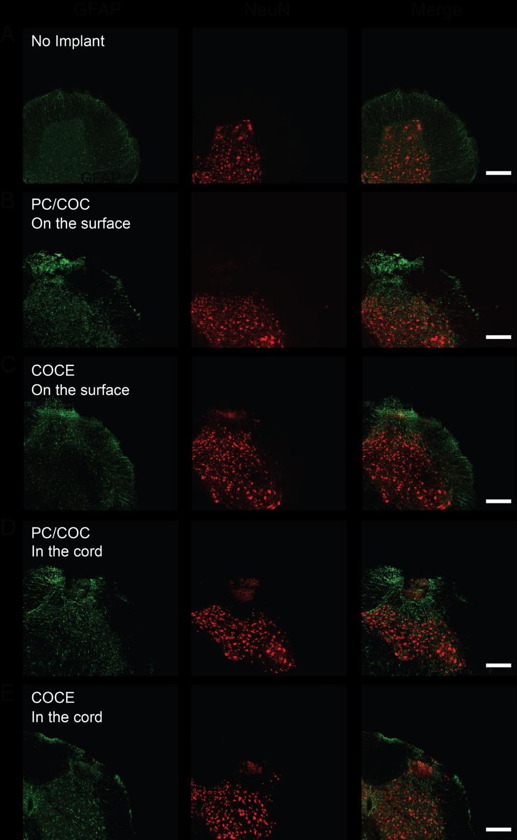
12 fig. S10. Immunohistochemical analysis of the dorsal horn 2 weeks after device implantation surgeries. GFAP (green) marks astrocytes, and NeuN (red) labels neurons. Scale bar is 100 μm. (A) Confocal micrographs of a transverse section of the lumbar spinal cord without an implant. (B) Transverse section of the lumbar spinal cord from a mouse implanted with a PC/COC/AgNW/PDMS probe for two weeks. The device was positioned on the spinal cord surface. (C) Confocal micrographs of a transverse section of the lumbar spinal cord from a mouse implanted with a PC/COC/AgNW/PDMS probe for two weeks. The device was positioned on the spinal cord surface. (D) Transverse section of the lumbar spinal cord from a mouse implanted with a PC/COC/AgNW/PDMS probe for two weeks. The device was inserted into the spinal cord. (E) Confocal micrographs of a transverse section of the lumbar spinal cord from a mouse implanted with a PC/COC/AgNW/PDMS probe for two weeks. The device was inserted into the spinal cord. No tissue erosion and negligible astrocyte proliferation were observed. video S1. Optical spinal control of muscle activity. Optical pulses (5 ms, 168 mw/mm 2 ) delivered to the lumbar region of the spinal cord of a Thy1-ChR2-YFP transgenic mouse through the cores of the PC/COC/AgNW/PDMS fiber probes evoke twitch muscle contractions at 10 and 100 Hz.
Nature Neuroscience: doi: /nn Supplementary Figure 1
 Supplementary Figure 1 Drd1a-Cre driven ChR2 expression in the SCN. (a) Low-magnification image of a representative Drd1a-ChR2 coronal brain section (n = 2) showing endogenous tdtomato fluorescence (magenta).
Supplementary Figure 1
 Supplementary Figure 1 Miniature microdrive, spike sorting and sleep stage detection. a, A movable recording probe with 8-tetrodes (32-channels). It weighs ~1g. b, A mouse implanted with 8 tetrodes in
Supplementary information - Table (1), Figures (12), and Videos (5)
, Figures (12), and Videos (5)") Supplementary information - Table (1), Figures (12), and Videos (5) A soft, transparent, freely accessible cranial window for chronic imaging and electrophysiology Chaejeong Heo 1, Hyejin Park 1, 2, Yong-Tae
Supplementary information - Table (1), Figures (12), and Videos (5) A soft, transparent, freely accessible cranial window for chronic imaging and electrophysiology Chaejeong Heo 1, Hyejin Park 1, 2, Yong-Tae
Nature Neuroscience: doi: /nn Supplementary Figure 1. Lick response during the delayed Go versus No-Go task.
 Supplementary Figure 1 Lick response during the delayed Go versus No-Go task. Trial-averaged lick rate was averaged across all mice used for pyramidal cell imaging (n = 9). Different colors denote different
Supplementary Figure 1. ACE robotic platform. A. Overview of the rig setup showing major hardware components of ACE (Automatic single Cell
 2 Supplementary Figure 1. ACE robotic platform. A. Overview of the rig setup showing major hardware components of ACE (Automatic single Cell Experimenter) including the MultiClamp 700B, Digidata 1440A,
Supplementary Figure 1
 8w Pia II/III IV V VI PV EYFP EYFP PV EYFP PV d PV EYFP Supplementary Figure a Spike probability x - PV-Cre d Spike probability x - RS RS b e Spike probability Spike probability.6......8..... FS FS c f
Supporting Information
 Supporting Information An efficient broadband and omnidirectional light-harvesting scheme employing the hierarchical structure based on ZnO nanorod/si 3 N 4 -coated Si microgroove on 5-inch single crystalline
Supplementary Figure 1. Recording sites.
 Supplementary Figure 1 Recording sites. (a, b) Schematic of recording locations for mice used in the variable-reward task (a, n = 5) and the variable-expectation task (b, n = 5). RN, red nucleus. SNc,
Development of Ultrasound Based Techniques for Measuring Skeletal Muscle Motion
 Development of Ultrasound Based Techniques for Measuring Skeletal Muscle Motion Jason Silver August 26, 2009 Presentation Outline Introduction Thesis Objectives Mathematical Model and Principles Methods
How we study the brain: a survey of methods used in neuroscience
 How we study the brain: a survey of methods used in neuroscience Preparing living neurons for recording Large identifiable neurons in a leech Rohon-Beard neurons in a frog spinal cord Living slice of a
Supplemental Information. A Visual-Cue-Dependent Memory Circuit. for Place Navigation
 Neuron, Volume 99 Supplemental Information A Visual-Cue-Dependent Memory Circuit for Place Navigation Han Qin, Ling Fu, Bo Hu, Xiang Liao, Jian Lu, Wenjing He, Shanshan Liang, Kuan Zhang, Ruijie Li, Jiwei
Supplementary Figure 1 Information on transgenic mouse models and their recording and optogenetic equipment. (a) 108 (b-c) (d) (e) (f) (g)
 108 (b-c) (d) (e) (f) (g)") Supplementary Figure 1 Information on transgenic mouse models and their recording and optogenetic equipment. (a) In four mice, cre-dependent expression of the hyperpolarizing opsin Arch in pyramidal cells
Supplementary Figure 1 Information on transgenic mouse models and their recording and optogenetic equipment. (a) In four mice, cre-dependent expression of the hyperpolarizing opsin Arch in pyramidal cells
Nature Neuroscience: doi: /nn Supplementary Figure 1. Confirmation that optogenetic inhibition of dopaminergic neurons affects choice
 Supplementary Figure 1 Confirmation that optogenetic inhibition of dopaminergic neurons affects choice (a) Sample behavioral trace as in Figure 1d, but with NpHR stimulation trials depicted as green blocks
A genetically targeted optical sensor to monitor calcium signals in astrocyte processes
 A genetically targeted optical sensor to monitor calcium signals in astrocyte processes 1 Eiji Shigetomi, 1 Sebastian Kracun, 2 Michael V. Sofroniew & 1,2 *Baljit S. Khakh Ψ 1 Departments of Physiology
Nature Neuroscience: doi: /nn Supplementary Figure 1. Trial structure for go/no-go behavior
 Supplementary Figure 1 Trial structure for go/no-go behavior a, Overall timeline of experiments. Day 1: A1 mapping, injection of AAV1-SYN-GCAMP6s, cranial window and headpost implantation. Water restriction
Microcircuitry coordination of cortical motor information in self-initiation of voluntary movements
 Y. Isomura et al. 1 Microcircuitry coordination of cortical motor information in self-initiation of voluntary movements Yoshikazu Isomura, Rie Harukuni, Takashi Takekawa, Hidenori Aizawa & Tomoki Fukai
Supporting Online Material for
 www.sciencemag.org/cgi/content/full/317/5841/183/dc1 Supporting Online Material for Astrocytes Potentiate Transmitter Release at Single Hippocampal Synapses Gertrudis Perea and Alfonso Araque* *To whom
1- Cochlear Impedance Telemetry
 INTRA-OPERATIVE COCHLEAR IMPLANT MEASURMENTS SAMIR ASAL M.D 1- Cochlear Impedance Telemetry 1 Cochlear implants used presently permit bi--directional communication between the inner and outer parts of
bio-mof-1 DMASM Wavenumber (cm -1 ) Supplementary Figure S1 FTIR spectra of bio-mof-1, DMASMI, and bio-mof-1 DMASM.
 Supplementary Figure S1 FTIR spectra of bio-mof-1, DMASMI, and bio-mof-1 DMASM.") bio-mof-1 Transmittance bio-mof-1 DMASM DMASMI 2000 1500 1000 500 Wavenumber (cm -1 ) Supplementary Figure S1 FTIR spectra of bio-mof-1, DMASMI, and bio-mof-1 DMASM. Intensity (a.u.) bio-mof-1 DMASM as
bio-mof-1 Transmittance bio-mof-1 DMASM DMASMI 2000 1500 1000 500 Wavenumber (cm -1 ) Supplementary Figure S1 FTIR spectra of bio-mof-1, DMASMI, and bio-mof-1 DMASM. Intensity (a.u.) bio-mof-1 DMASM as
SUPPLEMENTARY INFORMATION
 doi: 10.1038/nature06310 SUPPLEMENTARY INFORMATION www.nature.com/nature 1 www.nature.com/nature 2 www.nature.com/nature 3 Supplementary Figure S1 Spontaneous duration of wake, SWS and REM sleep (expressed
Supplementary Materials
 Supplementary Materials Fig. S1. Weights of full-dose treatment groups comparing 1 st, 2 nd, and 3 rd generation gene replacement therapy. Mice were treated at p1 with 4x10 11 GC of the three different
SUPPLEMENTARY INFORMATION
 SUPPLEMENTARY INFORMATION doi:10.1038/nature11306 Supplementary Figures Supplementary Figure 1. Basic characterization of GFP+ RGLs in the dentate gyrus of adult nestin-gfp mice. a, Sample confocal images
Development of a neural prosthesis for motor rehabilitation
 CSE599E Brain-Computer Interfaces, Spring 2006 Development of a neural prosthesis for motor rehabilitation Andy Jackson 1 and Jaideep Mavoori 2 1 Dept of Physiology and Biophysics and Washington National
Electromyography (EMG)
") Introduction In this laboratory, you will explore the electrical activity of skeletal muscle by recording an electromyogram (EMG) from a volunteer. You will examine the EMG of both voluntary and evoked
Introduction In this laboratory, you will explore the electrical activity of skeletal muscle by recording an electromyogram (EMG) from a volunteer. You will examine the EMG of both voluntary and evoked
BSL PRO Lesson H03: Nerve Conduction Velocity: Along the Ulnar Nerve of a Human Subject
 Updated 12-22-03 BSL PRO Lesson H03: Nerve Conduction Velocity: Along the Ulnar Nerve of a Human Subject This PRO lesson describes hardware and software setup of the BSL PRO System to record and measure
GLOSSARY OF TERMS ASSOCIATED WITH TENS
 GLOSSARY OF TERMS ASSOCIATED WITH TENS ATP Adenosine Triphosphate that helps to promote protein synthesis. Accommodation Becoming accustomed to stimulation resulting in nerve and muscle fatigue. Acute
Light-evoked hyperpolarization and silencing of neurons by conjugated polymers
 Light-evoked hyperpolarization and silencing of neurons by conjugated polymers Paul Feyen 1,, Elisabetta Colombo 1,2,, Duco Endeman 1, Mattia Nova 1, Lucia Laudato 2, Nicola Martino 2,3, Maria Rosa Antognazza
The Nervous System: The
 C h a p t e r 14 The Nervous System: The Spinal Cord and Spinal Nerves PowerPoint Lecture Slides prepared by Jason LaPres North Harris College Houston, Texas Copyright 2009 Pearson Education, Inc., publishing
Supplementary Figure 1
 Supplementary Figure 1 The average sigmoid parametric curves of capillary dilation time courses and average time to 50% peak capillary diameter dilation computed from individual capillary responses averaged
Supplementary Figure 1. Sample preparation schematic. First (Stage I), square islands of MoO 3 are prepared by either photolithography followed by
, square islands of MoO 3 are prepared by either photolithography followed by") Supplementary Figure 1. Sample preparation schematic. First (Stage I), square islands of MoO 3 are prepared by either photolithography followed by thermal evaporation and liftoff or by a process where
Supplementary Figure 1. Sample preparation schematic. First (Stage I), square islands of MoO 3 are prepared by either photolithography followed by thermal evaporation and liftoff or by a process where
Department of Orthopaedic Surgery, Tohoku University Graduate School of Medicine, Sendai, Japan, 2
 Low-energy Extracorporeal Shock Wave Therapy Promotes VEGF Expression and Angiogenesis and Improve Locomotor and Sensory Functions after spinal cord injury Kenichiro Yahata 1, Hiroshi Ozawa, M.D., Ph.D.
Short- and long-lasting consequences of in vivo nicotine treatment
 Short- and long-lasting consequences of in vivo nicotine treatment on hippocampal excitability Rachel E. Penton, Michael W. Quick, Robin A. J. Lester Supplementary Figure 1. Histogram showing the maximal
Behavioral generalization
 Supplementary Figure 1 Behavioral generalization. a. Behavioral generalization curves in four Individual sessions. Shown is the conditioned response (CR, mean ± SEM), as a function of absolute (main) or
Graduate School of Integrated Design Engineering, Keio University, Yokohama, Kanagawa, Japan (2)
") AMPLITUDE AND FREQUENCY FEATURE EXTRACTION OF NEURAL ACTIVITY IN MOUSE VENTROLATERAL STRIATUM UNDER DIFFERENT MOTIVATIONAL STATES USING FIBER PHOTOMETRIC SYSTEM S. Imai (1), Y. Mitsukura (2), K. Yoshida
AMPLITUDE AND FREQUENCY FEATURE EXTRACTION OF NEURAL ACTIVITY IN MOUSE VENTROLATERAL STRIATUM UNDER DIFFERENT MOTIVATIONAL STATES USING FIBER PHOTOMETRIC SYSTEM S. Imai (1), Y. Mitsukura (2), K. Yoshida
Nature Methods: doi: /nmeth Supplementary Figure 1. Activity in turtle dorsal cortex is sparse.
 Supplementary Figure 1 Activity in turtle dorsal cortex is sparse. a. Probability distribution of firing rates across the population (notice log scale) in our data. The range of firing rates is wide but
Supplementary Information
 Supplementary Information Title Degeneration and impaired regeneration of gray matter oligodendrocytes in amyotrophic lateral sclerosis Authors Shin H. Kang, Ying Li, Masahiro Fukaya, Ileana Lorenzini,
Cellular Bioelectricity
 ELEC ENG 3BB3: Cellular Bioelectricity Notes for Lecture #30 Thursday, March 30, 2006 Nerve excitation: To evaluate the pattern of nerve activation that is produced by a particular electrode configuration,
EE 4BD4 Lecture 20. Therapeutic Stimulation
 EE 4BD4 Lecture 20 Therapeutic Stimulation 1 2 Extracellular Stimulation (at cathode) 3 4 Design of FES (cont.): Example stimulus waveform shapes: monophasic, biphasic, chopped, triphasic, and asymmetric,
Supplementary Figure 1
 Supplementary Figure 1 Localization of virus injections. (a) Schematic showing the approximate center of AAV-DIO-ChR2-YFP injection sites in the NAc of Dyn-cre mice (n=8 mice, 16 injections; caudate/putamen,
EEG, ECG, EMG. Mitesh Shrestha
 EEG, ECG, EMG Mitesh Shrestha What is Signal? A signal is defined as a fluctuating quantity or impulse whose variations represent information. The amplitude or frequency of voltage, current, electric field
Supplementary Materials for
 advances.sciencemag.org/cgi/content/full/1/10/e1500775/dc1 Supplementary Materials for Structural-functional connectivity deficits of neocortical circuits in the Fmr1 /y mouse model of autism Matthias
Neuro-MS/D DIAGNOSTICS REHABILITATION TREATMENT STIMULATION. Transcranial Magnetic Stimulator. of motor disorders after the stroke
 Neuro-MS/D Transcranial Magnetic Stimulator DIAGNOSTICS of corticospinal pathway pathology REHABILITATION of motor disorders after the stroke TREATMENT of depression and Parkinson s disease STIMULATION
File name: Supplementary Information Description: Supplementary Figures, Supplementary Table and Supplementary References
 File name: Supplementary Information Description: Supplementary Figures, Supplementary Table and Supplementary References File name: Supplementary Data 1 Description: Summary datasheets showing the spatial
Supplementary Materials for
 advances.sciencemag.org/cgi/content/full/2/11/e1601007/dc1 Supplementary Materials for A conducting polymer with enhanced electronic stability applied in cardiac models Damia Mawad, Catherine Mansfield,
Supplementary Figure 1: Kv7 currents in neonatal CA1 neurons measured with the classic M- current voltage-clamp protocol.
 Supplementary Figures 1-11 Supplementary Figure 1: Kv7 currents in neonatal CA1 neurons measured with the classic M- current voltage-clamp protocol. (a), Voltage-clamp recordings from CA1 pyramidal neurons
Supplementary figure 1: LII/III GIN-cells show morphological characteristics of MC
 1 2 1 3 Supplementary figure 1: LII/III GIN-cells show morphological characteristics of MC 4 5 6 7 (a) Reconstructions of LII/III GIN-cells with somato-dendritic compartments in orange and axonal arborizations
Supplementary Figure 1. SybII and Ceb are sorted to distinct vesicle populations in astrocytes. Nature Neuroscience: doi: /nn.
 Supplementary Figure 1 SybII and Ceb are sorted to distinct vesicle populations in astrocytes. (a) Exemplary images for cultured astrocytes co-immunolabeled with SybII and Ceb antibodies. SybII accumulates
Social transmission and buffering of synaptic changes after stress
 SUPPLEMENTARY INFORMATION Articles https://doi.org/10.1038/s41593-017-0044-6 In the format provided by the authors and unedited. Social transmission and buffering of synaptic changes after stress Toni-Lee
SUPPLEMENTARY INFORMATION. Supplementary Figure 1
 SUPPLEMENTARY INFORMATION Supplementary Figure 1 The supralinear events evoked in CA3 pyramidal cells fulfill the criteria for NMDA spikes, exhibiting a threshold, sensitivity to NMDAR blockade, and all-or-none
Principles of Electrical Currents. HuP 272
 Principles of Electrical Currents HuP 272 Electricity is an element of PT modalities most frightening and least understood. Understanding the basis principles will later aid you in establishing treatment
Quantitative Electrophysiology
 ECE 795: Quantitative Electrophysiology Notes for Lecture #10 Wednesday, November 22, 2006 14. FUNDAMENTALS OF FUNCTIONAL ELECTRICAL STIMULATION (FES) We will look at: Design issues for FES Subthreshold
Nature Neuroscience: doi: /nn Supplementary Figure 1. Large-scale calcium imaging in vivo.
 Supplementary Figure 1 Large-scale calcium imaging in vivo. (a) Schematic illustration of the in vivo camera imaging set-up for large-scale calcium imaging. (b) High-magnification two-photon image from
SUPPLEMENTARY INFORMATION
 SUPPLEMENTARY INFORMATION doi:10.1038/nature12024 entary Figure 1. Distribution of the number of earned cocaine Supplementary Figure 1. Distribution of the number of earned cocaine infusions in Shock-sensitive
The Nervous System 12/11/2015
 The Nervous System Biology 12 Unit 3: Homeostasis December 11, 2015 The nervous system is an elaborate communication system that contains more than 100 billion nerve cells in the brain alone There are
SUPPLEMENTARY INFORMATION. Rett Syndrome Mutation MeCP2 T158A Disrupts DNA Binding, Protein Stability and ERP Responses
 SUPPLEMENTARY INFORMATION Rett Syndrome Mutation T158A Disrupts DNA Binding, Protein Stability and ERP Responses Darren Goffin, Megan Allen, Le Zhang, Maria Amorim, I-Ting Judy Wang, Arith-Ruth S. Reyes,
Supplementary Figure 1
 Supplementary Figure 1 Global TeNT expression effectively impairs synaptic transmission. Injection of 100 pg tent mrna leads to a reduction of vesicle mediated synaptic transmission in the spinal cord
Supplementary Figure 1. Nature Neuroscience: doi: /nn.4547
 Supplementary Figure 1 Characterization of the Microfetti mouse model. (a) Gating strategy for 8-color flow analysis of peripheral Ly-6C + monocytes from Microfetti mice 5-7 days after TAM treatment. Living
Astrocyte signaling controls spike timing-dependent depression at neocortical synapses
 Supplementary Information Astrocyte signaling controls spike timing-dependent depression at neocortical synapses Rogier Min and Thomas Nevian Department of Physiology, University of Berne, Bern, Switzerland
Biomedical Instrumentation
 University of Zagreb Faculty of Electrical Engineering and Computing Biomedical Instrumentation Electrical stimulation prof.dr.sc. Ratko Magjarević December 2015 Electrical stimulation The effect of electric
Neurostyle. Medical Innovation for Better Life
 Neurostyle Medical Innovation for Better Life Neurostyle Pte Ltd is a company dedicated to design, develop, manufacture and distribute neurological and neuromuscular medical devices. Strategically located
Analysis of in-vivo extracellular recordings. Ryan Morrill Bootcamp 9/10/2014
 Analysis of in-vivo extracellular recordings Ryan Morrill Bootcamp 9/10/2014 Goals for the lecture Be able to: Conceptually understand some of the analysis and jargon encountered in a typical (sensory)
Unique functional properties of somatostatin-expressing GABAergic neurons in mouse barrel cortex
 Supplementary Information Unique functional properties of somatostatin-expressing GABAergic neurons in mouse barrel cortex Luc Gentet, Yves Kremer, Hiroki Taniguchi, Josh Huang, Jochen Staiger and Carl
Supplementary Figure 1
 Supplementary Figure 1 Supplementary Figure 1. Short latency of the fepsp evoked in CA3 by electrical stimulation of perforant path inputs (a) Single and superimposed representative perforant pathway-ca3
Rapamycin suppresses astrocytic and microglial activation and reduces development of neuropathic pain after spinal cord injury in mice.
 Rapamycin suppresses astrocytic and microglial activation and reduces development of neuropathic pain after spinal cord injury in mice. Satoshi Tateda, MD, Haruo Kanno, MD, PhD, Hiroshi Ozawa, MD, PhD,
Nature Neuroscience: doi: /nn.4642
 Supplementary Figure 1 Recording sites and example waveform clustering, as well as electrophysiological recordings of auditory CS and shock processing following overtraining. (a) Recording sites in LC
Part 1 Making the initial neuron connection
 To begin, follow your teacher's directions to open the Virtual Neurons software. On the left side of the screen is a group of skin cells. On the right side of the screen is a group of muscle fibers. In
Intravital Microscopic Interrogation of Peripheral Taste Sensation
 Supplementary Information Intravital Microscopic Interrogation of Peripheral Taste Sensation Myunghwan Choi 1, Woei Ming Lee 1,2, and Seok-Hyun Yun 1 * 1 Harvard Medical School and Wellman Center for Photomedicine,
Quick Guide - eabr with Eclipse
 What is eabr? Quick Guide - eabr with Eclipse An electrical Auditory Brainstem Response (eabr) is a measurement of the ABR using an electrical stimulus. Instead of a traditional acoustic stimulus the cochlear
Technologies and architectures" Stimulator, electrodes, system flexibility, reliability, security, etc."
 March 2011 Introduction" Basic principle (Depolarization, hyper polarization, etc.." Stimulation types (Magnetic and electrical)" Main stimulation parameters (Current, voltage, etc )" Characteristics (Muscular
Tuning properties of individual circuit components and stimulus-specificity of experience-driven changes.
 Supplementary Figure 1 Tuning properties of individual circuit components and stimulus-specificity of experience-driven changes. (a) Left, circuit schematic with the imaged component (L2/3 excitatory neurons)
HHS Public Access Author manuscript Nat Neurosci. Author manuscript; available in PMC 2014 September 19.
 Selective optical drive of thalamic reticular nucleus generates thalamic bursts & cortical spindles Michael M. Halassa 1,2,4, Joshua H. Siegle 2,4, Jason T. Ritt 3, Jonathan T. Ting 2, Guoping Feng 2,
Supplementary Information
 Hyperpolarization-activated cation channels inhibit EPSPs by interactions with M-type K + channels Meena S. George, L.F. Abbott, Steven A. Siegelbaum Supplementary Information Part 1: Supplementary Figures
Supporting Information
 Supporting Information Toward High-Efficient Red Emissive Carbon Dots: Facile Preparation, Unique Properties, and Applications as Multifunctional Theranostic Agents Shan Sun,, Ling Zhang, Kai Jiang, Aiguo
Supplementary Table I Blood pressure and heart rate measurements pre- and post-stroke
 SUPPLEMENTARY INFORMATION doi:10.1038/nature09511 Supplementary Table I Blood pressure and heart rate measurements pre- and post-stroke Pre Post 7-days Systolic Diastolic BPM Systolic Diastolic BPM Systolic
Fluorescent Carbon Dots as Off-On Nanosensor for Ascorbic Acid
 Electronic Supplementary Material (ESI) for RSC Advances. This journal is The Royal Society of Chemistry 2014 Fluorescent Carbon Dots as Off-On Nanosensor for Ascorbic Acid Jun Gong, Xin Lu, Xueqin An*
Katherine Gibson-Corley DVM, PhD, DACVP. Pathology Grand Rounds March 27, 2014
 Katherine Gibson-Corley DVM, PhD, DACVP Pathology Grand Rounds March 27, 2014 Outline Introduction to spinal cord stimulation (SCS) Current SCS devices The Iowa-Patch TM (I-Patch) Post-surgical pathologies
Selective optical drive of thalamic reticular nucleus generates thalamic bursts & cortical spindles
 Selective optical drive of thalamic reticular nucleus generates thalamic bursts & cortical spindles Michael M. Halassa, Joshua H. Siegle, Jason T. Ritt, Jonathan T. Ting, Guoping Feng and Christopher I.
Fig. S4. Current-voltage relations of iglurs. A-C: time courses of currents evoked by 100 ms pulses
 Fig. S1. Immunohistochemical detection of iglur2 protein in single islet cells. A: α cells identified using glucagon-specific antibody express the iglur2 subtype of AMPA receptor. 24 out of 26 identified
ABR assesses the integrity of the peripheral auditory system and auditory brainstem pathway.
 By Prof Ossama Sobhy What is an ABR? The Auditory Brainstem Response is the representation of electrical activity generated by the eighth cranial nerve and brainstem in response to auditory stimulation.
Supplemental Information. In Vivo Optogenetic Stimulation. of Neocortical Excitatory Neurons. Drives Brain-State-Dependent Inhibition
 Current Biology, Volume 21 Supplemental Information In Vivo Optogenetic Stimulation of Neocortical Excitatory Neurons Drives Brain-State-Dependent Inhibition Celine Mateo, Michael Avermann, Luc J. Gentet,
Nature Neuroscience: doi: /nn Supplementary Figure 1
 Supplementary Figure 1 Relative expression of K IR2.1 transcript to enos was reduced 29-fold in capillaries from knockout animals. Relative expression of K IR2.1 transcript to enos was reduced 29-fold
Supplementary Figure 1. Example of an amygdala neuron whose activity reflects value during the visual stimulus interval. This cell responded more
 1 Supplementary Figure 1. Example of an amygdala neuron whose activity reflects value during the visual stimulus interval. This cell responded more strongly when an image was negative than when the same
ELECTROMYOGRAPHY (EMG) AND NERVE CONDUCTION STUDIES (NCS)
 AND NERVE CONDUCTION STUDIES (NCS)") ELECTROMYOGRAPHY (EMG) AND NERVE CONDUCTION STUDIES (NCS) Non-Discrimination Statement and Multi-Language Interpreter Services information are located at the end of this document. Coverage for services,
ELECTROMYOGRAPHY (EMG) AND NERVE CONDUCTION STUDIES (NCS) Non-Discrimination Statement and Multi-Language Interpreter Services information are located at the end of this document. Coverage for services,
Dopamine in Ube3a m-/p+ mice. Online Supplemental Material
 Online Supplemental Material S1 Supplemental Figure 1. Schematic of rate-dependent intracranial self-stimulation (ICSS) (A) Mice implanted with monopolar stimulating electrodes to the medial forebrain
mm Distance (mm)
") b a Magnet Illumination Coverslips MPs Objective 2575 µm 1875 µm 1575 µm 1075 µm 875 µm 545 µm 20µm 2 3 0.5 0.3mm 1 1000 100 10 1 0.1 1000 100 10 1 0.1 Field Induction (Gauss) 1.5 0 5 10 15 20 Distance
b a Magnet Illumination Coverslips MPs Objective 2575 µm 1875 µm 1575 µm 1075 µm 875 µm 545 µm 20µm 2 3 0.5 0.3mm 1 1000 100 10 1 0.1 1000 100 10 1 0.1 Field Induction (Gauss) 1.5 0 5 10 15 20 Distance
Structural and Optical Properties of Single- and Few-Layer Magnetic
 SUPPORTING INFORMATION Structural and Optical Properties of Single- and Few-Layer Magnetic Semiconductor CrPS 4 Jinhwan Lee 1, Taeg Yeoung Ko 2, Jung Hwa Kim 3, Hunyoung Bark 4, Byunggil Kang 4, Soon-Gil
Rapamycin Suppresses Astrocytic and Microglial Activation and Reduced Development of Neuropathic Pain after Spinal Cord Injury in Mice.
 Rapamycin Suppresses Astrocytic and Microglial Activation and Reduced Development of Neuropathic Pain after Spinal Cord Injury in Mice. Satoshi Tateda, M.D., Haruo Kanno, M.D., Ph.D., Hiroshi Ozawa, M.D.,
SUPPLEMENTARY INFORMATION
 Supplementary Figure 1. Normal AMPAR-mediated fepsp input-output curve in CA3-Psen cdko mice. Input-output curves, which are plotted initial slopes of the evoked fepsp as function of the amplitude of the
SUPPLEMENTARY FIG. S2. Representative counting fields used in quantification of the in vitro neural differentiation of pattern of dnscs.
 Supplementary Data SUPPLEMENTARY FIG. S1. Representative counting fields used in quantification of the in vitro neural differentiation of pattern of anpcs. A panel of lineage-specific markers were used
Supplementary Materials for
 advances.sciencemag.org/cgi/content/full/2/12/e1601838/dc1 Supplementary Materials for General and programmable synthesis of hybrid liposome/metal nanoparticles Jin-Ho Lee, Yonghee Shin, Wooju Lee, Keumrai
Electromyography II Laboratory (Hand Dynamometer Transducer)
") (Hand Dynamometer Transducer) Introduction As described in the Electromyography I laboratory session, electromyography (EMG) is an electrical signal that can be recorded with electrodes placed on the surface
(Hand Dynamometer Transducer) Introduction As described in the Electromyography I laboratory session, electromyography (EMG) is an electrical signal that can be recorded with electrodes placed on the surface
Nerve Conduction Studies NCS
 Nerve Conduction Studies NCS Nerve conduction studies are an essential part of an EMG examination. The clinical usefulness of NCS in the diagnosis of diffuse and local neuropathies has been thoroughly
Wenqin Hu, Cuiping Tian, Tun Li, Mingpo Yang, Han Hou & Yousheng Shu
 Distinct contributions of Na v 1.6 and Na v 1.2 in action potential initiation and backpropagation Wenqin Hu, Cuiping Tian, Tun Li, Mingpo Yang, Han Hou & Yousheng Shu Supplementary figure and legend Supplementary
High Voltage Pulsed Current (HVPC) Mohammed Taher Ahmed Associate professor of PT Mobile phone :
 Mohammed Taher Ahmed Associate professor of PT Mobile phone :") High Voltage Pulsed Current (HVPC) Mohammed Taher Ahmed Associate professor of PT E-Mail: momarar@ksu.edu.sa Mobile phone : 0542115404 Objective To review the core concepts and terminology used in high
High Voltage Pulsed Current (HVPC) Mohammed Taher Ahmed Associate professor of PT E-Mail: momarar@ksu.edu.sa Mobile phone : 0542115404 Objective To review the core concepts and terminology used in high
Nerve Conduction Studies NCS
 Nerve Conduction Studies NCS Nerve conduction studies are an essential part of an EMG examination. The clinical usefulness of NCS in the diagnosis of diffuse and local neuropathies has been thoroughly
Neurosoft TMS. Transcranial Magnetic Stimulator DIAGNOSTICS REHABILITATION TREATMENT STIMULATION. of motor disorders after the stroke
 Neurosoft TMS Transcranial Magnetic Stimulator DIAGNOSTICS REHABILITATION TREATMENT of corticospinal pathways pathology of motor disorders after the stroke of depression and Parkinson s disease STIMULATION
BNP mrna expression in DR and DS rat left ventricles (n = 5). (C) Plasma norepinephrine
. (C) Plasma norepinephrine") Kanazawa, et al. Supplementary figure legends Supplementary Figure 1 DS rats had congestive heart failure. (A) DR and DS rat hearts. (B) QRT-PCR analysis of BNP mrna expression in DR and DS rat left ventricles
Kanazawa, et al. Supplementary figure legends Supplementary Figure 1 DS rats had congestive heart failure. (A) DR and DS rat hearts. (B) QRT-PCR analysis of BNP mrna expression in DR and DS rat left ventricles
Introduction. Device Description. Dual-Band Spectral Output
 Optimum Spectrum and Pulse Shape for Vascular Lesion Treatment: The Science Behind MaxG James Childs, Ph.D.; Andrei Erofeev, Ph.D.; Mikhail Smirnov, Ph.D.; and Gregory Altshuler, Ph.D., Sc.D. Introduction