GROSS ANATOMY. Unit #3: Head and Neck. Lecture Syllabus 2008
|
|
|
- Roderick Anderson
- 6 years ago
- Views:
Transcription
1 GROSS ANATOMY Lecture Syllabus 2008 Unit #3: Head and Neck ANAT Gross Anatomy Department of Neurobiology and Anatomy University of Utah School of Medicine David A. Morton, Ph.D. 1
2 Unit #3- Neck and Head G15- Neck Overview G16-17A: Triangles of the Neck Posterior Triangle Anterior Triangle Visceral Triangle G17B- Brain and Base of the Skull G18- Cranial Nerves and Autonomics- A Systemic Overview G19- Orbit G20A- Superficial Face and Infratemporal Fossa G20B- Pterygopalatine Fossa, Nasal Cavity, and Paranasal Sinuses G21A- Oral Cavity, Tongue and Teeth G21B- Pharynx G22- Larynx G23- Ear 2
3 G15: Neck Overview At the end of this lecture, students should be able to master the following: 1)Fascia a) Describe the location and anatomical structures associated with the following fascial layers (1) Superficial cervical fascia- same as subcutaneous layer of skin; contains the platysma m. Platysma m.: innervated by the cervical branch of CN VII (2) Deep cervical fascia Investing fascia- envelopes sternocleidomastoid and trapezius muscles; occipital bone, mastoid process, zygomatic arch inferiorly to the scapular spine, acromion, clavicle and manubrium Pretracheal fascia-the muscular portion encloses the infrahyoid muscles, while the visceral portion surrounds the thyroid gland, larynx, trachea, pharynx and esophagus Prevertebral fascia- surrounds the cervical vertebral column, and envelopes deep neck muscles including prevertebral, scalenes, and deep back muscles Carotid sheath- formed by the investing, pretracheal and prevertebral fascial layers; surrounds internal jugular vein, common carotid artery, and vagus nerve b) Describe the location of the retropharyngeal space and its relation to the deep cervical fascial layers 3
4 4
5 2) Cervical Plexus Draw and label the cervical plexus including all spinal levels (C1-4): Sensory nerve branches Lesser occipital (C2) Great auricular (C2-C3) Transverse cervical (C2-C3) Supraclavicular (C3-C4) Motor nerve branches Ansa cervicalis (C1 superior root) (C2-C3 inferior root) Nerve to geniohyoid and thyrohyoid mm. (C1) Nerve to superior belly of omohyoid m. (C1) Nerve to sternothyroid and sternohyoid mm. (C1-C3) Nerve to inferior belly of omohyoid m. (C2-C3) Phrenic (C3-C5) 5
6 6
7 3) Vessels of the Head and Neck a) Common Carotid Artery i) Internal carotid artery- courses through neck in carotid sheath without any branching, then travels through the carotid canal to enter to skull Carotid sinus - baroreceptor; CN IX visceral sensory Carotid body - chemoreceptor; CN IX and X visceral sensory; located at bifurcation ii) External carotid artery Describe the origin, course, and destination of the following branches of the external carotid artery: Superior thyroid Ascending pharyngeal Lingual Facial Occipital Posterior auricular Maxillary Superficial temporal b) The IJV and EJV are the primary venous channels for the head and neck i) Internal jugular vein- located in carotid sheath Anterior jugular vein ii) External jugular vein - formed posterior to the angle of the mandible as the posterior auricular vein and retromandibular vein Retromandibular vein is formed by the superficial temporal and maxillary veins Anterior division - joins the facial vein to form the common facial vein, which joins the IJV Posterior division - joins the posterior auricular vein to form the external jugular vein 7

8 8
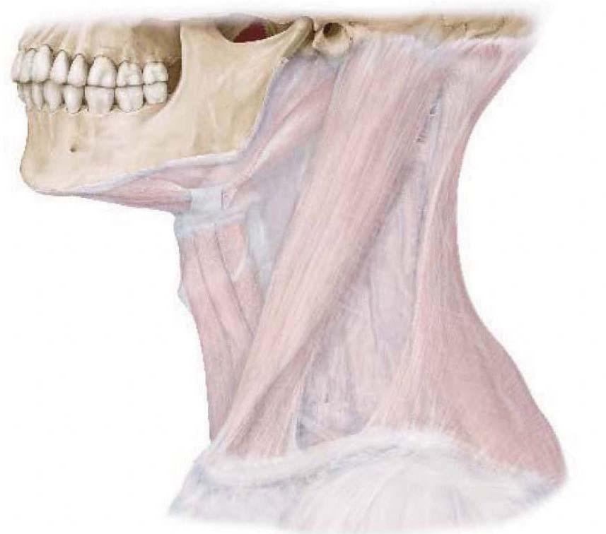
9 4) Muscles of the Neck Superficial Neck Muscles - Platysma m. - Trapezius m. - Sternocleidomastoic m. Unilateral: Tilts the head to the same side; rotates the head to the opposite side Bilateral: Extends the head; assists in respiration when the head is fixed Innervated by CN XI Ventral Strap Muscles Suprahyoid muscles - Digastric, geniohyoid, mylohyoid, stylohyoid Infrahyoid muscles - Sternohyoid, sternothyroid, thyrohyoid, omohyoid Prevertebral Muscles: stretch between the cervical spine and skull acting on both Longus captis m. Longus colli m. Lateral, Deep Neck Muscles Scalenes: lateral, deep neck muscles; attach between the cervical spine and upper two ribs and assist in respiration. the anterior and middle scalene are separated by the interscalene space; a topgraphicallly important interval traversed by the brachial plexus and subclavian artery Posterior scalene Middle scalene Axillary sheath Brachial plexus Subclavian artery - courses posterior to the anterior scalene m. Anterior scalene Subclavian vein - courses across the anterior scalene m. 9
10 10
11 G16-17A: Triangles of the Neck 1) Posterior Triangle of the Neck Describe the anatomical structures that create the following boundaries of the posterior triangle of the neck: Borders: sternocleidomastoid m., trapezius m., clavicle, occipital bone Roof: Investing layer of deep cervical fascia that surrounds the SCM and trapezius mm. Floor: Prevertebral fascia (splenius capitis, levator scapulae, posterior, middle and anterior sclane mm.) 2) Anterior Triangle of the Neck Describe the boundaries and major anatomical contents of the following subdivisions of the anterior triangle of the neck a) Submental Triangle: body of hyoid, anterior belly of digastricus, mandibular symphysis - Contains submental lymph nodes and mylohyoid muscle b) Submandibular (digastric) Triangle: inferior mandible, anterior and posterior bellies of digastricus - Contains the submandibular gland and portions of the hypoglossal nerve, facial artery, facial vein c) Carotid Triangle: superior belly of the omohyoid, anterior SCM, posterior belly of digastricus - Contains portions of the common carotid artery, internal jugular vein, and vagus nerve d) Muscular (omotracheal) Triangle: midline of neck, superior belly of omohyoid, anterior SCM - Contains: longus colli, longus capitis, rectus capitis anterior, rectus capitis lateralis, sternocleidomastoid, digastric, mylohyoid, geniohyoid, thyrohyoid, omohyoid, sternothyroid, sternohyoid 3 Visceral Triangle of the Neck Describe the boundaries and major anatomical contents of the visceral triangle of the neck a) Anterior: contains thyroid and parathyroid glands, pharynx/esophagus 11

12 12
13 1) Posterior Triangle of the Neck a) Describe the following topographical relations: i) The relation of the cervical plexus to the sternocleidomastoid muscle ii) The cutaneous distribution of the superficial branches of the cervical plexus (lesser occipital, great auricular, transverse cervical, supraclavicular) iii) External jugular vein formed by posterior auricular and retromandibular vein. Courses down the neck in superficial fascia external to the SCM and pierces the investing deep cervical fascia entering the subclavian vein. Clinical correlations with EJV - jugular venous pulse iv) The relation between the anterior scalene and the phrenic nerve v) The location of the brachial plexus nerves (C5-T1 ventral rami) in the posterior triangle of the neck vi) The pathway of the following vessels through the posterior triangle of the neck: suprascapular artery, transverse cervical artery, occipital artery, external jugular vein, and subclavian vein vii) The relation of the prevertebral fascia to the spinal accessory nerve (CN XI) 13


14 14
15 2) Anterior Triangle of the Neck a) Submandibular triangle i) Superior to the digastric bellies ii) Contents: submandibular gland, submandibular nodes, facial a. and v., hypglossal n. b) Carotid triangle i) Posterior digastric, inferior omohyoid and SCM ii) Contents: carotid sheath and related structures (carotids, CN X, IJV, carotid body and sinus) c) Muscular triangle i) Formed by the infrahyoid mm. ii) Contents: infrahyoid mm. d) Submental triangle i) Anterior digastric bellies and hyoid bone ii) Contents: submental lymph nodes 15

16 16
17 2) Continued... Nerves Associated with the Anterior Triangle of the Neck a) Cervical plexus i) Know the branches to geniohyoid and thyrohyoid mm., to superior belly of omohyoid m., to sternothyroid m., to sternohyoid m., to inferior belly of the omohyoid m. ii) Describe the relation of the ansa cervicalis to the carotid sheath, common carotid artery, internal jugular vein, and sympathetic trunk iii) Describe the relationship of the ansa cervicalis (superior root) to CN XII b) Glossopharyngeal nerve (CN IX) i) Describe the course/function of the glossopharyngeal nerve (CN XI) to the carotid sinus and carotid body Carotid branch- descends to the carotid sinus and carotid body to monitor arterial blood pressure and oxygen content (VS) c) Vagus nerve (CN X) i) Describe the relation of the vagus nerve to the common carotid artery and internal jugular vein ii) Describe the course, destination, and nerve fiber contents of the following branches of the vagus nerve: superior laryngeal, internal laryngeal, external laryngeal, recurrent laryngeal iii) Describe the course and function of the vagus nerve (CN X) supply to the carotid body Carotid body branch- branches from the inferior vagal ganglion and descends to the carotid body to monitor arterial oxygen content (VS) d) Sympathetics i) Describe the relation of the sympathetic trunk (chain) to the prevertebral fascia, longus colli muscle, longus capitis muscle and cervical vertebrae ii) Describe the location and nerve fiber contents of the following sympathetic trunk ganglia: inferior cervical, middle cervical, and superior cervical ganglia iii) Describe the function of the cervical sympathetic trunk in innervation of the head and neck 17
18 18
19 3) Visceral Triangle and Root of the Neck a) Thyroid gland i) Identify the parts (lobes and isthmus) and general function ii) Describe the arteries and veins of the thyroid gland iii) Describe the relationship between the superior thyroid artery and superior laryngeal nerve, and the inferior thyroid artery and recurrent laryngeal nerve b) Parathyroid glands i) Describe the location and general function of the parathyroid glands c) Trachea i) Describe the general function of the trachea and topography d) Esophagus i) Describe the general function of the esophagus and topography 4) Root of the Neck a) Describe the anatomical structures that create the following boundaries of the root of the neck: Manubrium, rib 1 and T1 vertebra b) Describe the arterial asymmetry of the aortic arch branches in the root of the neck c) Describe the origin, course, and destination of the following branches of the subclavian arteries: vertebral, thyrocervical, dorsal scapular arteries d) Describe the origin, course, and destination of the following veins of the root of the neck: external jugular, anterior jugular, jugular venous arch, subclavian, internal jugular, brachiocephalic veins e) Describe the relation of the thoracic lymphatic and right lymphatic ducts to the cervical veins 19
20 20
21 G17B: Brain and Base of Skull At the end of this lecture, students should be able to master the following: 1) Scalp a) Describe the layers of the scalp. The first three layers are held tightly together. Skin Connective tissue- dense connective tissue; distributes neurovascular supply to the scalp Aponeurosis - consists of the occipitalis muscle, galea aponeurotica and frontalis muscle Loose connective tissue - separates the aponeurotic layer from the pericraneum Pericranium - periosteum b) Describe the arterial vascularization of the scalp Internal carotid artery - ophthalmic artery - supratrochlear and supraorbital branches External carotid artery - superficial temporal, posterior auricular, and occipital arteries c) Map the cutaneous innervation of the scalp Trigeminal nerve branches- supraorbital (CN V-1), supratrochlear (CN V-1), zygomaticotemporal (CN V-2), auriculotemporal (CN V-3) Lesser occipital nerve (C2 ventral ramus) Greater occipital nerve (C2 dorsal ramus) Third (least) occipital nerve (C3 anterior ramus) 2) Skull a) Identify the bones of the neurocranium (frontal, parietal, temporal, occipital, sphenoid, and ethmoid bones) b) Identify the following sutures and related landmark on the skull: sagittal suture, lamboid suture, coronal suture, and pterion 21


22 C2 Spinal cord level 22
23 3) Cranial Meninges The brain is surrounded and protected by meninges, coupled with real and potential spaces within the cranial cavity. a) Dura Mater Describe the 2 layers of dura mater The periosteal layer is attached to the internal skull and sutures The meningeal layer is in close contact with the arachnoid mater and is continuous with the spinal dura The 2 layers of dura separate at numerous locations to form partitions which project inward and separate parts of the brain (falx cerebri, falx cerebelli, tentorium cerebelli, sellar diaphragm) Describe the composition and function of the dural venous sinuses and contrast them to the typical veins of the body Formed between two layers of dura Lined with endothelium, no tunica media or externa, no valves Eventually lead to the IJV Diploic veins coursing in the spongy bone of the skull and emissary veins coursing outside of the skull may drain into the dural venous sinuses Understand the potential spaces associated with the dura (epidural and subdural spaces) Describe the vascularization of the dura mater (primarily the middle meningeal artery with small meningeal branches from the ophthalmic, occipital, and vertebral arteries) Describe the general sensory innervation of the dura mater (by small meningeal branches from all three divisions of CN V and C1-C2 cervical nerves; detect pain from stretching dura mater b) Arachnoid Mater Describe the location and components of the arachnoid mater Arachnoid mater, arachnoid trabeculae, subarachnoid space, and archnoid villi/granulations The subarachnoid space contains CSF and blood vessels and surrounds the brain and spinal cord and in certain locations enlarges into cisterns) c) Pia Mater Describe the location of the pia mater and its relation to the arachnoid mater, the cerebrospinal fluid, and the gray matter of the central nervous system Pia mater follows the surface and contour of the brain into the grooves and fissures 23


24 24
25 d) Venous Drainage Describe venous drainage of the brain Small veins, lead to larger cerebral and cerebellar veins, which eventually empty into dural venous sinuses Identify the major dural venous sinues (as outlined in the figure) Describe the importance of the cavernous sinus The cavernous sinuses receive blood from cerebral veins, ophthalmic veins and emissary veins from the pterygoid plexus These connections provide pathways for infections to spread from extracranial to intracranial Describe the relation of the cavernous sinus to the following structures: - Structures passing through the cavernous sinus: internal carotid artery and CN VI - Structures in the lateral wall of the cavernous sinus from superior to inferior: CN III, IV, V-1 and V-2 25

26 26
27 4) Brain a) Parts of the Brain Describe the general location and function of the following parts of the brain including associated ventricles and cranial nerves Cerebrum- cerebral hemispheres with lateral ventricles Brainstem Diencephalon and third ventricle Midbrain- nuclei of CN III, IV Cerebral aqueduct Pons- nuclei of CN V, VI, VII, VIII Medulla oblongata- nuclei of CN IX, X, XI, XII Cerebellum- fourth ventricle Corpus callosum - commisure b) Ventricles of the Brain and Cerebrospinal Fluid Describe the location and function of the choroid plexuses and cerebrospinal fluid Trace the flow of cerebrospinal fluid through the ventricular system - CSF is produced by choroid plexus in the ventricles - CSF is clear, colorless, cell-free fluid that circulates through the subarachnoid space surrounding the brain and spinal cord - CSF returns to the venous system through arachnoid villi into the superior sagittal sinus 27

28 28
29 5) Blood Supply to the Brain a) Internal Carotid Artery Trace the pathway of the internal carotid artery into the skull (cervical portion passes through carotid sheath, petrous portion travels through the carotid canal, cavernous portion then bends through the cavernous sinus to give rise to the cerebral portion of the internal carotid) Describe the pathway and distribution of the major intracranial branches of the internal carotid artery Ophthalmic artery- courses towards orbit, supplies eye and other orbital structures Anterior cerebral artery- courses anteriorly, supplies medial surface of brain (leg-foot area of motor and sensory cortices) Middle cerebral artery- courses laterally, supplies lateral surface of brain (trunk-arm-face are of motor and sensory cortices, Broca s and Wernicke s speech areas) Anterior communicating artery- connects the two anterior cerebral arteries in the circle of Willis b) Vertebral Arteries Trace the pathway of the vertebral artery from branching off the subclavian artery into the skull, to the anastamosis to form the basilar artery (transverse foramina of cervical vertebrae, floor of sub-occipital triangle, foramen magnum, anastamosis) Describe the pathway and distribution of the major branches of the basilar artery Posterior and anterior inferior cerebellar arteries- (posterior branching off of the vertebral arteries) Supply the cerebellum and brain stem Posterior cerebral arteries- terminal branches of the basilar artery, supply the occipital lobes of the cerebrum Posterior communication artery- connects the middle cerebral artery with the posterior cerebral arteries c) Circle of Willis Explain the function of having redundancies in cerebral circulation If one part of the circle or artery supplying the circle become blocked or narrowed, blood flow from the other blood vessels can often preserve the cerebral perfusion well enough to avoid ischemia. 29


30 30
31 G18: Cranial Nerves and Autonomics- A Systemic Overview At the end of this lecture students should be able to master the following: 1) Introduction to Cranial Nerve Modalities a) Describe the difference between a nerve and a neuron (a nerve is a bundle of neurons - A neuron can be sensory, or motor - A nerve can contain only sensory neurons, only motor neurons or a mixture of both b) Describe the difference between a nucleus and a ganglion - Nucleus: A collection of nerve cell bodies in the CNS - Ganglion: A collection of nerve cell bodies in the PNS (sensory or autonomic) c) Compare and contrast the following sensory (afferent) neuron modalities: General Somatic Afferent (GSA) = General Sensory (GS) - pain, temperature, touch, proprioception (CN V, VII, X) Special Afferent (SA) = Special Sensory (SS) - smell, sight, hearing, taste and balance (CN I, II, VII, VIII, IX, X) General Visceral Afferent (GVA) = Visceral Sensory (VS) - sensory input from viscera (CN IX, X) d) Compare and contrast the following motor (efferent) neuron modalities: General Somatic Efferent (GSE) = Somatic Motor (SM) - innervate voluntary muscles derived from somites (CN III, IV, VI, XII Branchial efferent (BE) = Branchial Motor (BM) - innervate voluntary muscles derived from branchial (pharyngeal) arches (CN V, VII, IX, X, XI) General Visceral Efferent (GVE) = Visceral Motor (VM) - motor innervation to smooth muscle, heart muscle and glands (CN III, VII, IX, X) * only parasympathetics arise from the brainstem via CN III, VII, IX and X; sympathetics to the head arise from the T1 level of the spinal cord* **Memorise the cranial nerves by Roman numeral as well as name** ***The Tables on pgs , 805, 807 in GAFS are very helpful*** 31


32 32
33 2) The Cranial Nerves Describe the course, distribution, function, and common manifestations of lesions for each of the CN s i) Olfactory nerve (CN I)- olfactory epithelium in upper nasal cavity for smell (SS) Lesions- anosmia ii) Optic nerve (CN II)- optic chiasm, optic canal, orbit, retina of eye for sight (SS) Lesions- visual field defects (anopsia), loss of light reflex with CN III, blindness, only CN affected by MS iii) Oculomotor nerve (CN III) SM- superior orbital fissure, orbit to 4 of the 6 extraoccular muscles (superior, medial and inferior rectus, inferior oblique plus the levator palpebrae superioris) VM- constricts pupil (parasympathetic fibers to cilliary ganglion) via the sphincter pupillae muscle; lens accommodation (ciliary muscles) Lesions- diplopia (external strabismus), injured eye positioned down and out, loss of parallel gaze, dilated pupil, loss of light reflex with CN II, loss of near response, ptosis iv) Trochlear nerve (CN IV)- superior oblique muscle (SM) to move the eye down and out Lesions- inability to look inferiorly when eye is adducted, head tilts away from lesioned side; difficulty walking down stairs vi) Abducens nerve (CN VI)- lateral rectus muscle (SM) to abduct eye Lesions- inability of lateral eye movement, diplopia (internal strabismus), loss of parallel gaze 33

34 34
35 v) Trigeminal nerve (CN V)- trigeminal ganglion with sensory cell bodies; possesses three branches named for cranial location, supplies eyes, maxilla, and mandible (1) Ophthalmic nerve (CN V-1)- orbit, surface of the cornea and skin of the forehead and scalp (GS) Lesions- loss of sensation in the skin of the forehead and scalp, loss of blink reflex with VII (2) Maxillary nerve (CN V-2)- palate, nasal cavity, skin over maxillary face and maxillary teeth (GS) Lesions- loss of sensation in skin over maxilla and maxillary teeth, trigeminal neuralgia (pain) (3) Mandibular nerve (CN V-3) GS- sensory to the anterior tongue, mandibular teeth, and skin over mandibular face BM- motor to muscles of mastication (masseter, temporalis, pterygoids), anterior digastricus, mylohyoid, and 2 tensors (tensor tympani, tensor veli palatini) Lesions- Loss of sensation in the mandibular skin, teeth, and tongue, weakness in chewing, jaw deviates to one side, trigeminal neuralgia (pain) vii) Facial nerve (CN VII) BM- muscles of facial expression, posterior digastricus, stylohyoid, stapedius SS- chorda tympani to the anterior tongue for taste VM- submandibular and sublingual salivary glands (chorda tympani) via V-3, lacrimal, nasal and palatine glands (greater petrosal) via V-2 Lesions- paralysis of facial muscles (Bell s palsy) on ipsilateral side (peripheral CN VII injury), paralysis of contralateral facial muscles below the eye (brainstem injury), loss of blink reflex, hyperacusis, alteration or loss of taste (ageusia), red and dry eye viii) Vestibulocochlear (CN VIII) SS- hearing (cochlea) and balance (vestibular apparatus) Lesions- hearing loss, loss of balance, tinnitus (ringing in the ear) 35

36 36
37 ix) Glossopharyngeal nerve (CN IX) GS- mucous of the oropharynx, tympanic cavity and auditory tube SS- taste for posterior tongue VM- parotid gland (lesser petrosal nerve to the otic ganglion) to the auriculotemporal n. (CN V-3) BM- stylopharyngeus VS- carotid body/sinus Lesions- loss of gag reflex with CN X, alteration or loss of taste to posterior tongue; altered vaso-vagal reflex x) Vagus nerve (CN X) BM- palatoglossus, soft palate muscles (except tensor veli pal), pharynx (except stylopharyngeus) and laryngeal muscles GS- skin posterior to ear and external acoustic meatus and posterior dura VS- aortic body, chemo- and baroreceptors in aortic arch, mucous membranes of pharynx, larynx, esophagus, bronchi, lungs, heart, and abdominal viscera of foregut and midgut VM- smooth muscle and glands in the pharynx, larynx, thoracic and abdominal viscera (fore-/midgut) Lesions- loss of gag reflex with CN IX, loss of cough reflex, uvula pointing away from affected side, xi) Accessory nerve (CN XI) BM- trapezius and sternocleidomastoid Lesions- weakness turning head to opposite side, shoulder drop xii) Hypoglossal nerve (CN XII) (BM)- innervates all extrinsic and intrinsic tongue muscles (except palatoglossus) Lesions- tongue pointing towards affected side on protrusion 37

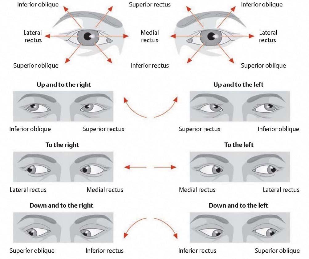
38 38
39 3) Autonomics a) Sympathetic innervation to the head Describe the origin, course, distribution, function, and common manifestations of lesions of sympathetic innervation in the head - Preganglionic sympathetic fibers- originate from T1 of the spinal cord, ascend in the sympathetic trunk, synapse with postganglionic fibers in the superior cervical ganglion - Postganglionic sympathetic fibers- follow internal and external carotid artery branches throughout the head to serve primarily blood vessels and sweat glands, but also the superior tarsal muscle to raise the upper eye lid and dilatator pupillae muscle to dilate the pupil Lesions- Horner syndrome, eyelid droop (ptosis), constricted pupil (miosis), loss of sweating (anhydrosis), flushed face b) Parasympathetic innervation to the head Describe the origin and distribution of parasympathetic innervation in the head, include location of synapse CN III - Oculomotor nerve - Preganglionic parasympathetic neurons originate in Edinger-Westphal nucleus and synapse in the ciliary ganglion - Postganglionic parasympathetic neurons serve the ciliary muscles and sphincter pupillae for light accommodation and constriction of the pupil CN VII - Facial nerve Greater petrosal nerve branch - Preganglionic parasympathetic neurons originate in the superior salvatory nucleus and synapse in the pterygopalatine ganglion - Postganglionic parasympathetic neurons serve the lacrimal gland, nasal glands, and nasal mucosa; Chorda tympani nerve branch - Preganglionic parasympathetic neurons originate in the superior salvatory nucleus and synapse in the submandibular ganglion - Postganglionic parasympathetic neurons serve the submandibular and sublingual salivary glands CN IX - Glossopharyngeal nerve - Preganglionic parasympathetic neurons originate in the inferior salvatory nucleus and synapse in the otic ganglion - Postganglionic parasympathetic neurons serve the parotid gland via the auriculotemporal n. (CN V-3) CN X - Vagus nerve - Preganglionic parasympathetic neurons originate in the dorsal vagal nucleus and synapse with intramural ganglia at or near target organ - Postganglionic parasympathetic neurons serve smooth muscle and glands of the pharynx and larynx, heart, lungs and GI tract to the transverse colon 39
40 40
41 G19: Obit At the end of this lecture students should be able to master the following: 1) Eyelid and Conjunctiva a) Describe the action and innervation of the following eyelid muscles: Orbiularis oculi- closes eyelid, CN VII Levator palpebrae superioris- opens upper eyelid, CN III Superior tarsal (Muller s) muscle- smooth muscle portion under sympathetic control; elevates eyelid b) Describe the function and differences in location and innervation of the palpebral and bulbar conjunctiva 2) Lacrimal Apparatus a) Describe the location of the lacrimal gland b) Trace tears flowing from the orbit to the nasal cavity c) Describe the innervation of the lacrimal gland (VM from CN VII via the greater petrosal n.) - Preganglionic parasympathetic cell bodies in brain, internal acoustic meatus, facial, canal, greater petrosal nerve, nerve of the pterygoid canal, pterygopalatine fossa, synapse in pterygopalatine ganglion - Postganglionic fibers jump hitch-hike on the zygomatic nerve (CN V-2), on lacrimal nerve, lacrimal gland 41

42 42
43 3) Eyeball a) Fibrous Layer Describe the location and function of the components of the fibrous layer of the eye Cornea- anterior surface, bends light, encases most of anterior chamber (sensory innervation by CN V-1) Sclera- surrounds posterior surface, gives strength and structure to eye along with vitreous body, allows for muscle attachment b) Choroid Layer i) Describe the function of the ciliary apparatus (lens accommodation) Ciliary muscle control through parasympathetic innervation (CN III) lens accommodation (see below) ii) Describe the function and innervation of the following muscles of the iris: Sphincter pupillae- constricts the pupil (para CN III) - Preganglionic cell bodies in Edinger Westphal nucleus, cavernous sinus, superior orbital fissure, synapse in ciliary ganglion - Postganglionic fibers in short ciliary nerves to ciliary muscle and sphincter pupillae Dilator pupillae- dilates the pupil (sympathetic control from T1) - Preganglionic fibers from T1, sympathetic trunk, synapse in superior cervical ganglion - Postganglionic fibers run along internal carotid artery, ophthalmic artery, short or long ciliary nerves or muscular branches of CN III to dilator pupillae and/or superior tarsal muscles (elevate eyelid) c) Retina Describe the structure and function of the retina- pigmented region with photoreceptors sensitive to light Optic disc- where optic nerve enters, blind spot Macula lutea and fovea centralis- most acute vision d) Chambers of the eye Identify the chambers of the eye and their associated humors Anterior and posterior chambers-aqueous humor Vitreous chamber- vitreous humour 43

44 44
45 4) Extraoccular Eye Muscles a) Distinguish between optical axis and orbital axis - Orbital axis: directed laterally from back to front by an angle of 23 degrees - Optical axis: Each eyeball is directed anteriorly As a result, the pull of some extraoccular eye muscles has multiple effects on the movement of the eyeball, while that of others has only a single effect b) Describe the action, innervation, and topography for each of the extraoccular eye muscles i) CN III - Medial rectus m. (adduct) - Superior rectus m. (up and in) - Inferior rectus m. (down and in) - Inferior oblique m. (up and out) - Levator palpebrae superioris (eye lid up) ii) CN IV - Superior oblique m. (down and out); tendon courses through a tendinous pulley called the trochlea to double back and insert into the sclera on the top of the globe iii) CN VI - Lateral rectus m. (abduct) 45

46 46
47 c) Contrast the difference between anatomical actions of the extraoccular eye muscles and how to clinically test them Clinical testing Ask the patient to follow your finger drawing a large H pattern in the air in front of their face. - The two vertical lines of the H isolate and test the superior and inferior rectus muscles and superior and inferior oblique muscles. - The horizontal lines of the H will test the medial and lateral rectus muscles. Explanation The lateral rectus muscle abducts the eye (away from the nose) and the medial rectus muscle adducts the eye (towards the nose). The lateral and medial muscles move the eye only in the horizontal or axial plane. Therefore, the action of these muscles and clinically testing these two muscles is the same. However, the vertical motion is a little more complex. Four muscles (superior rectus, inferior rectus, superior oblique and inferior oblique) control the vertical motion. To clinically test the superior and inferior rectus muscles you first need to isolate them from the superior and inferior oblique muscles. When the right eye is fully abducted (away from the nose), only the superior and inferior rectus muscles can elevate and depress the eye. This is purely a mechanical property due to the axis of the eye lining up parallel to the superior and inferior rectus muscles. When the right eye is fully adducted (towards the nose), only the superior and inferior oblique muscles can elevate and depress the eye. This is again due to the axis of the muscles paralleling the axis of the eye. Using the picture at the bottom of the page observe how it is possible for the superior oblique muscle to depress the eye when the eye is adducted. 47

48 48
49 5) Vasculature of the Orbit a) Trace the pathway and distribution of the major branches of the ophthalmic artery b) Describe the pathway of the central artery of the retina c) Describe the venous anastamosis between the superior ophthalmic, inferior ophthalmic and facial veins near the orbit. Describe the three possible drainage pathways. 6) Innervation of the Orbit a) CN II Exits/Enters Cranium: optic canal with ophthalmic artery Function: sight (SS) b) CN III Exits/Enters Cranium: Superior orbital fissure along with IV, V-1, VI and sup. ophthalmic v. Function: innervates 4 of the 6 extraoccular eye mm. and lev. palp. sup. m. (SM); constricts the pupil and accomodates the lens via the ciliary ganglion (VM) c) CN IV Exits/Enters Cranium: Superior orbital fissure Function: innervates superior oblique m. d) CN V Exits/Enters Cranium: V-1 Superior orbital fissure Function: general sensory to the forehead, scalp, cornea and bridge of the nose (GS) e) CN VI Exits/Enters Cranium: Superior orbital fissure Function: Innervates lateral rectus m. (SM) f) CN VII Exits/Enters Cranium: Internal acoustic meatus, greater petrosal hiatus, greater petrosal nerve, pterygopalatine ganglion, V-2, V-1 to lacrimal gland Function: Innervates the lacrimal gland (VM) 49
50 50
51 G20A: Superficial Face and Infratemporal Fossa At the end of this lecture, students should be able to master the following: 1) Cutaneous Innervation Map out the distribution of the major cutaneous branches of the trigeminal nerve (CN V) Ophthalmic division (CN V-1) - Superior orbital fissure to orbit Frontal nerve- supraorbital and supratrochlear nerves Nasociliary nerve- infratrochlear and anterior ethmoidal (external nasal) nerve Maxillary division (CN V-2) - Foramen rotundum - pterygopalatine fossa - infraorbital canal and foreman Zygomatic nerve- zygomaticotemporal and zygomaticofacial nerves Infraorbital nerve- inferior palpebral, lateral nasal, and superior labial nerves Mandibular division (CN V-3) - Foramen ovale - infratemporal fossa - mandibular canal - mental foramen Buccal nerve Auriculotemporal nerve Inferior alveolar nerve- mental nerve 51

52 52
53 2) Parotid Gland a) Describe the function and location of the parotid gland including relations to the external carotid artery, retromandibular vein, facial nerve, masseter, bucal fat pad, and buccinator muscle b) Trace the parasympathetic innervation to the parotid gland along the glossopharyngeal nerve (CN IX) naming all nerves and anatomical spaces traveled (preganglionic cell bodies in brain, CN IX, jugular foramen, tympanic canal into temporal bone, middle ear, tympanic plexus, lesser petrosal nerve, foramen ovale, motormotor synapse in otic ganglion, postganglionic fibers to parotid gland) 3) Facial Expression Describe the location, and basic attachments and actions of the following muscles of facial expression: Zygomaticus major and minor Levator anguli oris Levator labii superioris Levator labii superioris alaeque nasi Buccinator Risorius Orbicularis oris Depressor anguli oris Depressor labii inferioris Mentalis Platysma Occipitofrontalis Corrugator supercilii Procerus Nasalis Innervation of the muscles of facial expression (BM) CN VII- internal acoustic meatus, facial canal, stylomastoid foramen, through parotid gland - Muscular branches: Temporal, Zygomatic, Buccal, Mandibular, Cervical nerves 53

54 54
55 4) Blood Supply to the Face Arteries Describe the origin, course, and destination of the following external carotid arterial branches to the face Facial artery- curls around mandible, banches into superior and inferior labial, lateral nasal, and angular areries Superficial temporal- branches out on lateral surface of cranium anterior to ear Transverse facial- extends anteriorly along zygomatic arch Maxillary artery- deep to mandibular ramus gives rives to many branches (inferior alveolar, superior alveolar, infraorbital, deep temporal) Veins Describe the origin, course, and destination of the following veins of the face External jugular tributaries Posterior auricular vein- drains skin posterior to the ear Retromandibular vein- drain skin anterior to the ear, through the parotid Internal jugular tributaries Facial vein- drains most of facial skin Anterior vein- drains anterior cervical skin 55
 Describe the openings that communicate with the infratemporal fossa Foramen ovale-")
56 5) Infratemporal Fossa Topography a) Describe the boundaries of the infratemporal fossa Medial- lateral pterygoid plate of the sphenoid Lateral- ramus of the mandible Anterior- posterior aspect of the maxilla Posterior- condyle of the mandible, mastoid process and styloid process of the temporal bone b) Describe the openings that communicate with the infratemporal fossa Foramen ovale- mandibular nerve (CN V-3) Foramen spinosum- middle meningeal artery Pterygomaxillary fissure- maxillary artery Inferior orbital fissure- infraorbital nerve 56
57 c) Temporomandibular joint (TMJ) Describe the structure and movements of the temporomandibular joint, including bony components, fibrous capsule, synovial membrane, and the articular disc d) Describe the actions and muscles that produce the actions of the TMJ - Elevation: temporalis, masseter, medial pterygoid - Depression: gravity, digastric, geniohyoid and mylohyoid - Protrusion: lateral pterygoid m. (assisted by medial pterygoid) - Retraction: posterior termporalis, deep masseter geniohyoid, digastric, e) Muscles of Mastication Describe the location, function, basic attachments, and innervation for each of the following muscles of mastication: - Masseter - Temporalis - Medial pterygoid - Lateral pterygoid f) Arteries of the Infratemorpal Fossa Maxillary artery Describe how the maxillary artery is broken into three regions relative to the lateral pterygoid muscle Describe the course and distribution of the principle branches of the three regions of the maxillary artery First region- middle meningeal, inf alveolar (deep auricular, anterior tympanic, accessory meningeal) Second region- deep temporal (buccal, pterygoid arteries, masseteric) Third region- descending palatine, sphenopalatine, infraorbital (artery of the pterygoid canal) g) Pterygoid plexus of veins Describe the origin, course destination, and relations of the pterygoid plexus of veins and drainage to the maxillary, deep facial and ophthalmic vv.; include its potential drainage into the cavernous sinus 57
58 h) Nerves of the Infratemporal Fossa Mandibular division of the trigeminal nerve (CN V-3) (anterior 2/3 of tongue, mandibular face, and teeth) Main trunk of V-3- through foramen ovale to infratemporal fossa then spilts into anterior and posterior divisions (nerve to tensor tympani, nerve to tensor veli palatini, nerve to medial pterygoid) Anterior division Sensory: buccal Motor: nerve to lateral pterygoid, masseter, deep temporal nerves Posterior division Sensory: Auriculotemporal, inferior alveolar and mental, lingual Motor: Nerve to mylohyoid Chorda tympani - CN VII SS hitches to lingual n. for taste on anterior 2/3 of tongue Submandibular ganglion - CN VII VM hitches to lingual to submandibular and sublingual salivary glands Otic Ganglion - CN IX VM hitches to CN V-3 to the parotid gland; synapses in otic ganglion 58
59 G21B: Pterygopalatine Fossa, Nasal Cavity, and Paransal Sinuses At the end of this lecture, students should be able to master the following: 1) Boundaries, Osteology and Contents of the Pterygopalatine Fossa Describe the bony boundaries of the pterygopalatine fossa - Medial to the infratemporal fossa through the pterygomaxillary fissure Describe the principle contents of the pterygopalatine fossa - Maxillary artery - Maxillary nerve (CN V-2) - Pterygopalatine ganglion) 2) Pterygopalatine Fossa a) Know the main branches of the third part of the maxillary artery - Posterior superior alveolar artery - Descending palatine artery - Infraorbital artery: in the infraorbital canal, the infraorbital nerve gives rise to the: - Anterior superior alveolar - Middle superior alveolar - Sphenopalatine artery- passes through the sphenopalatine foramen to supply the nasal mucosa 59

60 60
61 b) Nerves of the pterygopalatine fossa Describe the course and distribution of the branches of CN V-2 - Posterior superior alveolar nerve (PSA) - Zygomatic nerve: passes through the inferior orbital fissure towards the lateral orbital wall, gives rise to a communicating branch that carries parasympathetic autonomic fibers from CN VII to the lacrimal nerve; sends off zygomaticofacial and zygomaticotemporal branches to skin in temporal region - Greater and lesser palatine nerves - Nasoplalatine nerve- courses through the sphenopalatine foramen, to the medial and lateral wall of the nasal cavity, then travels anteriorly along the nasal septum supplying general sense and parasympathetic postganglionic visceral motor (CN VII) to the mucosa, traversing the incisive canal to the hard palate - Infraorbital nerve- While in the infraorbital groove/canal, the nerve gives rives to the: - Middle superior alveolar nerve- GS to the upper premolars and maxillary sinus - Anterior superior alveolar nerve- GS to the upper incisors and canine - Pharyngeal nerve- courses through the palatovaginal canal to supply GS and VM (postganglionic parasympathetic via CN VII) to the mucosa and glands of the nasopharynx - Nerve of the pterygoid canal (Vidian nerve)- courses into the pterygopalatine fossa as a union of greater petrosal nerve (parasympathetic preganglionic fibers from CN VII) and the deep petrosal nerve (sympathetic postganglionic fibers from the carotid plexus) through the pterygoid canal and synapses with postganglionic parasympathetic fibers in the pterygopalatine ganglion; which are then distributed to the mucosal glands of the nasal cavity, palate and VM fibers hitch-hike on CN V-2 (zygomatic n.) and then CN V-1 (lacrimal gland) Pterygopalatine ganglion Trace the following autonomic pathways to the following areas: Parasympathetics - Preganglionic fibers- cell bodies in brain, CN VII, internal acoustic meatus, facial canal, greater petrosal nerve, joined by deep petrosal nerve to form the nerve of the pterygoid canal (Vidian nerve) passing through the pterygoid canal, pterygopalatine fossa, synapse in pterygopalatine ganglion - Postganglionic fibers to: Lacrimal gland- zygomatic nerve through the inferior orbital fissure, communicating branch to lacrimal nerve, lacrimal gland Smooth muscle and glands of the nasal cavity- nasopalatine nerve through the sphenopalatine foramen to the nasal mucosa Smooth muscle and glands of the palate- greater and lesser palatine nerves through the greater and lesser palatine canals, respectively Sympathetic fibers - Preganglionic fibers- originate from spinal levels T1-L2, ascend to synapse in sup cervical ganglion - Postganglionic fibers- carotid nerve runs with internal carotid artery, deep petrosal nerve, nerve of pterygoid canal through pterygoid canal, pterygopalatine fossa, pterygopalatine ganglion, travels with parasympathetic postganglionic fibers to same target organs (lacrimal gland, nasal. and palatal mucosa) 61

62 62


63 3) Nasal Cavity a) Boundaries and osteology of the nasal cavity Describe the bony boundaries of the nasal cavity - Medial boundary (septum)- perpendicular plate of the ethmoid, vomer and septal nasal cartilage - Floor- palatine process of the maxilla and palatine bone - Roof- nasal bones, cribriform plate of the ethmoid, and sphenoid bone - Lateral boundary- inferior nasal concha, middle and superior nasal concha, frontal process of maxilla, perpendicular plate of the palatine bone, medial pterygoid plate of the sphenoid, lacrimal bone b) Describe the topography of the lateral wall of the nasal cavity - Superior nasal concha- superior meatus, sphenoethmoidal recess, sphenoid sinus, post. ethmoid air cells - Middle nasal concha- middle meatus, hiatus semilunaris, ethmoidal bulla, uncinate process, middle and anterior ethmoidal air cells, maxillary sinus, frontal sinus - Inferior nasal concha- inferior meatus, nasolacrimal duct 63
64 c) Arteries of the nasal cavity Describe the pathway and distribution of the carotid branches that serve the nasal cavity Maxillary artery- branches from the external carotid artery, passes through the infratemporal fossa, then through the pterygomaxillary fissure to the pterygopalatine fossa to give rise to: - Sphenopalatine artery - Greater palatine artery Ophthalmic artery- internal carotid artery, travels through the superior orbital fissure to the orbit, gives rise to anterior and posterior ethmoidal arteries Facial artery branches to nasal cavity - Kiesselbach s plexus: region in the antero-inferior portion of nasal septum where all arteries to the nasal cavity anastomose (common site for epistaxis) 64
65 d) Nerves of the nasal cavity Olfactory nerve (CN I)- olfactory bulb lies superior to the cribriform plate and houses sensory cell bodies, sends olfactory nerves through the cribriform foramina to the olfactory epithelium in the roof of the nasal cavity to detect smell (SS) Opthalmic division of the trigeminal nerve (CN V-1) Nasocilliary nerve- courses to the superior, medial aspect of the orbit, gives rise to the anterior ethmoidal nerve Anterior ethmoidal nerve- -passes through the anterior ethmoidal canal, supplies the ethmoidal air cell and frontal sinus before it splits to supply both medial and lateral aspects of the anterior nasal cavity. It will continue anterior and terminate as the external nasal nerve on the skin around the nares. Maxillary division of the trigeminal nerve (CN V-2)- passes through the foramen rotundum to the pterygopalatine fossa and gives rise to several nasal branches that travel through the sphenophalatine foramen, the largest being the nasopalatine - Nasopaltine nerve- sends branches to inferior and posterior septum and lateral walls 65
 Ethmoidal sinus- ethmoidal labrynth (anterior middle and posterior), anterior drain into ethmoidal infundibulum or frontonasal")
66 4) Paranasal Sinuses Describe the location, drainage pathway, relation to nasal structures, and innervation for each of the following paranasal sinuses Frontal sinus- frontal bone, drains via frontonasal duct to the hiatus semilunaris, supraorbital nerve (CN V-1) Ethmoidal sinus- ethmoidal labrynth (anterior middle and posterior), anterior drain into ethmoidal infundibulum or frontonasal duct, middle drain into ethmoidal bulla, posterior drain onto superior nasal meatus, innervated by anterior and posterior ethmoidal nerves (CN V-1) and orbital branches of the pterygopalatine ganglion (CN V-2) Sphenoidal sinus- with the body of the sphenoid, drains into the spheno-ethmoidal recess, innervated by posterior ethmoidal nerve (CN V-1) and orbital branches of the pterygopalatine ganglion (CN V-2) Maxillary sinus- maxilla, drains in to the hiatus semilunaris, innervated by the infra-orbital and superior alveolar branches of the maxillary nerve (CN V-2) 66
, frenula (upper and lower lips), oral vestibule 2) Tongue a) Surface anatomy- describe the location and significance of the")
67 G21A: Oral Cavity At the end of this lecture, students should be able to master the following: 1) Oral Cavity Overview Contents Locate the contents of the oral cavity: tongue, hard palate, soft palate (palatoglossal arch, palatopharyngeal arch, palatine tonsil, uvula), frenula (upper and lower lips), oral vestibule 2) Tongue a) Surface anatomy- describe the location and significance of the following surface lingual structures - Sulcus terminalis- V-shaped groove, boundary between oral and pharyngeal regions of the tongue - Foramen caecum- fossa at center of sulcus terminalis, embryonic remnant of thyroid gland invagination - Vallate papillae- anterior to sulcus ternimalis, contain taste buds - Lingual tonsil- lymphoid tissue in submucosa of the pharyngeal surface of the tongue - Pharyngeal part of the tongue- posterior 1/3 of the tongue - Oral of the tongue- anterior 2/3 of the tongue - Root of the tongue- attaches to the mandible and hyoid 67
68 b) Tongue muscles Describe the location and function of each of the following extrinsic tongue muscles Genioglossus- runs from superior mental tubercles to tongue and hyoid, superior to geniohyoid, protrudes tongue, depresses center of tongue (CN XII) Hyoglossus- runs from hyoid to lateral surface of tongue, depresses tongue (CN XII) Styloglossus- runs from the styloid process to the lateral surface of tongue, elevates and retracts tongue (CN XII) Palatoglossus- runs from palatine aponeurosis to lateral margin of tongue, medial to styloglossus, depresses palate, elevates posterior tongue (CN X) 68
69 c) Arteries of the tongue Lingual artery- branches from the external carotid artery just superior to the hyoid bone, runs anterior between the superior and middle pharyngeal constrictors, then travels anterior to the oral cavity between the hyoglossus and the genioglossus, then branches Dorsal lingual arteries- stay in floor of oral cavity Deep lingual artery- branches superiorly toward tongue Facial artery Sublingual branch- branches near body of mandible, forms anastamosis with dorsal lingual arteries d) Innervation of the tongue Motor Describe the pathway and distribution of the following motor nerves Hypoglossal nerve (CN XII)- hypoglossal canal, descends through the submandibular triangle of the neck, joins ansa cervicalis, deep to mylohyoid innervating all tongue muscles (except palatoglossus) Vagus nerve (CN X) (somatic motor)- jugular foramen, pharyngeal nerve branches off of inferior vagal ganglion, fibers join pharyngeal plexus, supplies palatoglossus m. Sensory General sensory- Describe the pathway and distribution of the following GS nerves Mandibular nerve (CN V-3)- cell bodies in trigeminal ganglion in cranial cavity, foramen ovale, intratemporal fossa, lingual nerve between mandible and medial pterygoid, supplies general sense to anterior 2/3 of tongue Glossopharyngeal nerve (CN IX)- brain, jugular foramen, cell bodies in superior glossopharyngeal ganglion, along posterior aspect of pharynx, between superior and middle pharyngeal constrictors to enter oral cavity, supplies general sense to posterior 1/3 of tongue Special sensory (taste)- Describe the pathway and distribution of the following SS nerves Facial nerve (CN VII)- brain, internal acoustic meatus, cell bodies in geniculate ganglion, facial canal, chorda tympani branches in middle ar, petrotympanic fissure, infratemporal fossa, lingual nerve, taste to anterior 2/3 of tongue Glossopharyngeal nerve (CN IX)- brain, jugular foramen, cell bodies in superior and inferior glossopharyngeal ganglia, taste to posterior 1/3 of tongue 69
70 70
71 3) Palate a) Muscles of the Soft palate Muscles of the soft palate- describe the location and function of the following palatal muscles Tensor veli palatini- runs from the sphenoid bone to the palatal aponeurosis, lateral to the levator veli palatini, tenses the soft palate, opens the pharyngotympanic tube (CN V-3) Levator veli palatini- runs from the temporal bone to the palatal aponeurosis, medial to the tensor veli palatini, elevates the soft palate (CN X) Palatopharyngeus- runs from palatine aponeurosis to pharyngeal wall, posterior to the palatine tonsil, depresses soft palate, elevates pharynx (CN X) Palatoglossus- runs from palatine aponeurosis to lateral margin of tongue, medial to styloglossus, anterior to the palatine tonsil, depresses palate, elevates posterior tongue (CN X) Musculus uvulae- runs from hard palate to connective tissue of uvula, elevates uvula (CN X) b) Palatine tonsils Describe the location and function of the palatine tonsils (between the palatoglossal and palatopharyngeal arches for body defense) 71
72 c) Arteries of the palate Describe the pathway and distribution of the arteries that serve the palate Maxillary artery Descending palatine artery Greater palatine artery Lesser palatine artery Facial artery Ascending palatine artery- ascends along the pharynx, passes superior to the superior pharyngeal constrictor, perforates the pharyngeal fascia to enter the soft palate Ascending pharyngeal artery- branches directly off of external carotid artery, runs with ascending palatine artery 72
- brain, foramen ovale, infratemporal fossa to the tensor veli palatini Sensory Maxillary nerve (CN V-2)- brain, sensory cell bodies in trigeminal")
73 d) Innervation of the palate Motor - Vagus nerve (CN X)- brain, jugular foramen, pharyngeal nerve branches off of inferior vagal ganglion, pharyngeal nerve plexus to all soft palate muscles except tensor veli palatini - Mandibular nerve (CN V-3)- brain, foramen ovale, infratemporal fossa to the tensor veli palatini Sensory Maxillary nerve (CN V-2)- brain, sensory cell bodies in trigeminal ganglion, foramen rotundum, pterygopalatine fossa, pterygopalatine ganglion Greater and lesser palatine nerves Nasoplalatine nerve 4) Teeth Describe the number and position of incisors, canines, premolars, and molars found in an adult s and a child s mouth Understand directional terminology specific to teeth Describe the innervation of the upper and lower teeth 73


74 74
75 G21B: Pharynx At the end of this lecture, students should be able to master the following: 1) Pharynx a) Regions of the pharynx Describe the topographical relations of the three regions of the pharynx: nasopharynx, oropharynx, laryngophaynx b) Describe the attachments and functions of each of the the following pharyngeal muscles: - Superior pharyngeal constrictor- pterygoid hamulus to pterygomandibular raphe to mandible, constricts the pharynx during swallowing (CN X) - Middle pharyngeal constrictor- hyoid bone to central raphe, constricts the pharynx during swallowing (CN X) - Inferior pharyngeal constrictor- thyroid and cricoid cartilages to central raphe, constricts the pharynx during swallowing (CN X) - Stylopharyngeus- styloid process of the temporal bone to the pharyngeal wall in between the superior and middle pharyngeal constrictors, elevates the pharynx during swallowing (CN IX) - Salpingopharyngeus- pharyngotympanic tube to pharyngeal wall, elevates pharynx during swallowing (CN X) - Palatopharyngeus- palatine aponeurosis to paryngeal wall, elevates pharynx during swallowing (CN X) c) Vascularization of the Pharynx -Pharyngeal artery: branch of the maxillary artery through the palatovaginal canal - Facial artery: - Ascending palatine artery: ascends along the pharynx, passes superior to the sup. pharyng constr. - Tonsillar artery: branches from the ascending palatine artery, penetrates the sup. pharyng constr. and supplies the palatine tonsil - Ascending pharyngeal artery: branches directly off of the external carotid artery, runs with ascending palatine artery d) Pharyngeal tonsil (adenoids) Describe the location and function of the pharyngeal tonsils 75
76 76
77 e) Innervation of the Pharynx Somatic Motor (SM) - CN IX: stylopharyngeus m. - CN X: all pharyngeal muscles (except stylopharyngeus m.) General Sensory (GS) (Note: some texts may call CN IX and X s sensory input from the pharynx VS) - CN V-2: Nasopharynx; maxillary division of CN V via the pharyngeal branch from pterygopalatine fossa - CN IX: Orogpharynx; pharyngeal plexus - CN X: Laryngopharynx; pharyngeal plexus f) Structures passing through the gaps Between the floor of the sphenoid bone and superior pharyngeal constrictor (pharyngeal fascia) - Levator veli palatini - Auditory tube - Ascending palatine artery Between superior and middle pharyngeal constrictors - Stylopharyngeus - CN IX - Stylohyoid ligament Between middle and inferior pharyngeal constrictors - Internal laryngeal nerve on route to larynx - Superior laryngeal artery and vein on route to larynx Below inferior pharyngeal constrictor - Recurrent laryngeal nerve - Inferior laryngeal artery 77
78 78
79 G22: Larynx At the end of this lecture, students should be able to master the following: 1) Cartilages Describe the location and relation between the cartilages of the larynx Thyroid Cricoid Arytenoid - identify the vocal process and muscular process Corniculate Epiglottis Movements of the laryngeal cartilages Arrows on the illustration indicate the directions of movement in each joint. The thyroid cartilage can tilt relative to the cricoid cartilage in the cricothyroid joint The base of the arytenoid cartilage on each side can transfer or rotate relative to the upper edge of the cricoid cartilage at the cricoarytenoid joint The arytenoid cartilages move during talking 2) Folds of the Larynx Describe the location, anatomical contents and relations of the following folds of the larynx Aryepiglottic fold - formed by the aryepiglotticus muscles and overlying mucosa; create the lateral borders of the laryngeal inlet Vestibular folds - formed by the vestibular ligament and overlying mucosa; creates the boundary between the vestibule and the ventricle, also the rima vestibuli between the two vestibular folds Laryngeal ventricle - expanded region of mucosa between the vestibular and vocal folds Vocal folds - formed by the vocal ligament and overlying mucosa, creates the boundary between the vestibule and the infraglottic space, also rima glottidis between the two volds 79


80 80
function - sensory & postganglionic sympathetic [communication from the internal carotid plexus in the cavernous sinus] innervation of the mucosa of
![function - sensory & postganglionic sympathetic [communication from the internal carotid plexus in the cavernous sinus] innervation of the mucosa of](/thumbs/74/71276096.jpg "function - sensory & postganglionic sympathetic [communication from the internal carotid plexus in the cavernous sinus] innervation of the mucosa of") Nerves I. Cranial nerves A. Olfactory (CN I) 1. Olfactory bulb 2. Olfactory tract B. Optic n. (CNII) function - carries visual sensory information from the neural retina to the diencephalon & midbrain
Nerves I. Cranial nerves A. Olfactory (CN I) 1. Olfactory bulb 2. Olfactory tract B. Optic n. (CNII) function - carries visual sensory information from the neural retina to the diencephalon & midbrain
PTERYGOPALATINE FOSSA
 PTERYGOPALATINE FOSSA Outline Anatomical Structure and Boundaries Foramina and Communications with other spaces and cavities Contents Pterygopalatine Ganglion Especial emphasis on certain arteries and
Cranial nerves.
 Cranial nerves eaglezhyxzy@163.com Key Points of Learning Name Components Passing through Peripheral distribution Central connection Function Cranial nerves Ⅰ olfactory Ⅱ optic Ⅲ occulomotor Ⅳ trochlear
Bony orbit Roof The orbital plate of the frontal bone Lateral wall: the zygomatic bone and the greater wing of the sphenoid
 Bony orbit Roof: Formed by: The orbital plate of the frontal bone, which separates the orbital cavity from the anterior cranial fossa and the frontal lobe of the cerebral hemisphere Lateral wall: Formed
Maxilla, ORBIT and infratemporal fossa. Neophytos C Demetriades MD, DDS, MSc Associate professor European University of Cyprus School of Medicine
 Maxilla, ORBIT and infratemporal fossa Neophytos C Demetriades MD, DDS, MSc Associate professor European University of Cyprus School of Medicine MAXILLA Superior, middle, and inferior meatus Frontal sinus
Veins of the Face and the Neck
 Veins of the Face and the Neck Facial Vein The facial vein is formed at the medial angle of the eye by the union of the supraorbital and supratrochlear veins. connected through the ophthalmic veins with
CN I Olfactory. CN II Optic. CN III Oculomotor. Special Sensory Efferent fibers to Olfactory Bulb. Cribiform Plate of Ethmoid
 CN I Olfactory Efferent fibers to Olfactory Bulb Olfactory Tract Olfactory Bulb Cribiform Plate of Ethmoid Anosmia Loss of sense of smell Uncinate Fits olfactory hallucinations To Olfactory Epithelium
The Neck the lower margin of the mandible above the suprasternal notch and the upper border of the clavicle
 The Neck is the region of the body that lies between the lower margin of the mandible above and the suprasternal notch and the upper border of the clavicle below Nerves of the neck Cervical Plexus Is formed
Biology 323 Human Anatomy for Biology Majors Week 10; Lecture 1; Tuesday Dr. Stuart S. Sumida. Cranial Nerves and Soft Tissues of the Skull
 Biology 323 Human Anatomy for Biology Majors Week 10; Lecture 1; Tuesday Dr. Stuart S. Sumida Cranial Nerves and Soft Tissues of the Skull FOREBRAIN MIDBRAIN HINDBRAIN Forebrain: Cerebrum Perception,
human anatomy 2016 lecture fifteen Dr meethak ali ahmed neurosurgeon
 Cranial Nerves Organization of the Cranial Nerves The cranial nerves are named as follows: I. Olfactory II. Optic III. Oculomotor IV. Trochlear V. Trigeminal VI. Abducent VII. Facial VIII. Vestibulocochlear
Nose & Mouth OUTLINE. Nose. - Nasal Cavity & Its Walls. - Paranasal Sinuses. - Neurovascular Structures. Mouth. - Oral Cavity & Its Contents
 Dept. of Human Anatomy, Si Chuan University Zhou hongying eaglezhyxzy@163.com Nose & Mouth OUTLINE Nose - Nasal Cavity & Its Walls - Paranasal Sinuses - Neurovascular Structures Mouth - Oral Cavity & Its
Tracing the Cranial Nerves Osteologically
 CN I II III IV V 1 Supra-orbital ethmoidal nn. Ext. nasal V 2 Tracing the Cranial Nerves Osteologically Nucleus of Origin Olfactory tracts of frontal lobe of cerebrum Optic tracts from optic chiasma and
SCHOOL OF ANATOMICAL SCIENCES Mock Run Questions. 4 May 2012
 SCHOOL OF ANATOMICAL SCIENCES Mock Run Questions 4 May 2012 1. With regard to the muscles of the neck: a. the platysma muscle is supplied by the accessory nerve. b. the stylohyoid muscle is supplied by
C h a p t e r PowerPoint Lecture Slides prepared by Jason LaPres North Harris College Houston, Texas
 C h a p t e r 15 The Nervous System: The Brain and Cranial Nerves PowerPoint Lecture Slides prepared by Jason LaPres North Harris College Houston, Texas Copyright 2009 Pearson Education, Inc., publishing
Parotid Gland, Temporomandibular Joint and Infratemporal Fossa
 M1 - Anatomy Parotid Gland, Temporomandibular Joint and Infratemporal Fossa Jeff Dupree Sanger 9-057 jldupree@vcu.edu Parotid gland: wraps around the mandible positioned between the mandible and the sphenoid
Temporal region. temporal & infratemporal fossae. Zhou Hong Ying Dept. of Anatomy
 Temporal region temporal & infratemporal fossae Zhou Hong Ying Dept. of Anatomy Temporal region is divided by zygomatic arch into temporal & infratemporal fossae. Temporal Fossa Infratemporal fossa Temporal
University of Palestine. Midterm Exam 2013/2014 Total Grade:
 Course No: DNTS2208 Course Title: Head and Neck Anatomy Date: 09/11/2013 No. of Questions: (50) Time: 1hour Using Calculator (No) University of Palestine Midterm Exam 2013/2014 Total Grade: Instructor
Omran Saeed. Luma Taweel. Mohammad Almohtaseb. 1 P a g e
 2 Omran Saeed Luma Taweel Mohammad Almohtaseb 1 P a g e I didn t include all the photos in this sheet in order to keep it as small as possible so if you need more clarification please refer to slides In
Mohammad Hisham Al-Mohtaseb. Lina Mansour. Reyad Jabiri. 0 P a g e
 2 Mohammad Hisham Al-Mohtaseb Lina Mansour Reyad Jabiri 0 P a g e This is only correction for the last year sheet according to our record. If you already studied this sheet just read the yellow notes which
Temporal fossa Infratemporal fossa Pterygopalatine fossa Terminal branches of external carotid artery Pterygoid venous plexus
 Outline of content Temporal fossa Infratemporal fossa Pterygopalatine fossa Terminal branches of external carotid artery Pterygoid venous plexus Boundary Content Communication Mandibular division of trigeminal
Introduction to Head and Neck Anatomy
 Introduction to Head and Neck Anatomy Nervous Tissue Controls and integrates all body activities within limits that maintain life Three basic functions 1. sensing changes with sensory receptors 2. interpreting
Prevertebral Region, Pharynx and Soft Palate
 Unit 20: Prevertebral Region, Pharynx and Soft Palate Dissection Instructions: Step1 Step 2 Step 1: Insert your fingers posterior to the sternocleidomastoid muscle, vagus nerve, internal jugular vein,
MAXILLA, ORBIT & PTERYGOPALATINE FOSSA. Neophytos C Demetriades MD, DDS, MSc Associate professor European University of Cyprus School of Medicine
 MAXILLA, ORBIT & PTERYGOPALATINE FOSSA Neophytos C Demetriades MD, DDS, MSc Associate professor European University of Cyprus School of Medicine Maxilla MAXILLA Superior, middle, and inferior meatus Frontal
Dr.Ban I.S. head & neck anatomy 2 nd y. جامعة تكريت كلية طب االسنان املرحلة الثانية أ.م.د. بان امساعيل صديق 6102/6102
 جامعة تكريت كلية طب االسنان التشريح مادة املرحلة الثانية أ.م.د. بان امساعيل صديق 6102/6102 Parotid region The part of the face in front of the ear and below the zygomatic arch is the parotid region. The
University of Palestine. Final Exam 1 st Semester 2014/2015 Total Grade: 60
 Question One: MCQ: 1- The coronal suture joins the a) frontal and parietal bones. b) left and right parietal bones. c) parietal and occipital bones. d) parietal, squamous temporal and greater wing of the
Trigeminal Nerve Worksheets, Distributions Page 1
 Trigeminal Nerve Worksheet #1 Distribution by Nerve Dr. Darren Hoffmann Dental Gross Anatomy, Spring 2013 We have drawn out each of the branches of CN V in lecture and you have an idea now for their basic
Infratemporal fossa: Tikrit University college of Dentistry Dr.Ban I.S. head & neck Anatomy 2 nd y.
 Infratemporal fossa: This is a space lying beneath the base of the skull between the lateral wall of the pharynx and the ramus of the mandible. It is also referred to as the parapharyngeal or lateral pharyngeal
University of Palestine. Midterm Exam 2013/2014 Total Grade:
 [ Course No: DNTS2208 Course Title: Head and Neck Anatomy Date: 17/11/1024 No. of Questions: (52) Time: 2hours Using Calculator (No) University of Palestine Midterm Exam 2013/2014 Total Grade: Instructor
Bisection of Head & Nasal Cavity 頭部對切以及鼻腔. 解剖學科馮琮涵副教授 分機
 Bisection of Head & Nasal Cavity 頭部對切以及鼻腔 解剖學科馮琮涵副教授 分機 3250 E-mail: thfong@tmu.edu.tw Outline: The structure of nose The concha and meatus in nasal cavity The openings of paranasal sinuses Canals, foramens
For the following questions, indicate the letter that corresponds to the SINGLE MOST APPROPRIATE ANSWER
 GROSS ANATOMY EXAMINATION May 15, 2000 For the following questions, indicate the letter that corresponds to the SINGLE MOST APPROPRIATE ANSWER 1. Pain associated with an infection limited to the middle
HEAD & NECK ANATOMY - MCQ HEAD & NECK ANATOMY
 . ' HEAD & NECK ANATOMY I. Deep investing layer of cervical fascia splits to enclose: A. Sternocleidomastoid B. Trapezius C. Parotid gland D. Omohyoid 2. Regarding the prevertebral fascia, the following
3-Deep fascia: is absent (except over the parotid gland & buccopharngeal fascia covering the buccinator muscle)
") The Face 1-Skin of the Face The skin of the face is: Elastic Vascular (bleed profusely however heal rapidly) Rich in sweat and sebaceous glands (can cause acne in adults) It is connected to the underlying
The Face 1-Skin of the Face The skin of the face is: Elastic Vascular (bleed profusely however heal rapidly) Rich in sweat and sebaceous glands (can cause acne in adults) It is connected to the underlying
By : Prof Saeed Abuel Makarem & Dr.Sanaa Alshaarawi
 By : Prof Saeed Abuel Makarem & Dr.Sanaa Alshaarawi OBJECTIVES By the end of the lecture, students shouldbe able to: List the nuclei of the deep origin of the trigeminal and facial nerves in the brain
Tikrit University collage of dentistry Dr.Ban I.S. head & neck anatomy 2 nd y. Lec [5] / Temporal fossa :
![Tikrit University collage of dentistry Dr.Ban I.S. head & neck anatomy 2 nd y. Lec [5] / Temporal fossa :](/thumbs/88/115294566.jpg "Tikrit University collage of dentistry Dr.Ban I.S. head & neck anatomy 2 nd y. Lec [5] / Temporal fossa :") Lec [5] / Temporal fossa : Borders of the Temporal Fossa: Superior: Superior temporal line. Inferior: gap between zygomatic arch and infratemporal crest of sphenoid bone. Anterior: Frontal process of the
Lec [5] / Temporal fossa : Borders of the Temporal Fossa: Superior: Superior temporal line. Inferior: gap between zygomatic arch and infratemporal crest of sphenoid bone. Anterior: Frontal process of the
Cranial Nerve VII - Facial Nerve. The facial nerve has 3 main components with distinct functions
 Cranial Nerve VII - Facial Nerve The facial nerve has 3 main components with distinct functions Somatic motor efferent Supplies the muscles of facial expression; posterior belly of digastric muscle; stylohyoid,
Parotid Gland. Parotid Gland. Largest of 3 paired salivary glands (submandibular; sublingual) Ramus of Mandible. Medial pterygoid.
 Ramus of Mandible. Medial pterygoid.") Parotid region Parotid Gland Largest of 3 paired salivary glands (submandibular; sublingual) Ramus of Mandible Medial pterygoid Cross section of mandible Masseter D S SCM Parotid Gland Mastoid Process
Parotid region Parotid Gland Largest of 3 paired salivary glands (submandibular; sublingual) Ramus of Mandible Medial pterygoid Cross section of mandible Masseter D S SCM Parotid Gland Mastoid Process
INTRODUCTION: ANATOMY UNDERLYING CLINICAL TESTS OF CRANIAL NERVES
 INTRODUCTION: ANATOMY UNDERLYING CLINICAL TESTS OF CRANIAL NERVES CRANIAL NERVE I - OLFACTORY I - OLFACTORY NERVE - SMELL TEST: SMELL ODORS (note: not ammonia; pain in nasal cavity CN5 DAMAGE: LOSS OF
Head and Face Anatomy
 Head and Face Anatomy Epicranial region The Scalp The soft tissue that covers the vault of skull. Extends from supraorbital margin to superior nuchal line. Layers of the scalp S C A L P = skin = connective
PERIPHERAL NERVOUS SYSTEM
 CHAPTER 13 PERIPHERAL NERVOUS SYSTEM Functional division of nervous system = afferent info to the CNS ascending spinal cord = efferent info from CNS descending spinal cord somatic skin, muscles visceral
The sebaceous glands (glands of Zeis) open directly into the eyelash follicles, ciliary glands (glands of Moll) are modified sweat glands that open
 open directly into the eyelash follicles, ciliary glands (glands of Moll) are modified sweat glands that open") The Orbital Region The orbits are a pair of bony cavities that contain the eyeballs; their associated muscles, nerves, vessels, and fat; and most of the lacrimal apparatus upper eyelid is larger and more
The Orbital Region The orbits are a pair of bony cavities that contain the eyeballs; their associated muscles, nerves, vessels, and fat; and most of the lacrimal apparatus upper eyelid is larger and more
Subdivided into Vestibule & Oral cavity proper
 Extends from the lips to the oropharyngeal isthmus The oropharyngeal isthmus: Is the junction of mouth and pharynx. Is bounded: Above by the soft palate and the palatoglossal folds Below by the dorsum
The orbit-1. Dr. Heba Kalbouneh Assistant Professor of Anatomy and Histology
 The orbit-1 Dr. Heba Kalbouneh Assistant Professor of Anatomy and Histology Orbital plate of frontal bone Orbital plate of ethmoid bone Lesser wing of sphenoid Greater wing of sphenoid Lacrimal bone Orbital
A. The supraclavicular nerves supply sensory fibers to the skin of the clavicular area
 YR 1 GROSS ANATOMY WRITTEN EXAM 2 -- October 10, 1997. CHOOSE THE SINGLE BEST ANSWER FOR QUESTIONS 1-42. 1. Each of the following statements is CORRECT EXCEPT: A. The supraclavicular nerves supply sensory
REVIEW OF HEAD AND NECK CRANIAL NERVES AND EVERYTHING ELSE
 REVIEW OF HEAD AND NECK CRANIAL NERVES AND EVERYTHING ELSE OLFACTORY NERVE CN I ANTERIOR CRANIAL FOSSA CRISTA GALLI OF ETHMOID OLFACTORY FORAMINA IN CRIBIFORM PLATE OF ETHMOID BONE CN I OLFACTORY NERVE
Face and Scalp 解剖學科鄭授德
 Face and Scalp 解剖學科鄭授德 本教材之圖片取自於 1 Gray s Anatomy for Students, 3rd ed, 2015, by Drake, Vogl, and Mitchell 2 Clinically Oriented Anatomy, 7th ed, 2014, by Moore, Dalley, and Agur 3 Clinically Oriented
Bones Ethmoid bone Inferior nasal concha Lacrimal bone Maxilla Nasal bone Palatine bone Vomer Zygomatic bone Mandible
 splanchnocranium - Consists of part of skull that is derived from branchial arches - The facial bones are the bones of the anterior and lower human skull Bones Ethmoid bone Inferior nasal concha Lacrimal
Neck-2. Dr. Heba Kalbouneh Associate Professor of Anatomy and Histology
 Neck-2 ` Dr. Heba Kalbouneh Associate Professor of Anatomy and Histology Triangles of the neck Side of the neck Midline Lower border of mandible Line between angle of mandible and mastoid Superior nuchal
Trigeminal Nerve (V)
") Trigeminal Nerve (V) Lecture Objectives Discuss briefly how the face is developed. Follow up the course of trigeminal nerve from its point of central connections, exit and down to its target areas. Describe
Trigeminal Nerve (V) Lecture Objectives Discuss briefly how the face is developed. Follow up the course of trigeminal nerve from its point of central connections, exit and down to its target areas. Describe
The orbit-2. Dr. Heba Kalbouneh Assistant Professor of Anatomy and Histology
 The orbit-2 Dr. Heba Kalbouneh Assistant Professor of Anatomy and Histology Eyelids The eyelids (act like the curtains) protect the eye from injury and excessive light by their closure The upper eyelid
Anatomic Relations Summary. Done by: Sohayyla Yasin Dababseh
 Anatomic Relations Summary Done by: Sohayyla Yasin Dababseh Anatomic Relations Lecture 1 Part-1 - The medial wall of the nose is the septum. - The vestibule lies directly inside the nostrils (Nares). -
Dr. Sami Zaqout, IUG Medical School
 The skull The skull is composed of several separate bones united at immobile joints called sutures. Exceptions? Frontal bone Occipital bone Vault Cranium Sphenoid bone Zygomatic bones Base Ethmoid bone
Anatomy: head and Neck (6 questions) 1. Prevertebral Flexor Musculature (lying in front of the vertebrae) include all, EXCEPT: Longus Colli.
 1. Prevertebral Flexor Musculature (lying in front of the vertebrae) include all, EXCEPT: Longus Colli.") Anatomy: head and Neck (6 questions) 1. Prevertebral Flexor Musculature (lying in front of the vertebrae) include all, EXCEPT: Longus Colli. Rectus Capitis Anterior. Rectus Capitis Lateralis. Rectus Capitis
Anatomy: head and Neck (6 questions) 1. Prevertebral Flexor Musculature (lying in front of the vertebrae) include all, EXCEPT: Longus Colli. Rectus Capitis Anterior. Rectus Capitis Lateralis. Rectus Capitis
The Neck. BY: Lina Abdullah & Rahaf Jreisat
 The Neck BY: Lina Abdullah & Rahaf Jreisat Boundaries of the Neck: generally from base of the skull to root of the neck Superior margin :From superior nuchal line of occipital bone up to mastoid process
Cranial Cavity REFERENCES: OBJECTIVES OSTEOLOGY. Stephen A. Gudas, PT, PhD
 Stephen A. Gudas, PT, PhD Cranial Cavity REFERENCES: Moore and Agur, Essential Clinical Anatomy (ECA), 3rd ed., pp. 496 498; 500 507; 512 514 Grant s Atlas 12 th ed., Figs 7.6; 7.19 7.30. Grant s Dissector
Basic Anatomy and Physiology of the Lips and Oral Cavity. Dr. Faghih
 Basic Anatomy and Physiology of the Lips and Oral Cavity Dr. Faghih It is divided into seven specific subsites : 1. Lips 2. dentoalveolar ridges 3. oral tongue 4. retromolar trigone 5. floor of mouth 6.
HEAD & NECK BY NUMBERS THIRD EDITION Copyright 2013, Anatomy Numbers (Phoenix, AZ) All rights reserved
 All rights reserved") HEAD & NECK BY NUMBERS THIRD EDITION Copyright 2013, Anatomy Numbers (Phoenix, AZ) All rights reserved No part of this publication may be reproduced or transmitted in any form or by any means, electronic
HEAD & NECK BY NUMBERS THIRD EDITION Copyright 2013, Anatomy Numbers (Phoenix, AZ) All rights reserved No part of this publication may be reproduced or transmitted in any form or by any means, electronic
Face. Definition: The area between the two ears and from the chin to the eye brows. The muscles of the face
 Face Definition: The area between the two ears and from the chin to the eye brows. The muscles of the face The muscle of facial expression (include the muscle of the face and the scalp). All are derived
213: HUMAN FUNCTIONAL ANATOMY: PRACTICAL CLASS 12 Cranial cavity, eye and orbit
 213: HUMAN FUNCTIONAL ANATOMY: PRACTICAL CLASS 12 Cranial cavity, eye and orbit OSTEOLOGY Identify the bones which comprise the walls of the orbit: maxilla, zygomatic, ethmoid, lachrymal, frontal, and
Anatomy and Physiology. Bones, Sutures, Teeth, Processes and Foramina of the Human Skull
 Anatomy and Physiology Chapter 6 DRO Bones, Sutures, Teeth, Processes and Foramina of the Human Skull Name: Period: Bones of the Human Skull Bones of the Cranium: Frontal bone: forms the forehead and the
Anatomy of the Trigeminal Nerve
 19 Anatomy of the Trigeminal Nerve.1 Introduction 0. The Central Part of the Trigeminal Nerve 1..1 Origin 1.. Trigeminal Nuclei.3 The Peripheral Part of the Trigeminal Nerve 4.3.1 Ophthalmic Nerve 4.3.
CRANIAL NERVES. Dr. Amani A. Elfaki Associate Professor Department of Anatomy
 CRANIAL NERVES Dr. Amani A. Elfaki Associate Professor Department of Anatomy LEARNING OBJECTIVES Named the cranial nerves Identify the funcunal component of each cranial nerve Identify the effect of each
Cranial nerve Dept. of Anatomy Zhou Hong Ying
 Cranial nerve Dept. of Anatomy Zhou Hong Ying Key Points of Learning Name Components Passing through Peripheral distribution Central connection Function Cranial nerve Ⅰ olfactory Ⅱ optic Ⅲ occulomotor
Posterior Triangle of the Neck By Prof. Dr. Muhammad Imran Qureshi
 Posterior Triangle of the Neck By Prof. Dr. Muhammad Imran Qureshi For the purpose of anatomical description the neck is sub divided into two major triangles, the Anterior and the Posterior by muscle bellies
Trigeminal Nerve Anatomy. Dr. Mohamed Rahil Ali
 Trigeminal Nerve Anatomy Dr. Mohamed Rahil Ali Trigeminal nerve Largest cranial nerve Mixed nerve Small motor root and large sensory root Motor root Nucleus of motor root present in the pons and medulla
ACTIVITY 7: NERVOUS SYSTEM HISTOLOGY, BRAIN, CRANIAL NERVES
 ACTIVITY 7: NERVOUS SYSTEM HISTOLOGY, BRAIN, CRANIAL NERVES LABORATORY OBJECTIVES: 1. Histology: Identify structures indicated on three different slides or images of nervous system tissue. These images
Brain and spinal nerve. By: shirin Kashfi
 Brain and spinal nerve By: shirin Kashfi Nervous system: central nervous system (CNS) peripheral nervous system (PNS) Brain (cranial) nerves Spinal nerves Ganglions (dorsal root ganglions, sympathetic
*in general the blood supply of the nose comes from branches of the internal and external carotid arteries.
 In the previous lecture we talked about the anatomy of the nasal cavity, today we will talk about its blood supply, venous drainage, innervations, and finally about the paranasal sinuses. When we describe
Lec [8]: Mandibular nerve:
![Lec [8]: Mandibular nerve:](/thumbs/94/121295776.jpg "Lec [8]: Mandibular nerve:") Lec [8]: Mandibular nerve: The mandibular branch from the trigeminal ganglion lies in the middle cranial fossa lateral to the cavernous sinus. With the motor root of the trigeminal nerve [motor roots lies
Lec [8]: Mandibular nerve: The mandibular branch from the trigeminal ganglion lies in the middle cranial fossa lateral to the cavernous sinus. With the motor root of the trigeminal nerve [motor roots lies
The Pharynx. Dr. Nabil Khouri MD. MSc, Ph.D
 The Pharynx Dr. Nabil Khouri MD. MSc, Ph.D Introduction The pharynx is the Musculo-fascial halfcylinder that links the oral and nasal cavities in the head to the larynx and esophagus in the neck Common
Biology 218 Human Anatomy. Adapted from Martini Human Anatomy 7th ed. Chapter 6 The Skeletal System: Axial Division
 Adapted from Martini Human Anatomy 7th ed. Chapter 6 The Skeletal System: Axial Division Introduction The axial skeleton: Composed of bones along the central axis of the body Divided into three regions:
EXAM NUMBER STRUCTURAL BASIS OF MEDICAL PRACTICE EXAMINATION 7 October 28, PART l. Answer in the space provided. (9 pts)
") STRUCTURAL BASIS OF MEDICAL PRACTICE EXAMINATION 7 October 28, 2005 PART l. Answer in the space provided. (9 pts) 1. Identify the structures. (3 pts) a. _Frontal Sinus b. _Lateral Posterior Inferior Nasal
STRUCTURAL BASIS OF MEDICAL PRACTICE EXAMINATION 7 October 28, 2005 PART l. Answer in the space provided. (9 pts) 1. Identify the structures. (3 pts) a. _Frontal Sinus b. _Lateral Posterior Inferior Nasal
Brain ميهاربا لض اف دمح ا د The Meninges 1- Dura Mater of the Brain endosteal layer does not extend meningeal layer falx cerebri tentorium cerebelli
 .احمد د فاضل ابراهيم Lecture 15 Brain The Meninges Three protective membranes or meninges surround the brain in the skull: the dura mater, the arachnoid mater, and the pia mater 1- Dura Mater of the Brain
Anatomy of Oral Cavity DR. MAAN AL-ABBASI
 Anatomy of Oral Cavity DR. MAAN AL-ABBASI By the end of this lecture you should be able to: 1. Differentiate different parts of the oral cavity 2. Describe the blood and nerve supply of mucosa and muscles
Spinal nerves and cervical plexus Prof. Abdulameer Al Nuaimi. E mail: a.al E. mail:
 Spinal nerves and cervical plexus Prof. Abdulameer Al Nuaimi E mail: a.al nuaimi@sheffield.ac.uk E. mail: abdulameerh@yahoo.com Branches of ophthalmic artery Muscles of face A spinal nerve Spinal
Oral cavity : consist of two parts: the oral vestibule and the oral cavity proper. Oral vestibule : is slit like space between.
 Oral cavity Oral cavity : consist of two parts: the oral vestibule and the oral cavity proper Oral vestibule : is slit like space between the teeth, buccal gingiva, lips, and cheeks 1 Oral cavity Oral
Introduction to Local Anesthesia and Review of Anatomy
 5-Sep Introduction and Anatomy Review 12-Sep Neurophysiology and Pain 19-Sep Physiology and Pharmacology part 1 26-Sep Physiology and Pharmacology part 2 Introduction to Local Anesthesia and Review of
Anterior triangle of neck
 Anterior triangle of neck Dept. of Anatomy Zhou Hong Ying Outline boundary and subdivisions of ant. triangle contents of the triangle Muscles: suprahyoid muscles, infrahyoid muscles Nerves: CNⅩ, CNⅪ, CNⅫ,
Functional components
 Facial Nerve VII cranial nerve Emerges from Pons Two roots Functional components: 1. GSA (general somatic afferent) 2. SA (Somatic afferent) 3. GVE (general visceral efferent) 4. BE (Special visceral/branchial
Structure Location Function
 Frontal Bone Cranium forms the forehead and roof of the orbits Occipital Bone Cranium forms posterior and inferior portions of the cranium Temporal Bone Cranium inferior to the parietal bone forms the
Major Anatomic Components of the Orbit
 Major Anatomic Components of the Orbit 1. Osseous Framework 2. Globe 3. Optic nerve and sheath 4. Extraocular muscles Bony Orbit Seven Bones Frontal bone Zygomatic bone Maxillary bone Ethmoid bone Sphenoid
Learning Outcomes. The Carotid 20/02/2013. Scalp, Face, Parotid. Layers of the Scalp. The Parotid Gland. The Scalp. The Carotid The Facial Artery
 Learning Outcomes The Scalp Layers of the Scalp Bleeding from the Scalp The Carotid The Facial Artery Major Muscles of the Face and Jaw(s) Muscles of Mastication Muscles of Facial Expression The Parotid
Dr.Ban I.S. head & neck anatomy 2 nd y جامعة تكريت كلية طب االسنان مادة التشريح املرحلة الثانية أ.م.د. بان امساعيل صديق 6102/6102
 جامعة تكريت كلية طب االسنان مادة التشريح املرحلة الثانية أ.م.د. بان امساعيل صديق 6102/6102 Pterygopalatine fossa: The pterygopalatine fossa is a cone-shaped depression, It is located between the maxilla,
Lab 16: PNS: Nerves and Autonomic NS Hamilton Answers to Pre- Lab Assignments
 Lab 16: PNS: Nerves and Autonomic NS Hamilton Answers to Pre- Lab Assignments Pre-Lab Activity 1: 1. a. olfactory nerve b. optic nerve c. oculomotor nerve d. abducens nerve e. trochlear nerve f. trigeminal
Skull-2. Norma Basalis Interna Norma Basalis Externa. Dr. Heba Kalbouneh Associate Professor of Anatomy and Histology
 Skull-2 Norma Basalis Interna Norma Basalis Externa Dr. Heba Kalbouneh Associate Professor of Anatomy and Histology Norma basalis interna Base of the skull- superior view The interior of the base of the
HEAD AND NECK ANATOMY PRACTICE QUESTIONS
 HEAD AND NECK ANATOMY PRACTICE QUESTIONS 1. A patient complains that he has lost sensation on his face and that the skin of his face feels numb. The physician tests tactile acuity by touching the forehead
Trigeminal nerve. Slide in bold and please go back to see the pictures, if I skipped any part of record that because it wasn t clear to me
 Trigeminal nerve Slide in bold and please go back to see the pictures, if I skipped any part of record that because it wasn t clear to me Hala nsour 2/26/2018 P a g e 1 this lecture contain two topics
Chapter 7 Part A The Skeleton
 Chapter 7 Part A The Skeleton Why This Matters Understanding the anatomy of the skeleton enables you to anticipate problems such as pelvic dimensions that may affect labor and delivery The Skeleton The
mistake ;slides in bold but you still have to go back to our slides to see the figure, tables and some scheme
 Khozama jehad : I am doing my best and I am sorry for any unintended mistake ;slides in bold but you still have to go back to our slides to see the figure, tables and some scheme The Orbit, Orbital Contents
Biology 218 Human Anatomy. Adapted from Martini Human Anatomy 7th ed. Chapter 10 The Muscular System Axial Musculature
 Adapted from Martini Human Anatomy 7th ed. Chapter 10 The Muscular System Axial Musculature Introduction The skeletal muscle of the body can be subdivided into: Axial musculature Muscles that position
HBA THE BODY Head & Neck Written Examination October 23, 2014
 HBA 531 - THE BODY Head & Neck Written Examination October 23, 2014 Name: NOTE 2: When asked to trace nerve, artery, or vein pathways, do so by using arrows, e.g., structure a structure b structure c...
Head & Neck Radiology (I) Waseem Jerjes
 Waseem Jerjes") Head & Neck Radiology (I) Waseem Jerjes Lamboid Suture Frontal Sinus Left Orbit (roof) Left Supraorbital Margin Left Frontozygomatic Suture Left Superior Orbital Fissure Rt Petrous Ridge of Temporal Bone
Head & Neck Radiology (I) Waseem Jerjes Lamboid Suture Frontal Sinus Left Orbit (roof) Left Supraorbital Margin Left Frontozygomatic Suture Left Superior Orbital Fissure Rt Petrous Ridge of Temporal Bone
Unit VIII Problem 3 Neuroanatomy: Brain Stem, Cranial Nerves and Scalp
 Unit VIII Problem 3 Neuroanatomy: Brain Stem, Cranial Nerves and Scalp - Brain stem: It is connected to the cerebellum and cerebral hemispheres. Rostral end of brain stem: diencephalon is the area which
The Seventh Cranial Nerve The Facial By Prof. Dr. Muhammad Imran Qureshi
 The Seventh Cranial Nerve The Facial By Prof. Dr. Muhammad Imran Qureshi Functional Components: SVE: Fibers originate from nucleus of facial nerve, and supply facial muscles GVE: Fibers derived from superior
APRIL
 APRIL - 2003 OCTOBER - 2003 February 2009 [KU 652] Sub. Code : 4131 FIRST B.D.S DEGREE EXAMINATION (Modified Regulations III) Paper I HUMAN ANATOMY, HISTOLOGY AND EMBRYOLOGY Time : Three hours
Laith Sorour. Facial nerve (vii):
:") Laith Sorour Cranial nerves 7 & 8 Hello, there are edited slides please go back to them to see pictures, they are not that much important in this lecture but still, and yes slides are included :p Let s
Laith Sorour Cranial nerves 7 & 8 Hello, there are edited slides please go back to them to see pictures, they are not that much important in this lecture but still, and yes slides are included :p Let s
GNK485 The eye and related structures. Prof MC Bosman 2012
 GNK485 The eye and related structures Prof MC Bosman 2012 Surface anatomy Bony orbit Eyeball and Lacrimal apparatus Extra-ocular muscles Movements of the eye Innervation Arterial supply and venous drainage