Neurohistology II: Synapses, Meninges, & Receptors
|
|
|
- Claribel Perkins
- 6 years ago
- Views:
Transcription

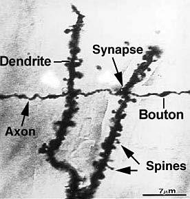
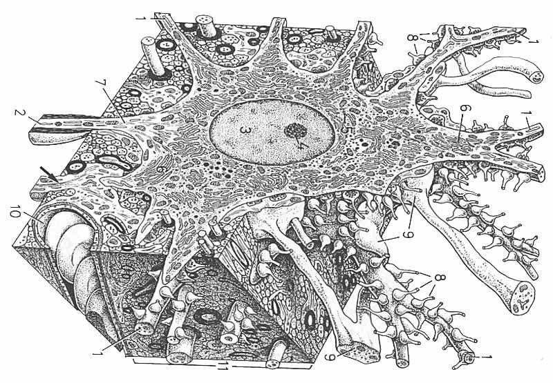
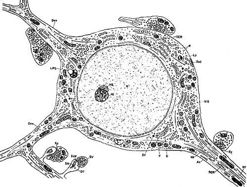
1 Lecture 2 Neurohistology II: Synapses, Meninges, & Receptors Overall Objectives: To understand the concept of the synapse; to understand the concept of axonal transport; to learn to identify the three layers of the meninges; and to understand how receptors are classified. I. The Synapse: The synapse is a specialized point of functional contact between neurons or between a neuron and a target organ (i.e., muscle) that allows neurons to communicate with one another or with their target cells. Synaptic Anatomy... The synpase is a site of apposition between a presynaptic element of one neuron and a postsynaptic membrane of a target neuron (or an effector organ); where, typically, a presynaptic axon enlargement releases transmitter molecules that diffuse across a synaptic cleft and bind to receptor channels in the postsynaptic membrane. Synapses are comprised of three elements: a) Presynaptic nerve terminal contains synaptic vesicles which house a chemical neurotransmitter that is released after vesicle fusion with the presynaptic terminal plasma membrane. b) Postsynaptic element a dendrite, a cell body, or a target cell receiving the synaptic input. Receptor protein molecules, to which neurotransmitter molecules bind, are embedded in the postsynaptic plasma membane. c) Synaptic Cleft a gap between pre- and post-synaptic elements into which neurotransmitter molecules are released. 11
2
3
4
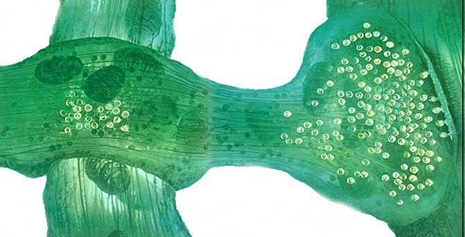
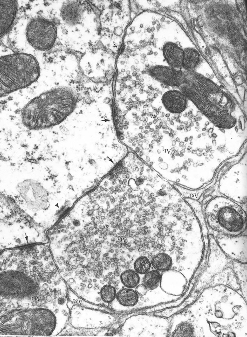
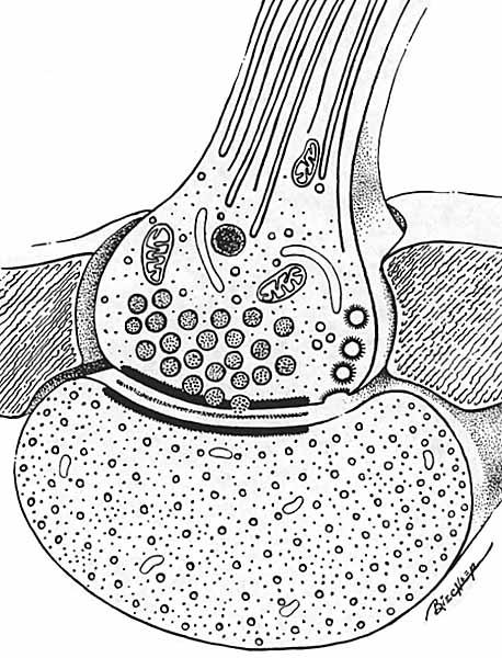
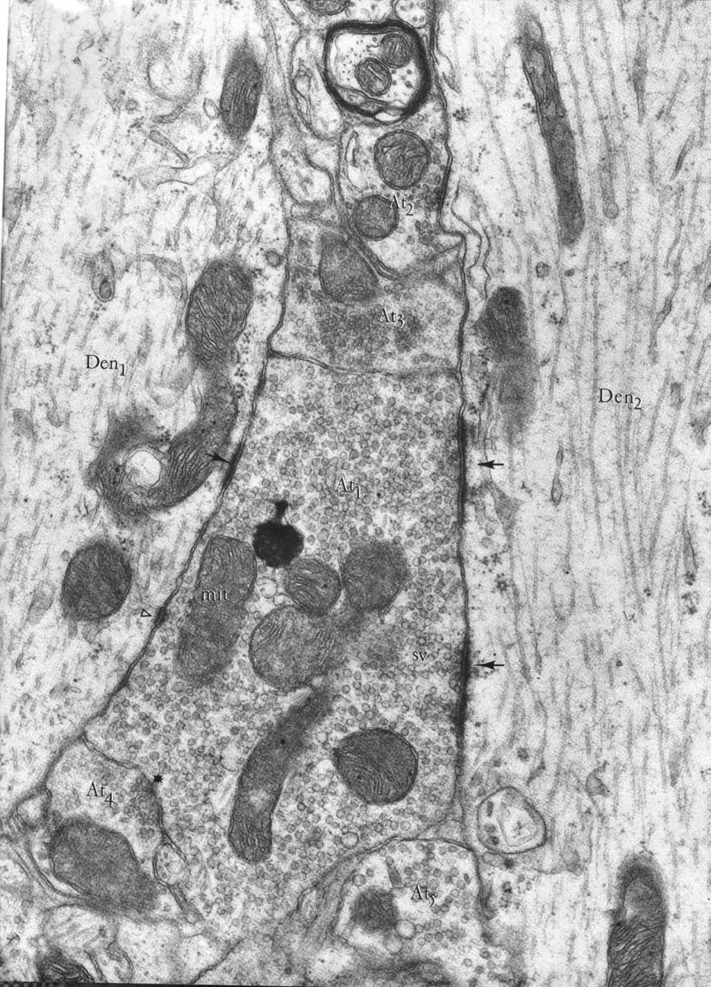
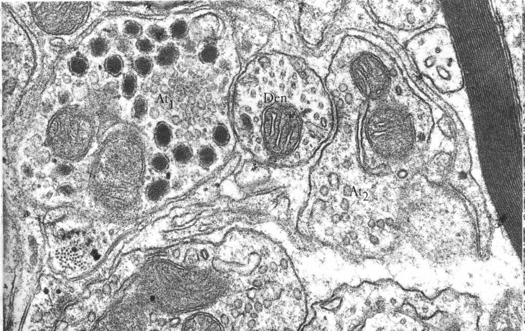
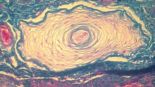
5 Common presynaptic arrangements: 1) axon terminal branches have terminal enlargements (called boutons or bulbs) 2) axon terminal branches feature varicosities (for synapses in passing ) 3) neuromuscular synapse: axon branches have terminal ramifications that form motor end plates on skeletal muscle fibers. Terminal bulbs En passant varicosities Neuromuscular end plates Classification of synaptic types: 1] axodendritic axon terminal branch (presynaptic element) synapses on a dendrite; 2] axosomatic axon terminal branch synapses on a soma (cell body); 3] axoaxonic axon terminal branch synapses on another axon terminal branch (for presynaptic inhibition) or beside the initial segment of an axon; 4] dendrodendritic dendrite synapsing on another dendrite (very localized effect). Synaptic ultrastructure: The presynaptic enlargement (bouton, varicosity, or end plate) contains synaptic vesicles (20 nm diameter), clustered around an electron dense active zone (protein-rich plasma membrane). Vesicles are anchored in place by actin microfilaments. Microtubule Neurofilament Synaptic vesicle Mitochondrion Presynaptic terminal bulb Astrocyte Pre- and postsynaptic plasma membranes are separated by a synaptic cleft (20 nm wide). The cleft contains glycoprotein linking material and is surrounded by glial cell processes. Synaptic cleft Postsynaptic dendrite The postsynaptic plasma membrane may appear unremarkable or thickened (electron dense). Receptor proteins (typically ligand-gated channels) are embedded in the plasma membrane. 12
6
7
8 Common presynaptic arrangements: 1) axon terminal branches have terminal enlargements (called boutons or bulbs) 2) axon terminal branches feature varicosities (for synapses in passing ) 3) neuromuscular synapse: axon branches have terminal ramifications that form motor end plates on skeletal muscle fibers. Terminal bulbs En passant varicosities Neuromuscular end plates Classification of synaptic types: 1] axodendritic axon terminal branch (presynaptic element) synapses on a dendrite; 2] axosomatic axon terminal branch synapses on a soma (cell body); 3] axoaxonic axon terminal branch synapses on another axon terminal branch (for presynaptic inhibition) or beside the initial segment of an axon; 4] dendrodendritic dendrite synapsing on another dendrite (very localized effect). Synaptic ultrastructure: The presynaptic enlargement (bouton, varicosity, or end plate) contains synaptic vesicles (20 nm diameter), clustered around an electron dense active zone (protein-rich plasma membrane). Vesicles are anchored in place by actin microfilaments. Microtubule Neurofilament Synaptic vesicle Mitochondrion Presynaptic terminal bulb Astrocyte Pre- and postsynaptic plasma membranes are separated by a synaptic cleft (20 nm wide). The cleft contains glycoprotein linking material and is surrounded by glial cell processes. Synaptic cleft Postsynaptic dendrite The postsynaptic plasma membrane may appear unremarkable or thickened (electron dense). Receptor proteins (typically ligand-gated channels) are embedded in the plasma membrane. 12
9
10
11
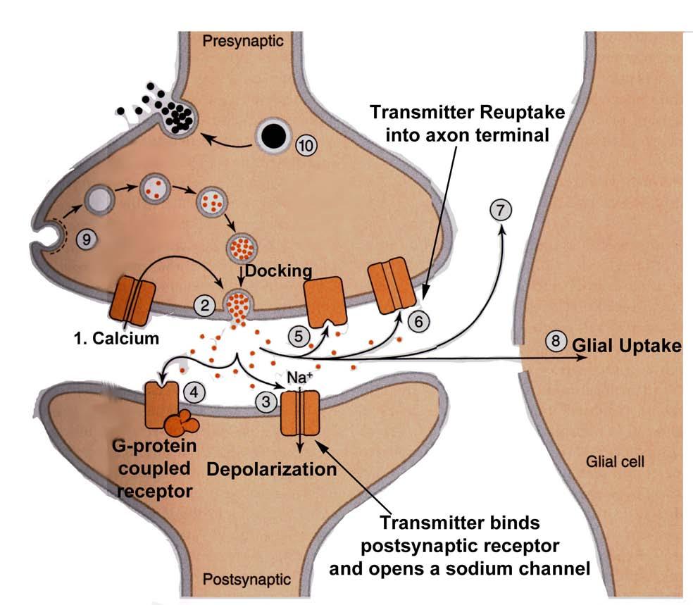
12 Synaptic Physiology... Presynaptic events: Neurotransmitter molecules are released in proportion to the amount of Ca ++ influx, in turn proportional to the amount of presynaptic membrane depolarization, i.e., in the resting state, the presynaptic membrane is polarized when an action potential arrives at the end of the axon, the adjacent presynaptic membrane is passively depolarized (toward zero transmembrane potential) voltage-gated Ca ++ channels allow Ca ++ influx (driven by [Ca ++ ] gradient). elevated [Ca ++ ] triggers vesicle mobilization and docking with the plasma membrane a number of vesicles fuse with presynaptic plasma membrane and release neurotransmitter molecules (about 5,000 per vesicle) by exocytosis. transmitter molecules diffuse across the cleft & bind with postsynaptic receptor proteins neurotransmitter molecules are eliminated from synaptic clefts via pinocytotic uptake by presynaptic or glial processes and/or via enzymatic degradation at the postsynaptic membrane. The molecules are recycled. subsequently, presynaptic plasma membrane repolarizes (due to K + channel conductance). Postsynaptic events: Neurotransmitter binding results in a proportional ion flux across the postsynaptic membrane. The particular excitability effect depends on the nature of the ion flux which depends on the nature of the ion channels in the particular postsynaptic membrane, i.e., in the resting state, postsynaptic plasma membrane is polarized (voltage activated K + channels dominate conductance) arriving neurotransmitter molecules bind briefly/repeatedly to ligand-gated receptors, which opens ion channels directly or by means of second messengers activation of [Na + & K + ] channels > leads to depolarization toward zero potential; activation of Cl - or K + channels > hyperpolarization of postsynaptic membrane. a postsynaptic potential (PSP) results from the altered membrane conductance EPSP = Excitatory PSP = depolarization toward zero potential, excites the postsynaptic cell IPSP = Inhibitory PSP = hyperpolarization (serves to cancel EPSPs), inhibits the postsynaptic cell following the removal/degradation of Electrotonic Conduction neurotransmitter molecules, the postsynaptic membrane is -70 re-polarized (K + channel conductance again dominates.) Note: PSPs constitute electrotonic conduction, a passive voltage spread (in contrast to the regenerative conduction of which axons are capable). PSPs decay exponentially, over distance and with time. The magnitude of a PSP depends on the number of open ion channels which, in turn, depends on the amount of neurotransmitter released. 0 mv EPSP Distance T i m e 13
13
14 Synaptic Physiology... Presynaptic events: Neurotransmitter molecules are released in proportion to the amount of Ca ++ influx, in turn proportional to the amount of presynaptic membrane depolarization, i.e., in the resting state, the presynaptic membrane is polarized when an action potential arrives at the end of the axon, the adjacent presynaptic membrane is passively depolarized (toward zero transmembrane potential) voltage-gated Ca ++ channels allow Ca ++ influx (driven by [Ca ++ ] gradient). elevated [Ca ++ ] triggers vesicle mobilization and docking with the plasma membrane a number of vesicles fuse with presynaptic plasma membrane and release neurotransmitter molecules (about 5,000 per vesicle) by exocytosis. transmitter molecules diffuse across the cleft & bind with postsynaptic receptor proteins neurotransmitter molecules are eliminated from synaptic clefts via pinocytotic uptake by presynaptic or glial processes and/or via enzymatic degradation at the postsynaptic membrane. The molecules are recycled. subsequently, presynaptic plasma membrane repolarizes (due to K + channel conductance). Postsynaptic events: Neurotransmitter binding results in a proportional ion flux across the postsynaptic membrane. The particular excitability effect depends on the nature of the ion flux which depends on the nature of the ion channels in the particular postsynaptic membrane, i.e., in the resting state, postsynaptic plasma membrane is polarized (voltage activated K + channels dominate conductance) arriving neurotransmitter molecules bind briefly/repeatedly to ligand-gated receptors, which opens ion channels directly or by means of second messengers activation of [Na + & K + ] channels > leads to depolarization toward zero potential; activation of Cl - or K + channels > hyperpolarization of postsynaptic membrane. a postsynaptic potential (PSP) results from the altered membrane conductance EPSP = Excitatory PSP = depolarization toward zero potential, excites the postsynaptic cell IPSP = Inhibitory PSP = hyperpolarization (serves to cancel EPSPs), inhibits the postsynaptic cell following the removal/degradation of Electrotonic Conduction neurotransmitter molecules, the postsynaptic membrane is -70 re-polarized (K + channel conductance again dominates.) Note: PSPs constitute electrotonic conduction, a passive voltage spread (in contrast to the regenerative conduction of which axons are capable). PSPs decay exponentially, over distance and with time. The magnitude of a PSP depends on the number of open ion channels which, in turn, depends on the amount of neurotransmitter released. 0 mv EPSP Distance T i m e 13

15 Additional Comments synaptic transmission is unidirectional (vesicles are located on only one side). glutamate is the major excitatory neurotransmitter in the nervous system; GABA and glycine are the major inhibitory neurotransmitters. synaptic transmission is slower than axonal conduction; each synapse introduces delay into a neural pathway (at least 0.5 msec/synapse). synapses are more susceptible to fatigue, hypoxia, and drug effects than are axons (generally pathways fail first at synapses). different kinds of drugs (tranquilizers, anesthetics, narcotics, anticonvulsants, muscle relaxants, etc.) work by modifying activity selectively among the different kinds of chemical synapses. certain diseases are manifestations of selective synaptic dysfunction; e.g., Parkinson's disease, tetanus, myasthenia gravis, various intoxications, etc. II. Connective Tissue Coverings of Axons in the PNS: 1. Endoneurium-- surrounds each myelinated axon, or a group of nonmyelinated axons. 2. Perineurium surrounds each nerve fascicle (a bundle of axons); consists of a perineural epithelium and associated collagenous connective tissue. The perineurium participates in forming a blood-nerve barrier which limits the passage of water-soluble substances and proteins from blood into the endoneurial compartment. (The integrity of this barrier is altered in certain neuropathies and following nerve trauma.) 3. Epineurium surrounds the entire nerve 14

16

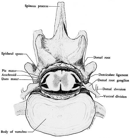
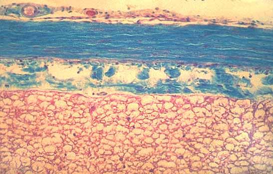
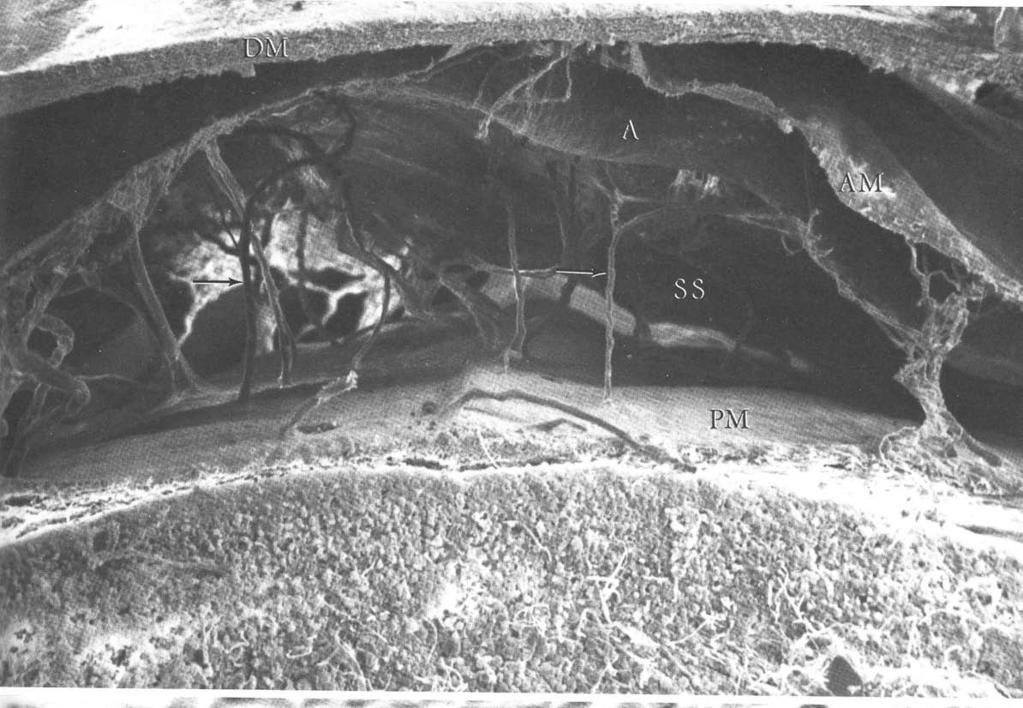
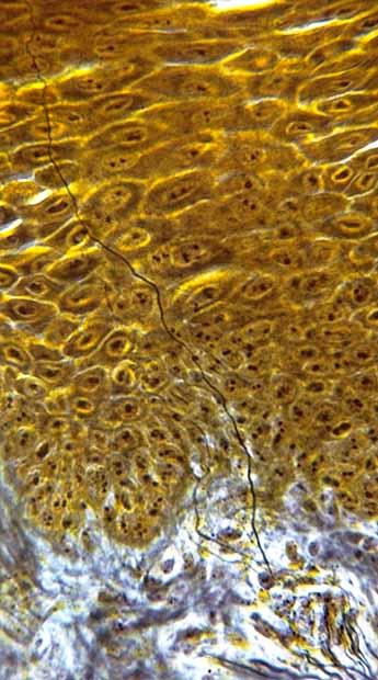
17 III. Axonal Transport: 1. The net movement of substances along the axon; 2 rates: A. Fast Axonal Transport mm/day B. Slow Axonal Transport 1-10 mm/day 2. Anterograde Transport transport of materials down the axon away from the cell body; important for renewing proteins along the axon and thus maintaining the axon. 3. Retrograde Transport transport from the axon terminal toward the cell body; important mechanism by which virus particles (rabies) and neurotoxins (tetanus toxin) gain access to the CNS. [Note: Tetanus and Botulinum toxins are proteases which cleave neuronal SNARE-proteins.] IV. Meninges: protective connective tissue sheaths surrounding the brain and spinal cord. There are three layers of meninges: 1. Dura Mater the outermost layer consisting of coarse, irregular connective tissue; composed of collagen and elastic fibers. 2. Arachnoid middle layer of the meninges; it consists of a distinct membrane and numerous fibrous trabeculae on its inner surface. This trabecular network forms the structural framework for the subarachnoid space which lies between the arachnoid proper and the underlying pia mater. The subarachnoid space contains cerebrospinal fluid (CSF). At certain points the subarachnoid space is dilated and forms cisterns. The cisterna magna and lumbar cisterns are important clinically because that is where CSF taps are performed. [Note: CSF is a clear colorless fluid that surrounds and permeates the entire central nervous system. It functions to protect, support and nourish the CNS.] 3. Pia Mater (from the latin term meaning tender mother ), the innermost layer of the meninges, it forms a thin protective membrane which adheres to the surface of the brain and spinal cord. It consists of flattened fibrocytes superficial to elastic and collagen fine fibers that extends into the numerous depressions and fissures on the surface of the brain and cord. It is very vascular. 15
18
19
20
21 Subarachnoid space V. Receptors: 1. Receptor = a specialized region located on a peripheral terminal branch of an axon of a primary afferent neuron, that can serve as a transducer converting environmental energy (sensory stimuli) into depolarizing ionic current (nerve signals). The number of receptors per neuron ranges from several (small receptive field) to several dozen (large receptive field). vs. Sense organ = an organized collection of receptor cells, with which the dendritic zones of afferent neurons synapse. The excitability of receptor cells is modified by environmental energy, i.e., the receptor cells act as transducers. Sense organs are: retina, cochlea, vestibular apparatus, taste buds, and olfactory epithelium. Neurons that synapse on receptor cells are SSA or SVA in type and commonly bipolar rather than unipolar. 2. Classification of receptor populations: Receptor classification based on Morphology: 1) free nerve endings terminal branches ramifying among epithelial cells, very common especially in the skin (mediate pain sensation, itch thermal sensations). 2) tactile discs consists of a terminal expansions of an afferent axon which are joined to modified epidermal cells (found in skin and mucous membranes). 3) encapsulated each receptor is encapsulated by lemmocytes and perineural epithelium (examples: pacinian corpuscles, tactile corpuscles, muscle spindles). Receptor classification based on Location: Cranial Meninges Arachnoid villus Dorsal sagittal venous sinus Dura mater Arachnoid Arachnoid trabecula Pia mater Cerebral cortex Falx cerebri White matter 1) 2) 3) 16
22
23
: 1) mechanoreceptors detect mechanical deformation (touch, pressure, vibration) 2) thermoreceptors detect changes in temperature (some")
24 1) Exteroceptors associated with skin and subcutaneous tissue (GSA) 2) Proprioceptors associated with muscles, tendons and joints (GSA) 3) Interoceptors located in viscera (GVA) Receptor and sense organ classification based on Modality (energy sensitivity): 1) mechanoreceptors detect mechanical deformation (touch, pressure, vibration) 2) thermoreceptors detect changes in temperature (some detect warmth, some detect cold) 3) nociceptors detect damage to tissue (pain receptors); also detect itch 4) electromagnetic detect light on the retina of the eye 5) chemoreceptors detect chemical molecules, including: taste receptors, olfactory receptors, arterial oxygen receptors in the aortic arch and carotid bodies, blood osmolarity in the hypothalamus and blood glucose and fatty acid receptors in the hypothalamus. Schematic diagram illustrating various types of peripheral receptors: 17
浙江大学医学院基础医学整合课程 各论 III. The Nervous System. Dr. ZHANG Xiong Dept. of Physiology ZJU School of Medicine
 The Nervous System Dr. ZHANG Xiong Dept. of Physiology ZJU School of Medicine xiongzhang@zju.edu.cn http://10.202.77.12/ 1 Part 1. Summary of the nervous system 2 The Nervous System Central Nervous System
The Nervous System. Dr. ZHANG Xiong Dept. of Physiology ZJU School of Medicine.
 The Nervous System Dr. ZHANG Xiong Dept. of Physiology ZJU School of Medicine Http://10.10.10.151 Part 1. Summary of the nervous system The Nervous System Central Nervous System Brain + Spinal Cord Peripheral
Nervous System. Master controlling and communicating system of the body. Secrete chemicals called neurotransmitters
 Nervous System Master controlling and communicating system of the body Interacts with the endocrine system to control and coordinate the body s responses to changes in its environment, as well as growth,
NERVOUS TISSUE. 1. Functional units of the nervous system; receive, process, store and transmit information to other neurons, muscle cells or glands.
 NERVOUS TISSUE LEARNING OBJECTIVES 1. Characterize and contrast the structure of neuronal cell bodies, dendrites and axons 2. List the classification of synapses and identify the basic structures of a
Major Structures of the Nervous System. Brain, cranial nerves, spinal cord, spinal nerves, ganglia, enteric plexuses and sensory receptors
 Major Structures of the Nervous System Brain, cranial nerves, spinal cord, spinal nerves, ganglia, enteric plexuses and sensory receptors Nervous System Divisions Central Nervous System (CNS) consists
Chapter 11 Introduction to the Nervous System and Nervous Tissue Chapter Outline
 Chapter 11 Introduction to the Nervous System and Nervous Tissue Chapter Outline Module 11.1 Overview of the Nervous System (Figures 11.1-11.3) A. The nervous system controls our perception and experience
Chapter 12 Nervous Tissue. Copyright 2009 John Wiley & Sons, Inc. 1
 Chapter 12 Nervous Tissue Copyright 2009 John Wiley & Sons, Inc. 1 Terms to Know CNS PNS Afferent division Efferent division Somatic nervous system Autonomic nervous system Sympathetic nervous system Parasympathetic
Nervous System. Electrical Signals.III Signal Transmission at Synapses Neurotransmitters.V Neural Circuits.VI
 Nervous System Overview.I Histology.II Electrical Signals.III Signal Transmission at Synapses Neurotransmitters.V Neural Circuits.VI Repairs.VII Pathology.VIII.IV 1 Controls and integrates all body activities
Nervous system. 1. Neurons :
 Nervous system nervous system is composed of billions of cells, the most essential being the nerve cells or neurons. There are estimated to be as many as 100 billion neurons in our nervous system. Two
NEURONS COMMUNICATE WITH OTHER CELLS AT SYNAPSES 34.3
 NEURONS COMMUNICATE WITH OTHER CELLS AT SYNAPSES 34.3 NEURONS COMMUNICATE WITH OTHER CELLS AT SYNAPSES Neurons communicate with other neurons or target cells at synapses. Chemical synapse: a very narrow
ANATOMY AND PHYSIOLOGY OF NEURONS. AP Biology Chapter 48
 ANATOMY AND PHYSIOLOGY OF NEURONS AP Biology Chapter 48 Objectives Describe the different types of neurons Describe the structure and function of dendrites, axons, a synapse, types of ion channels, and
What is Anatomy and Physiology?
 Introduction BI 212 BI 213 BI 211 Ecosystems Organs / organ systems Cells Organelles Communities Tissues Molecules Populations Organisms Campbell et al. Figure 1.4 Introduction What is Anatomy and Physiology?
5-Nervous system II: Physiology of Neurons
 5-Nervous system II: Physiology of Neurons AXON ION GRADIENTS ACTION POTENTIAL (axon conduction) GRADED POTENTIAL (cell-cell communication at synapse) SYNAPSE STRUCTURE & FUNCTION NEURAL INTEGRATION CNS
Chapter 11: Functional Organization of Nervous Tissue
 Chapter 11: Functional Organization of Nervous Tissue I. Functions of the Nervous System A. List and describe the five major nervous system functions: 1. 2. 3. 4. 5. II. Divisions of the Nervous System
Synapses. Excitatory synapses
 Synapses Sensory cells located at the periphery of the body, initiate and conduct signals to the brain and provide various sensory inputs such as vision, hearing, posture, and so on. Providing information
The Nervous System: Neural Tissue Pearson Education, Inc.
 13 The Nervous System: Neural Tissue Introduction Nervous System Characteristics Controls and adjust the activity of the body Provides swift but brief responses The nervous system includes: Central Nervous
Module H NERVOUS SYSTEM
 Module H NERVOUS SYSTEM Topic from General functions of the nervous system Organization of the nervous system from both anatomical & functional perspectives Gross & microscopic anatomy of nervous tissue
Chapter 7. The Nervous System: Structure and Control of Movement
 Chapter 7 The Nervous System: Structure and Control of Movement Objectives Discuss the general organization of the nervous system Describe the structure & function of a nerve Draw and label the pathways
Chapter 7. Objectives
 Chapter 7 The Nervous System: Structure and Control of Movement Objectives Discuss the general organization of the nervous system Describe the structure & function of a nerve Draw and label the pathways
Anatomy Review. Graphics are used with permission of: Pearson Education Inc., publishing as Benjamin Cummings (
 Anatomy Review Graphics are used with permission of: Pearson Education Inc., publishing as Benjamin Cummings (http://www.aw-bc.com) Page 1. Introduction Neurons communicate with other cells at junctions
2/27/2019. Functions of the Nervous System. Nervous Tissue and Neuron Function. Fundamentals Of The Nervous System And Nervous Tissue
 Nervous Tissue and Neuron Function Fundamentals Of The Nervous System And Nervous Tissue Learn and Understand 1. Like muscle cells, neurons use membrane polarity upset (AP) as a signal therefore keeping
Human Anatomy & Physiology
 PowerPoint Lecture Slides prepared by Barbara Heard, Atlantic Cape Community College Ninth Edition Human Anatomy & Physiology C H A P T E R 11 Annie Leibovitz/Contact Press Images 2013 Pearson Education,
8.2. Types of Neurons
 Chapter 8 Nervous Tissue The neuron is the functional and the structural unit of the nervous system. It displays two highly developed physiological traits: 1. Irritability - the capacity to generate a
Outline. Neuron Structure. Week 4 - Nervous System. The Nervous System: Neurons and Synapses
 Outline Week 4 - The Nervous System: Neurons and Synapses Neurons Neuron structures Types of neurons Electrical activity of neurons Depolarization, repolarization, hyperpolarization Synapses Release of
Chapter 45: Synapses Transmission of Nerve Impulses Between Neurons. Chad Smurthwaite & Jordan Shellmire
 Chapter 45: Synapses Transmission of Nerve Impulses Between Neurons Chad Smurthwaite & Jordan Shellmire The Chemical Synapse The most common type of synapse used for signal transmission in the central
Lecture 22: A little Neurobiology
 BIO 5099: Molecular Biology for Computer Scientists (et al) Lecture 22: A little Neurobiology http://compbio.uchsc.edu/hunter/bio5099 Larry.Hunter@uchsc.edu Nervous system development Part of the ectoderm
Introduction to Neurobiology
 Biology 240 General Zoology Introduction to Neurobiology Nervous System functions: communication of information via nerve signals integration and processing of information control of physiological and
Physiology of synapses and receptors
 Physiology of synapses and receptors Dr Syed Shahid Habib Professor & Consultant Clinical Neurophysiology Dept. of Physiology College of Medicine & KKUH King Saud University REMEMBER These handouts will
Nervous System Dr. Naim Kittana Department of Biomedical Sciences Faculty of Medicine & Health Sciences An-Najah National University
 Nervous System Department of Biomedical Sciences Faculty of Medicine & Health Sciences An-Najah National University Declaration The content and the figures of this seminar were directly adopted from the
MOLECULAR AND CELLULAR NEUROSCIENCE
 MOLECULAR AND CELLULAR NEUROSCIENCE BMP-218 November 4, 2014 DIVISIONS OF THE NERVOUS SYSTEM The nervous system is composed of two primary divisions: 1. CNS - Central Nervous System (Brain + Spinal Cord)
Dendrites Receive impulse from the axon of other neurons through synaptic connection. Conduct impulse towards the cell body Axon
 Dendrites Receive impulse from the axon of other neurons through synaptic connection. Conduct impulse towards the cell body Axon Page 22 of 237 Conduct impulses away from cell body Impulses arise from
Chapter 4 Neuronal Physiology
 Chapter 4 Neuronal Physiology V edit. Pg. 99-131 VI edit. Pg. 85-113 VII edit. Pg. 87-113 Input Zone Dendrites and Cell body Nucleus Trigger Zone Axon hillock Conducting Zone Axon (may be from 1mm to more
Nervous system is the most complex system in our body. It is formed by a network of more than 100 million nerve cells (neurons) assisted by many more
 assisted by many more") Nervous system Nervous system is the most complex system in our body. It is formed by a network of more than 100 million nerve cells (neurons) assisted by many more glial cells. Devoid from connective
Nervous system Nervous system is the most complex system in our body. It is formed by a network of more than 100 million nerve cells (neurons) assisted by many more glial cells. Devoid from connective
action potential afferent neuron Weblike; specifically, the weblike middle layer of the three meninges. arachnoid astrocytes autonomic nervous system
 action potential A large transient depolarization event, including polarity reversal, that is conducted along the membrane of a muscle cell or a nerve fiber. afferent neuron Nerve cell that carries impulses
The Nervous System. PowerPoint Lecture Slides C H A P T E R 7. Prepared by Patty Bostwick-Taylor, Florence-Darlington Technical College
 PowerPoint Lecture Slides Prepared by Patty Bostwick-Taylor, Florence-Darlington Technical College C H A P T E R 7 The Nervous System NERVOUS SYSTEM OVERVIEW Essential Question: What are the primary functions
My green thumb came only as a result of the mistakes I made while learning to see things from the plant s point of view. -H. Fred Ale Nervous System 1
 My green thumb came only as a result of the mistakes I made while learning to see things from the plant s point of view. -H. Fred Ale Nervous System 1 Classroom Rules You'll get one warning, then you'll
Nervous Tissue and Neurophysiology
 Nervous Tissue and Neurophysiology Objectives Describe the two major divisions of the nervous system and their characteristics. Identify the structures/functions of a typical neuron. Describe the location
The Nervous System. Nervous System Functions 1. gather sensory input 2. integration- process and interpret sensory input 3. cause motor output
 The Nervous System Nervous System Functions 1. gather sensory input 2. integration- process and interpret sensory input 3. cause motor output The Nervous System 2 Parts of the Nervous System 1. central
BIOL Week 6. Nervous System. Transmission at Synapses
 Collin County Community College BIOL 2401 Week 6 Nervous System 1 Transmission at Synapses Synapses are the site of communication between 2 or more neurons. It mediates the transfer of information and
BIOLOGY 2050 LECTURE NOTES ANATOMY & PHYSIOLOGY I (A. IMHOLTZ) FUNDAMENTALS OF THE NERVOUS SYSTEM AND NERVOUS TISSUE P1 OF 5
 FUNDAMENTALS OF THE NERVOUS SYSTEM AND NERVOUS TISSUE P1 OF 5") P1 OF 5 The nervous system controls/coordinates the activities of cells, tissues, & organs. The endocrine system also plays a role in control/coordination. The nervous system is more dominant. Its mechanisms
P1 OF 5 The nervous system controls/coordinates the activities of cells, tissues, & organs. The endocrine system also plays a role in control/coordination. The nervous system is more dominant. Its mechanisms
Biology Dr. Khalida Ibrahim Nervous system The nervous system is responsible for communication between different regions of the body, it is divided
 Biology Dr. Khalida Ibrahim Nervous system The nervous system is responsible for communication between different regions of the body, it is divided into: CNS (central nervous system) = brain + spinal cord
STRUCTURAL ELEMENTS OF THE NERVOUS SYSTEM
 STRUCTURAL ELEMENTS OF THE NERVOUS SYSTEM STRUCTURE AND MAINTENANCE OF NEURONS (a) (b) Dendrites Cell body Initial segment collateral terminals (a) Diagrammatic representation of a neuron. The break in
Branches of the Nervous System
 The Nervous System Branches of the Nervous System There are 2 main branches of the nervous system Central Nervous System Brain Spinal Cord Peripheral Nervous System All nerves leading to rest of body Anatomy
3) Most of the organelles in a neuron are located in the A) dendritic region. B) axon hillock. C) axon. D) cell body. E) axon terminals.
 Most of the organelles in a neuron are located in the A) dendritic region. B) axon hillock. C) axon. D) cell body. E) axon terminals.") Chapter 48 Neurons, Synapses, and Signaling Multiple-Choice Questions 1) A simple nervous system A) must include chemical senses, mechanoreception, and vision. B) includes a minimum of 12 ganglia. C) has
Chapter 48 Neurons, Synapses, and Signaling Multiple-Choice Questions 1) A simple nervous system A) must include chemical senses, mechanoreception, and vision. B) includes a minimum of 12 ganglia. C) has
Chapter 7 Nerve tissue 1 Liu Jiamei
 Chapter 7 Nerve tissue 1 Liu Jiamei General description: nerve tissue nerve cells (neurons): show numerous long processes receive the stimulation make contact with each other, conduct the nerve impulse
Human Histology The Nervous System. Dr. Rawaa Salim Hameed
 Human Histology The Nervous System Dr. Rawaa Salim Hameed The organization of the nervous system Anatomically, the nervous system is divided into:- Neurohistology Structurally, nerve tissue consists of
Neurobiology. Cells of the nervous system
 Neurobiology Cells of the nervous system Anthony Heape 2010 1 The nervous system Central nervous system (CNS) Peripheral nervous system (PNS) 2 Enteric nervous system (digestive tract, gall bladder and
Nervous System. 2. Receives information from the environment from CNS to organs and glands. 1. Relays messages, processes info, analyzes data
 Nervous System 1. Relays messages, processes info, analyzes data 2. Receives information from the environment from CNS to organs and glands 3. Transmits impulses from CNS to muscles and glands 4. Transmits
Portions from Chapter 6 CHAPTER 7. The Nervous System: Neurons and Synapses. Chapter 7 Outline. and Supporting Cells
 CHAPTER 7 The Nervous System: Neurons and Synapses Chapter 7 Outline Neurons and Supporting Cells Activity in Axons The Synapse Acetylcholine as a Neurotransmitter Monoamines as Neurotransmitters Other
CHAPTER 44: Neurons and Nervous Systems
 CHAPTER 44: Neurons and Nervous Systems 1. What are the three different types of neurons and what are their functions? a. b. c. 2. Label and list the function of each part of the neuron. 3. How does the
Synaptic Communication. Steven McLoon Department of Neuroscience University of Minnesota
 Synaptic Communication Steven McLoon Department of Neuroscience University of Minnesota 1 Course News The first exam is next week on Friday! Be sure to checkout the sample exam on the course website. 2
Neurons, Synapses and Signaling. Chapter 48
 Neurons, Synapses and Signaling Chapter 48 Warm Up Exercise What types of cells can receive a nerve signal? Nervous Organization Neurons- nerve cells. Brain- organized into clusters of neurons, called
Action potential. Definition: an all-or-none change in voltage that propagates itself down the axon
 Action potential Definition: an all-or-none change in voltage that propagates itself down the axon Action potential Definition: an all-or-none change in voltage that propagates itself down the axon Naturally
Neurons, Synapses, and Signaling
 Neurons, Synapses, and Signaling The Neuron is the functional unit of the nervous system. Neurons are composed of a cell body, which contains the nucleus and organelles; Dendrites which are extensions
SYNAPTIC COMMUNICATION
 BASICS OF NEUROBIOLOGY SYNAPTIC COMMUNICATION ZSOLT LIPOSITS 1 NERVE ENDINGS II. Interneuronal communication 2 INTERNEURONAL COMMUNICATION I. ELECTRONIC SYNAPSE GAP JUNCTION II. CHEMICAL SYNAPSE SYNAPSES
Nervous tissue. Lab. 7
 Nervous tissue Lab. 7 Nervous tissue :- is responsible for transport nervous impulse (motor and sensory impulse), and it is formed by network more than 100 million nerve cell (neurons), nerve fiber and
Chapter 2: Cellular Mechanisms and Cognition
 Chapter 2: Cellular Mechanisms and Cognition MULTIPLE CHOICE 1. Two principles about neurons were defined by Ramón y Cajal. The principle of connectional specificity states that, whereas the principle
SOME BASIC TERMINOLOGY CNS: Central Nervous System: Brain + Spinal Cord
 SOME BASIC TERMINOLOGY CNS: Central Nervous System: Brain + Spinal Cord CEREBROSPINAL FLUID (CSF): The fluid filling the ventricles, cerebral aqueduct, central canal, and subarachnoid space. It is a filtrate
Neurons Chapter 7 2/19/2016. Learning Objectives. Cells of the Nervous System. Cells of the Nervous System. Cells of the Nervous System
 Learning Objectives Neurons Chapter 7 Identify and describe the functions of the two main divisions of the nervous system. Differentiate between a neuron and neuroglial cells in terms of structure and
Synaptic transmission
 Outline Synaptic transmission Sompol Tapechum M.D., Ph.D. Department of Physiology Faculty of Medicine Siriraj Hospital, Bangkok, Thailand. sisth@mahidol.ac.th 2 Structure of synapse Modes of synaptic
Ameen Alsaras. Ameen Alsaras. Mohd.Khatatbeh
 9 Ameen Alsaras Ameen Alsaras Mohd.Khatatbeh Nerve Cells (Neurons) *Remember: The neural cell consists of: 1-Cell body 2-Dendrites 3-Axon which ends as axon terminals. The conduction of impulse through
Nerve Cell Flashcards
 1. What does the word innervates mean? Refers to a nerve supplying a muscle or organ. For example, The phrenic nerve innervates the diaphragm muscle. 2. 3 parts of the Nervous System 1. Central Nervous
Chapter 17 Nervous System
 Chapter 17 Nervous System 1 The Nervous System Two Anatomical Divisions Central Nervous System (CNS) Brain and Spinal Cord Peripheral Nervous System (PNS) Two Types of Cells Neurons Transmit nerve impulses
The Nervous System -The master controlling and communicating system of the body
 The Nervous System -The master controlling and communicating system of the body Functions: -Sensory input -Integration -Motor output Organization of the Nervous System Central nervous system (CNS) -Brain
Unit Three. I. General Functions of the Nervous System. I. General Functions of the Nervous System
 10 Refer to the following URLs. It is a good idea to print them and bring them to class. Be sure to study these along with your book. http://www.sirinet.net/~jgjohnso/nervous.html http://faculty.washington.edu/chudler/ap.html
Outline. Animals: Nervous system. Neuron and connection of neurons. Key Concepts:
 Animals: Nervous system Neuron and connection of neurons Outline 1. Key concepts 2. An Overview and Evolution 3. Human Nervous System 4. The Neurons 5. The Electrical Signals 6. Communication between Neurons
Endocrine System Nervous System
 Cells Endocrine System Nervous System Tissues Controls Organs Nervous System vs Endocrine System Electrical signals (graded potentials and action potentials) and chemical signals (neurotransmitters) Fast
Chapter 7 Nerve Cells and Electrical Signaling
 Chapter 7 Nerve Cells and Electrical Signaling 7.1. Overview of the Nervous System (Figure 7.1) 7.2. Cells of the Nervous System o Neurons are excitable cells which can generate action potentials o 90%
What Cell Make Up the Brain and Spinal Cord
 What Cell Make Up the Brain and Spinal Cord Jennifer LaVail, Ph.D. (http://anatomy.ucsf.edu/pages/lavaillab/index.html) What kinds of cells are these?" Neuron?" Epithelial cell?" Glial cell?" What makes
10.1: Introduction. Cell types in neural tissue: Neurons Neuroglial cells (also known as neuroglia, glia, and glial cells) Dendrites.
 Dendrites.") 10.1: Introduction Copyright The McGraw-Hill Companies, Inc. Permission required for reproduction or display. Cell types in neural tissue: Neurons Neuroglial cells (also known as neuroglia, glia, and glial
10.1: Introduction Copyright The McGraw-Hill Companies, Inc. Permission required for reproduction or display. Cell types in neural tissue: Neurons Neuroglial cells (also known as neuroglia, glia, and glial
EE 791 Lecture 2 Jan 19, 2015
 EE 791 Lecture 2 Jan 19, 2015 Action Potential Conduction And Neural Organization EE 791-Lecture 2 1 Core-conductor model: In the core-conductor model we approximate an axon or a segment of a dendrite
Na + K + pump. The beauty of the Na + K + pump. Cotransport. The setup Cotransport the result. Found along the plasma membrane of all cells.
 The beauty of the Na + K + pump Na + K + pump Found along the plasma membrane of all cells. Establishes gradients, controls osmotic effects, allows for cotransport Nerve cells have a Na + K + pump and
BIOH111. o Cell Module o Tissue Module o Integumentary system o Skeletal system o Muscle system o Nervous system o Endocrine system
 BIOH111 o Cell Module o Tissue Module o Integumentary system o Skeletal system o Muscle system o Nervous system o Endocrine system Endeavour College of Natural Health endeavour.edu.au 1 TEXTBOOK AND REQUIRED/RECOMMENDED
Biology 218 Human Anatomy
 Chapter 17 Adapted form Tortora 10 th ed. LECTURE OUTLINE A. Overview of the Nervous System (p. 537) 1. The nervous system and the endocrine system are the body s major control and integrating centers.
EM: myelin sheath shows a series of concentrically arranged lamellae
 EM: myelin sheath shows a series of concentrically arranged lamellae ---- how to form myelin sheath? Schwann cell invagination and envelop the axon form mesaxon mesaxon become longer and longer winding
ANSWERS TO PRE- LAB ASSIGNMENTS
 Lab 14 Introduction to Nervous System Hamilton ANSWERS TO PRE- LAB ASSIGNMENTS Pre-Lab Activity 1: 1. a. orbicularis oculi b. sternocleidomastoid c. deltoid d. pectoralis major e. biceps brachii f. rectus
AP Biology Unit 6. The Nervous System
 AP Biology Unit 6 The Nervous System Branches of the Nervous System There are 2 main branches of the nervous system Central Nervous System Brain Spinal Cord Peripheral Nervous System All nerves leading
Human Anatomy and Physiology - Problem Drill 11: Neural Tissue & The Nervous System
 Human Anatomy and Physiology - Problem Drill 11: Neural Tissue & The Nervous System Question No. 1 of 10 The human body contains different types of tissue. The tissue is formed into organs and organ systems.
BIO 115 Anatomy & Physiology II Practice Assignment 4: The Nervous System & The Senses This is not a required assignment but it is recommended.
 BIO 115 Anatomy & Physiology II Practice Assignment 4: The Nervous System & The Senses This is not a required assignment but it is recommended. 1. This figure depicts a typical neuron. What structures
Functions of Nervous System Neuron Structure
 Chapter 10 Nervous System I Divisions of the Nervous System Cell Types of Neural Tissue neurons neuroglial cells Central Nervous System brain spinal cord Peripheral Nervous System nerves cranial nerves
Collin County Community College BIOL Week 5. Nervous System. Nervous System
 Collin County Community College BIOL 2401 Week 5 Nervous System 1 Nervous System The process of homeostasis makes sure that the activities that occur in the body are maintained within normal physiological
Chapter 8 Nervous System
 Chapter 8 Nervous System Two message centers: Functions of these systems: 1. * 2. * Overview of the Nervous System Parts: General Functions: Functions Sensory input: Sensation via nerves Integration: interpretation
Introduction to Physiological Psychology
 Introduction to Physiological Psychology Review Kim Sweeney ksweeney@cogsci.ucsd.edu www.cogsci.ucsd.edu/~ksweeney/psy260.html Today n Discuss Final Paper Proposal (due 3/10) n General Review 1 The article
NEURAL TISSUE (NEUROPHYSIOLOGY) PART I (A): NEURONS & NEUROGLIA
 PART I (A): NEURONS & NEUROGLIA") PART I (A): NEURONS & NEUROGLIA Neural Tissue Contains 2 kinds of cells: neurons: cells that send and receive signals neuroglia (glial cells): cells that support and protect neurons Neuron Types Sensory
PART I (A): NEURONS & NEUROGLIA Neural Tissue Contains 2 kinds of cells: neurons: cells that send and receive signals neuroglia (glial cells): cells that support and protect neurons Neuron Types Sensory
Function of the Nervous System
 Nervous System Function of the Nervous System Receive sensory information, interpret it, and send out appropriate commands to form a response Composed of neurons (functional unit of the nervous system)
Nervous Tissue and Histology of CNS
 Nervous Tissue and Histology of CNS Functions of Nervous System Like the CPU of a computer, the nervous system is the master controlling system of the body. It is designed to constantly and rapidly adjust
Chapter 9. Nervous System
 Chapter 9 Nervous System Central Nervous System (CNS) vs. Peripheral Nervous System(PNS) CNS Brain Spinal cord PNS Peripheral nerves connecting CNS to the body Cranial nerves Spinal nerves Neurons transmit
Section: Chapter 5: Multiple Choice. 1. The structure of synapses is best viewed with a(n):
:") Section: Chapter 5: Multiple Choice 1. The structure of synapses is best viewed with a(n): p.155 electron microscope. light microscope. confocal microscope. nissle-stained microscopic procedure. 2. Electron
Section: Chapter 5: Multiple Choice 1. The structure of synapses is best viewed with a(n): p.155 electron microscope. light microscope. confocal microscope. nissle-stained microscopic procedure. 2. Electron
Neurons, Synapses, and Signaling
 Chapter 48 Neurons, Synapses, and Signaling PowerPoint Lecture Presentations for Biology Eighth Edition Neil Campbell and Jane Reece Lectures by Chris Romero, updated by Erin Barley with contributions
Nervous Tissue. Dr. Heba Kalbouneh Associate Professor of Anatomy and Histology
 Nervous Tissue Dr. Heba Kalbouneh Associate Professor of Anatomy and Histology Controls and integrates all body activities within limits that maintain life Three basic functions 1. sensing changes with
Functions of the Nervous System. Fundamentals of the Nervous System & Nervous Tissue
 Fundamentals of the Nervous System & Nervous Tissue Overview Structure cell types & structures Neurophysiology membrane potential Synapse, neurotransmitters & receptors Functions of the Nervous System
Biology 201-Worksheet on Nervous System (Answers are in your power point outlines-there is no key!)
") Bio 201 Tissues and Skin 1 March 21, 2011 Biology 201-Worksheet on Nervous System (Answers are in your power point outlines-there is no key!) 1. The study of the normal functioning and disorders of the
Bio 201 Tissues and Skin 1 March 21, 2011 Biology 201-Worksheet on Nervous System (Answers are in your power point outlines-there is no key!) 1. The study of the normal functioning and disorders of the
BIOH111. o Cell Module o Tissue Module o Integumentary system o Skeletal system o Muscle system o Nervous system o Endocrine system
 BIOH111 o Cell Module o Tissue Module o Integumentary system o Skeletal system o Muscle system o Nervous system o Endocrine system Endeavour College of Natural Health endeavour.edu.au 1 Textbook and required/recommended
Chapter 11: Nervous System and Nervous Tissue
 Chapter 11: Nervous System and Nervous Tissue I. Functions and divisions of the nervous system A. Sensory input: monitor changes in internal and external environment B. Integrations: make decisions about
DO NOW: ANSWER ON PG 73
 DO NOW: ANSWER ON PG 73 1. Name 1 neurotransmitter that we have learned about. 2. Draw a basic graph of a neuron action potential. Label resting potential, threshold, depolarization, and repolarization
Nervous System (Part A-1) Module 8 -Chapter 14
 Module 8 -Chapter 14") Nervous System (Part A-1) Module 8 -Chapter 14 Overview Susie Turner, M.D. 1/9/13 Cellular structure of the nervous system Neurons Neuroglia Nervous System Divisions Central nervous system Peripheral nervous
Nervous System (Part A-1) Module 8 -Chapter 14 Overview Susie Turner, M.D. 1/9/13 Cellular structure of the nervous system Neurons Neuroglia Nervous System Divisions Central nervous system Peripheral nervous
Functions of the Nervous System
 Chapter 11 Functional Organization of Nervous Tissue 11-1 Functions of the Nervous System 1. Sensory input. Monitor internal and external stimuli 2. Integration. Brain and spinal cord process sensory input
The 7 th lecture. Anatomy and Physiology For the. 1 st Class. By Dr. Ala a Hassan Mirza
 The 7 th lecture In Anatomy and Physiology For the 1 st Class By Dr. Ala a Hassan Mirza Nervous System (part I) The Nerve Tissue and the Nervous System The Tissues of the Body There are 4 types of tissues
Human Brain and Senses
 Human Brain and Senses Outline for today Levels of analysis Basic structure of neurons How neurons communicate Basic structure of the nervous system Levels of analysis Organism Brain Cell Synapses Membrane
Fundamentals of the Nervous System and Nervous Tissue. Nervous System. Basic Divisions of the Nervous System C H A P T E R 12.
 C H A P T E R 12 Fundamentals of the Nervous System and Nervous Tissue Nervous System Sensory input Integration Motor output Figure 12.1 Basic Divisions of the Nervous System Brain CNS Spinal cord Nerves