Tauopathies Alzheimer s disease Down s syndrome Dementia with only tangles (tangle only dementia) Pick s disease Progressive supranuclear palsy
|
|
|
- Darrell Ward
- 5 years ago
- Views:
Transcription
1 Tauopathies Prof. Isidro Ferrer, Institut Neuropatologia, Servei Anatomia Patològica, IDIBELL-Hospital Universitari de Bellvitge, Universitat de Barcelona, CIBERNED, Hospitalet de LLobregat; Spain
2
3 Tauopathies Alzheimer s disease Down s syndrome Dementia with only tangles (tangle only dementia) Pick s disease Progressive supranuclear palsy Corticobasal degeneration Argyrophilic grain disease Frontotemporal lobar degeneration (and parkinsonism) due to mutations in MAPT (tau gene) Parkinson-dementia-ALS complex guamanian and non-guamanian Dementia pugilistica Myotonic dystrophy Rare tauopathies: tau panencephalopathy, AD-like with astrocytes, Niemann-Pick disease type C, certain prion diseases (GSS with tau), mutations in LRRK2, familial British dementia, Hallervorden-Spatz disease, postencephalitic parkinsonism Hyperphosphorylated tau in muscular diseases: sporadic Inclusion body myositis (sibm), myofibrillar myopathies (MFM)
4 Post-translational modifications of tau 1. O-Glycosylation: O-linked N-Acetylglucosamine (O-GlcNAc) 2. Phosphorylation: 2a.- Sites: about 79 putative sites, 30 described; most of them localized outside the microtubulebinding domains; most of them on the Ser-Pro and Thr-Pro motives. 2b.- Kinases: mitogen-activated protein kinase (MAPK) extracellular-regulated kinase (MAPK/ERK); glycogen synthase kinase (GSK3); tau-tubulin kinase; cyclin-dependent kinases cdk2 and cdk5; stress-activated protein kinases (SAPK/JNKs), stress-kinase of 38 kda (p38); microtubule-affinity regulating kinase (MARK): calcium/calmodulin-dependent protein kinase II (CaMPK II), cyclic- AMP-dependent kinase (PKA); and casein kinase II. 2c.- Phosphatases: Ser/Thr phosphatase proteins 1, 2A, 2B (calcineurin) and 2C.
5 0N 1N 2N 3R MBDs 2N 1N 0N 4R Exon 2 Exon 3 Exon 10 Tau isoforms expressed in the brain result from alternative splicing; 3Rtau contain 3 repeats, whereas 4Rtau contain four repeats. Big tau (about 120 kda) is expressed in the peripheral nervous system and muscle.
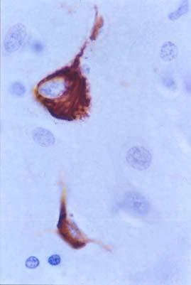
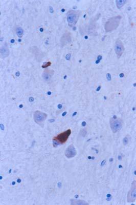
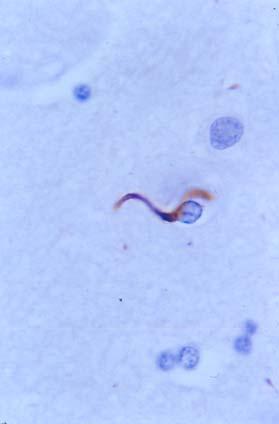
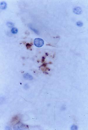
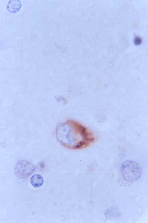
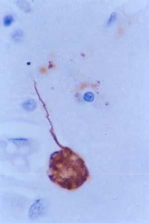
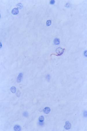
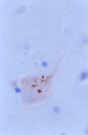
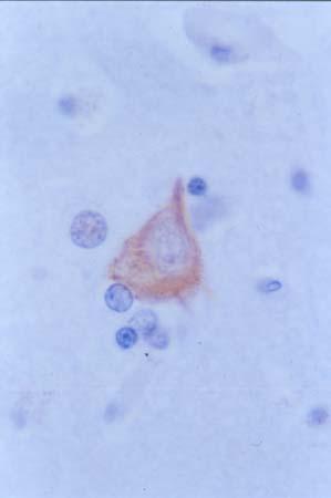
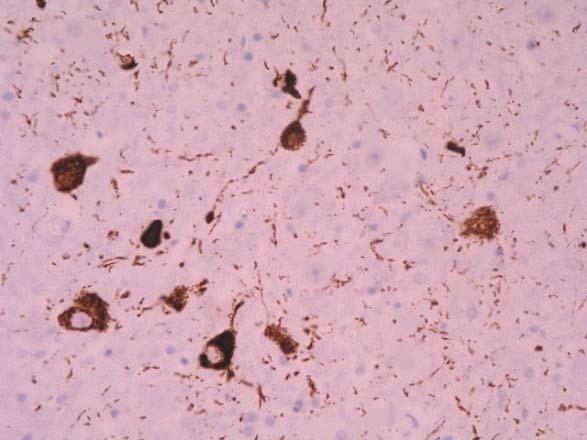
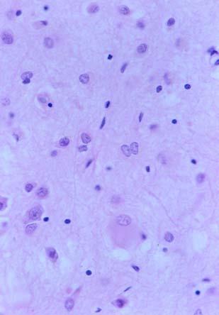
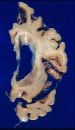
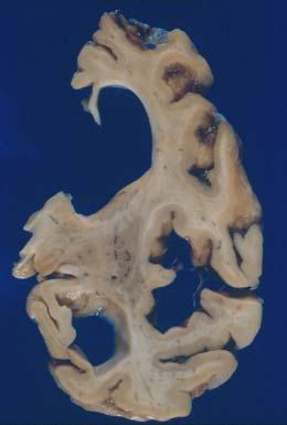
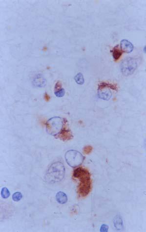
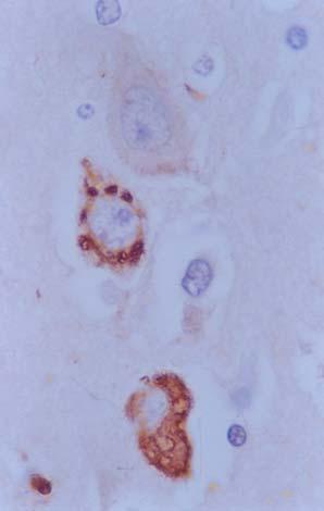
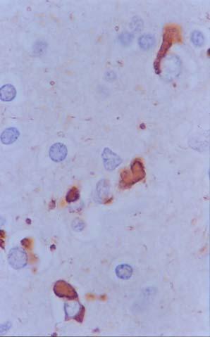
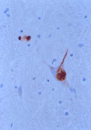
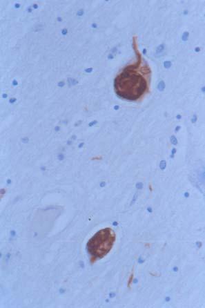
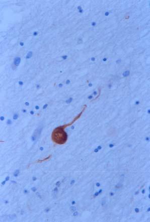
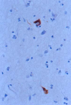
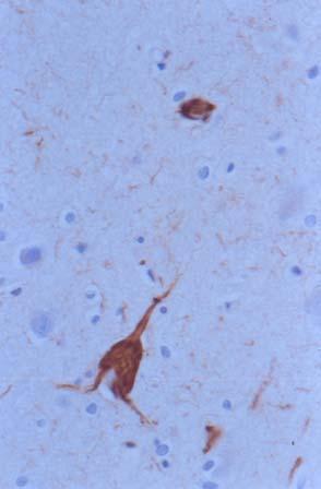
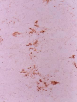
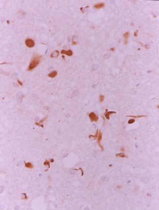
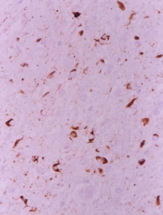
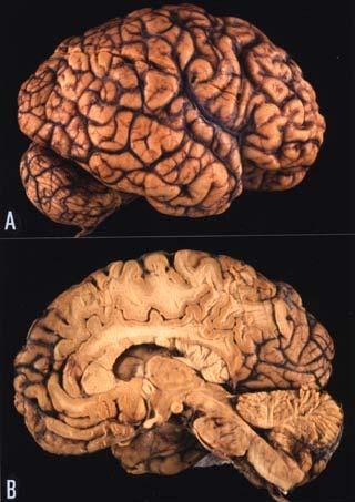
6 Pick disease Atrophy of the anterior temporal and frontal lobes, the orbital frontal lobe and the medial temporal lobes ( knife-edge atrophy). Severe degeneration in the hippocampus and amygdala. Degeneration in the corpus striatum, pallidum and substantia nigra may be detected. Neuron loss, vacuolation of the neuropil, astrocytic gliosis in the affected regions. Loss of dendritic spines and pre-synaptic markers. NF-P (+), tau (-) ballooned neurons (swollen chromatolytic neurons or Pick cells) in cerebral cortex. Pick bodies, NF-P (+), tau-p (+), in cerebral cortex (cingulate, insular, inferior temporal, inferior parietal and fusiform), hippocampal complex (pyramidal neurons of the hippocampus, granule cells of the dentate gyrus). tau-positive (ramified) inclusions in astrocytes. tau-positive coiled bodies and threads in oligodendrocytes.

, silver")
.")
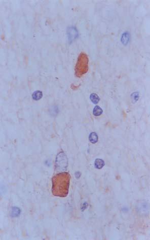
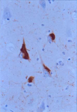
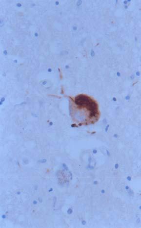
7 Pick disease A B C Atrophy of the frontal and temporal lobes, and caudate (A). Pick bodies (arrow), silver stain (B) and electron microscopy (C). Pick bodies, antiphosphorylated tau antibodies (D) D

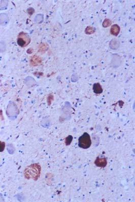
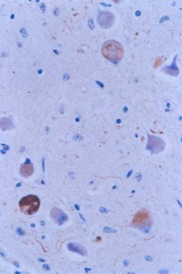
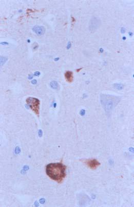
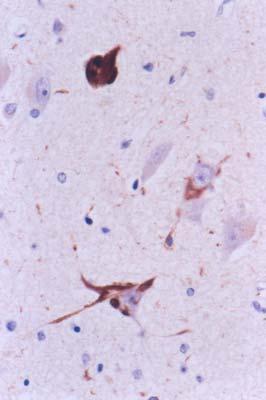
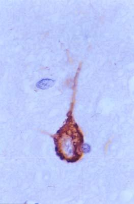
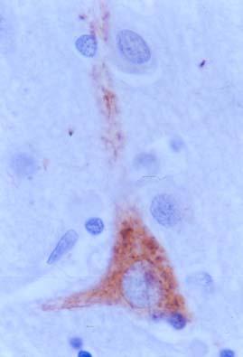
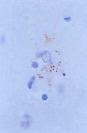
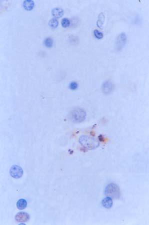
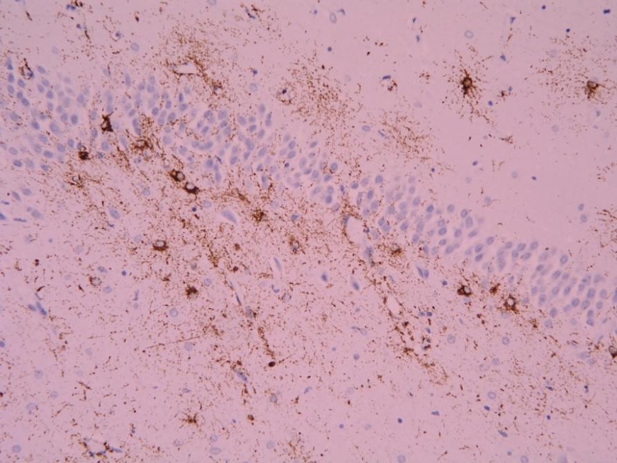
8 Pick disease A B C D E F G H I Pick bodies and tau-immunoreactive inclusions (AT8 antibody) in the hilus of the hippocampus (A), dentate gyrus (B), CA1 area (C), subiculum (D), entorhinal cortex (E), temporal cortex (F), amygdala (G), reticular nuclei of the brain stem (H)
 and coiled bodies (I) (E) a few of them reminiscent of")
9 Pick disease A B C D E F G H I Atypical Pick disease: Pick bodies, tau-immunoreactive astrocytes astrocytes (F) and coiled bodies (I) (E) a few of them reminiscent of tufted

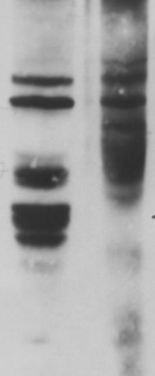
10 Band pattern of phospho-tau as revealed by several anphospho-specific anti-tau antibodies. Bands of 64 and 60 kda PiD 64 KDa 60 KDa Thr 181 Ser 202 Ser 214 Ser 396 Ser 422 Ser 262 Tau kinases in sarkosyl insoluble fractions Tau-Thr 181 MAPK/ERK-P SAPK/JNK-P p38- P CAMKβ-II 64 KDa 60 KDa Immunoprecipitation (IP) of p38-p from Pick sarkosyl-insoluble fractions p38-p kinase assay of Pick IP. ATF-2 phosphorylation 38 kda 45 kda p38 P C C PiD PiD CT CT - ATF-2 +ATF-2 -ATF-2 +ATF-2 ATF-2 +ATF-2
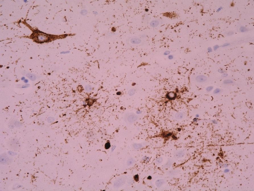
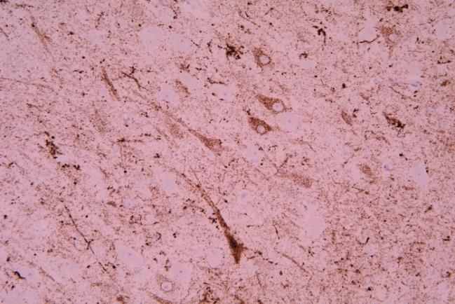
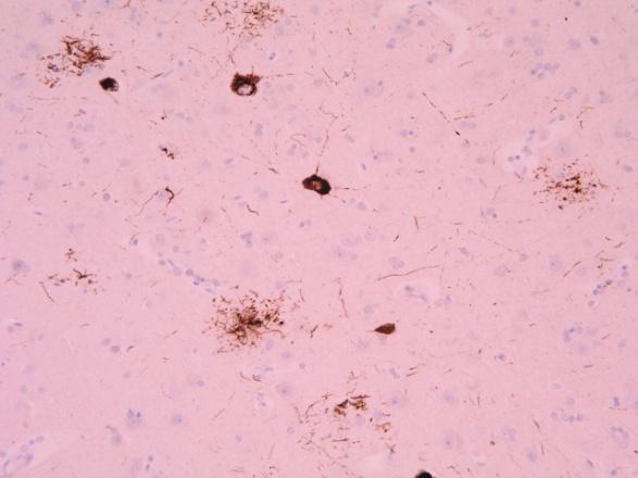
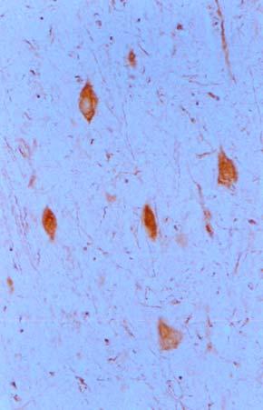
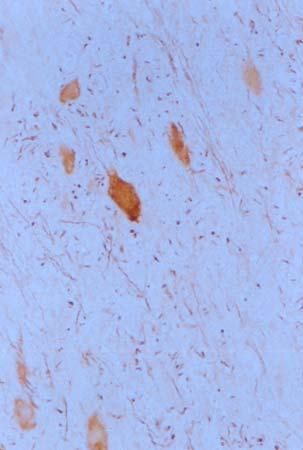
11 Progressive supranuclear palsy (PSP) Neuron loss and astrocytosis are consistently found in the brainstem, cerebellum, pallidum, subthalamic nucleus and thalamus. Cortical involvement may be detected in patients who had suffered from cognitive decline. PSP subgroups: Cortical type Subcortical type tau (+) globose neurofibrillary tangles (NFTs) and neuropil threads in the basal ganglia, nucleus basalis of Meynert, brainstem and dentate nucleus. NFTs may be observed in small pyramidal neurons of the cerebral neocortex and pyramidal cells of the hippocampus (but distribution differs from that in AD). tau (+) inclusions in astrocytes: astrocytic tufts (star-like tufts), thorn-shaped inclusions. tau (+) oligodendrocytic inclusions: coiled bodies and threads.

 (A-D).")
")
 and white matter (wm) in")

12 Progressive supranuclear palsy (PSP) Neurofibrillary tangles and threads in locus ceruleus (lc) and cerebral cortex (cc) (A-D). Astrocytes and tufted astrocytes stained with anti-p-tau antibodies (right panel) P-tau-immunoreactive bands in the pons (p) and white matter (wm) in sarkosyl-insoluble fraction in PSP p tau Ser 214 p tau Ser kda 68 kda 66 kda 62 kda triplet at 37 kda 33 kda P WM P WM

,")
;")
 in locus")

")

 are seen in")
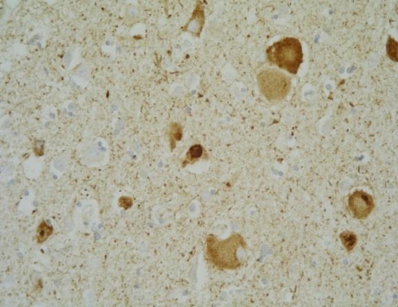
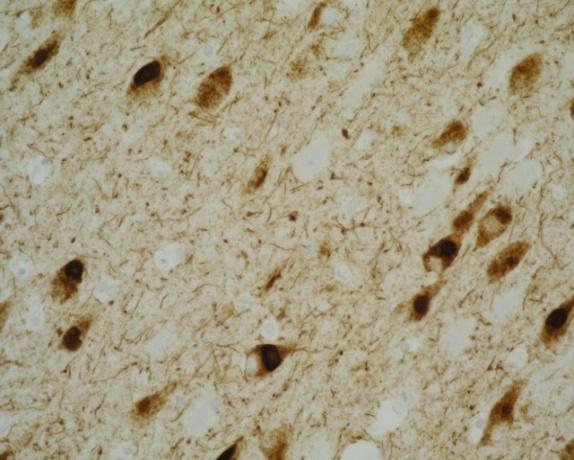
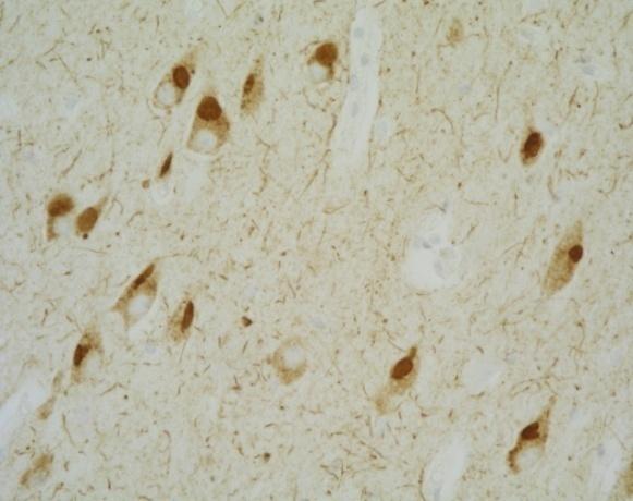
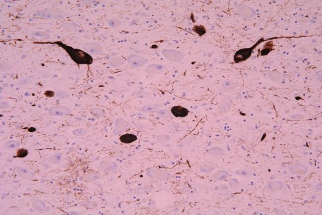
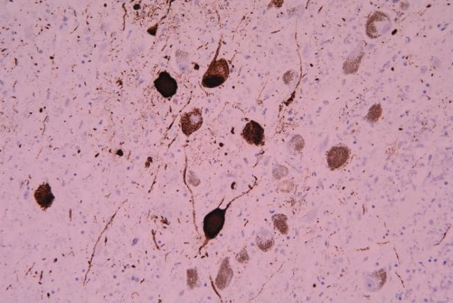
13 Progressive supranuclear palsy A B C D E F G H Neuron loss in the globus pallidus (A), subthalamus (B) and substantia nigra (C); neurofibrillary tangle (arrow) in locus ceruleus (D). Hyperphosphorylated tau in the striatum (E, F), superior colliculus (G) and locus ceruleus (H). Positive astrocytes and oligodendrocytes (coiled bodies) are seen in addition to tau-immunorecative neurons.
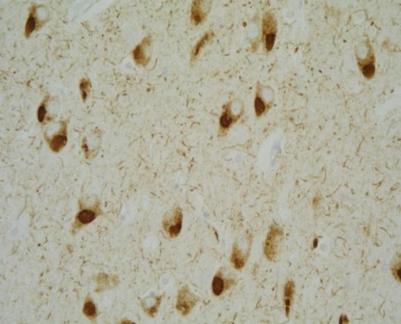
14 Progressive supranuclear palsy A B C D E F G H Neuronal involvement in PSP (A-C). Entorhinal cortex (D); pons (E); locus ceruleus (F); lateral reticular formation (G); dentate nucleus (H)
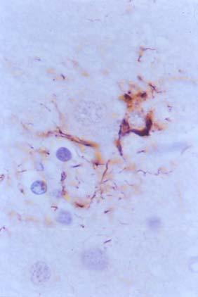
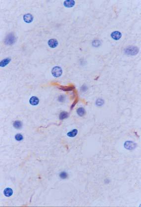
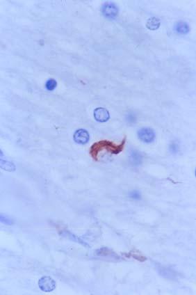
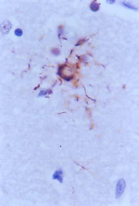
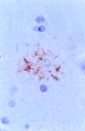
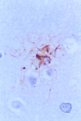
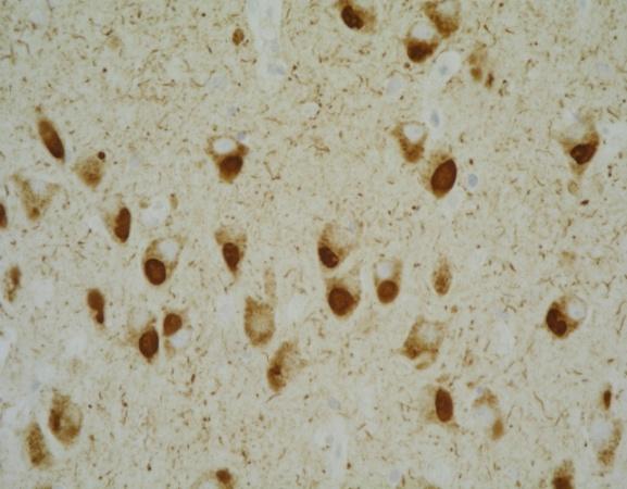
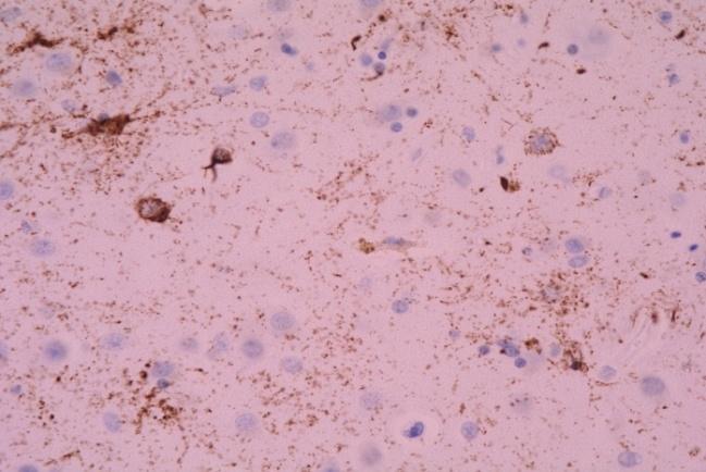
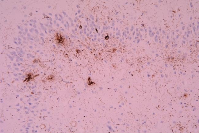
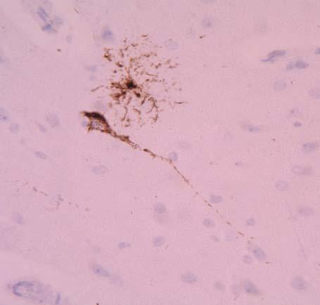
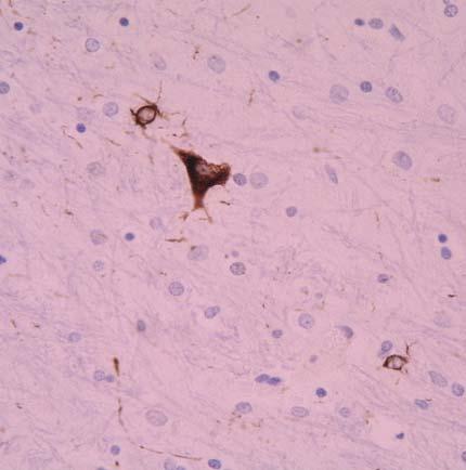
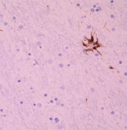
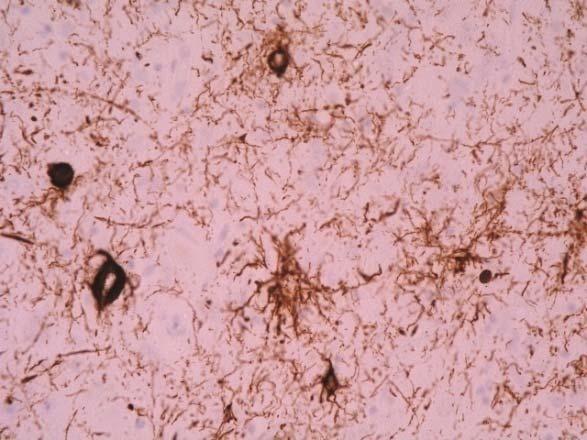
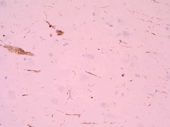
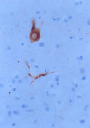
15 Progressive supranuclear palsy A B C D E F G H Hyperphosphorylated-tau-immunoreactive inclusions in astrocytes (A-E) and oligodendrocytes: coiled bodies (F-H). C-E: tufted astrocytes
16 Progressive supranuclear palsy A B C D E F G H Expression of active tau-kinases in PSP. A-D: SAPK/JNK-P; E-G: p38-p; H: GSK-3β-P


17 PSP A B C D Early PSP, pre-clinical stage. A, B: caudate; C,: globus pallidus; D: subthalamus. A, GFAP; B-D: AT8


18 PSP A B C D Early PSP, pre-clinical stage. A: caudate; B: amygdala; C: CA1; D: DG

19 PSP A B C D E F G H I J PSP, intermediate stages. A: putamen; B: caudate; C: amygdala; D: CA1; E: dentate gyrus; F: Meynert nucleus; G: pons; H, midbrain (tufted astrocyte); I: locus ceruleus; J: gyrus cinguli


20 PSP A B C D E F G H I PSP; AT8. A: caudate; B: amygdala; C: pallidus; D: dorsomedial thalamus; E: subthalamus; F: superior colliculus; G: pons; H: frontal cortex; I: CA1 area of the hippocampus.


21 PSP CASE 1 CASE 2 CASE 3 CASE 4 CASE 5 p-tau Ser p-tau Thr p-tau Ser FC S P WM FC S P WM FC S P WM FC S P WM FC CP P WM


, striatum (S), pons (P) and white")


22 PSP CASE 1 CASE 2 CASE 3 CASE 4 CASE 5 p-tau Ser p-tau Ser FC S P WM FC S P WM FC S P WM FC S P WM FC S P WM Band patterns of phospho-tau, as seen with different phospho-specific anti-tau antibodies in the frontal cortex (FC), striatum (S), pons (P) and white matter (WM) in five PSP cases. Note the two bands of 68 and 64 kda practically in all cases and regions, and the different bands of lower molecular weight in the different regions and different cases.


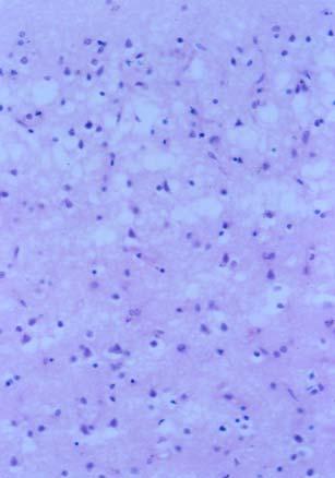
23 A B C D Loss of neurons and spongiosis in the upper cortical layers (A) together with ballooned neurons filled willed with phosphorylated neurofilaments and αb-crystallin (C, D) can be seen in cortical type PSP. (B): locus ceruleus with neurofibrillary tangles (arrow)
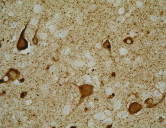
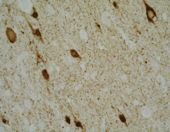
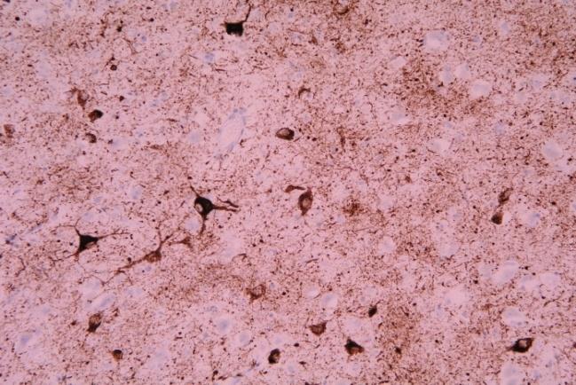
24 Corticobasal degeneration (CBD) (Corticodentatonigral degeneration with neuronal achromasia, corticobasal ganglionic degeneration, corticonigral degeneration with neuronal achromasia). Cortical atrophy (frontal lobe > temporal lobe > parietal lobe), often asymmetrical. Moderate atrophy of the striatum. Degeneration in the substantia nigra. Neuron loss, vacuolation of the neuropil, astrocytic gliosis in the cerebral cortex, striatum, pallidum, dorsomedial nucleus of the thalamus, amygdala and substantia nigra. Mild gliosis in the dentate nucleus. Large numbers of NF-P (+), tau (-) huge ballooned neurons (achromatic neurons in Nissl stains) in cerebral cortex, also containing a B crystallin. tau (+) intraneuronal inclusions (pre-tangles), granular deposits, rarely neurofibrillary tangles. tau (+) neuropil threads. tau (+) astrocytic inclusions: astrocytic plaques; tau (+) astrocytic end-feet in subependymal, subpial and perivascular areas. tau (+) oligodendrocytic inclusions: coiled bodies and threads.

,")

, globose neurons in the")
25 Corticobasal degeneration Atrophy of the cerebral cortex and enlargemenmt of the lateral ventricle. Swollen cortical neeurons (achromatic neurons), as seen with haematoxylin and eosin, are filled with phosphorylated neurofilaments Hyper-phosphorylated tau is found in neurons (horizontal arrowheads), globose neurons in the brain stem, and in astrocytic plaques (vertical arrowheads)

26 Corticobasal degeneration Ballooned neurons (arrows) contain phosphorylated neurofilaments

27 Corticobasal degeneration Ballooned neurons contain phosphorylated αb-crystallin A, B: frontal cortex; C, D: AD, amygdala. Ballooned neurons. A, C: αb-crystallin; B, D: phosphorylated αbcrystallin
 and phosphorylatyed neurofilaments (D- F).")
28 Corticobasal degeneration A B C D A E F G H I Ballooned neurons contain αb-crystallin that co-localizes with hsp-27 (A-C) and phosphorylatyed neurofilaments (D- F). Negative controls (G-I)


; B: balloooned")


29 Corticobasal degeneration A B C D E F G H I A: frontal cortex: espongiosis in the upper layers (left); B: balloooned neurons in the cortex stained with anti B- crystallin antibodies; C: frontal cortex; D: striatum; E: CA1 region of the hippocampus; F: Dentate gyrus; G: amygdala; H: substantia nigra; I: medial reticular nucleus, medulla oblongata. C-I: phospho-tau (AT8 antibody)


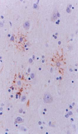
30 Corticobasal degeneration Astrocytic plaques in cerebral cortex


31 Corticobasal degeneration Frontotemporal atrophy. Band pattern of phospho-tau in PHF fractions reveals two bands of 68 and 64 kda


32 Corticobasal degeneration A B C D E F G H A, B: Ballooned neurons in the cerebral cortex, NF-P; αb-crystallin; C: frontal cortex; D: hippocampus; E: caudate; F, G: astrocytic plaques; H: coiled body in the white matter. C-H: AT8 antibody
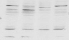
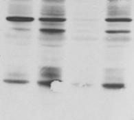
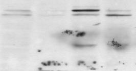
33 Phospho-tau bands, as visualized with different phospho-specific anti-tau antibodies, in fractions enriched with abnormal aggregates of filaments in Alzheimer disease (AD), progressive supranuclear palsy (PSP), Pick disease (PiD) and corticobasal degeneration (CBD). AD is characterized by four bands of 73, 68, 64 and 60 kda; PSP and CBD by two bands of 68 and 64 kda, and PiD by two bands of about 64 and 60 kda


34 Rare tauopathies: Mixed tauopathy with incomplete PSP and CBD characteristics


.")

, white matter")
, pons")
35 Rare tauopathies: Mixed tauopathy with incomplete PSP and CBD characteristics A B C D E F G H Ballooned neurons in cerebral cortex (A and B: αb-crystallin). C-H: hyperphosphorylated tau in striatum (C), white matter (D), motor ocular nucleus (E), pons (F), ventral reticular (G), and lateral reticular (H).


36 Rare tauopathies: Multiple system tauopathy A B C D Tau-immunoreactive inclusions in neurons and glial cells as revealed with several phospho-specific anti-tau antibodies Ser422 (A), Ser202 (C), Ser396 (D) and with the phosphorylation-independent tau antibody 7.51 (B).
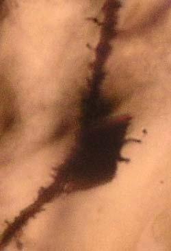
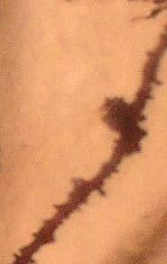
37 Argyrophilic grain disease Argyrophilic grains may be the only alteration in rare demented individuals, but they are also found in some cognitive normal subjects over age 65 years Association with AD stages I-IV of Braak, common. Argyrophilic grains may be found in Progressive supranuclear palsy, Corticobasal Degeneration, Pick disease, Diffuse lewy body disease, Multiple system atrophy, Parkinson disease and Motoneuron disease Neuropathology Dendritic-derived, irregulary shaped structures with filiform or knobby appendages (grains). Electron microscopy: 9-19 nm straight filaments or smooth tubules. Immunohistochemistry: phosphorylated tau tau-positive coiled bodies in oligodendrocytes in deep cortical layers and subcortical white matter.

;")
 B 4R tau CA1 (A), entorhinal")

, and are stained with")


38 AGD A AGD, Gallyas stain. Pretangles, tangles and grains (A, B). Ballooned neurons in amygdala; Gallyas-positive astrocytes (D); coiled bodies (oligodendrocytes) (E, F) B 4R tau CA1 (A), entorhinal cortex (B) Ballooned neurons in the amygdala contain phosphorylated neurofilaments and αb-crystallin (A-C), and are stained with antibodies to active tau kinases; MAPK/ERK, SAPK/JNK, p38, GSK-3β 73 KDa 68 KDa 64 KDa 60 KDa AD AGD Ser 262 Thr 181 Ser 202 Ser 214 Ser 396 Ser 422 AGD, bands of 68 and 64 kda of phospho-tau


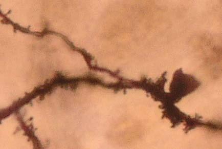
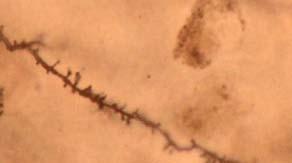
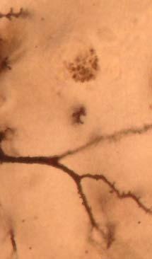


39 AGD AGD AD Grains: Golgi method Band pattern of phospho-tau in PHF fractions in AD and AGD. Note in AGD the upper bands of the 68 and 64 kda and the lower bands which differ from those found in AD
, nucleus tuberalis (C), mammilllary bodies (D).")
40 AGD A B C D Argyrophilic grains in CA1 (A), entorhinal cortex (B), nucleus tuberalis (C), mammilllary bodies (D). AT8 antibody
41 AGD A B C D E F Argyrophilic grains in CA1 (A), subiculum (B), amygdala (C), Meynert nucleus (D), cingular cortex (E), locus ceruleus (F). AT8 antibody


42 AGD AGD. A: CA1; B:dentate gyrus; C: entorhinal cortex; D: amygdala; E: accumbens; F: septum; G: putamen; H: substantia nigra; I: white matter; J: CA1 tau4r; K: EC tau4r; I: amyg, αb-crystallin


43 AGD AGD. A-C: αb-crystallin in ballooned neurons in amygdala; D-F: phosphorylated αb-crystallin in ballooned neurons in amygdala; G: αb-crystallin in reactive astrocytes in the temporal white matter (these cells do not contain phosphorylated B-crystallinB; H: α B-crystallin in cortical oligodendroglia; I: phosphorylated αbcrystallin is absent in cortical oligodendroglia.

44 Dementia with only tangles A B C D E F G H Hyper-phosphorylated tau (AT8 antibody. A: entorhinal cortex; B: CA1 area of the hippocampus; C: putamen; D: Meynert nucleus; E: superior colliculus; F: locus ceruleus; G: motor ocular III; H: lateral reticular nucleus of the brain stem
45 Frontotemporal lobar degeneration (dementia) with mutations in the tau gene: familial tauopathies (FTLD-tau) N279K N296N R5H R5L L266V K257T G372V ΔN296 N296H S305S S305N 10 Intronic mutations S320F V337M S352L G389R R406W 1 9 L284L P301L P301S K369I L315R E342V I260V Splicing Pick-like Tangle predominant Other patterns
46 Mechanisms of MAPT mutations 1. Splicing mutations: Alterations in the 3R:4R ratio a. In intron following exon 10 b. In exon 10 i. Pure (silent) L284L, S305S, N296N ii. Mixed splicing and structural ΔN296, N296H, N279K, S305N 2. Mutations affecting the structure of the tau protein a. In exon 10 P301L, P301S b. In exons 1, 9, 11, 12, or 13 i. With neurofibrillary tangles: V337M, R406W ii. With Pick bodies: K257, L266V, G272V, L315R, S320F, 336R, E342V, K369I, G389R iii. With other patterns R5H, R5L, I260V, S352L11


.")


47 FTLD-tau P301L A B C D Frontotemporal atrophy. Loss of neurons and spongiosis in the upper layers (A) with occasional ballooned neurons (B) in the cerebral cortex (CC). Inclusions are also seen in the dentate gyrus (arrow, DG, C) Hyperphosphorylated tau deposits in neurons and glial cells
, and with antibodies directed against active forms of tau-kinases JNK, p38 and")
48 FTLD-tau P301L Intraneuronal inclusions are stained with phospho-specific anti-tau antibodies (214, 396), and with antibodies directed against active forms of tau-kinases JNK, p38 and GSK-3


49 FTLD-tau P301L Two different cases. Left pannel: intraneuronal inclusions as revealed with different phospho-specific anti-tau antibodies, tubulin (tub) and ubiquitin (ubi). Right pannel; different regions: cc: cerebral cortex; EC: entorhinal cortex; CA1 area of the hippocampus; Str: striatum; DG: dentate gyrus; Mey: Meynert nucleus; As: astrocytes; WM: coiled bodies in the white matter


50 FTLD deln296 A B C D E F Widespread neuronal and glial hyperphosphorylated tau deposits Frontotemporal atrophy, atrophy of the mesencephalon and pons

51 FTLD deln296 Tangles Astrocytes Coiled bodies Thr Ser Ser Ser Ser MAPK/ERK-P SAPK/JNK-P P38-P GSK-3β-P tau-kinases associated with tau phosphorylation in FTLD del296: MAPK-ERK-P; SAPK-JNK-P; p38-p; GSK-3β-P


.")


52 FTLD K317M A B C Frontotemporal atrophy D E F Marked spongiosis in the upper layers of the cerebral cortex (A). Widespread neuronal and glial hyperphorylated tau-immunoreactive deposits (B-F). Tufted astrocytes, fibrous astrocytes, globose oligodendoglial inclusions and coiled bodies are common.
.")
53 FTLD K317M Generalized neuronal and glial hyperphosphorylated tau deposits in the cerebral cortex (cc), thalamus (thal), striatum (str), mammillary bodies (mam) and cerebral white matter (wm). Deposits in astrocytes are reminiscent of tufted astrocytes and fibrous astrocytes, and inclusions in oligodendrocytes are globose or coiled.

54 Identification of phospho-tau-immunoreactive bands in sarkosyl-insoluble fractions in FTDP P-tau Ser Case Region FC FC Hip FC Tau mutation P301L P301L R406W Phospho-tau bands in P301L mutation differ from those in R406W mutation
55 Myotonic dystrophy A B C D E F G H I A: parietal cortex; B: temporal cortex; C: cingulate cortex; D: insula; E: CA1; F: mammillary body; G: locus ceruleus; H, I: entorhinal cortex. A-G: AT8; H: 3Rtau; I: 4Rtau
56 Myotonic dystrophy A B C D tau deposits in the entorhinal cortex, A, B: AT8; C: 3R tau; D: 4Rtau
 4R")
 compared with")

57 Myotonic dystrophy 50 kda 50 kda 50 kda 37 kda 37 kda 37 kda 25 kda 25 kda 25 kda AD MD1 MD2 MD3 AD MD1 MD2 MD3 AD AD AD MD1 MD2 MD3 Band pattern of 3R (left) 4R (middle) and phosphorylated tau (Ser214) (right) in fractions enriched in filaments in three cases of myotonic dystrophy (MD1, MD2, MD3) compared with Alzheimer disease (AD). AD is characterized by three bands of 68, 64 and 60 kda, whereas myotonic dystrophy shows bands of 50 kda and lower which are not found in any other tauopathy.
58 Prion diseases PRNP P102L-129V PRNP P102L-129V. Cerebral cortex; b: cerebellum, HE; C: cerebellum, PAS; D, E: PrP cortex; F: striatum; G: thalamus; H: cerebellum; I: spinal cord
59 Prion diseases PRNP P102L-129V PRNP P102L-129V. A: PrP; B: GFAP; C: CD68; D: αb-crystallin; E: AT8; F: AT8; G: tau3r; H: tau4r; I: ubiquitin
60 Prion diseases PRNP P102L-129V PRNP P102L-129V. Confocal microscopy PrP deposition (left column, green), phospho-tau (Thr181) (mid column, red), merge (right column). Hyperphosphorylated tau is accumulkated in cell processes surrrounding PrP deposits.

 and phospho-tau (Ser214; right) in PHF fractions")
 GSS1: PRNP Y218N 129V GSS2: PRNP")
61 Prion diseases 75 kda 75 kda 75 kda 50 kda 50 kda 50 kda 37 kda 37 kda 37 kda 25 kda 25 kda 25 kda AD AD AD GSS1 GSS2 AD AD AD GSS1 GSS2 AD AD AD GSS 1 GSS2 Band pattern of 3Rtau (left), 4Rtau (middle) and phospho-tau (Ser214; right) in PHF fractions in two cases of Gerstmann- Straüssler-Scheinker (GSS) syndrome and in Alzheimer disease (AD) GSS1: PRNP Y218N 129V GSS2: PRNP P102L-129V
62 Myofibrillar myopathies(mfm): myotilinopathies and desminopathies sarcolemma integrins MLP myotilin Filamin C actin zyxin α-actinin telethonin calsarcin titin CapZ ZASP MLP nebulin αb-crystallin myopalladin desmin mink Myotilinopaty Z-line Z-line Thick filaments Thin filaments I band A band I band Desminopathy

63 sibm and MFM sibm. Positive fibers immunoreactive for A: 4Rtau; B: 3Rtau; C: P-tau Thr 181; D: Alz50; E: AT8; F: P-tau Ser422; G: AKT-P; H: MAPK/ERK-P; I: SAPK/JNK-P; J: p38-p; K: GSK-3βTyr; L: GSK-3βSer9; : J: p38-p; K: GSK-3βTyr; L: GSK-3βSer9

64 sibm and MFM Myotilinopathy. Positive fibers immunoreactive for A: 3Rtau; B: 4Rtau; C: P-tau Thr181; D: Alz50; E: P-tau Ser422; F: AT8; G: 3Rtau; H: P-tau Ser396; I: P-tau-Thr181.

65 sibm and MFM Myotilinopathy. Positive fibers immunoreactive for A: AKT-P: B: MAPK/ERK-P; C: SAPK/JNK-P; D: p38-p; E: GSK-3βTyr; F: GSK-3βSer9

66 sibm and MFM Desminopathy. Positive fibers immunoreactive fo A: 3Rtau; B: 4Rtau; C: Alz50; D. P-tau Thr181; E: P-tau Ser 422; F: P-tau-Ser 396; G: SAPK/JNK-P; H: p38-p; I: GSK-3βSer9
")

67 sibm and MFM. Big tau (about 120 kda) and GSK-3α/β levels. Myoblastic C2C12 line used as a control
68
Neuropathology of Neurodegenerative Disorders Prof. Jillian Kril
 Neurodegenerative disorders to be discussed Alzheimer s disease Lewy body diseases Frontotemporal dementia and other tauopathies Huntington s disease Motor Neuron Disease 2 Neuropathology of neurodegeneration
FDG-PET e parkinsonismi
 Parkinsonismi FDG-PET e parkinsonismi Valentina Berti Dipartimento di Scienze Biomediche, Sperimentali e Cliniche Sez. Medicina Nucleare Università degli Studi di Firenze History 140 PubMed: FDG AND parkinsonism
The Spectrum of Age-Associated Astroglial Tauopathies. Dennis W. Dickson MD Department of Neuroscience Mayo Clinic, Jacksonville, FL
 The Spectrum of Age-Associated Astroglial Tauopathies Dennis W. Dickson MD Mayo Clinic, Jacksonville, FL Thorn-shaped astrocytes TSA were first reported by Ikeda (1995), as tau-positive astrocytes in various
Chronic Traumatic Encephalopathy Provider and Parent Essentials
 Chronic Traumatic Encephalopathy Provider and Parent Essentials Concussion Global Cast July 30, 2014 John Lockhart, MD Seattle Children s Hospital Chronic Traumatic Encephaly (CTE) Working Definition Chronic
Invited review: Neuropathology of tauopathies: principles and practice
 Neuropathology and Applied Neurobiology (2015), 41, 3 23 doi: 10.1111/nan.12208 Invited review: Neuropathology of tauopathies: principles and practice G. G. Kovacs Institute of Neurology, Medical University
NACC Vascular Consortium. NACC Vascular Consortium. NACC Vascular Consortium
 NACC Vascular Consortium NACC Vascular Consortium Participating centers: Oregon Health and Science University ADC Rush University ADC Mount Sinai School of Medicine ADC Boston University ADC In consultation
! slow, progressive, permanent loss of neurologic function.
 UBC ! slow, progressive, permanent loss of neurologic function.! cause unknown.! sporadic, familial or inherited.! degeneration of specific brain region! clinical syndrome.! pathology: abnormal accumulation
Evaluating the Patterns of Aging-Related Tau Astrogliopathy Unravels Novel Insights Into Brain Aging and Neurodegenerative Diseases
 J Neuropathol Exp Neurol Vol. 76, No. 4, April 2017, pp. 270 288 doi: 10.1093/jnen/nlx007 ORIGINAL ARTICLE Evaluating the Patterns of Aging-Related Tau Astrogliopathy Unravels Novel Insights Into Brain
Clinicopathologic and genetic aspects of hippocampal sclerosis. Dennis W. Dickson, MD Mayo Clinic, Jacksonville, Florida USA
 Clinicopathologic and genetic aspects of hippocampal sclerosis Dennis W. Dickson, MD Mayo Clinic, Jacksonville, Florida USA The hippocampus in health & disease A major structure of the medial temporal
Dementia syndrome. Manifestation DISORDERS & DEMENTIA. Reasons of demencia
 Manifestation DEGENERATIVE DISORDERS & DEMENTIA Roman Beňačka, MD,PhD Department of Pathophysiology Medical Faculty, Šafarik University Košice Increase in time required to retrieve information Less able
NACC Neuropathology (NP) Diagnosis Coding Guidebook
 Diagnosis Coding Guidebook") Department of Epidemiology, School of Public Health and Community Medicine, University of Washington 4311 11 th Avenue NE #300 Seattle, WA 98105 phone: (206) 543-8637; fax: (206) 616-5927 e-mail: naccmail@u.washington.edu
Department of Epidemiology, School of Public Health and Community Medicine, University of Washington 4311 11 th Avenue NE #300 Seattle, WA 98105 phone: (206) 543-8637; fax: (206) 616-5927 e-mail: naccmail@u.washington.edu
Argyrophilic grain disease
 Argyrophilic grain disease Prof. Isidro Ferrer, Institut Neuropatologia, Servei Anatomia Patològica, IDIELLHospital Universitari de ellvitge, Universitat de arcelona, IERNED, Hospitalet de LLobregat; Spain
Altered proteins in the aging brain
 Digital Comprehensive Summaries of Uppsala Dissertations from the Faculty of Medicine 1182 Altered proteins in the aging brain ADILA ELOBEID ACTA UNIVERSITATIS UPSALIENSIS UPPSALA 2016 ISSN 1651-6206 ISBN
Chapter 3. Structure and Function of the Nervous System. Copyright (c) Allyn and Bacon 2004
 Allyn and Bacon 2004") Chapter 3 Structure and Function of the Nervous System 1 Basic Features of the Nervous System Neuraxis: An imaginary line drawn through the center of the length of the central nervous system, from the
Chapter 3 Structure and Function of the Nervous System 1 Basic Features of the Nervous System Neuraxis: An imaginary line drawn through the center of the length of the central nervous system, from the
Systems Neuroscience Dan Kiper. Today: Wolfger von der Behrens
 Systems Neuroscience Dan Kiper Today: Wolfger von der Behrens wolfger@ini.ethz.ch 18.9.2018 Neurons Pyramidal neuron by Santiago Ramón y Cajal (1852-1934, Nobel prize with Camillo Golgi in 1906) Neurons
Introduction to the Central Nervous System: Internal Structure
 Introduction to the Central Nervous System: Internal Structure Objective To understand, in general terms, the internal organization of the brain and spinal cord. To understand the 3-dimensional organization
Brain dissection protocol for amyotrophic lateral sclerosis/motor neurone disease
 Brain dissection protocol for amyotrophic lateral sclerosis/motor neurone disease Prepared by Approved by Approved by Revised by Name Signature Date Sampling and biomarker OPtimization and Harmonization
Do pathological changes in tau protein isoforms manifest in cerebrospinal fluid of tauopathy patients?
 Do pathological changes in tau protein isoforms manifest in cerebrospinal fluid of tauopathy patients? Development and validation of sensitive immuno-pcr assays Barcelona, November 2012 Rohan de Silva,
Nsci 2100: Human Neuroanatomy 2017 Examination 3
 Name KEY Lab Section Nsci 2100: Human Neuroanatomy 2017 Examination 3 On this page, write your name and lab section. On your bubble answer sheet, enter your name (last name, space, first name), internet
Biological Bases of Behavior. 3: Structure of the Nervous System
 Biological Bases of Behavior 3: Structure of the Nervous System Neuroanatomy Terms The neuraxis is an imaginary line drawn through the spinal cord up to the front of the brain Anatomical directions are
Lecture 42: Final Review. Martin Wessendorf, Ph.D.
 Lecture 42: Final Review Martin Wessendorf, Ph.D. Lecture 33 cortex Heilbronner 5 lobes of the cortex Lateral view (left side) Mid-saggital view (right side) Cellular organization of cortex White matter
FTD basics! Etienne de Villers-Sidani, MD!
 FTD basics! Etienne de Villers-Sidani, MD! Frontotemporal lobar degeneration (FTLD) comprises 3 clinical syndromes! Frontotemporal dementia (behavioral variant FTD)! Semantic dementia (temporal variant
Anatomy and Physiology (Bio 220) The Brain Chapter 14 and select portions of Chapter 16
 The Brain Chapter 14 and select portions of Chapter 16") Anatomy and Physiology (Bio 220) The Brain Chapter 14 and select portions of Chapter 16 I. Introduction A. Appearance 1. physical 2. weight 3. relative weight B. Major parts of the brain 1. cerebrum 2.
Anatomy and Physiology (Bio 220) The Brain Chapter 14 and select portions of Chapter 16 I. Introduction A. Appearance 1. physical 2. weight 3. relative weight B. Major parts of the brain 1. cerebrum 2.
Brain anatomy and artificial intelligence. L. Andrew Coward Australian National University, Canberra, ACT 0200, Australia
 Brain anatomy and artificial intelligence L. Andrew Coward Australian National University, Canberra, ACT 0200, Australia The Fourth Conference on Artificial General Intelligence August 2011 Architectures
Atypical Parkinsonian Disorders
 APDs: Neuropathology and Nosology 111 Atypical Parkinsonian Disorders Neuropathology and Nosology 8 Charles Duyckaerts INTRODUCTION In many neurological diseases the topography of the lesion, whatever
Exam 2 PSYC Fall (2 points) Match a brain structure that is located closest to the following portions of the ventricular system
 Match a brain structure that is located closest to the following portions of the ventricular system") Exam 2 PSYC 2022 Fall 1998 (2 points) What 2 nuclei are collectively called the striatum? (2 points) Match a brain structure that is located closest to the following portions of the ventricular system
Exam 2 PSYC 2022 Fall 1998 (2 points) What 2 nuclei are collectively called the striatum? (2 points) Match a brain structure that is located closest to the following portions of the ventricular system
Bacterial, viral, protoozal and fungal infections of the CNS
 Bacterial, viral, protoozal and fungal infections of the CNS Prof. Isidro Ferrer, Institut Neuropatologia, Servei Anatomia Patològica, IDIBELL-Hospital Universitari de Bellvitge, Universitat de Barcelona,
The relationship between development of neuronal and astrocytic tau pathologies. in subcortical nuclei and progression of argyrophilic grain disease
 Ikeda et al. The relationship between development of neuronal and astrocytic tau pathologies in subcortical nuclei and progression of argyrophilic grain disease Chikako Ikeda ), Osamu Yokota,,), Shigeto
The neurvous system senses, interprets, and responds to changes in the environment. Two types of cells makes this possible:
 NERVOUS SYSTEM The neurvous system senses, interprets, and responds to changes in the environment. Two types of cells makes this possible: the neuron and the supporting cells ("glial cells"). Neuron Neurons
10/3/2016. T1 Anatomical structures are clearly identified, white matter (which has a high fat content) appears bright.
 appears bright.") H2O -2 atoms of Hydrogen, 1 of Oxygen Hydrogen just has one single proton and orbited by one single electron Proton has a magnetic moment similar to the earths magnetic pole Also similar to earth in that
H2O -2 atoms of Hydrogen, 1 of Oxygen Hydrogen just has one single proton and orbited by one single electron Proton has a magnetic moment similar to the earths magnetic pole Also similar to earth in that
FRONTOTEMPORAL DEGENERATION: OVERVIEW, TRENDS AND DEVELOPMENTS
 FRONTOTEMPORAL DEGENERATION: OVERVIEW, TRENDS AND DEVELOPMENTS Norman L. Foster, M.D. Director, Center for Alzheimer s Care, Imaging and Research Chief, Division of Cognitive Neurology, Department of Neurology
Prof. Saeed Abuel Makarem & Dr.Sanaa Alshaarawy
 Prof. Saeed Abuel Makarem & Dr.Sanaa Alshaarawy 1 Objectives By the end of the lecture, you should be able to: Describe the anatomy and main functions of the thalamus. Name and identify different nuclei
DEMENTIA 101: WHAT IS HAPPENING IN THE BRAIN? Philip L. Rambo, PhD
 DEMENTIA 101: WHAT IS HAPPENING IN THE BRAIN? Philip L. Rambo, PhD OBJECTIVES Terminology/Dementia Basics Most Common Types Defining features Neuro-anatomical/pathological underpinnings Neuro-cognitive
PSY 302: CHAPTER 3 NOTES THE BRAIN (PART II) - 9/5/17. By: Joseline
 - 9/5/17. By: Joseline") PSY 302: CHAPTER 3 NOTES THE BRAIN (PART II) - 9/5/17 By: Joseline Left 3 MAJOR FISSURES : 2HEMISPHERES Right Lateral Ventricle Central Fissure Third Ventricle Sulcus Lateral Fissure Gyros Fissure- Fissures
PSY 302: CHAPTER 3 NOTES THE BRAIN (PART II) - 9/5/17 By: Joseline Left 3 MAJOR FISSURES : 2HEMISPHERES Right Lateral Ventricle Central Fissure Third Ventricle Sulcus Lateral Fissure Gyros Fissure- Fissures
The Central Nervous System I. Chapter 12
 The Central Nervous System I Chapter 12 The Central Nervous System The Brain and Spinal Cord Contained within the Axial Skeleton Brain Regions and Organization Medical Scheme (4 regions) 1. Cerebral Hemispheres
Current Concepts in the Classification and Diagnosis of Frontotemporal Lobar Degenerations
 Current Concepts in the Classification and Diagnosis of Frontotemporal Lobar Degenerations Frontotemporal lobar degenerations are clinically, genetically, and molecularly heterogeneous diseases characterized
I: To describe the pyramidal and extrapyramidal tracts. II: To discuss the functions of the descending tracts.
 Descending Tracts I: To describe the pyramidal and extrapyramidal tracts. II: To discuss the functions of the descending tracts. III: To define the upper and the lower motor neurons. 1. The corticonuclear
Regional and Lobe Parcellation Rhesus Monkey Brain Atlas. Manual Tracing for Parcellation Template
 Regional and Lobe Parcellation Rhesus Monkey Brain Atlas Manual Tracing for Parcellation Template Overview of Tracing Guidelines A) Traces are performed in a systematic order they, allowing the more easily
Announcement. Danny to schedule a time if you are interested.
 Announcement If you need more experiments to participate in, contact Danny Sanchez (dsanchez@ucsd.edu) make sure to tell him that you are from LIGN171, so he will let me know about your credit (1 point).
9.14 Class 32 Review. Limbic system
 9.14 Class 32 Review Limbic system 1 Lateral view Medial view Brainstem, sagittal section Sensory- Perceptual Motor Behavior Major functional modules of the CNS Motivation Courtesy of MIT Press. Used with
SPATIAL PATTERNS OF THE TAU PATHOLOGY IN PROGRESSIVE SUPRANUCLEAR PALSY
 SPATIAL PATTERNS OF THE TAU PATHOLOGY IN PROGRESSIVE SUPRANUCLEAR PALSY Richard A. Armstrong 1* and Nigel J. Cairns 2 1 Vision Sciences, Aston University, Birmingham B4 7ET, UK; 2 Departments of Neurology,
Amyotrophic lateral sclerosis (ALS) is a progressive neurodegenerative
 is a progressive neurodegenerative") ORIGINAL RESEARCH E. Matsusue S. Sugihara S. Fujii T. Kinoshita T. Nakano E. Ohama T. Ogawa Cerebral Cortical and White Matter Lesions in Amyotrophic Lateral Sclerosis with Dementia: Correlation with MR
ORIGINAL RESEARCH E. Matsusue S. Sugihara S. Fujii T. Kinoshita T. Nakano E. Ohama T. Ogawa Cerebral Cortical and White Matter Lesions in Amyotrophic Lateral Sclerosis with Dementia: Correlation with MR
CONSENSUS PAPER. Acta Neuropathol (2007) 114:5 22 DOI /s
 114:5 22 DOI /s") Acta Neuropathol (2007) 114:5 22 DOI 10.1007/s00401-007-0237-2 CONSENSUS PAPER Neuropathologic diagnostic and nosologic criteria for frontotemporal lobar degeneration: consensus of the Consortium for Frontotemporal
Acta Neuropathol (2007) 114:5 22 DOI 10.1007/s00401-007-0237-2 CONSENSUS PAPER Neuropathologic diagnostic and nosologic criteria for frontotemporal lobar degeneration: consensus of the Consortium for Frontotemporal
Pathogenesis of Degenerative Diseases and Dementias. D r. Ali Eltayb ( U. of Omdurman. I ). M. Path (U. of Alexandria)
. M. Path (U. of Alexandria)") Pathogenesis of Degenerative Diseases and Dementias D r. Ali Eltayb ( U. of Omdurman. I ). M. Path (U. of Alexandria) Dementias Defined: as the development of memory impairment and other cognitive deficits
Pathogenesis of Degenerative Diseases and Dementias D r. Ali Eltayb ( U. of Omdurman. I ). M. Path (U. of Alexandria) Dementias Defined: as the development of memory impairment and other cognitive deficits
Dementia and Healthy Ageing : is the pathology any different?
 Dementia and Healthy Ageing : is the pathology any different? Professor David Mann, Professor of Neuropathology, University of Manchester, Hope Hospital, Salford DEMENTIA Loss of connectivity within association
Atypical Progressive Supranuclear Palsy With Corticospinal Tract Degeneration
 J Neuropathol Exp Neurol Copyright Ó 2006 by the American Association of Neuropathologists, Inc. Vol. 65, No. 4 April 2006 pp. 396Y405 ORIGINAL ARTICLE Atypical Progressive Supranuclear Palsy With Corticospinal
Role of TDP-43 in Non-Alzheimer s and Alzheimer s Neurodegenerative Diseases
 Role of TDP-43 in Non-Alzheimer s and Alzheimer s Neurodegenerative Diseases Keith A. Josephs, MD, MST, MSc Professor of Neurology 13th Annual Mild Cognitive Impairment (MCI) Symposium: Alzheimer and Non-Alzheimer
BASAL GANGLIA. Dr JAMILA EL MEDANY
 BASAL GANGLIA Dr JAMILA EL MEDANY OBJECTIVES At the end of the lecture, the student should be able to: Define basal ganglia and enumerate its components. Enumerate parts of Corpus Striatum and their important
NEUROPATHOLOGY BRAIN CUTTING MANUAL LAST UPDATED ON 6/22/2015
 NEUROPATHOLOGY BRAIN CUTTING MANUAL LAST UPDATED ON 6/22/2015 Neuropathology Faculty involved in Brain Cutting: Dr. Sandra Camelo-Piragua Dr. Andrew Lieberman (Chief of the Division) Dr. Kathryn A. McFadden
CASE 49. What type of memory is available for conscious retrieval? Which part of the brain stores semantic (factual) memories?
 memories?") CASE 49 A 43-year-old woman is brought to her primary care physician by her family because of concerns about her forgetfulness. The patient has a history of Down syndrome but no other medical problems.
CASE 49 A 43-year-old woman is brought to her primary care physician by her family because of concerns about her forgetfulness. The patient has a history of Down syndrome but no other medical problems.
Parts of the Brain. Hindbrain. Controls autonomic functions Breathing, Heartbeat, Blood pressure, Swallowing, Vomiting, etc. Upper part of hindbrain
 Parts of the Brain The human brain is made up of three main parts: 1) Hindbrain (or brainstem) Which is made up of: Myelencephalon Metencephalon 2) Midbrain Which is made up of: Mesencephalon 3) Forebrain
Perspectives on Frontotemporal Dementia and Primary Progressive Aphasia
 Perspectives on Frontotemporal Dementia and Primary Progressive Aphasia Bradley F. Boeve, M.D. Division of Behavioral Neurology Department of Neurology Mayo Clinic Rochester, Minnesota Alzheimer s Disease
Psyc 311A, fall 2008 Conference week 3 TA: Jürgen Germann
 Psyc 311A, fall 2008 Conference week 3 TA: Jürgen Germann e-mail: jurgen.germann@mcgill.ca Overview: 1. Meninges 2. Cerebral cortex-cytoarchitecture 3. Diencephalon (thalamus/hypothalamus) (this replaces
Prion diseases or transmissible spongiform encephalopathies (TSEs)
") Prion diseases or transmissible spongiform encephalopathies (TSEs) rare progressive neurodegenerative disorders that affect both humans and animals. They are distinguished by long incubation periods, characteristic
Prion diseases or transmissible spongiform encephalopathies (TSEs) rare progressive neurodegenerative disorders that affect both humans and animals. They are distinguished by long incubation periods, characteristic
Histology of the CNS
 Histology of the CNS Lecture Objectives Describe the histology of the cerebral cortex layers. Describe the histological features of the cerebellum; layers and cells of cerebellar cortex. Describe the elements
Brainstem. Steven McLoon Department of Neuroscience University of Minnesota
 Brainstem Steven McLoon Department of Neuroscience University of Minnesota 1 Course News Change in Lab Sequence Week of Oct 2 Lab 5 Week of Oct 9 Lab 4 2 Goal Today Know the regions of the brainstem. Know
Study Guide Unit 2 Psych 2022, Fall 2003
 Study Guide Unit 2 Psych 2022, Fall 2003 Subcortical Anatomy 1. Be able to locate the following structures and be able to indicate whether they are located in the forebrain, diencephalon, midbrain, pons,
Final Scientific Progress Report
 CUREPSP Final Scientific Progress Report Tau in Peripheral Tissues of PSP and CBD. Brittany Dugger, PhD; University of California San Francisco Specific Aim: Using immunohistochemical methods on autopsy
CEREBRUM & CEREBRAL CORTEX
 CEREBRUM & CEREBRAL CORTEX Seonghan Kim Dept. of Anatomy Inje University, College of Medicine THE BRAIN ANATOMICAL REGIONS A. Cerebrum B. Diencephalon Thalamus Hypothalamus C. Brain Stem Midbrain Pons
Neurodegenerative Disease. April 12, Cunningham. Department of Neurosciences
 Neurodegenerative Disease April 12, 2017 Cunningham Department of Neurosciences NEURODEGENERATIVE DISEASE Any of a group of hereditary and sporadic conditions characterized by progressive dysfunction,
Lewy Bodies in the Amygdala
 ORIGINAL CONTRIBUTION Lewy Bodies in the Amygdala Increase of -Synuclein Aggregates in Neurodegenerative Diseases With Tau-Based Inclusions Anca Popescu, MD; Carol F. Lippa, MD; Virginia M.-Y. Lee, PhD;
PETER PAZMANY CATHOLIC UNIVERSITY Consortium members SEMMELWEIS UNIVERSITY, DIALOG CAMPUS PUBLISHER
 PETER PAZMANY CATHOLIC UNIVERSITY SEMMELWEIS UNIVERSITY Development of Complex Curricula for Molecular Bionics and Infobionics Programs within a consortial* framework** Consortium leader PETER PAZMANY
The Nervous System: Sensory and Motor Tracts of the Spinal Cord
 15 The Nervous System: Sensory and Motor Tracts of the Spinal Cord PowerPoint Lecture Presentations prepared by Steven Bassett Southeast Community College Lincoln, Nebraska Introduction Millions of sensory
Medical Neuroscience Tutorial Notes
 Medical Neuroscience Tutorial Notes Blood Supply to the Brain MAP TO NEUROSCIENCE CORE CONCEPTS 1 NCC1. The brain is the body's most complex organ. LEARNING OBJECTIVES After study of the assigned learning
14 - Central Nervous System. The Brain Taft College Human Physiology
 14 - Central Nervous System The Brain Taft College Human Physiology Development of the Brain The brain begins as a simple tube, a neural tube. The tube or chamber (ventricle) is filled with cerebrospinal
Dementia. Stephen S. Flitman, MD Medical Director 21st Century Neurology
 Dementia Stephen S. Flitman, MD Medical Director 21st Century Neurology www.neurozone.org Dementia is a syndrome Progressive memory loss, plus Progressive loss of one or more cognitive functions: Language
Basal Ganglia. Today s lecture is about Basal Ganglia and it covers:
 Basal Ganglia Motor system is complex interaction between Lower motor neurons (spinal cord and brainstem circuits) and Upper motor neurons (pyramidal and extrapyramidal tracts) plus two main regulators
Chapter 2: Studies of Human Learning and Memory. From Mechanisms of Memory, second edition By J. David Sweatt, Ph.D.
 Chapter 2: Studies of Human Learning and Memory From Mechanisms of Memory, second edition By J. David Sweatt, Ph.D. Medium Spiny Neuron A Current Conception of the major memory systems in the brain Figure
DISSECTION OF THE SHEEP'S BRAIN
 Sheep Brain Dissection Guide Page 1 DISSECTION OF THE SHEEP'S BRAIN Introduction The purpose of the sheep brain dissection is to familiarize you with the threedimensional structure of the brain and teach
Distinct clinical and neuropathological features of G51D SNCA mutation cases compared with SNCA duplication and H50Q mutation
 Distinct clinical and neuropathological features of G51D SNCA mutation cases compared with SNCA duplication and H50Q mutation Article Published Version Creative Commons: Attribution 4.0 (CC BY) Open Access
The Neuroscience of Music in Therapy
 Course Objectives The Neuroscience of Music in Therapy Unit I. Learn Basic Brain Information Unit II. Music in the Brain; Why Music Works Unit III. Considerations for Populations a. Rehabilitation b. Habilitation
Stanley Pruisinger 1980's
 Neuroanatomy Prion disease cerebellum chapter b/c cerebellar ataxia here as a warning for obvious reasons. Creutzfeldt - Jakob Disease (CJD) "Spongiform" (brain turns to sponge) Jews in Lybia who ate
212 Index C-SB-13,
 Index A Acetylcholinesterase inhibitor, treatment, 15 Age-associated memory impairment (AAMI), 5 Alzheimer s disease (AD), 40, 95 96 apolipoprotein E genotype and risk for, 58 cellular neurodegeneration
Nervous System. 1. What N.S. division controls skeletal muscles? 3. What kind of neuroglia myelinates axons in the PNS?
 . What N.S. division controls skeletal muscles? Nervous System SRS Review %. Central nervous system %. Peripheral nervous system %. Afferent division %. Somatic division %. Autonomic division %. Sympathetic
The Carroll A. Campbell, Jr. Neuropathology Laboratory: A Tool for Dementia Discovery in South Carolina
 The Carroll A. Campbell, Jr. Neuropathology Laboratory: A Tool for Dementia Discovery in South Carolina Pathology in the Cerebral Cortex H&E stain of mature neuritic plaque Modified Bielschowsky stain
Neuroanatomy lecture (1)
") Neuroanatomy lecture (1) Introduction: Neuroanatomy has two parts: the central and peripheral nervous system. The central nervous system is composed of brain and spinal cord. The brain has the following
Neuroanatomy lecture (1) Introduction: Neuroanatomy has two parts: the central and peripheral nervous system. The central nervous system is composed of brain and spinal cord. The brain has the following
Overview of Brain Structures
 First Overview of Brain Structures Psychology 470 Introduction to Chemical Additions Steven E. Meier, Ph.D. All parts are interrelated. You need all parts to function normally. Neurons = Nerve cells Listen
Department of Cognitive Science UCSD
 Department of Cognitive Science UCSD Verse 1: Neocortex, frontal lobe, Brain stem, brain stem, Hippocampus, neural node, Right hemisphere, Pons and cortex visual, Brain stem, brain stem, Sylvian fissure,
Original Article Typical or atypical progressive supranuclear palsy: a comparative clinicopathologic study of three Chinese cases
 Int J Clin Exp Pathol 2015;8(1):867-874 www.ijcep.com /ISSN:1936-2625/IJCEP0003790 Original Article Typical or atypical progressive supranuclear palsy: a comparative clinicopathologic study of three Chinese
Objectives. Overview. Why FTD and AD? FTD May Mimic AD. Introduction and Process Norman L. Foster, MD. Introduction and Process 7BS.
 Introduction and Process Norman L. Foster, MD 7BS.006 IMPROVING ACCURACY OF DEMENTIA DIAGNOSIS: CASE STUDIES WITH NEUROPATHOLOGY Norman L. Foster, MD University of Utah Salt Lake City, UT Edward Zamrini,
Motor System Hierarchy
 Motor Pathways Lectures Objectives Define the terms upper and lower motor neurons with examples. Describe the corticospinal (pyramidal) tract and the direct motor pathways from the cortex to the trunk
Supplementary Material S3 Further Seed Regions
 Supplementary Material S3 Further Seed Regions Figure I. Changes in connectivity with the right anterior insular cortex. (A) wake > mild sedation, showing a reduction in connectivity between the anterior
Brainstem. Amadi O. Ihunwo, PhD School of Anatomical Sciences
 Brainstem Amadi O. Ihunwo, PhD School of Anatomical Sciences Lecture Outline Constituents Basic general internal features of brainstem External and Internal features of Midbrain Pons Medulla Constituents
Gross Morphology of the Brain
 Gross Morphology of the Brain Done by : Marah Marahleh & Razan Krishan *slides in bold Principal Parts of the Brain Cerebrum : largest part of the brain Diencephalon Thalamus & hypothalamus Cerebellum
PROPERTY OF ELSEVIER SAMPLE CONTENT - NOT FINAL. Gross Anatomy and General Organization of the Central Nervous System
 3 Gross Anatomy and General Organization of the Central Nervous System C h a p t e r O u t l i n e The Long Axis of the CNS Bends at the Cephalic Flexure Hemisecting a Brain Reveals Parts of the Diencephalon,
Ch 13: Central Nervous System Part 1: The Brain p 374
 Ch 13: Central Nervous System Part 1: The Brain p 374 Discuss the organization of the brain, including the major structures and how they relate to one another! Review the meninges of the spinal cord and
Detailed protocol Only dissected human brain samples are stored. The microdissection is performed on frozen brains and the samples are kept on -70 C.
 2008 Detailed protocol Only dissected human brain samples are stored. The microdissection is performed on frozen brains and the samples are kept on -70 C. BrainNet Europe II Project Co-ordinator: Prof.
Neocortex. Cortical Structures in the Brain. Neocortex Facts. Laminar Organization. Bark-like (cortical) structures: Shepherd (2004) Chapter 12
 structures: Shepherd (2004) Chapter 12") Neocortex Shepherd (2004) Chapter 12 Rodney Douglas, Henry Markram, and Kevan Martin Instructor: Yoonsuck Choe; CPSC 644 Cortical Networks Cortical Structures in the Brain Bark-like (cortical) structures:
Neocortex Shepherd (2004) Chapter 12 Rodney Douglas, Henry Markram, and Kevan Martin Instructor: Yoonsuck Choe; CPSC 644 Cortical Networks Cortical Structures in the Brain Bark-like (cortical) structures:
b. The groove between the two crests is called 2. The neural folds move toward each other & the fuse to create a
 Chapter 13: Brain and Cranial Nerves I. Development of the CNS A. The CNS begins as a flat plate called the B. The process proceeds as: 1. The lateral sides of the become elevated as waves called a. The
NIH Public Access Author Manuscript Semin Neurol. Author manuscript; available in PMC 2014 November 14.
 NIH Public Access Author Manuscript Published in final edited form as: Semin Neurol. 2013 September ; 33(4): 386 416. doi:10.1055/s-0033-1359312. Neuroimaging Biomarkers of Neurodegenerative Diseases and
9.01 Introduction to Neuroscience Fall 2007
 MIT OpenCourseWare http://ocw.mit.edu 9.01 Introduction to Neuroscience Fall 2007 For information about citing these materials or our Terms of Use, visit: http://ocw.mit.edu/terms. 9.01 Recitation (R02)
THE CENTRAL NERVOUS SYSTEM. The Brain & Spinal Cord
 THE CENTRAL NERVOUS SYSTEM The Brain & Spinal Cord Review: Nervous System Parallel Distributed Processing Composition of the CNS Nuclei: Clusters of neurons in the CNS ( neighborhoods ) Fiber Tracts/Pathways:
brain MRI for neuropsychiatrists: what do you need to know
 brain MRI for neuropsychiatrists: what do you need to know Christoforos Stoupis, MD, PhD Department of Radiology, Spital Maennedorf, Zurich & Inselspital, University of Bern, Switzerland c.stoupis@spitalmaennedorf.ch
UNIT 5 REVIEW GUIDE - NERVOUS SYSTEM 1) State the 3 functions of the nervous system. 1) 2) 3)
 State the 3 functions of the nervous system. 1) 2) 3)") UNIT 5 REVIEW GUIDE - NERVOUS SYSTEM State the 3 functions of the nervous system. Briefly describe the general function(s) of each of the following neuron types: a) SENSORY NEURONS: b) INTERNEURONS: c)
UNIT 5 REVIEW GUIDE - NERVOUS SYSTEM State the 3 functions of the nervous system. Briefly describe the general function(s) of each of the following neuron types: a) SENSORY NEURONS: b) INTERNEURONS: c)
Autopsy Committee Sample Autopsy Case. Alzheimer Disease. Authors Ashley Thorburn, MD. Joseph E. Parisi, MD Autopsy Committee
 Autopsy Committee Sample Autopsy Case Alzheimer Disease Authors Ashley Thorburn, MD Joseph E. Parisi, MD Autopsy Committee Clinical Summary: A 75-year-old man presented to his primary care physician with
Supplementary Online Material Supplementary Table S1 to S5 Supplementary Figure S1 to S4
 Supplementary Online Material Supplementary Table S1 to S5 Supplementary Figure S1 to S4 Table S1: Brain regions involved in the adapted classification learning task Brain Regions x y z Z Anterior Cingulate
SHORT ANSWER. Write the word or phrase that best completes each statement or answers the question.
 Exam Name 1) A change in the conditions in the synaptic terminal can influence the soma as a result of axoplasmic transport. 2) The nervous system is composed of the brain and spinal cord. A) efferent
Telencephalon (Cerebral Hemisphere)
") Telencephalon (Cerebral Hemisphere) OUTLINE The Cortex - Lobes, Sulci & Gyri - Functional Subdivisions - Limbic Lobe & Limbic System The Subcortex - Basal Ganglia - White Matter (Internal Capsule) - Relations
Telencephalon (Cerebral Hemisphere) OUTLINE The Cortex - Lobes, Sulci & Gyri - Functional Subdivisions - Limbic Lobe & Limbic System The Subcortex - Basal Ganglia - White Matter (Internal Capsule) - Relations
For more information about how to cite these materials visit
 Author(s): Peter Hitchcock, PH.D., 2009 License: Unless otherwise noted, this material is made available under the terms of the Creative Commons Attribution Non-commercial Share Alike 3.0 License: http://creativecommons.org/licenses/by-nc-sa/3.0/
Diagnosis before NIA AA The impact of FDG PET in. Diagnosis after NIA AA Neuropathology and PET image 2015/10/16
 The impact of FDG PET in degenerative dementia diagnosis Jung Lung, Hsu MD, Ph.D (Utrecht) Section of dementia and cognitive impairment Department of Neurology Chang Gung Memorial Hospital, Linkou, Taipei