Spine and spinal cord
|
|
|
- Shona Davis
- 5 years ago
- Views:
Transcription
1 NEURORADIOLOGY Spine and spinal cord Erika Vörös University of Szeged Department of Radiology SZEGED
2 DISEASES OF SPINE AND SPINAL CORD I. Non-tumourous diseases developmental anomalies vascular disorders inflammatory processes degenerative changes trauma II. Tumours extradural intradural - extramedullary intramedullary
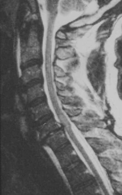
3 SPINE AND SPINAL CORD - imaging methods Plain film radiography Ultrasonography Computer tomography (CT) Magnetic resonance imaging (MRI) Digital subtraction angiography (DSA) Myelography
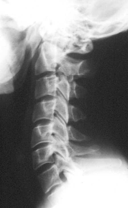
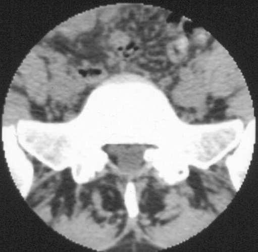
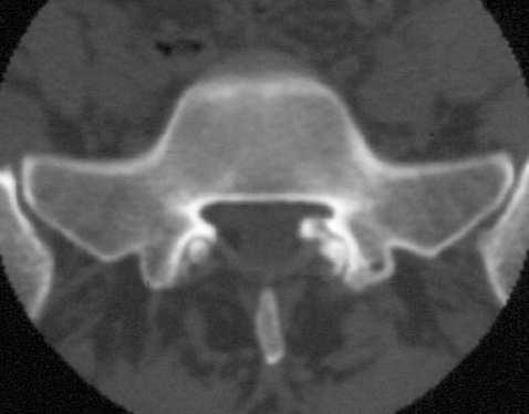
4 Imaging modalities - indications Plain film radiography developmental anomalies: bone inflammations: spondylitis / spondylodiscitis degenerative: spondylarthrosis trauma: fractures / dislocations tumours: bone changes
5 Imaging modalities - indications Computer tomography (CT) developmental anomalies: bones / spinal canal inflammations: bones / discs degenerative changes: spondylarthrosis, disc herniations trauma: bones, epi - subdural haemorrhages tumours: bone changes Myelo-CT trauma: dural and radicular injuries etc.
6 Imaging modalities - indications Magnetic resonance imaging (MRI) spinal cord lesions: all! developmental anomalies vascular disorders inflammatory processes degenerative diseases: disc herniations trauma: intramedullary lesions, haemorrhages tumours
7 Imaging modalities - indications Digital subtraction angiography (DSA) preoperative dg, diff.dg vascular malformations hypervascularized tumours intervention (embolization) vascular malformations hypervascularised tumours
8 Imaging modalities - indications Myelography if MRI not available: degenerative diseases, spaceoccupying lesions, trauma etc. severe scoliosis arachnoiditis
9 DEVELOPMENTAL ANOMALIES: (some injury in fetal life) Stage 1: Weeks 3-4 Dorsal Induction: Formation and Closure of the Neural Tube Three phases: Neurulation, canalization, retrogressive differentiation Failure: Anencephaly, Cephalocele, Chiari, Spinal dysraphism Stage 2: Weeks 5-10 Ventral Induction: Formation of the Brain Segments and Face Three vesicles (prosencephalon, mesencephalon, and rhombencephalon) form the cerebrum, mid-brain, cerebellum, and lower brain stem. Division into two hemispheres. Failure: Holoprosencephalies, Corpus callosum agenesis, Dandy Walker,Facial anomalies Stage 3: Months 2-5. Migration and Histogenesis Neuronal migration from germinal matrix to the cortex. Cortical organization. Disorders: Heterotopias, agyria-pachygyria, polymicrogyria, vascular malformations, teratomas, phakomatosis. Stage 4: 5-15 months; matures by 3 years. Myelination Inferior to superior; posterior to anterior. Failure: developmental delay, dysmyelinating disease


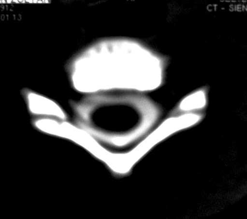
10 Developmental anomalies Generally complex (several layers and several levels involved) bones meninges spinal cord Chiari malformation, Tethered cord, syringomyelia spina bifida


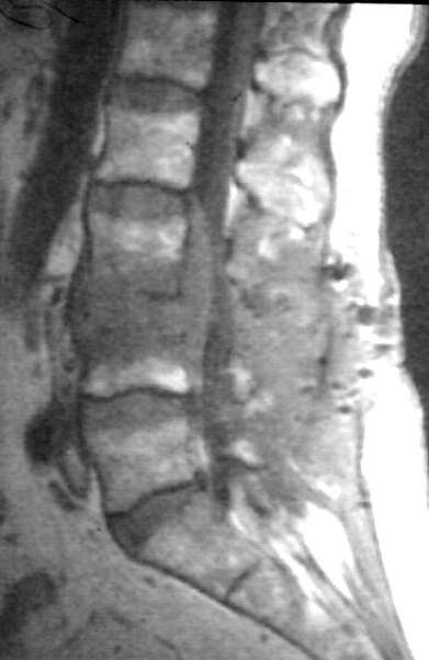
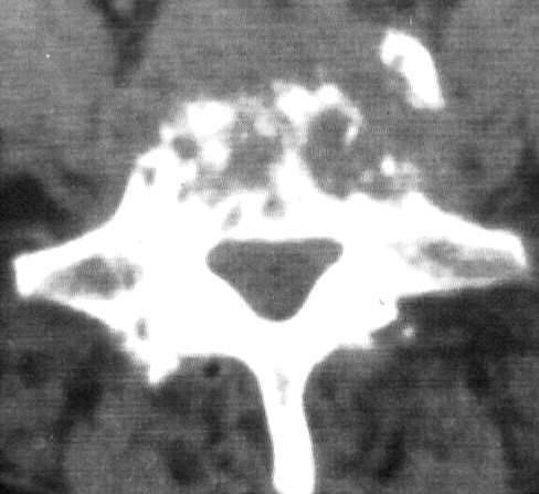
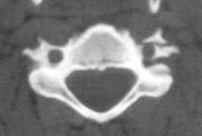
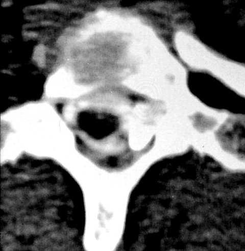
11 Developmental anomalies Generally complex (several layers and several levels involved) bones meninges spinal cord Diastematomyelia with block vertebrae


12 Vascular disorders Developmental malformations Acquired infarcts Spinal arterio-venous fistula

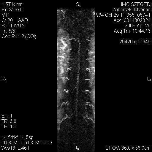
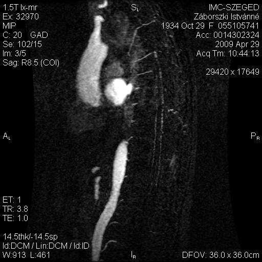
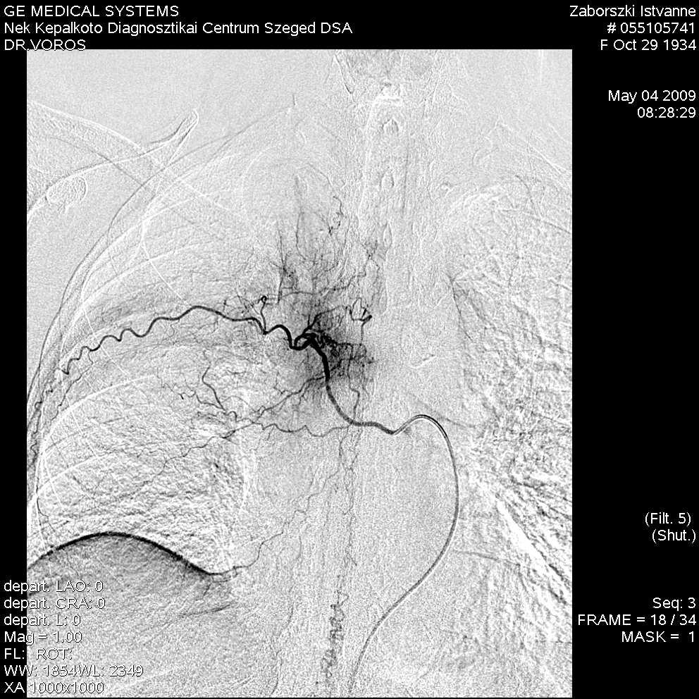
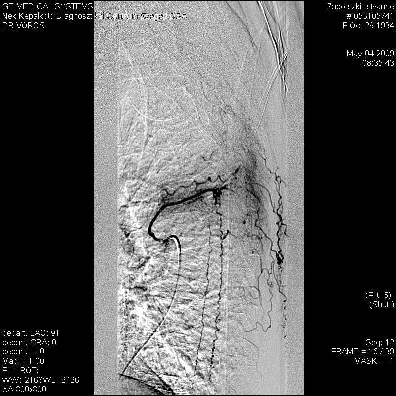

13 Vascular disorders Malformations Therapy planning Spinal dural arterio-venous fistula

 infarcts")
14 Vascular disorders Developmental malformations Acquired (stenoocclusive, thromboembolic) infarcts Ischaemic infarct

15 Inflammatory processes Spondylitis/spondylodiscitis Meningitis Myelitis Multiple sclerosis

16 Gyulladásos folyamatok Spondylitis/spondylodiscitis Meningitis/arachnoiditis Myelitis Sclerosis multiplex


17 Inflammatory processes Spondylitis/spondylodiscitis Meningitis Myelitis/radiculitis Guillain-Barré syndrome Multiple sclerosis

18 Inflammatory processes Spondylitis/spondylodiscitis Meningitis Myelitis Multiple sclerosis
19 Inflammatory processes Spondylitis/spondylodiscitis Meningitis Myelitis Multiple sclerosis
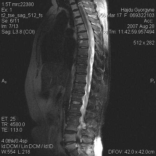
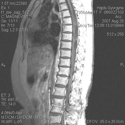
20 Degenerative changes Spondylarthrosis Disc herniations

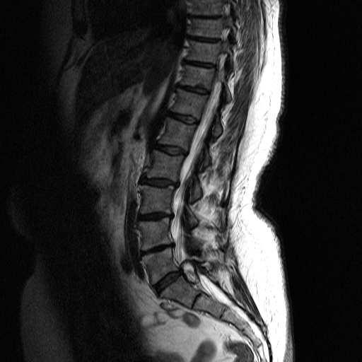
21 Degenerative changes Spondylarthrosis synovial cyst Disc hernia
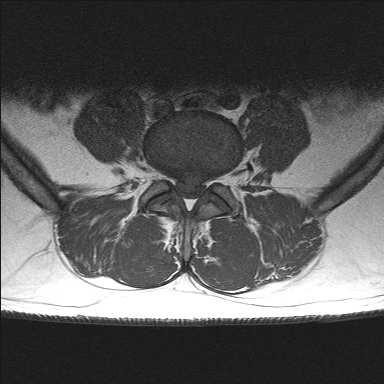
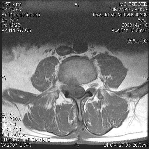
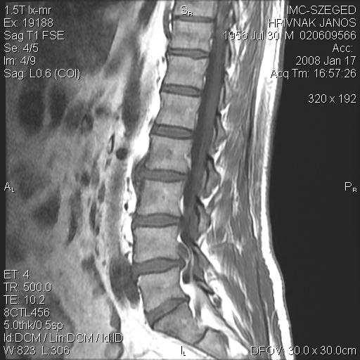
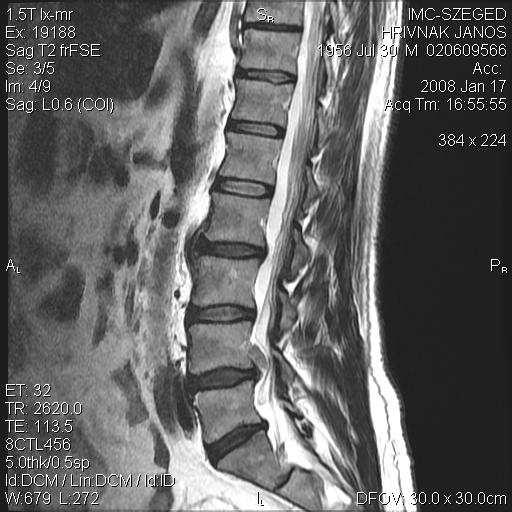
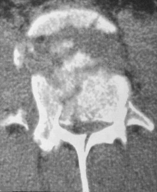
22 Degenerative changes Spondylarthrosis Disc herniations CT MR myelography

23 Degenerative changes Spondylarthrosis Disc herniations CT MR myelography


24 Degenerative changes Spondylarthrosis Disc herniations - locations medial paramedial lateral (foraminal and extraforaminal)


25 Degenerative changes Spondylarthrosis Disc herniations postoperative scar

26 Degenerative changes Spondylarthrosis Discus hernia postoperative scar recurrent/residual DH Preop. Postop.

27 Traumatic lesions Fractures/dislocations Haemorrhages Spinal cord injuries Dural tear/root avulsion


28 Traumatic lesions Fractures/dislocations Haemorrhages Spinal cord injuries Dural tear/root avulsion


29 Traumatic lesions Fractures/dislocations Haemorrhages Spinal cord injuries Dural tear/root avulsion


30 Traumás eltérések Törések / luxatiók Haemorrhagiák Gerincvelő/dura sérülések Cord herniation Dura/ gyöksérülés

31 Traumatic lesions Fractures/dislocations Haemorrhages Spinal cord injuries Dural tear/root avulsion


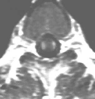
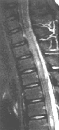
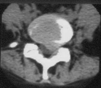
32 Tumours Extradural Bone metastasis Intradural - extramedullary meningiomas neurinomas Intramedullary astrocytomas ependymomas haemangioblastomas T2W T1W FLAIR


33 Tumours Extradural Other bone tumours Intradural - extramedullary meningiomas neurinomas Intramedullary astrocytomas ependymomas haemangioblastomas Myeloma


34 Tumours Extradural Intradural - extramedullary meningeoma neurinoma Intramedullary astrocytoma ependymoma haemangioblastoma Meningioma causing cord compression


35 Tumours Extradural bone tumours metastases Intradural - extramedullary meningiomas neurinomas Intramedullary astrocytomas ependymomas haemangioblastomas Dumbbell-shaped tumour


36 Tumours Extradural bone tumours metastases Intradural - extramedullary meningiomas neurinomas Intramedullary astrocytomas ependymomas haemangioblastomas Cystic intramedullary tumour


37 Tumours Extradural bone tumours metastases Intradural - extramedullary meningiomas neurinomas Intramedullary astrocytomas ependymomas haemangioblastomas Intramedullary tumour + syringohydromyelia
NEURORADIOLOGY. Part III. Angela Csomor University of Szeged Department of Radiology
 NEURORADIOLOGY Part III Angela Csomor University of Szeged Department of Radiology DISEASES OF SPINE AND SPINAL CORD I. Non-tumourous diseases developmental anomalies vascular disorders inflammatory processes
NEURORADIOLOGY Part I
 NEURORADIOLOGY Part I Vörös Erika University of Szeged Department of Radiology SZEGED DISEASES OF CNS BRAIN Developmental anomalies Cerebrovascular disorders Tumours Inflammatory diseases Trauma DISEASES
Neuroimaging. spine / spinal cord
 Neuroimaging spine / spinal cord Spine & spinal cord imaging methodology Plain x-ray of spine Computed tomography CT - traditional ( normal CT) - reconstructions - myelo-ct Magnetic resonance MR - standard
Introduction to Neuroimaging spine. John J. McCormick MD
 Introduction to Neuroimaging spine John J. McCormick MD Neuroanatomy Netter drawings Radiographic Anatomy Cervical Spine Cervical Spine Oblique View Cervical Spine Dens View Thoracic Spine Lumbar Spine
NEURORADIOLOGY Part I
 NEURORADIOLOGY Part I Vörös Erika University of Szeged Department of Radiology SZEGED BRAIN IMAGING METHODS Plain film radiography Ultrasonography (US) Computer tomography (CT) Magnetic resonance imaging
Spine. Neuroradiology. Spine. Spine Pathology. Distribution of fractures. Radiological algorithm. Role of radiology 18/11/2015
 Spine Neuroradiology Spine Prof.Dr.Nail Bulakbaşı X Ray: AP/L/Oblique Vertebra & disc spaces CT & CTA Vertebra, discs, vessels MRI & MRA Vertebra, disc, vessels, meninges Spinal cord & nerves Myelography
Pediatric Spinal Anomalies
 Department of Radiology University of California San Diego Pediatric Spinal Anomalies John R. Hesselink, M.D. Spine Embryogenesis 1. Primitive streak 2. Proliferation of cells at primitive pit (Hensen's
Malformations of the Nervous System November 10, Dr. Peter Ostrow
 Malformations of the Nervous System November 10, 2016 Dr. Peter Ostrow Malformations of the Nervous System 1. Abnormal closure of the neural tube 1. Disorders of forebrain formation 1. Cortical anomalies
Central Nervous System Congenital Abnormalities
 Central Nervous System Congenital Abnormalities Eva Brichtova, M.D., Ph.D., Department of Pediatric Sugery, Orthopaedics and Traumatology, University Hospital Brno Neural tube defects Dysraphism uncomplete
Imaging the Spinal Cord & Intradural Disease
 Department of Radiology University of California San Diego Imaging the Spinal Cord & Intradural Disease John R. Hesselink, M.D. Spinal Cord Diseases Tumors Syringohydromyelia Trauma Ischemia / Infarction
A Journey Down The Canal
 A Journey Down The Canal Radiological Assessment of Spinal Cord Masses John Berry-Candelario HMS III Gillian Lieberman, MD BIDMC Objectives Patient review Anatomy of the spine Imaging techniques Classification
Neuroanatomy. Assistant Professor of Anatomy Faculty of Medicine The University of Jordan Dr Maha ELBeltagy
 Neuroanatomy Dr. Maha ELBeltagy Assistant Professor of Anatomy Faculty of Medicine The University of Jordan 2018 Development of the Central Nervous System Development of the nervous system Development
Index. aneurysm, 92 carotid occlusion, 94 ICA stenosis, 95 intracranial, 92 MCA, 94
 A ADC. See Apparent diffusion coefficient (ADC) Aneurysm cerebral artery aneurysm, 93 CT scan, 93 gadolinium, 93 Angiography, 13 Anoxic brain injury, 25 Apparent diffusion coefficient (ADC), 7 Arachnoid
SPINAL MAGNETIC RESONANCE IMAGING INTERPRETATION
 CLINICAL VIGNETTE 2017; 3:2 SPINAL MAGNETIC RESONANCE IMAGING INTERPRETATION Editor-in-Chief: Idowu, Olufemi E. Neurological surgery Division, Department of Surgery, LASUCOM/LASUTH, Ikeja, Lagos, Nigeria.
SPLIT NOTOCHORD SYNDROME ASSOCIATION. DR. Hasan Nugud Consultant Paediatric Surgeon
 SPLIT NOTOCHORD SYNDROME ASSOCIATION DR. Hasan Nugud Consultant Paediatric Surgeon CASE PRESENTATION :- New born baby, boy, referred to the paediatric surgical team at the age of 14 hours. Birth History
In-Training Examination for Diagnostic Radiology Residents Rationales
 28th Annual In-Training Examination for Diagnostic Radiology Residents Rationales Sponsored by: Commission on Education Committee on Residency Training in Diagnostic Radiology February 3, 2005 The American
Adult - Cerebrovascular. Adult - Cranio-Cervical Junction. Adult - Epilepsy. Adult - Hydrocephalus
 list for SET and IMG Neurosurgery Adult - Cerebrovascular Aneurysm - Clipping: Anterior circulation Aneurysm - Clipping: Posterior circulation AVM excision Carotid endarterectomy Carotid trapping Cavernoma
Supplementary Online Content
 Supplementary Online Content Honein MA, Dawson AL, Petersen E, et al; US Zika Pregnancy Registry Collaboration. Birth Defects Among Fetuses and Infants of US Women With Laboratory Evidence of Possible
The Brain: Prenatal and Postnatal Effects of Congenital Heart Disease. Dianna M. E. Bardo, M D Swedish Cherry Hill Radia, Inc.
 The Brain: Prenatal and Postnatal Effects of Congenital Heart Disease Dianna M. E. Bardo, M D Swedish Cherry Hill Radia, Inc. Seattle, WA embryology We recognize the VACTERL association and frequency of
SPINAL CORD DISEASE IN DOGS PART TWO: MOST LIKELY CAUSES
 Vet Times The website for the veterinary profession https://www.vettimes.co.uk SPINAL CORD DISEASE IN DOGS PART TWO: MOST LIKELY CAUSES Author : RITA GONÇALVES Categories : Vets Date : April 7, 2014 RITA
CNS Embryology 5th Menstrual Week (Dorsal View)
") Imaging of the Fetal Brain; Normal & Abnormal Alfred Abuhamad, M.D. Eastern Virginia Medical School CNS Embryology 5th Menstrual Week (Dorsal View) Day 20 from fertilization Neural plate formed in ectoderm
Imaging of the Fetal Brain; Normal & Abnormal Alfred Abuhamad, M.D. Eastern Virginia Medical School CNS Embryology 5th Menstrual Week (Dorsal View) Day 20 from fertilization Neural plate formed in ectoderm
A Retrospective Analysis of Clinical Profile and Surgical Outcome in Patients with Spinal Dysraphism at Tertiary Care Center
 Original Research Article A Retrospective Analysis of Clinical Profile and Surgical Outcome in Patients with Spinal Dysraphism at Tertiary Care Center Premlal KV * Assistant Professor, Department of Neurosurgery,
Spinal canal stenosis Degenerative diseases F 06
 What is spinal canal stenosis? The condition known as spinal canal stenosis is a narrowing (stenosis) of the spinal canal that in most cases develops due to the degenerative (wear-induced) deformation
Prenatal Diagnosis of Central Nervous System (CNS) Pathologies: does Fetal MRI help in their management?
 Pathologies: does Fetal MRI help in their management?") Prenatal Diagnosis of Central Nervous System (CNS) Pathologies: does Fetal MRI help in their management? Daniela Prayer, Division of Neuroradiology and Musculoskeletal Radiology Medical University Vienna/Austria
Prenatal Diagnosis of Central Nervous System (CNS) Pathologies: does Fetal MRI help in their management? Daniela Prayer, Division of Neuroradiology and Musculoskeletal Radiology Medical University Vienna/Austria
Introduction to Neurosurgical Subspecialties:
 Introduction to Neurosurgical Subspecialties: Pediatric Neurosurgery Brian L. Hoh, MD 1 and Gregory J. Zipfel, MD 2 1 University of Florida, 2 Washington University Pediatric Neurosurgery Pediatric neurosurgeons
Spinal cord tumours Luc van den Hauwe et al.
 overview spinal cord tumours L. van den Hauwe 1,2, D. Balériaux 3, J.W. Van Goethem 2, C. Venstermans 2, F. De Belder 2, P.M. Parizel 2 introduction imaging spinal tumour classification spinal cord tumours
Spinal Imaging. Bearbeitet von Herwig Imhof. 1. Auflage Taschenbuch. 312 S. Paperback ISBN Format (B x L): 12,5 x 19 cm
: 12,5 x 19 cm") Spinal Imaging Bearbeitet von Herwig Imhof 1. Auflage 2007. Taschenbuch. 312 S. Paperback ISBN 978 3 13 144071 6 Format (B x L): 12,5 x 19 cm Weitere Fachgebiete > Medizin > Sonstige Medizinische Fachgebiete
Spinal Imaging Bearbeitet von Herwig Imhof 1. Auflage 2007. Taschenbuch. 312 S. Paperback ISBN 978 3 13 144071 6 Format (B x L): 12,5 x 19 cm Weitere Fachgebiete > Medizin > Sonstige Medizinische Fachgebiete
IMAGING OF A CASE OF SPINAL MENINGIOMA- A CASE REPORT
 IMAGING OF A CASE OF SPINAL MENINGIOMA- A CASE REPORT Ramneet Wadi 1, Anil Kumar Shukla 2, Seetha Pramila V. V 3, Sabyasachi Basu 4, Sonam Sanjay 5 1Postgraduate Student, Department of Radiodiagnosis,
Spinal Vascular Lesions
 Spinal Vascular Lesions Spinal Vascular Lesions Spinal cord infarction Hemangioblastoma Cavernous malformation Vascular malformations (Type 1-4) Spinal artery aneurysm Troy Hutchins, MD Assistant Professor
Pediatric Spine Tumors (and other masses)
") Pediatric Spine Tumors (and other masses) Francisco A Perez, MD, PhD Assistant Professor Neuroradiology and Pediatric Radiology Seattle Children s Hospital University of Washington, Seattle Commercial
Pediatric Spine Tumors (and other masses) Francisco A Perez, MD, PhD Assistant Professor Neuroradiology and Pediatric Radiology Seattle Children s Hospital University of Washington, Seattle Commercial
Spine Surgery: Techniques, Complication Avoidance, and Management. 2 Volume Set
 Spine Surgery: Techniques, Complication Avoidance, and Management. 2 Volume Set Benzel, E ISBN-13: 9781437705874 Table of Contents SECTION 1 - HISTORY 1 - History 2 - History of Spine Instrumentation -
Intracranial hypotension secondary to spinal CSF leak: diagnosis
 Intracranial hypotension secondary to spinal CSF leak: diagnosis Spinal cerebrospinal fluid (CSF) leak is an important and underdiagnosed cause of new onset headache that is treatable. Cerebrospinal fluid
Spinal meningioma imaging
 Spinal meningioma imaging Poster No.: C-0448 Congress: ECR 2018 Type: Educational Exhibit Authors: M. Smoljan, D. Zadravec ; Zagreb/HR, Zageb/HR Keywords: Neoplasia, Imaging sequences, Education, MR, CT,
The surgical treatment of metastatic disease of the spine
 The surgical treatment of metastatic disease of the spine Péter Banczerowski National Institute of Neurosurgery, Budapest Spine tumours 15% of the primary tumours of the CNS affect the spine The spine
EANS Training Course Edinburgh, 28 th - 31 st January 2018 Spine and Peripheral Nerves
 EANS Training Course Edinburgh, 28 th - 31 st January 2018 Spine and Peripheral Nerves SUNDAY 28 th January 2018 09:00-09:10 Welcome and introduction 09:10-10:20 Basics Chair: Smrcka 09:10-09:25 Anatomical
A Case of Naso-Ethmoidal Meningoencephalocele
 A Case of Naso-Ethmoidal Meningoencephalocele Divyanshu Dubey, Sonjjay Pande, Pradeep Dubey, Anshudha Sawhney Vol. 3 No. 8 (August 2011) International Journal of Collaborative Research on Internal Medicine
Giant thoracolumbar extradural arachnoid cyst : A case report
 Acta Orthop. Belg., 2008, 74, 709-713 CASE REPORT Giant thoracolumbar extradural arachnoid cyst : A case report Muddassir RASHID, Anjum SYED, Ibne AHMAD, EKRAMULLAH From the Jawaharlal Nehru Medical College
Developmental Neuropathology
 Developmental Neuropathology Pathology, Radiology, and Clinical Correlations Reid Heffner MD Distinguished Teaching Professor Department of Pathology and Anatomy I HAVE NO CONFLICTS OF INTEREST OR DISCLOSURES
Spinal Neoplasms. First Things First!! Localize the Lesion!! Ependymomas. Common Intramedullary Lesions
 Acta Radiológica Portuguesa, Vol.XXIII, nº 90, pág. 101-114, Abr.-Jun., 2011 Spinal Neoplasms Bruno A Policeni University of Iowa Hospitals and Clinics Assistant Professor of Radiology Disclosure of Commercial
Symposium: OB/GY US (Room B) CNS Anomalies
 CNS Anomalies") 82 Symposium: OB/GY US (Room B) 11 : 50 1 2 : 10 CNS Anomalies Brain area Midline structure S u p r a t e n t o r i a l ventricular system Cerebral hemisphere Posterior fossa Head size and shape Image
82 Symposium: OB/GY US (Room B) 11 : 50 1 2 : 10 CNS Anomalies Brain area Midline structure S u p r a t e n t o r i a l ventricular system Cerebral hemisphere Posterior fossa Head size and shape Image
Epidemiology of Low back pain
 Low Back Pain Definition Pain felt in your lower back may come from the spine, muscles, nerves, or other structures in that region. It may also radiate from other areas like the mid or upper back, a inguinal
A Very Unusual Case of a Dorsal Heteropagus Twin
 PRG A Very Unusual Case of a Dorsal Heteropagus Twin Nathan David P. Concepcion, MD 1, Bernard F. Laya, DO 1, Eduardo P. Manrique, MD 2 and Faith Caroline D. Bayabos, MD 1 1 Section of Pediatric Radiology,
AMERICAN ACADEMY OF NEUROLOGY SPINE FELLOWSHIP CORE CURRICULUM
 AMERICAN ACADEMY OF NEUROLOGY SPINE FELLOWSHIP CORE CURRICULUM Introduction Spine conditions affect virtually everyone at some time during their life. Surveys indicate a yearly prevalence of spine-related
Neuroradiology. J.Lisý
 Neuroradiology J.Lisý X-ray of skull/spine trauma (2 perpendicular projections) congenital developemental errors (scoliosis, spina bifida) Perimyelography (PMG) Lumbar puncture, isoosmolar iodine CM Dural
Prenatal Prediction of The Neurologically Impaired Neonate By Ultrasound
 Prenatal Prediction of The Neurologically Impaired Neonate By Ultrasound Robert H. Debbs, D.O.,F.A.C.O.O.G. Professor of OB-GYN Perelman School of Medicine, University of Pennsylvania Director, Pennsylvania
Role of Magnetic Resonance Imaging in the Evaluation of Compressive Myelopathy in Rohilkhand Region, India
 Mohit Agarwal et al Original article 10.5005/jp-journals-10050-10091 Role of Magnetic Resonance Imaging in the Evaluation of Compressive Myelopathy in Rohilkhand Region, India 1 Mohit Agarwal, 2 Pramod
Treatment of cervicodorsalepidermiod cyst
 Treatment of cervicodorsalepidermiod cyst Ayied Motteb Turkey Department of Medicine, College of medicine, University of Tikret, Tekrit, Iraq Received 22 / 9/2011 Accepted 22/11/2011 Abstract Intramedullary
Neuroimaging Core Curriculum
 Neuroimaging Core Curriculum Program Content The purpose of the training program is to prepare the physician for the independent practice of neuroimaging. Neuroimaging is the subspecialty of Neurology
Brain Imaging. Bearbeitet von Klaus Sartor, Stefan Hähnel, Bodo Kress
 Brain Imaging Bearbeitet von Klaus Sartor, Stefan Hähnel, Bodo Kress 1. Auflage 2007. Taschenbuch. 312 S. Paperback ISBN 978 3 13 143961 1 Format (B x L): 12,5 x 19 cm Weitere Fachgebiete > Medizin > Sonstige
Development of the Nervous System. Leah Militello, class of 2018
 Development of the Nervous System Leah Militello, class of 2018 Learning Objectives 1. Describe the formation and fate of the neural tube and neural crest including timing and germ layer involved. 2. Describe
Ibtisam Nasir Ahmed. MBChB. DMRD. Specialist Radiological Diagnosis. Al-sadr Teaching Hospital. Basrah-Iraq.
 THE VALUE OF CORONAL IMAGE IN DETECTING EXTRA SPINAL LESION IN MAGNETIC RESONANCE IMAGING OF THE SPINE Ibtisam Nasir Ahmed. MBChB. DMRD. Specialist Radiological Diagnosis. Al-sadr Teaching Hospital. Basrah-Iraq.
Clinico-Mri Correlation of Compressive Myelopathy (Retrospective Study)
") 2015; 1(7): 60-64 ISSN Print: 2394-7500 ISSN Online: 2394-5869 Impact Factor: 5.2 IJAR 2015; 1(7): 60-64 www.allresearchjournal.com Received: 18-04-2015 Accepted: 16-05-2015 Sreeramulu Diguvinti Associate
2015; 1(7): 60-64 ISSN Print: 2394-7500 ISSN Online: 2394-5869 Impact Factor: 5.2 IJAR 2015; 1(7): 60-64 www.allresearchjournal.com Received: 18-04-2015 Accepted: 16-05-2015 Sreeramulu Diguvinti Associate
Overview. Spinal Anatomy Spaces & Meninges Spinal Cord. Anatomy of the dura. Anatomy of the arachnoid. Anatomy of the spinal meninges
 European Course in Neuroradiology Module 1 - Anatomy and Embryology Dubrovnik, October 2018 Spinal Anatomy Spaces & Meninges Spinal Cord Johan Van Goethem Overview spinal meninges & spaces spinal cord
Symptomatic Multiple Level Lateral Meningoceles with Intraspinal Meningocele: A Case Study and Its Surgical Management
 THIEME Original Article 15 Symptomatic Multiple Level Lateral Meningoceles with Intraspinal Meningocele: A Case Study and Its Surgical Management Vernon Velho 1 Sachin Guthe 1 Pravin Survashe 1 Poonam
Index. F FLAIR sequence, 9 Flow diversion, aneurysms, Fluoroscopy, epilepsy, 193 Fractures, Frontal lobe tumors, 31
 A Acromegaly, 40 Adjuvant therapy chemotherapy, 15 malignant brain tumors, 34 radiation, 14 15 Adrenocorticotropic hormone (ACTH), 40 Allograft, 176 Aneurysms bypass surgery, 98 100 compression symptoms,
Ligaments of the vertebral column:
 In the last lecture we started talking about the joints in the vertebral column, and we said that there are two types of joints between adjacent vertebrae: 1. Between the bodies of the vertebrae; which
Dumbbell Shaped Thoracic Spine Cavernous Hemangioma: A Case Report and Review of the Literature
 ISPUB.COM The Internet Journal of Neurosurgery Volume 3 Number 1 Dumbbell Shaped Thoracic Spine Cavernous Hemangioma: A Case Report and Review of the Literature J Gonzalez-Cruz, A Nanda Citation J Gonzalez-Cruz,
Accuracy of intraoperative frozen section diagnosis in spinal cord lesions
 Accuracy of intraoperative frozen section diagnosis in spinal cord lesions Department of Orthopedic Surgery Niigata University Medical and Dental Hospital Toru Hirano, Kei Watanabe, Keiichi Katsumi, Masayuki
ESSENTIALS OF PLAIN FILM INTERPRETATION: SPINE DR ASIF SAIFUDDIN
 ESSENTIALS OF PLAIN FILM INTERPRETATION: SPINE DR ASIF SAIFUDDIN Consultant Musculoskeletal Radiologist Royal National Orthopaedic Hospital Stanmore,UK. INTRODUCTION 2 INTRODUCTION 3 INTRODUCTION Spinal
Fetal Medicine. Case Presentations. Dr Ermos Nicolaou Fetal Medicine Unit Chris Hani Baragwanath Hospital. October 2003
 Case Presentations Dr Ermos Nicolaou Fetal Medicine Unit Chris Hani Baragwanath Hospital October 2003 Case 1 Ms A M 22year old P0 G1 Referred from Sebokeng Hospital at 36w for polyhydramnios On Ultrasound:
The neurvous system senses, interprets, and responds to changes in the environment. Two types of cells makes this possible:
 NERVOUS SYSTEM The neurvous system senses, interprets, and responds to changes in the environment. Two types of cells makes this possible: the neuron and the supporting cells ("glial cells"). Neuron Neurons
Case SCIWORA in patient with congenital block vertebra
 Case 15428 SCIWORA in patient with congenital block vertebra Lucas Walgrave 1, Charlotte Vanhoenacker 1-2, Thomas Golinvaux 3, Filip Vanhoenacker3-5 1: Leuven University Hospital, Department of Radiology,
Sonography of the Neonatal Spine: Part 2, Spinal Disorders
 Neonatal Spine Sonography Pediatric Imaging Pictorial Essay Downloaded from www.ajronline.org by 148.251.232.83 on 04/11/18 from IP address 148.251.232.83. Copyright RRS. For personal use only; all rights
Radiography of the Spine
 Radiography of the Spine Radiography of the Spine Attila ARANY-TóTH, DVM Complex anatomy Vertebrae: 7 cervical, 13 thoracal, 7 lumbal, 3 sacral, n caudal Thorough neurological examination - localization!!!
1/9/2013 EXTRAMEDULLARY TUMORS OF THE PEDIATRIC SPINE. Introduction. Classification for Extramedullary Tumors
 EXTRAMEDULLARY TUMORS OF THE PEDIATRIC SPINE Eugene Wang 1/20/12 Dent Neurologic Institute Introduction 2/3 of all intraspinal tumors of childhood are extramedullary 50% Extradural 10-15% Intradural Back
Essentials of Clinical MR, 2 nd edition. 51. Primary Neoplasms
 51. Primary Neoplasms As with spinal central canal neoplasms in other regions, those of the lumbar spine may be classified as extradural, intradural extramedullary, and medullary. If an extradural lesion
Ependymoma of the spine
 Ependymoma of the spine Tenny Zhang, MS-3 Harvard Medical School 1 Case presentation: history and exam HPI: A 30-year-old man with no significant past medical history presents with one week of bilateral
Department of Cognitive Science UCSD
 Department of Cognitive Science UCSD Verse 1: Neocortex, frontal lobe, Brain stem, brain stem, Hippocampus, neural node, Right hemisphere, Pons and cortex visual, Brain stem, brain stem, Sylvian fissure,
HEAD/NECK VESSELS. Objectives
 Objectives Arterial Supply to Head and Neck Arteries to Head Surrounding Brain Common carotid arteries Arteries to Head Surrounding Brain External carotid arteries Arteries to Head Surrounding Brain External
BRAIN STEM AND CEREBELLUM..
 Lecture Title: BRAIN STEM AND CEREBELLUM.. (CNS Block, Radiology) Dr. Hamdy Hassan Ass.Prof. Consultant Radiology Department KKHU King Saud University Lecture Objectives.. Students at the end of the lecture
22110 vertebral segment; cervical vertebral segment; thoracic vertebral segment; lumbar
 The following codes are authorized by Palladian Health for applicable product lines. Visit palladianhealth.com to request authorization and to access guidelines. Palladian Musculoskeletal Program Codes
ORIGINAL ARTICLE. Abstract. Aim. Materials and methods. Introduction. Results
 Is anatomical distribution helpful for differentiating TB spondylitis from neoplastic causes of extradural spinal cord compression in children? A pilot study Reena George, MD, MMed Rad, FRCR (UK) Savvas
Announcement. Danny to schedule a time if you are interested.
 Announcement If you need more experiments to participate in, contact Danny Sanchez (dsanchez@ucsd.edu) make sure to tell him that you are from LIGN171, so he will let me know about your credit (1 point).
Development of Brain Stem, Cerebellum and Cerebrum
 Development of Brain Stem, Cerebellum and Cerebrum The neural tube cranial to the 4th pair of somites develop into the brain. 3 dilatations and 2 flexures form at the cephalic end of the neural tube during
Role of helical CT and MRI in the evaluation of spinal dysraphism
 International Journal of Advances in Medicine Kumaran SK et al. Int J Adv Med. 2017 Feb;4(1):124-132 http://www.ijmedicine.com pissn 2349-3925 eissn 2349-3933 Original Research Article DOI: http://dx.doi.org/10.18203/2349-3933.ijam20170095
Johnson Rogers and colleagues- used Term LMM
 LIPOMENINGOMYELOCELE: CLASSIFICATION, MANAGEMENT AND CONTROVERSIES Definition : Lipomyelomeningocele is a form of OSD in which a subcutaneous fibrofatty mass traverses the lumbodorsal fascia, causes a
Two Consecutive Levels of Unilateral Cervical Spondylolysis on Opposite Sides 두개의연속된척추에서반대쪽편측에발생한경추척추분리증
 Case Report pissn 1738-2637 / eissn 2288-2928 http://dx.doi.org/10.3348/jksr.2015.73.3.181 Two Consecutive Levels of Unilateral Cervical Spondylolysis on Opposite Sides 두개의연속된척추에서반대쪽편측에발생한경추척추분리증 Kyeong
Long segment composite split cord malformation with double bony spur
 Long segment composite split cord malformation with double bony spur Anand Sharma, Achal Sharma, R.S. Mittal SMS Medical College, Jaipur, India Abstract: A composite type of SCM is very rare and only a
Fetal CNS MRI. Daniela Prayer. Division of Neuroradiology And Musculoskeletal Radiology. Medical University of Vienna, AUSTRIA
 Fetal CNS MRI Daniela Prayer Division of Neuroradiology And Musculoskeletal Radiology Medical University of Vienna, AUSTRIA Methods Normal development Malformations Acquired pathology MR- methods for assessment
CNS Imaging. Dr Amir Monir, MD. Lecturer of radiodiagnosis.
 CNS Imaging Dr Amir Monir, MD Lecturer of radiodiagnosis www.dramir.net Types of radiological examinations you know Plain X ray X ray with contrast GIT : barium (swallow, meal, follow through, enema) ERCP
Vascular Pattern in Tumours
 Acta Radiologica ISSN: 0001-6926 (Print) (Online) Journal homepage: https://www.tandfonline.com/loi/iaro20 Vascular Pattern in Tumours To cite this article: (1957) Vascular Pattern in Tumours, Acta Radiologica,
Student Lab #: Date. Lab: Gross Anatomy of Brain Sheep Brain Dissection Organ System: Nervous Subdivision: CNS (Central Nervous System)
") Lab: Gross Anatomy of Brain Sheep Brain Dissection Organ System: Nervous Subdivision: CNS (Central Nervous System) Student Lab #: Date 1 Objectives: 1. Learn the main components making up a motor neuron.
Lab: Gross Anatomy of Brain Sheep Brain Dissection Organ System: Nervous Subdivision: CNS (Central Nervous System) Student Lab #: Date 1 Objectives: 1. Learn the main components making up a motor neuron.
 Thermographic findings of spinal cord tumors by Ho Yeol Zhang Neurosurgery, National Health Insurance Service Ilsan Hospital, Yonsei University College of Medicine, Korea, hoyeolzhang@gmail.com Abstract
Neuroembryology II. Dr. Newton COPH G210
 Neuroembryology II Dr. Newton COPH G210 Anterior and posterior neuropore closure at E25 & E27, respectively, is essential for normal nervous system development. NTDs occur 1/1K births. Incidence can be
Chiari III Joseph Junewick, MD FACR
 Chiari III Joseph Junewick, MD FACR 07/02/2010 History Newborn with suboccipital mass. Diagnosis Chiari III Additional Clinical Surgery-Skin covered suboccipital cystic mass confined by the dura. Pathology-Leptomeningeal
Cervical spondylarthrotic myelopathy with early onset in Down's syndrome: five cases and a review of the literature
 283 Journal of Intellectual Disability Research VOLUME 43 PART 4 pp 283±288 AUGUST 1999 Cervical spondylarthrotic myelopathy with early onset in Down's syndrome: five cases and a review of the literature
Focal Anterior Displacement of the Thoracic Spinal Cord without Evidence of Spinal Cord Herniation or an Intradural Mass
 Original Article Musculoskeletal Imaging http://dx.doi.org/10.3348/kjr.2014.15.6.733 pissn 1229-6929 eissn 2005-8330 Korean J Radiol 2014;15(6):733-738 Focal Anterior Displacement of the Thoracic Spinal
Wound healing in trophic ulcers in spina bifida patients
 J Neurosurg 82:000 000, 1995 Wound healing in trophic ulcers in spina bifida patients VINOD KUMAR SRIVASTAVA, M.B.B.S, M.CH. Neurosurgical Unit, J. N. Medical College, Aligarh Muslim University, Aligarh,
Role of MRI in the Evaluation of Compressive Myelopathy
 IOSR Journal of Dental and Medical Sciences (IOSR-JDMS) e-issn: 2279-0853, p-issn: 2279-0861.Volume 15, Issue 4 Ver. XIII (Apr. 2016), PP 21-26 www.iosrjournals.org Role of MRI in the Evaluation of Compressive
Cutoff.Guru. Radiology Sample papers. 1.The following features are true for Tetralogy of Fallot, except: a. Ventricular septal defect
 Radiology Sample papers 1.The following features are true for Tetralogy of Fallot, except: a. Ventricular septal defect b. Right ventricular hypertrophy c. Atrial septal defect d. Pulmonary stenosis. 2.
Module 1: Basic Comprehensive Course
 The Hellenic Spine Society organize 5 modules according to the following program, which is based on the Eurospine program Module 1: Basic Comprehensive Course SESSION1: SPINE THE BIGGER PICTURE Evidence
Central nervous system
 Central nervous system By Dr. Mohsen Dashti Clinical Medicine & Pathology 316 7 th Lecture Lecture outline Review of structure & function. Symptoms, signs & tests. Specific diseases. Review of structure
Spinal ultrasound is considered medically necessary for ANY of the following indications:
 Medical Coverage Policy Effective Date...11/15/2017 Next Review Date...11/15/2018 Coverage Policy Number... 0246 Spinal Ultrasound Table of Contents Coverage Policy... 1 Overview... 1 General Background...
Central Nervous System Practical Exam. Chapter 12 Nervous System Cells. 1. Please identify the flagged structure.
 Central Nervous System Practical Exam Chapter 12 Nervous System Cells 1. Please identify the flagged structure. 2. Please identify the flagged structure. 3. Please identify the flagged structure. 4. A
CT Myelography and MR Imaging of Extramedullary Cysts of the Spinal Canal in Adult and Pediatric Patients
 Downloaded from www.ajronline.org by 37.44.199.154 on 12/23/17 from IP address 37.44.199.154. opyright RRS. For personal use only; all rights reserved T Myelography and MR Imaging of Extramedullary ysts
The central nervous system
 Sectc.qxd 29/06/99 09:42 Page 81 Section C The central nervous system CNS haemorrhage Subarachnoid haemorrhage Cerebral infarction Brain atrophy Ring enhancing lesions MRI of the pituitary Multiple sclerosis
Chapter 12b. Overview
 Chapter 12b Spinal Cord Overview Spinal cord gross anatomy Spinal meninges Sectional anatomy Sensory pathways Motor pathways Spinal cord pathologies 1 The Adult Spinal Cord About 18 inches (45 cm) long
Introduction and Basic structural organization of the nervous system
 Introduction and Basic structural organization of the nervous system **the slides are in bold and the book is in red Done by : razan krishan & marah marahleh INTRODUCTION The nervous system, along with
CNS Developmental. Anke van Eekelen, PhD. Telethon Institute for Child Health Research
 CNS Developmental Anke van Eekelen, PhD Telethon Institute for Child Health Research (Some slides are modified versions of Prof. Alan Harvey s Neuroscience lecture at ANHB and Dr. Joanne Britto s Dev Neuroscience