PET/CT in Paediatric Oncology Hybrid Imaging PET/CT & PET MRI
|
|
|
- Linda Washington
- 6 years ago
- Views:
Transcription


1 PET/CT in Paediatric Oncology Hybrid Imaging PET/CT & PET MRI Robert Howman-Giles MD FRACP FAANMS DDU The Children s Hospital at Westmead Department of Nuclear Medicine Westmead, Sydney, Australia Westmead Hospital Department of Nuclear Medicine, PET and Ultrasound Westmead, Sydney, Australia Discipline of Paediatrics and Child Health Discipline of Medical Imaging, Sydney Medical School University of Sydney ipet 2015 Vienna

2 The Children s Hospital at Westmead (Royal Alexandra Hospital for Children) Bed capacity: 340 Inpatients 2014: 31,833 Outpatients 2014: 734,030 Tertiary/Quaternary hospital Dept Nuclear Medicine 4203 studies NM, PET/CT, BMD, body composition Therapy: I131, MIBG

3 PET/CT June 2006 Dec 2014: >4,500 PET/CT studies 80% Oncology, 15% neurology / neuro-oncology, 5% Infection / PUO Siemens PET mct 128 Other adrenocorticocarcinoma, NPC, Germ Cell tumours, neuroendocrine tumours, melanoma, thyroid carcinoma, hepatoblastoma, HCC, LCH, NF1, PTLD etc.

4 Hybrid Imaging in Paediatric Oncology PET/CT & PET/MR or PET/CT & MR? Lymphoma Sarcoma bone, soft tissue Neurofibromatosis type 1 Neuroendocrine
5 PET/CT Oncology 2015 Common Paediatric Malignancy PET/CT Standard of Diagnostic Care Stage Response to Treatment Detection residual & recurrent cancer End of treatment stage Surveillance
6 PET/CT Oncology 2015 Definite role in management Lymphoma Hodgkins lymphoma Non-Hodgkins lymphoma Sarcoma Primary bone Soft tissue Rhabdomyosarcoma 55% Non rhabdomyosarcoma 45%
7 PET/CT Oncology 2015 Less well defined role Other & rare solid tumours Malignant brain tumours Neuroblastoma Hepatoblastoma, hepatocellular carcinoma Langerhan Cell Histiocytosis Germ Cell Tumours Malignant peripheral nerve sheath tumours Neuroendocrine Epithelial Neoplasms & Melanoma Adrenocortical Nasopharyngeal

8 PET, CT & MRI Technologies are rapidly changing and improving MRI: DWI, fmri and MRSI: anatomy, pathology, bio-physiology and chemistry PET/CT: faster, less radiation, more specific radiopharmaceuticals- biomarkers, metabolism, theranostics PET/MRI or PET/CT & MRI?

9 Journal of Magnetic Resonance Shah et al 229 (2013) Advances in multimodal neuroimaging: Hybrid MR PET and MR PET EEG at 3 T and 9.4T Strengths of modalities
 39:1745")
10 Diagnostic value of combined 18F-FDG PET & MRI for staging and restaging in paediatric oncology Pfluger et al Ludwig Maximilians University of Munich Eur J Nucl Med Mol Imaging (2012) 39:
11 MRI issues Primary Dx: Identifying viable bone/bone marrow tumour lesions due to limited size and/or soft tissue changes e.g. oedema False-positive findings - nonspecific inflammatory lymph node enlargement or infectious tissue changes Follow-up: MRI low specificity misinterpretation of posttherapeutic changes. o bone marrow appearance in children varies with age, bone marrow oedema, necrotic tissue, contrast enhancement in successfully treated lesions, o o difficulties in distinguishing between active tumour tissue and residual mass (necrosis or fibrosis on morphological imaging). lymph node enlargement without active tumour was regularly seen on a posttherapeutic MRI scan.

12 PET- Gold standard for Molecular Imaging F 18 Glucose-FDG Amino acids-fet, DOPA Cell hypoxia-f-miso Cell proliferation-flt Choline-CHT Ga 68 SSTR- Dotatate C 11 Cu 64 Lopci et al. EJNMMI 2015 DOI /s
13 Lymphoma & FDG PET/CT Children and Adolescents Standard of Diagnostic Care Valuable technique high sensitivity & specificity Essential for staging & impacts on management FDG PET/CT CI Sensitivity 95.9% 70.1% Specificity 99.7% 96.9% PET/CT correct modality in 86% discordant lesions PET/CT better specificity for predicting poor response London et al. 18F-FDG PET/CT in pediatric lymphoma : comparison with conventional imaging EJNMMI 2011;38:


14 13 yr F: Presented with painful R tibia? Primary bone malignancy Biopsied? Bx:Hodgkins Disease Stage IVB GG


15 Stage IVB Rx BEACOPP GG
16 Response J Clin Oncol 32: Cheson et al J Clin Oncol 32: Barrington et al

17 Completion of therapy march 2010 Complete response Note: abnormal bone on CT but not metabolic. Remains in remission May 2014 GG
18 Lymphoma-CI & MRI WBMR-high sensitivity 96% WBMR with DWI- helps in nodal disease and extranodal involvement Gu et al AJR 2011;197:W384-W391 Krohmer S et al Eur J Radiol 2010; Punwani et al. Radiology 255(1): , 2010 Good correlation PET/CT and WBMR (STIR & RARE imaging)
19 Paediatric HD Surveillance & Relapse Texas Childrens Hospital, Baylor College of Medicine, Texas Ped Hem Oncol 29: , 2012 Rathore N et al. 13/99 (13%) relapse 2-17 mths post Tx 11 (85%) within 12 mths (mean 5 mths) 2 pts at 16 and 17 mths post Tx The Children Hospital at Westmead, Sydney June 2006-June 2013 Patients: 64 PET/CT scans av 8.4 Relapse: 7.8% 5/64 <12 mths
20 NHL Highly proliferative malignant cells 85-99% Cure rates depends on pathological subtype and tumour stage 70-90% Histopathology 1. Burkitt or B cell Acute Lymphoblastic leukemia 40-50% 2. Lymphoblastic lymphoma (80% T cell 20% B cell) 20-25% 3. Anaplastic Large Cell Lymphoma (T cell or null cell) 10-15% 4. Diffuse Large B Cell Lymphoma 10% 5. Primary Mediastinal B cell lymphoma rare 6. Follicular Lymphoma rare

 jaw (endemic) head and neck (nonjaw) (sporadic) A Nodal, abdomen, bone, primary CNS, mediastinal t(8;14)(q24 q32), t(2;8) (p11;q24),")
(p23; q35) ALK, NMP E Anaplastic large cell lymphoma, cutaneous CD30+ (Ki-usually) Skin only; single or multiple lesions Lacks t(2;5) A B C D E All Types")
21 Major Histopath Categories of Non-Hodgkin s Lymphoma in Children and Adolescents Category WHO Classification/ Updated REAL Category (Working Formulation) Immuno-phenotype Clinical Presentation Chromosome Translocation Genes Affected A Lymphoblastic lymphoma, Precursor T/leukemia Lymphoblastic convoluted and nonconvoluted T cell Pre-B-cell Mediastinal, bone marrow Skin, bone MTS1/p16ink4a deletion TAL1 t(1;14)(p34; q11), t(11;14) (p13;q11) TAL1 TCRao*** RHOMB1, HOX11 B C Burkitt s and Burkitt s like lymphomas Diffuse large B-cell lymphoma ML small noncleaved cell ML large cell Mature B cell Mature B cell; maybe CD30+ Intra-abdominal (sporadic) jaw (endemic) head and neck (nonjaw) (sporadic) A Nodal, abdomen, bone, primary CNS, mediastinal t(8;14)(q24 q32), t(2;8) (p11;q24), t(8;22)(q24; q11) Not well characterized in children c-myc, IgH, IgK, Ig1** D Anaplastic large cell lymphoma, systemic ML immuno-blastic or ML large CD30+ (Ki-1+) T cell or null cell Variable, but systemic symptoms often prominent t(2;5)(p23; q35) ALK, NMP E Anaplastic large cell lymphoma, cutaneous CD30+ (Ki-usually) Skin only; single or multiple lesions Lacks t(2;5) A B C D E All Types Positive 18 F-FDG Considered high grade NHL NCI 2004
22 Stage & Response assessment 7 yr F Anaplastic Large Cell NHL STAGE III Very Good Response after 2 cycles chemotherapy Minimal activity in right axilla Increased bone marrow activity - Chemotherapy - G-CSF JV
:2106-2111,")
23 J Clin Oncology 33(18): , 2015

24 Sarcomas: Bone and Soft Tissue MRI preferred modality for Stage and extent of primary tumour directs protocols, surgery, radiotherapy Response primary +/- other sites residual mass? viable Post induction pre surgery Surveillance Chest CT- pulmonary metastases PET/CT Staging- extent of distant disease Response End of treatment Surveillance


25 15yr male pain left leg Bx Osteogenic sarcoma STIR T1FS SUV max 7.4 Staging: Pulmonary, bone and bone marrow metastases MF538631


26 STIR MRI A. 12 yr M pain in Left femur. Abn X-ray Bx:High grade osteogenic sarcoma B. 11yr M Pain proximal R tibia Bx:Osteoblastic osteogenic sarcoma *Growth plate Involvement?

27 PET/CT MIP A B SUV max 10.6 SUV max 17.8


28 Response 2 cycles chemotherapy LL A RL B
 Surgery: 25% viable cells Mx changes: more aggressive")

29 Response to Chemotherapy A Poor responder SUV max (42%) Surgery: 25% viable cells Mx changes: more aggressive chemotherapy SUV max 10.6 SUV max 6.1 B Very good response SUV max (88%) Surgical Resection: Histopath: no viable cells SUV max 17.8 SUV max 2.1
30 Response MRI Residual mass? Viable malignant tissue DWI or contrast enhancement may useful for tumour necrosis Uhl et al Invest Radiol 2006;41: Bone marrow Age differences, hyperactive stimulated marrow G-CSF Oedema and tumour infiltration
31 Relative reduction in SUVmax rather than the absolute value SUVmax following chemotherapy may be a stronger predictor of tumor response. Hamada K, Tomita Y, Inoue A et al. Evaluation of chemotherapy response in osteosarcoma with FDG- PET. Ann Nucl Med 23:89 95, 2009 (Osaka Uni) London K, Stege C, Cross S, Onikul E, Graf N, Kaspers G, Dalla-Pozza L, Howman-Giles R. 18F-FDG PET/CT compared to Conventional Imaging modalities in Pediatric Primary Bone Tumors. Pediatr Radiol 42(4):418-30,2012 PET/CT CI (inc regional MR) Sensitivity 98% 83% Specificity 97% 78%

32 Soft Tissue Sarcomas Histopathologically classified according to tissue they resemble. Rhabdomyosarcoma 55% Other 45%
33 Diagnosis & Staging MRI: preferred modality at diagnosis, extent of local involvement CT- pulmonary metastases Multicentre trial- Pediatric Sarcoma Volker et al J Clin Oncol 2007;25: (Berlin Dusseldorf) Marked added value of FDG PET/CT over CI Lymph node involvement (RMS sens PET 93% c/w CI 36%) Bone metastases OS Sensitivity PET/CT 90% v CI + Bone Scan 90% ES Sensitivity PET/CT 88% v CI+ Bone Scan 37%
34 Response Soft Tissue Sarcoma PET/CT More accurate than sized base criteria for histopathological response Modality of choice Evilevitch et al Clin Cancer Res 2008;14: MR + DWI tumour cellularity? Useful for response assessment Schnapauff et al J Magn Reson Imag 2009;29:


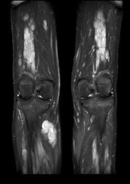
35 6 yr M IDDM Rx insulin Pain in L leg. Mass L lower medial leg above knee MRI: solid+necrotic mass R vastus medialis Bx.Alveolar Rhabdomyosarcoma T1 FS AX T1 FS SAG STIR COR


36 PET CT: Metabolic lesion with central necrosis (SUVmax 3.5) No regional LNs or distant metastases PET CT FDG coreg MRI- RTx planning CE


37 Post 2 cycles of chemotherapy MR COR STIR Mild reduction in size Partial response PET MR COREG SUVmax 2.3 (34%)


38 9 mths post Tx developed pain lower right back MRI metastatic disease COR STIR MRI CE


39 12 yr F presented with a mass on the right back July 2007 CT and MRI revealed a soft tissue mass with bone destruction and extension into the spinal canal Bx: High grade sarcoma Peripheral Nerve Sheath origin T2 AX T2 COR T2 SAG TL


40 SUVmax 5.3 Co-Registered MR and FDG PET Axial PET/ CT localised tumour Rx surgery, chemotherapy (ARST0332), radiotherapy TL


41 Follow up PET/CT Follow up- orthopaedic hardware artifacts
42 MRI Surveillance
43 MRI Surveillance PET/CT Resection Resection


44 SUVmax 4.6 Neurofibromatosis Type 1 8 yr F Multiple Plexiform Neurofibromas 2007 R paravertebral- Mn Peripheral Nerve Sheath Tumour SUVmax 2.4 R lower leg Benign PN SUVmax 4.6


45 Follow up MRI 2013: enlarging tumours 18F-FDG increased metabolic activity: Mass soft tissue deep to left 5 th intercostal space *SUVmax Grade 1 < 3 Grade 2 >3-<4 Grade 3 >4 *EJNMMI (2010) 37:1309 Moharir et al SUVmax 6.2 to 6.8 delay Histopath: MPNST WHO GrIV AB587652


46 15yr M NF1 Multiple plexiform Neurofibromas Rx Imatinib WBMR STIR Proximal R tibial lesion Larger than previous FDG PET


47 SUVmax 1.3 SUVmax 1.3 Delayed SUVmax 1.2 SUVmax 2.6 SUVmean 1.4
48 Other Paediatric Malignancy Neuroblastoma Neuroendocrine tumours Neurofibromatosis (NF1) Langerhan Cell Histiocytosis Hepatoblastoma, Hepatocellular carcinoma Adrenocortical carcinoma Nasopharyngeal carcinoma Germ cell tumours Yolk sac tumours Wilms tumour* Retinoblastoma Melanoma Thyroid carcinoma

49 Neuroendocrine Tumours Previous somatostatin agent 111 In-octreotide Replaced by PET agent 68 Ga DOTATATE or DOTATOC Taieb et al JNuclMed 53:264,2012

50 Management Peter MacCallum Hosp Melbourne


51 ES 9 yr F 2007 Hypertension, intermittent headaches, abdominal pain, hot and sweaty episodes Elevated catacheolamines CT.: 4cm vascular lobulated mass R para-aortic area 123 I- MIBG Study +ve Paraganglioma with lymphatic invasion Succinate Dehydrogenase B (SDH B) mutation ES991218


52 Represented 13 yrs June 2011 Recent increasing headaches, mass R paravertebral region near renal hilum on MRI. T2 MRI ES991218


53 Ga68 DOTATATE 2x2cm Lesion in hilum of R kidney: high uptake of Ga68 Incidental lesion in 6 th R rib R recurrence Co-Reg Ga68 & MR T2 PET CT L rib ES991218
54 Surgery 1. R paravertebral mass---- Paraganglioma 2. L 6 th Rib--- no abnormal pathology


55 Routine F/U 30 Aug Ga DOTATATE Multiple bone lesions 6 th L rib R pedicle T8 L1 vertebral body L iliac bone
56 Referred for Peptide Receptor Radionuclide therapy (PRRT) 177 Lu DOTATATE (Lutate) St George Hospital Sydney Uptake moderate Completed course Repeat Ga68 Dotatate- stable disease
")

57 16-yr F R adrenal Phaeochromocytoma (MIBG neg) Surgically removed 2010 Now presents 2012 biopsy proven metastases in the liver. FN


58 Nuclear medicine evaluation for biological markers for staging and therapy 123 I-MIBG 68 Ga DOTATATE NEGATIVE FN


59 18 F-FDG PET/CT MIP image Metastases Liver multiple R paravert near kidney Mid anterior vert L4 FN
60 Loss of SSR expression indicates poorly differentiated NET adverse prognosis higher responsiveness to chemotherapy: e.g.carboplatin and etoposide Other treatments Tyrosine kinase antagonists Sunitinib Chemotherapy Platinum and etoposide 5-Flurouracil (5-FU) Neo-vascularisation antagonists Avastin Radiotherapy bone and soft tissue Metastases localised to liver Surgery Inoperable Y-90 microspheres(sirt)
61 Conclusion PET/CT Paediatric Oncology 2015 PET/CT Standard of Diagnostic Care Stage Response to Treatment Detection residual & recurrent cancer End of treatment stage Surveillance Is complementary to MRI and other conventional imaging

62 Conclusion PET/CT has become standard of diagnostic care in the common paediatric solid tumours There is a significant but less well defined role in many of the uncommon and rare solid tumours The uptake of FDG relates to the degree of malignancy and cellular differentiation of these cancers
Dr Sneha Shah Tata Memorial Hospital, Mumbai.
 Dr Sneha Shah Tata Memorial Hospital, Mumbai. Topics covered Lymphomas including Burkitts Pediatric solid tumors (non CNS) Musculoskeletal Ewings & osteosarcoma. Neuroblastomas Nasopharyngeal carcinomas
PET/MR:Techniques, Indications and Applications
 PET/MR:Techniques, Indications and Applications Franz Wolfgang Hirsch Professor and Head of the Department of Pediatric Radiology University Hospital Leipzig / Germany Children s Hospital University Leipzig
SELF-ASSESSMENT MODULE REFERENCE SPR 2018 Oncologic Imaging Course Adrenal Tumors November 10, :00 12:10 p.m.
 SELF-ASSESSMENT MODULE REFERENCE SPR 2018 Oncologic Imaging Course Adrenal Tumors November 10, 2018 10:00 12:10 p.m. Staging Susan E. Sharp, MD 1. In the International Neuroblastoma Risk Group Staging
Pediatric cancer. Kleebsabai Sanpakit, MD. Hemato/Oncology division, Department of Pediatrics Faculty of Medicine Siriraj hospital
 Pediatric cancer เทคน คการให รห สโรค Siriraj Cancer ICD-O Registry ฉบ บม ออาช พ Kleebsabai Sanpakit, MD. Hemato/Oncology division, Department of Pediatrics Faculty of Medicine Siriraj hospital Different
PET-imaging: when can it be used to direct lymphoma treatment?
 PET-imaging: when can it be used to direct lymphoma treatment? Luca Ceriani Nuclear Medicine and PET-CT centre Oncology Institute of Southern Switzerland Bellinzona Disclosure slide I declare no conflict
Imaging: When to get MRI, CT or PET-CT?
 Imaging: When to get MRI, CT or PET-CT? Alina Uzelac, D.O. Assistant Clinical Professor Neuroradiology UCSF Department of Radiology and Biomedical Imaging San Francisco General Hospital Overview CT MRI
THE ROLE OF CONTEMPORARY IMAGING AND HYBRID METHODS IN THE DIAGNOSIS OF CUTANEOUS MALIGNANT MELANOMA(CMM) AND MERKEL CELL CARCINOMA (MCC)
 AND MERKEL CELL CARCINOMA (MCC)") THE ROLE OF CONTEMPORARY IMAGING AND HYBRID METHODS IN THE DIAGNOSIS OF CUTANEOUS MALIGNANT MELANOMA(CMM) AND MERKEL CELL CARCINOMA (MCC) I.Kostadinova, Sofia, Bulgaria CMM some clinical facts The incidence
THE ROLE OF CONTEMPORARY IMAGING AND HYBRID METHODS IN THE DIAGNOSIS OF CUTANEOUS MALIGNANT MELANOMA(CMM) AND MERKEL CELL CARCINOMA (MCC) I.Kostadinova, Sofia, Bulgaria CMM some clinical facts The incidence
Imaging of Pediatric MSK Tumors
 Imaging of Pediatric MSK Tumors Kirsten Ecklund, M.D. Boston Children s Hospital Harvard Medical School kirsten.ecklund@childrens.harvard.edu Tumor Imaging Goals Diagnosis Lesion characterization Benign
Prof. Dr. NAGUI M. ABDELWAHAB,M.D.; MARYSE Y. AWADALLAH, M.D. AYA M. BASSAM, Ms.C.
 Role of Whole-body Diffusion MR in Detection of Metastatic lesions Prof. Dr. NAGUI M. ABDELWAHAB,M.D.; MARYSE Y. AWADALLAH, M.D. AYA M. BASSAM, Ms.C. Cancer is a potentially life-threatening disease,
Radiation and Hodgkin s Disease: A Changing Field. Sravana Chennupati Radiation Oncology PGY-2
 Radiation and Hodgkin s Disease: A Changing Field Sravana Chennupati Radiation Oncology PGY-2 History of Present Illness 19 yo previously healthy male college student began having pain in his R shoulder
PET/CT Frequently Asked Questions
 PET/CT Frequently Asked Questions General Q: Is FDG PET specific for cancer? A: No, it is a marker of metabolism. In general, any disease that causes increased metabolism can result in increased FDG uptake
PET CT for Staging Lung Cancer
 PET CT for Staging Lung Cancer Rohit Kochhar Consultant Radiologist Disclosures Neither I nor my immediate family members have financial relationships with commercial organizations that may have a direct
Nuclear medicine in oncology. 1. Diagnosis 2. Therapy
 Nuclear medicine in oncology 1. Diagnosis 2. Therapy Diagnosis - Conventional methods - Nonspecific radiopharmaceuticals cumulating in tumours - Specific radiopharmaceuticals (receptor- and immunoscintigraphy)
Index. Surg Oncol Clin N Am 16 (2007) Note: Page numbers of article titles are in boldface type.
 Note: Page numbers of article titles are in boldface type.") Surg Oncol Clin N Am 16 (2007) 465 469 Index Note: Page numbers of article titles are in boldface type. A Adjuvant therapy, preoperative for gastric cancer, staging and, 339 B Breast cancer, metabolic
Surg Oncol Clin N Am 16 (2007) 465 469 Index Note: Page numbers of article titles are in boldface type. A Adjuvant therapy, preoperative for gastric cancer, staging and, 339 B Breast cancer, metabolic
Clinical indications for positron emission tomography
 Clinical indications for positron emission tomography Oncology applications Brain and spinal cord Parotid Suspected tumour recurrence when anatomical imaging is difficult or equivocal and management will
Effective local and systemic therapy is necessary for the cure of Ewing tumor Most chemotherapy regimens are a combination of cyclophosphamide,
 Ewing Tumor Perez Ewing tumor is the second most common primary tumor of bone in childhood, and also occurs in soft tissues Ewing tumor is uncommon before 8 years of age and after 25 years of age In the
Introduction Pediatric malignancies Changing trends & Radiation burden Radiation exposure from PET/CT Image gently PET & CT modification - PET/CT
 Introduction Pediatric malignancies Changing trends & Radiation burden Radiation exposure from PET/CT Image gently PET & CT modification - PET/CT protocols Tips Leukaemia / lymphoma: ~ 35% acute lymphoblastic
Quantitative Nuclear Medicine Imaging in Oncology. Susan E. Sharp, MD
 Quantitative Nuclear Medicine Imaging in Oncology Susan E. Sharp, MD Disclosures None Objectives Describe scoring systems used in oncologic nuclear medicine imaging Deauville scoring in lymphoma Curie
F NaF PET/CT in the Evaluation of Skeletal Malignancy
 F NaF PET/CT in the Evaluation of Skeletal Malignancy Andrei Iagaru, MD September 26, 2013 School of of Medicine Ø Introduction Ø F NaF PET/CT in Primary Bone Cancers Ø F NaF PET/CT in Bone Metastases
FDG PET/CT STAGING OF LUNG CANCER. Dr Shakher Ramdave
 FDG PET/CT STAGING OF LUNG CANCER Dr Shakher Ramdave FDG PET/CT STAGING OF LUNG CANCER FDG PET/CT is used in all patients with lung cancer who are considered for curative treatment to exclude occult disease.
CT PET SCANNING for GIT Malignancies A clinician s perspective
 CT PET SCANNING for GIT Malignancies A clinician s perspective Damon Bizos Head, Surgical Gastroenterology Charlotte Maxeke Johannesburg Academic Hospital Case presentation 54 year old with recent onset
performed to help sway the clinician in what the appropriate diagnosis is, which can substantially alter the treatment of management.
 Hello, I am Maura Polansky at the University of Texas MD Anderson Cancer Center. I am a Physician Assistant in the Department of Gastrointestinal Medical Oncology and the Program Director for Physician
Theranostics in Nuclear Medicine
 Theranostics in Nuclear Medicine Patrick FLAMEN, MD, PhD Head Nuclear Medicine Institut Jules Bordet Université Libre de Bruxelles (U.L.B.) n Theranostics in Nuclear Medicine n A form of (nuclear) diagnostic
All India Institute of Medical Sciences, New Delhi, INDIA. Department of Pediatric Surgery, Medical Oncology, and Radiology
 All India Institute of Medical Sciences, New Delhi, INDIA Department of Pediatric Surgery, Medical Oncology, and Radiology Clear cell sarcoma of the kidney- rare renal neoplasm second most common renal
Update in Lymphoma Imaging
 Update in Lymphoma Imaging Victorine V. Muse, MD Lymphoma Update in Lymphoma Imaging Victorine V Muse, MD Heterogeneous group of lymphoid neoplasms divided into two broad histological categories Hodgkin
Evaluation of Lung Cancer Response: Current Practice and Advances
 Evaluation of Lung Cancer Response: Current Practice and Advances Jeremy J. Erasmus I have no financial relationships, arrangements or affiliations and this presentation will not include discussion of
Lugano classification: Role of PET-CT in lymphoma follow-up
 CAR Educational Exhibit: ID 084 Lugano classification: Role of PET-CT in lymphoma follow-up Charles Nhan 4 Kevin Lian MD Charlotte J. Yong-Hing MD FRCPC Pete Tonseth 3 MD FRCPC Department of Diagnostic
Lymphoma Read with the experts
 Lymphoma Read with the experts Marc Seltzer, MD Associate Professor of Radiology Geisel School of Medicine at Dartmouth Director, PET-CT Course American College of Radiology Learning Objectives Recognize
UK Musculoskeletal Oncology: Something for All Ages. Lars Wagner, MD Pediatric Hematology/Oncology University of Kentucky
 UK Musculoskeletal Oncology: Something for All Ages Lars Wagner, MD Pediatric Hematology/Oncology University of Kentucky Pediatric-Type Sarcomas of Bone and Soft Tissue The incidence of sarcoma continues
The Importance of PET/CT in Human Health. Homer A. Macapinlac, M.D.
 The Importance of PET/CT in Human Health Homer A. Macapinlac, M.D. Learning objectives Provide an overview of the impact of PET/CT imaging on the management of patients and its impact on health care expenditures.
STAGING AND FOLLOW-UP STRATEGIES
 ATHENS 4-6 October 2018 European Society of Urogenital Radiology STAGING AND FOLLOW-UP STRATEGIES Ahmet Tuncay Turgut, MD Professor of Radiology Hacettepe University, Faculty of Medicine Ankara 2nd ESUR
objectives Pitfalls and Pearls in PET/CT imaging Kevin Robinson, DO Assistant Professor Department of Radiology Michigan State University
 objectives Pitfalls and Pearls in PET/CT imaging Kevin Robinson, DO Assistant Professor Department of Radiology Michigan State University To determine the regions of physiologic activity To understand
WHAT ARE PAEDIATRIC CANCERS
 WHAT ARE PAEDIATRIC CANCERS INTRODUCTION Childhood cancers are RARE 0.5% of all cancers in the West Overall risk that a child will develop cancer during first 15 years of life is 1 in 450 and 1 in 600
NET und NEC. Endoscopic and oncologic therapy
 NET und NEC Endoscopic and oncologic therapy Classification well-differentiated NET - G1 and G2 - carcinoid poorly-differentiated NEC - G3 - like SCLC well differentiated NET G3 -> elevated proliferation
Bone PET/MRI : Diagnostic yield in bone metastases and malignant primitive bone tumors
 Bone PET/MRI : Diagnostic yield in bone metastases and malignant primitive bone tumors Lars Stegger, Benjamin Noto Department of Nuclear Medicine University Hospital Münster, Germany Content From PET to
8/10/2016. PET/CT for Tumor Response. Staging and restaging Early treatment response evaluation Guiding biopsy
 PET/CT for Tumor Response Evaluation August 4, 2016 Wei Lu, PhD Department of Medical Physics www.mskcc.org Department of Radiation Oncology www.umaryland.edu FDG PET/CT for Cancer Imaging Staging and
Non-Hodgkin lymphomas (NHLs) Hodgkin lymphoma )HL)
 Hodgkin lymphoma )HL)") Non-Hodgkin lymphomas (NHLs) Hodgkin lymphoma )HL) Lymphoid Neoplasms: 1- non-hodgkin lymphomas (NHLs) 2- Hodgkin lymphoma 3- plasma cell neoplasms Non-Hodgkin lymphomas (NHLs) Acute Lymphoblastic Leukemia/Lymphoma
Non-Hodgkin lymphomas (NHLs) Hodgkin lymphoma )HL) Lymphoid Neoplasms: 1- non-hodgkin lymphomas (NHLs) 2- Hodgkin lymphoma 3- plasma cell neoplasms Non-Hodgkin lymphomas (NHLs) Acute Lymphoblastic Leukemia/Lymphoma
ADRENAL MEDULLARY DISORDERS: PHAEOCHROMOCYTOMAS AND MORE
 ADRENAL MEDULLARY DISORDERS: PHAEOCHROMOCYTOMAS AND MORE DR ANJU SAHDEV READER AND CONSULTANT RADIOLOGIST QUEEN MARY UNIVERSITY AND ST BARTHOLOMEW S HOSPITAL BARTS HEALTH, LONDON, UK DISCLOSURE OF CONFLICT
CLIC Sargent Eligibility Criteria
 1 Eligibility Criteria DOCUMENT GOVERNANCE: Eligibility criteria Produced by J. Hawkins & Grants Team Sponsored by Dara de Burca Version Approval by Executive Team 10 th June 2014 Board of Trustees 3 rd
LEUKAEMIA and LYMPHOMA. Dr Mubarak Abdelrahman Assistant Professor Jazan University
 LEUKAEMIA and LYMPHOMA Dr Mubarak Abdelrahman Assistant Professor Jazan University OBJECTIVES Identify etiology and epidemiology for leukemia and lymphoma. Discuss common types of leukemia. Distinguish
PET-MRI in malignant bone tumours. Lars Stegger Department of Nuclear Medicine University Hospital Münster, Germany
 PET-MRI in malignant bone tumours Lars Stegger Department of Nuclear Medicine University Hospital Münster, Germany Content From PET to PET/MRI General considerations Bone metastases Primary bone tumours
PET/CT F-18 FDG. Objectives. Basics of PET/CT Imaging. Objectives. Basic PET imaging
 Basics of PET/CT Imaging Kevin Robinson, DO Department of Radiology Michigan State University Objectives Basic PET imaging Evaluating the therapeutic response Evaluating the big 5 Lymphoma Breast Lung
4th INTERNATIONAL WORKSHOP ON INTERIM-PET IN LYMPHOMA Palais de l Europe, Menton Oct 4-5, 2012
 4th INTERNATIONAL WORKSHOP ON INTERIM-PET IN LYMPHOMA Palais de l Europe, Menton Oct 4-5, 2012 international workshop for PET in lymphoma staging and restaging Lale Kostakoglu Department of Radiology Mount
SOLID TUMOURS IN CHILDHOOD
 SOLID TUMOURS IN CHILDHOOD Fareed Omar Paediatric Oncology Steve Biko Academic hospital Introduction 1 Introduction Lymphomas and Leukemias make up about 40% of all childhood Cancers (Systemic cancers)
Cancer Prevention & Control in Adolescent & Young Adult Survivors
 + Cancer Prevention & Control in Adolescent & Young Adult Survivors NCPF Workshop July 15-16, 2013 Patricia A. Ganz, MD UCLA Schools of Medicine & Public Health Jonsson Comprehensive Cancer Center + Overview
GUIDELINES ON RENAL CELL CANCER
 20 G. Mickisch (chairman), J. Carballido, S. Hellsten, H. Schulze, H. Mensink Eur Urol 2001;40(3):252-255 Introduction is characterised by a constant rise in incidence over the last 50 years, with a predominance
FieldStrength. Leuven research is finetuning. whole body staging
 FieldStrength Publication for the Philips MRI Community Issue 40 May 2010 Leuven research is finetuning 3.0T DWIBS for whole body staging The University Hospital of Leuven is researching 3.0T whole body
Medical Policy An independent licensee of the Blue Cross Blue Shield Association
 PET Scanning: Oncologic Applications Page 1 of 88 Medical Policy An independent licensee of the Blue Cross Blue Shield Association Title: Positron Emission Tomography (PET) Scanning: Oncologic Applications
Update on RECIST and Staging of Common Pediatric Tumors Ethan A. Smith, MD
 Update on RECIST and Staging of Common Pediatric Tumors Ethan A. Smith, MD Section of Pediatric Radiology C.S. Mott Children s Hospital University of Michigan ethans@med.umich.edu Disclosures No relevant
Ronald C. Walker, MD, Prof of Radiology Vanderbilt University Medical Center Nashville, TN. Ga-DOTATATE PET/CT imaging Initial Vanderbilt experience
 Ronald C. Walker, MD, Prof of Radiology Vanderbilt University Medical Center Nashville, TN 68 Ga-DOTATATE PET/CT imaging Initial Vanderbilt experience Disclosures: No financial disclosures or conflicts
Los Angeles Radiological Society 62 nd Annual Midwinter Radiology Conference January 31, 2010
 Los Angeles Radiological Society 62 nd Annual Midwinter Radiology Conference January 31, 2010 Self Assessment Module on Nuclear Medicine and PET/CT Case Review FDG PET/CT IN LYMPHOMA AND MELANOMA Submitted
Pediatric Oncology. Vlad Radulescu, MD
 Pediatric Oncology Vlad Radulescu, MD Objectives Review the epidemiology of childhood cancer Discuss the presenting signs and symptoms, general treatment principles and overall prognosis of the most common
MRI in lymphoma: where are we in 2016
 MRI in lymphoma: where are we in 2016 Alain Rahmouni Henri Mondor Academic Hospital Paris Est Créteil University, France Plan Requirements DWI signal in lymphoma ADC measurement SNR: Surface coils 1.5
Systemic Therapy for Pheos/Paras: Somatostatin analogues, small molecules, immunotherapy and other novel approaches in the works.
 Systemic Therapy for Pheos/Paras: Somatostatin analogues, small molecules, immunotherapy and other novel approaches in the works. Arturo Loaiza-Bonilla, MD, FACP Assistant Professor of Clinical Medicine
New Visions in PET: Surgical Decision Making and PET/CT
 New Visions in PET: Surgical Decision Making and PET/CT Stanley J. Goldsmith, MD Director, Nuclear Medicine Professor, Radiology & Medicine New York Presbyterian Hospital- Weill Cornell Medical Center
Radiology Pathology Conference
 Radiology Pathology Conference Sharlin Johnykutty,, MD, Cytopathology Fellow Sara Majewski, MD, Radiology Resident Friday, August 28, 2009 Presentation material is for education purposes only. All rights
Colorectal Cancer and FDG PET/CT
 Hybrid imaging in colorectal & esophageal cancer Emmanuel Deshayes IAEA WorkShop, November 2017 Colorectal Cancer and FDG PET/CT 1 Clinical background Cancer of the colon and rectum is one of the most
Nuclear Medicine in Australia. Shaun Jenkinson
 Nuclear Medicine in Australia Shaun Jenkinson Landmark Infrastructure for Australian Science OPAL Research Reactor Australian Synchrotron Camperdown Cyclotron Bragg Institute Centre for Accelerator Science
Large cell immunoblastic Diffuse histiocytic (DHL) Lymphoblastic lymphoma Diffuse lymphoblastic Small non cleaved cell Burkitt s Non- Burkitt s
 Lymphoblastic lymphoma Diffuse lymphoblastic Small non cleaved cell Burkitt s Non- Burkitt s") Non Hodgkin s Lymphoma Introduction 6th most common cause of cancer death in United States. Increasing in incidence and mortality. Since 1970, the incidence of has almost doubled. Overview The types of
Non Hodgkin s Lymphoma Introduction 6th most common cause of cancer death in United States. Increasing in incidence and mortality. Since 1970, the incidence of has almost doubled. Overview The types of
The Use of PET Scanning in Urologic Oncology
 The Use of PET Scanning in Urologic Oncology Dr Nicholas C. Buchan Uro-oncology Fellow 1 2 Aims To understand the basic concepts underlying PET scanning. Understand the emerging role of PET Scanning for
Using PET/CT in Prostate Cancer
 Using PET/CT in Prostate Cancer Legal Disclaimer These materials were prepared in good faith by MITA as a service to the profession and are believed to be reliable based on current scientific literature.
Pediatric Thyroid Cancer Lung Metastases. Liora Lazar MD
 Pediatric Thyroid Cancer Lung Metastases Liora Lazar MD Differentiated thyroid cancer (DTC) The 3rd most common solid tumor in childhood and adolescence Accounting for 1.5%-3% of all childhood cancers
RFA of Tumors of the Lung: How and Why. Radiofrequency Ablation. Radiofrequency Ablation. RFA of pulmonary metastases. Radiofrequency Ablation of Lung
 RFA of Tumors of the Lung: How and Why Radiofrequency Ablation of Lung Ernest Scalzetti MD SUNY Upstate Medical University Syracuse NY FDA WARNING: Off-label use of a medical device Radiofrequency Ablation
PET Imaging in Langerhans Cell Histiocytosis
 PET Imaging in Langerhans Cell Histiocytosis Christiane Franzius Bremen, Germany Histiocytosis Histiocytosis Idiopathic proliferation of histiocytes Two types of histiocytes macrophages: antigen processing
Sarajevo (Bosnia Hercegivina) Monday June :30-16:15. PET/CT in Lymphoma
 Monday June :30-16:15. PET/CT in Lymphoma") Sarajevo (Bosnia Hercegivina) Monday June 16 2013 15:30-16:15 PET/CT in Lymphoma FDG-avidity Staging (nodal & extra nodal) Response evaluation Early assessment during treatment / interim (ipet) Remission
Sarajevo (Bosnia Hercegivina) Monday June 16 2013 15:30-16:15 PET/CT in Lymphoma FDG-avidity Staging (nodal & extra nodal) Response evaluation Early assessment during treatment / interim (ipet) Remission
Nuclear Sciences and Medicine
 Nuclear Sciences and Medicine Rethy Chhem, MD, PhD (Edu), PhD (His), FRCPC Division of Human Health Guest Professor, Medical University of Vienna International Atomic Energy Agency Medical Imaging X-rays
Oncology General Principles L A U R I E S I M A R D B R E A S T S U R G I C A L O N C O L O G Y F E L L O W D E C E M B E R
 Oncology General Principles L A U R I E S I M A R D B R E A S T S U R G I C A L O N C O L O G Y F E L L O W D E C E M B E R 2 0 1 2 Objectives Discuss Diagnostic and staging strategies in oncology Know
Esophageal Cancer. What is the value of performing PET scan routinely for staging of esophageal cancers
 Esophageal Cancer What is the value of performing PET scan routinely for staging of esophageal cancers What is the sensitivity and specificity of PET scan for metastatic lesions When should PET scan be
Thyroid Cancer: Imaging Techniques (Nuclear Medicine)
") Thyroid Cancer: Imaging Techniques (Nuclear Medicine) Andrei Iagaru, MD MIPS Molecular Imaging Program at Stanford Stanford University School of Medicine Department of Radiology Introduction Ø There are
Thyroid Cancer: Imaging Techniques (Nuclear Medicine) Andrei Iagaru, MD MIPS Molecular Imaging Program at Stanford Stanford University School of Medicine Department of Radiology Introduction Ø There are
Pediatric Lymphoma Update from the Children s Oncology Group
 Pediatric Lymphoma Update from the Children s Oncology Group Stephan D. Voss, MD, PhD Department of Radiology Boston Children s Hospital Harvard Medical School Staging Assessment Disclosures None Acknowledgements:
THYROID CANCER IN CHILDREN. Humberto Lugo-Vicente MD FACS FAAP Professor Pediatric Surgery UPR School of Medicine
 THYROID CANCER IN CHILDREN Humberto Lugo-Vicente MD FACS FAAP Professor Pediatric Surgery UPR School of Medicine Thyroid nodules Rare Female predominance 4-fold as likely to be malignant Hx Radiation exposure?
PET-CT in Peripheral T-cell Lymphoma: To Be or Not To Be
 PET-CT in Peripheral T-cell Lymphoma: To Be or Not To Be Bruce D. Cheson, M.D. Georgetown University Hospital Lombardi Comprehensive Cancer Center Washington, DC, USA So What is the Question(s)? What is
Pediatric Retroperitoneal Masses Radiologic-Pathologic Correlation
 Acta Radiológica Portuguesa, Vol.XVIII, nº 70, pág. 61-70, Abr.-Jun., 2006 Pediatric Retroperitoneal Masses Radiologic-Pathologic Correlation Marilyn J. Siegel Mallinckrodt Institute of Radiology, Washington
Primary bone tumors > metastases from other sites Primary bone tumors widely range -from benign to malignant. Classified according to the normal cell
 Primary bone tumors > metastases from other sites Primary bone tumors widely range -from benign to malignant. Classified according to the normal cell counterpart and line of differentiation. Among the
PET/CT in breast cancer staging
 PET/CT in breast cancer staging Anni Morsing Consultant, PhD, DMSc Rigshospitalet 1 18F- FDG PET/CT for breastcancer staging Where is the clinical impact? To which women should 18F- FDG PET/CT be offered?
Hybrid Imaging SPECT/CT PET/CT PET/MRI. SNMMI Southwest Chapter Aaron C. Jessop, MD
 Hybrid Imaging SPECT/CT PET/CT PET/MRI SNMMI Southwest Chapter 2014 Aaron C. Jessop, MD Assistant Professor, Department of Nuclear Medicine UT MD Anderson Cancer Center, Houston, Texas Complimentary role
Optimized. clinical pathway. propels high utilization of PET/MR at Pitié-Salpêtrière Hospital
 Optimized propels high utilization of PET/MR at Pitié-Salpêtrière Hospital clinical pathway As one of Europe s largest teaching hospitals, Pitié-Salpêtrière Hospital is renowned for its innovative research
Color Codes Pathology and Genetics Medicine and Clinical Pathology Surgery Imaging
 Saturday, November 5, 2005 8:30-10:30 a. m. Poorly Differentiated Endocrine Carcinomas Chairman: E. Van Cutsem, Leuven, Belgium 9:00-9:30 a. m. Working Group Sessions Pathology and Genetics Group leaders:
Imaging Pancreatic Neuroendocrine Tumors (PNETs): CT, MRI, EUS, Nuclear
: CT, MRI, EUS, Nuclear") Imaging Pancreatic Neuroendocrine Tumors (PNETs): CT, MRI, EUS, Nuclear Eric Tamm, M.D. Department of Diagnostic Radiology Division of Diagnostic Imaging MD Anderson Cancer Center Houston, TX Disclosure
Imaging Pancreatic Neuroendocrine Tumors (PNETs): CT, MRI, EUS, Nuclear Eric Tamm, M.D. Department of Diagnostic Radiology Division of Diagnostic Imaging MD Anderson Cancer Center Houston, TX Disclosure
Peptide Receptor Radionuclide Therapy (PRRT) of NET
 of NET") Peptide Receptor Radionuclide Therapy (PRRT) of NET Dr. Tuba Kendi Associate Prof of Radiology, Mayo Clinic, Rochester, MN 2014 MFMER slide-1 Relevant Financial Relationship(s) None Off Label Usage None
Peptide Receptor Radionuclide Therapy (PRRT) of NET Dr. Tuba Kendi Associate Prof of Radiology, Mayo Clinic, Rochester, MN 2014 MFMER slide-1 Relevant Financial Relationship(s) None Off Label Usage None
Nuclear Medicine: Basics to therapy
 Nuclear Medicine: Basics to therapy RCP Medical careers day Dr Sabina Dizdarevic MD MSc PhD FRCP Dr Deena Neriman MBBS FRCR Ms Charlotte Weston CEO BNMS On behalf of the British Nuclear Medicine Society
FDG-PET/CT in Gynaecologic Cancers
 Friday, August 31, 2012 Session 6, 9:00-9:30 FDG-PET/CT in Gynaecologic Cancers (Uterine) cervical cancer Endometrial cancer & Uterine sarcomas Ovarian cancer Little mermaid (Edvard Eriksen 1913) honoring
Radiology Pathology Conference
 Radiology Pathology Conference Nadia F. Yusaf, M.D. PGY-3 1/29/2010 Presentation material is for education purposes only. All rights reserved. 2010 URMC Radiology Page 1 of 90 Case 1 60 year- old man presents
PET/CT for Therapy Assessment in Oncology
 PET/CT for Therapy Assessment in Oncology Rodolfo Núñez Miller, M.D. Nuclear Medicine Section Division of Human Health International Atomic Energy Agency Vienna, Austria Clinical Applications of PET/CT
Molecular Imaging in the Development of Cancer Therapeutics. Johannes Czernin
 Molecular Imaging in the Development of Cancer Therapeutics Johannes Czernin Ahmanson Biological Imaging Division University of California Los Angeles Cancer Statistics Cancer Type 5-year Survival Rate
Gastrinoma: Medical Management. Haley Gallup
 Gastrinoma: Medical Management Haley Gallup Also known as When to put your knife down Gastrinoma Definition and History Diagnosis Historic Management Sporadic vs MEN-1 Defining surgical candidates Nonsurgical
Dr.Dafalla Ahmed Babiker Jazan University
 Dr.Dafalla Ahmed Babiker Jazan University Brain tumors are the second commonest malignancy in children Infratentorial tumors are more common As a general rule they do not metastasize out of the CNS, but
Pros and Cons: Interim PET in DLBCL Ulrich Dührsen Department of Hematology University Hospital Essen
 3rd International Workshop on in Lymphoma Menton, September 26, 2011 Afternoon Controversies Pros and Cons: in DLBCL Ulrich Dührsen Department of Hematology University Hospital Essen Pros Is there any
SOUTH THAMES CHILDREN S CANCER NETWORK GROUP. REFERRAL PROTOCOLS AND DIAGNOSIS AND STAGING PROTOCOLS October 2014
 SOUTH THAMES CHILDREN S CANCER NETWORK GROUP REFERRAL PROTOCOLS AND DIAGNOSIS AND STAGING PROTOCOLS October 2014 Contents 1. Leukaemia Referral, Diagnostic and Staging Guidelines 2. Lymphoma Referral,
Molecular Imaging and Cancer
 Molecular Imaging and Cancer Cancer causes one in every four deaths in the United States, second only to heart disease. According to the U.S. Department of Health and Human Services, more than 512,000
FDG-PET/CT in the management of lymphomas
 FDG-PET/CT in the management of lymphomas Olivier Gheysens, MD, PhD Dept. of Nuclear Medicine, UZ Leuven BHS, Feb 4th 2017 Brussels Dept. of Nuclear Overview Introduction on FDG-PET/CT Staging Response
Wilms Tumor and Neuroblastoma
 Wilms Tumor and Neuroblastoma Wilm s Tumor AKA: Nephroblastoma the most common intra-abdominal cancer in children. peak incidence is 2 to 3 years of age Biology somatic mutations restricted to tumor tissue
LYMPHOMA Joginder Singh, MD Medical Oncologist, Mercy Cancer Center
 LYMPHOMA Joginder Singh, MD Medical Oncologist, Mercy Cancer Center Lymphoma is cancer of the lymphatic system. The lymphatic system is made up of organs all over the body that make up and store cells
7 Omar Abu Reesh. Dr. Ahmad Mansour Dr. Ahmad Mansour
 7 Omar Abu Reesh Dr. Ahmad Mansour Dr. Ahmad Mansour -Leukemia: neoplastic leukocytes circulating in the peripheral bloodstream. -Lymphoma: a neoplastic process in the lymph nodes, spleen or other lymphatic
Radiological staging of lung cancer. Shukri Loutfi,MD,FRCR Consultant Thoracic Radiologist KAMC-Riyadh
 Radiological staging of lung cancer Shukri Loutfi,MD,FRCR Consultant Thoracic Radiologist KAMC-Riyadh Bronchogenic Carcinoma Accounts for 14% of new cancer diagnoses in 2012. Estimated to kill ~150,000
NCCN Practice Guidelines Narrative Summary of Indications for FDG PET and PET/CT
 NCCN Practice Guidelines Narrative Summary of Indications for FDG PET and PET/CT NCCN guidelines were reviewed on 2/14/2016 for utilization of 18F-fluorodeoxyglucose (FDG) PET and PET/CT (available at:
New imaging techniques: let there be light. Felix M. Mottaghy Department of Nuclear Medicine University Hospital KU Leuven
 New imaging techniques: let there be light Felix M. Mottaghy Department of Nuclear Medicine University Hospital KU Leuven Medical imaging and the pathology cascade Molecular/Cellular disturbance Alterations
Indications of PET/CT in oncology
 Monday, August 27, 2012 Session 1, 10:00-10:40 Indications of PET/CT in oncology Helle Westergren Hendel MD, PhD, assistant professor Bacelor in Leadership & Health Ecomomics Head of Clinical PET, Herlev
PET/CT in oncology. Positron emission tomography
 Clinical Medicine 2012, Vol 12, No 4: 368 72 PET/CT in oncology Fahim-Ul-Hassan, SpR Nuclear Medicine, Guy s Hospital, London; Gary J Cook, professor of Clinical PET, KCL Division of Imaging Sciences &
Page: 1 of 29. For this policy, PET scanning is discussed for the following 4 applications in oncology:
 Emission Tomography Scanning Page: 1 of 29 Last Review Status/Date: June 2015 Description Positron emission tomography (PET) scans are based on the use of positron-emitting radionuclide tracers coupled