Case 1 10/2/17. Myxoid Soft Tissue Tumors & Tumor-like Lesions. Myxofibro- or Fibromyxo-?: Myxoid Soft Tissue Tumours We Are All Mixed Up About
|
|
|
- Erik Allen
- 6 years ago
- Views:
Transcription
 Heterogenous grooup of lesions both benign & malignant characterized by abundant myxoid stroma Many have overlapping histologic and IHC features")
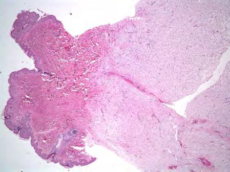
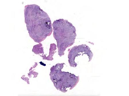
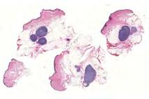
1 Myxoid Soft Tissue Tumors & Tumor-like Lesions Myxofibro- or Fibromyxo-?: Myxoid Soft Tissue Tumours We Are All Mixed Up About Rajiv M. Patel, M.D. RCPA NZ ASM 2017 (4:15-5:00pm, Saturday, ) Heterogenous grooup of lesions both benign & malignant characterized by abundant myxoid stroma Many have overlapping histologic and IHC features Their correct classification is important as many have significantly divergent biologic potential Talk will address the most common and/or clinically important myxoid lesions of soft tissue & the means by which they may be reliably identified Where appropriate the differential diagnosis & ancillary diagnostic methods needed for diagnosis will be addressed 2 Myxoid SST: Key features to evaluate Myxoid Soft Tissue Tumors & Tumor-like Lesions Cellularity Extremely low= myxoma, relatively high= fasciitis Arrangement of cells with respect to one another Little cell-cell contact= MLS, touching= ESMC Nuclear pleomorphism Absent= intramuscular myxoma, high degree of atypia= MFS Underlying vasculature Low vascularity= intramuscular myxoma, intricate vasculature= MLS & MFS (Alcian blue ph 2.5) evaluation of myxoid stroma (rarely) Hyaluronic acid-rich= intramuscular myxoma, MLS, MFS (pretreatment with hyaluronidase results in loss of positivity) Chondroitin sulfate-rich= ESMC & chordoma (hyaluronidase resistant positivity) Nodular fasciitis Intramuscular myxoma Angiomyxoma (aggressive angiomyxoma) Myxoid liposarcoma Myxofibrosarcoma Low-grade fibromyxoid sarcoma Extraskeletal myxoid chondrosarcoma 3 4 Case 1 20-year-old male with a right chest wall mass 6 1


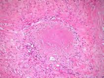
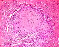
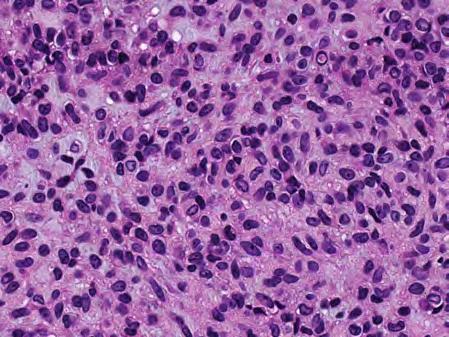
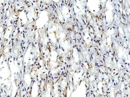
2 7 8 Diagnosis? Tram-track SMA Clinical: Nodular fasciitis Nodular fasciitis Pseudosarcomatous, self-limiting, reactive process composed of fibroblasts and myofibroblasts Common, typically subcutaneous Young adults M=F Upper extremities, trunk, head & neck Rapidly growing, sometimes painful Imaging may reveal calcifications <2% local recurrence Simple excision 2
 to collagenous (older lesions)")
; focal")

 Myxoid DFSP CD34 positive with COL1A1-PDGFRB translocation Cellular FH Collagen")

3 Histology: Nodular fasciitis Microcystic myxoid change Well circumscribe nodule (2-3 cm) Variably cellular, fascicular proliferation of fibroblasts & myofibroblasts Myxoid (early lesions) to collagenous (older lesions) Numerous mitoses especially in early lesions, not atypical forms Prominent inflammation around lesion Osteoclast-like giant cells in 10% of cases Postive for SMA, caldesmon, MSA & calponin (tram-track pattern); focal desmin; S100 negative Fasciitis Old fasciitis Differential: Nodular fasciitis Nodular fasciitis Myxomas Less cellular & mitotically active, finer more slender spindle cells with pyknotic nuclei Myxofibrosarcoma More atypia, mitoses with atypical forms, distinct curvilinear vessels MPNST More atypia, higher cellularity, SOX10 & S100 positive (typically focal) Myxoid DFSP CD34 positive with COL1A1-PDGFRB translocation Cellular FH Collagen trapping, foamy and hemosiderin laden macrophages, but may be indistinguishable from fasciitis Spindle cell carcinoma & melanoma Keratins and melanocytic markers, respectively Leiomyosarcoma Perinuclear vacuoles, stronger and more diffuse SMA and usually desmin positive 17 3


4 Ischemic fasciitis: zonation Granulation tissue surrouding areas of fibrinous & mxyoid change Reactive pseudosarcomatous myofibroblasts NF with USP6 Split Signal Case 2 33-year-old femal with a left thigh mass 4

5
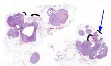
 Benign mesenchymal tumor composed of bland spindled cells embedded in a hypovascular, frequently cystic, myxoid matrix")
 Well-circumscribed myxoid appearing mass, cystic Abundant")
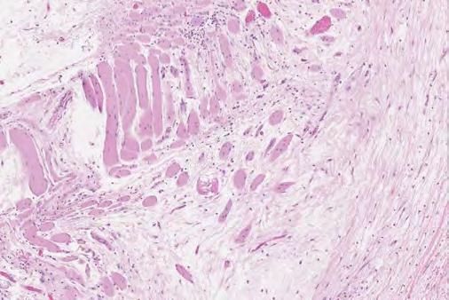
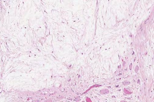
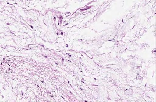
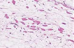
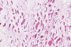
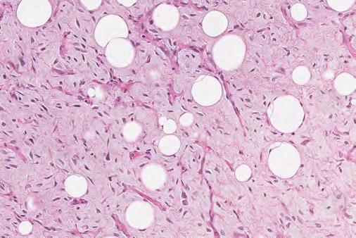
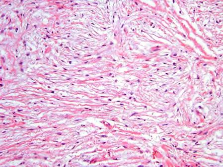
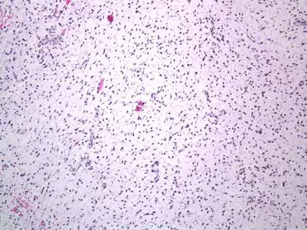
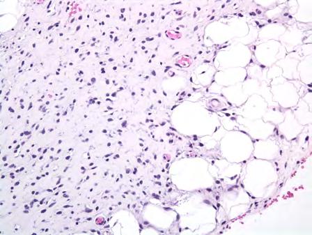
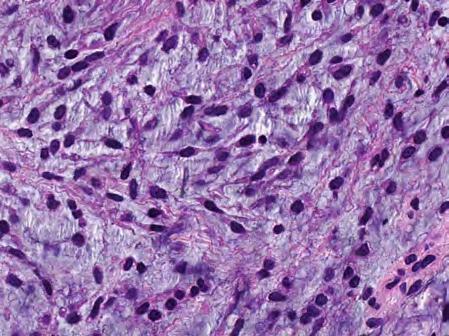
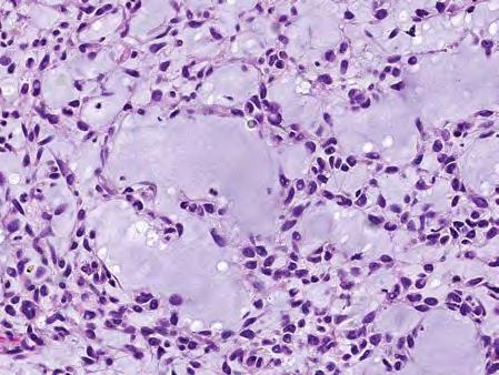
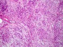
6 Diagnosis? Intramuscular myxoma Clinical: Myxoma (intramuscular & juxta-articular) Benign mesenchymal tumor composed of bland spindled cells embedded in a hypovascular, frequently cystic, myxoid matrix Relatively common Intramuscular: more common in women; large muscles of thigh & buttock; may be associated with fibrous dysplasia (Mazabraud syndrome) Juxta-articular: more common in men with DJD; near knees, shoulders, hips Painless soft tissue mass Cured by complete excision Histopathology: Myxoma (intramuscular & juxta-articular) Well-circumscribed myxoid appearing mass, cystic Abundant Alcian blue positive, hyaluronidase-sensitive myxoid matrix Splaying apart of skeletal muscle Hypovascular, without well-developed arborizing vasculature Slender spindle cells with pyknotic nuclei, do not tend to touch Juxta-articular myxomas often show areas of increased cellularity, with partial fascicular growth pattern 6

(q13;p11) resulting in FUS- DDIT3 (CHOP) gene")


7 Differential: Myxoma (intramuscular & juxta-articular) Myxofibrosarcoma Curvilinear vessels, atypia Myxoid liposarcoma Chicken wire vasculature, lipoblasts, t(12;16)(q13;p11) resulting in FUS- DDIT3 (CHOP) gene fusion ESMC Multinodular, cording and stranding of cells, NR4A3 gene fusions Myxoid nodular fasciitis Less myxoid, more cellular, multinucleated giant cells, extravasated RBCs Myxoid fibromatosis More cellular and fascicular, spindle cells have pinpoint nucleoli, lymphs scattered around distinct vessels Case 3 30-year-old female with a vaginal wall mass 40 Desmin ER


 May have a grenz zone if near skin/mucosa Abundantly myxoid Bland spindled to stellate cells Prominent hyalinized vasculature Small")
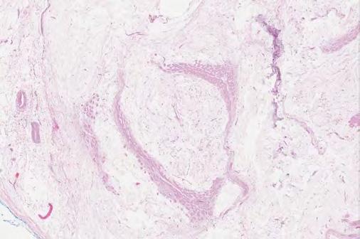
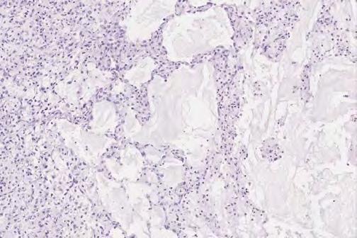
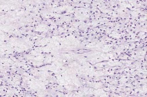
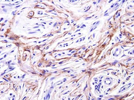
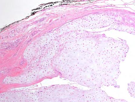
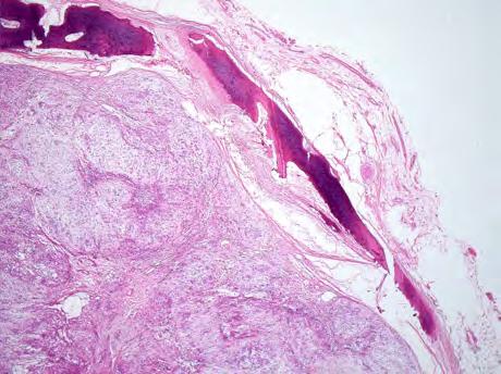
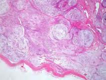
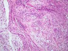
8 Diagnosis? Aggressive angiomyxoma Clinical: Aggressive angiomyxoma Rare, distinctive myxoid tumor of the genital region, most often seen in women of reproductive age (F:M ratio 6:1) Perineum, pelvis, genital region in women of reproductive age Scrotum, spermatic cord, and pelvis in adult men Benign, deep-seated, slow-growing neoplasm with significant potential for local recurrence given myxoid nature & infiltrative growth Wide local excision with free margins No know role for adjuvant therapy Pathology: Aggressive angiomyxoma Variably circumscribed, but infiltrative Large (up to 60cm) May have a grenz zone if near skin/mucosa Abundantly myxoid Bland spindled to stellate cells Prominent hyalinized vasculature Small smooth muscle aggregates near vessels Lymphod aggregates and mast cells Desmin, SMA, ER/PR, CD34+; CK & S100- Involvement of HMGA2 gene at 12q


9 Aggressive angiomyxoma: Desmin Aggressive angiomyxoma: PR 50 Differential: Aggressive angiomyxoma Angiomyofibroblastoma Angiomyofibroblastoma Plumper cells, not extensively myxoid Myxoid leiomyoma Better circumscribed, larger areas of typical smooth muscle, larger vessels Other myxomas Genital stromal polyps No grenz zone, more cellular variability, higher cellularity Myxofibrosarcoma Atypia, curvilinear vessels 52 Myxoid leiomyoma Case 4 41-year-old male with popliteal deep soft tissue mass 53 9


10 Diagnosis? 59 10


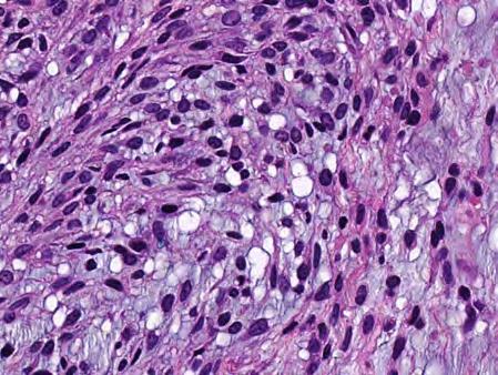
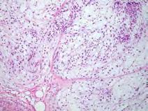
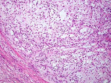
11 Clinical: Myxoid liposarcoma Myxoid liposarcoma Variably cellular, plexiform vasculature, myxoid, spindled or round cells, monovacuolated lipoblasts 30% liposarcoma; 10% of all sarcomas Slow-growing mass in deep soft tissues of limbs; may be multifocal M=F; peak incidence 4 th decade 90% overall 5 yr survival purely myxoid lesions, 40% for highgrade round cell lesions Adjuvant chemotherapy followed by wide excision Histopathology: Myxoid liposarcoma Multinodular, well-demarcated, gelatinous mass Proliferation of evenly distributed, monomorphic spindle cells, with little cell-to-cell contact Rich vascularized chicken wire vasculature Monovacuolated lipoblasts Abundant myxoid stroma Myxoid liposarcoma Plexiform vasculature 64 MLS MLS 11


12 MLS Monovacuolated lipoblasts High-grade myxoid (round cell) liposarcoma M/RCLS Ancillary Studies S-100 protein + t(12;16)(q13;p11) fusing DDIT3 with TLS t(12;22)(q13;q11) fusing DDIT3 and EWS DDIT3 (12q13) translocated DDIT3 (12q13) intact DDIT3[CHOP] (12q13) Break-Apart Probe 12


-EWS")


13 Differential: Myxoid liposarcoma Low-grade myxofibrosarcoma Distinct curvilinear vessels Pleomorphic tumor cells demonstrating perivascular condensation Lacks DDIT3 (CHOP)-TLS and DDIT3(CHOP)-EWS gene fusions ESMC Cording and stranding of cells end to end Inconspicuous vasculature NR4A3 gene fusions Case 5 A 4-year-old male presented with a flank mass 13
 and MUC4 Low grade fibromyxoid sarcoma(lgfms) Low-Grade Fibromyxoid Sarcoma (Evans tumor) Originally described by Harry Evans in 1987")
 4/10 died of disease Clinical: LGFMS 21 patients recurrences after intervals up to 15 years (median 3.")
14 Diagnosis? EMA MUC4 -ve for CD34, h-caldesmon, and SMA +ve for EMA (focal) and MUC4 Low grade fibromyxoid sarcoma(lgfms) Low-Grade Fibromyxoid Sarcoma (Evans tumor) Originally described by Harry Evans in 1987 (n=2) Both originally diagnosed as benign Both locally recurred and eventually metastasized One patient died from disease Follow-up study by Evans in 1993 (n=10) 10 new cases 8/10 diagnosed retrospectively after recurrence / metastasis 7/10 recurred 5/10 metastasized (some late) 4/10 died of disease Clinical: LGFMS 21 patients recurrences after intervals up to 15 years (median 3.5 yrs) 15 with metastasis after periods up to 45 years (median 5 yrs) 14 patients DOD Patients need lifelong follow-up Primarily affects young to middle-aged adults (median 35 yrs) 10-20% of cases present in children Predominantly present as deep soft tissue mass 20% present as superficial tumors of dermis or subcutis Superficial tumors relatively common in children (~40% of superficial tumors) Local recurrence 10%, metastasis in 5-10% Adverse factors: incomplete excision, possibly higher grade areas Wide excision with free margins, adjuvant radiotherapy Am J Surg Pathol Oct;35(10):


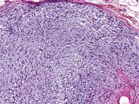
15 Histopathology: LGFMS Admixture of collagenous and myxoid zones, with abrupt transitions from one to the other Bland spindle cells with oval nuclei and pale eosinophilic cytoplasm Sweeping fascicles or storiform arrangement of cells Arcuate vessels, prominent in myxoid zones Low mitotic activity No necrosis Hypo- & hypercellular zones Curvilinear vessels 15


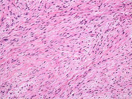
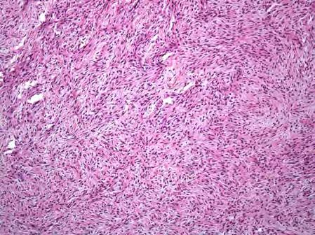
16 Bland spindle cells Giant collagenous rosettes in some cases (hyalinizing spindle cell tumor with giant rosettes) 16


 SMA +/- S100 -/+ CD34 rarely")
 t(7;16)(q33;p11)")


 S100+, MUC4-; FUS-CREB3L1, FUS-CREB3L2 or")
17 51 prospectively diagnosed as LGFMS 0 metastases 0 DOD LGFMS clinically behaves as low-grade sarcoma if accurately diagnosed. Caveat: short follow-up Histologic features: LGFMS/HSCT LGFMS/HSCT IHC & Molecular Pathology Vimentin + EMA -/+ (30%) SMA +/- S100 -/+ CD34 rarely focally +ve Desmin neg. MUC4 +ve - most useful marker (>90%) t(7;16)(q33;p11) FUS-CREB3L1; t(11;16)(p13;p11) FUS-CREB3L2 EWSR1-CREB3L1 rearrangements harbored by a smaller number of LGFMS FUS FISH Differential: LGFMS Perineurioma MUC4-; no FUS-CREB3L1, FUS-CREB3L2 or EWSR1-CREB3L1 Neuroblastoma-like schwannoma (for HSCTGR) S100+, MUC4-; FUS-CREB3L1, FUS-CREB3L2 or EWSR1-CREB3L1 Myxoid DFSP Strong and diffuse CD34, COL1A1-PDGFRB translocation Downs-Kelly et al Am J Surg Pathol 2008;32:

18 Soft tissue perineurioma EMA Myxoid DFSP 18


 Rare mesenchymal")


")
19 Key points: LGFMS Sarcoma of children and young adults Deep soft tissues of proximal extremities Deceptively bland histologic features Overlapping features between LGFMS/HSCT Both lesions demonstrate a t(7;16) reciprocal translocation Treatment: complete excision with tumor-free margins with adjuvant radiotherapy Outcome: low rate of recurrence and rare late metastases Sclerosing Epithelioid Fibrosarcoma (SEF) Rare mesenchymal fibroblastic malignant tumour Affects middle aged and elderly patients Deep soft tissue of limbs and limb girdle > trunk > head & neck Aggressive clinical behaviour: Local recurrence in 50% of cases Metastasis in upto 80% of cases Overall mortality of 25-57% Sclerosing epithelioid fibrosarcoma Sheets, nests and cords of monomorphic epithelioid fibroblastic cells embedded in a densely sclerotic collagenous matrix Myxoid change, calcification and metaplastic bone formation in places Haemangiopericytoma-like vasculature (focally) Conventional fibrosarcomatous areas or LGFMS may be present 19


(q33-34;p11),")


20 SEF - Immunohistochemistry EMA positive in 50% of cases MUC4 +ve in upto 65% t(7;16)(q33-34;p11), FUS-CREB3L2 in 38% of MUC4-positive cases Case 6 73-year-old male with a history of an enlarging mass of the left distal posterior leg Gisselsen 1998; Donner 2000; Jiao 2002; Ogose 2004; Guillou 2007; Doyle

, no sex predilection Slow growing, painless mass, limbs & limb girdles; 2/3 in")

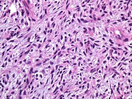
21 Diagnosis? 123 Myxofibrosarcoma Myxofibrosarcoma Fibroblastic lesion with pleomorphic cells, variable myxoid stroma & distinct vascular pattern One of the most common sarcomas of the elderly (50-70 yrs), no sex predilection Slow growing, painless mass, limbs & limb girdles; 2/3 in subcutis & lower dermis, 1/3 deep soft tissues Recurrences 50% to 60%, regardless of grade Metastases 20% to 35%, only in intermediate to high-grade lesions (lungs, bone, lymph nodes) Complete excision +/- adjuvant therapy based on grade and margin status 21


22 Myxofibrosarcoma: Pathologic Features Atypical cells with enlarged, hyperchromatic, pleomorphic nuclei Variable myxoid stroma Lipoblast-like cells Low mitotic activity Prominent arcuate vascular with condensation of atypical cells Grades 1-3 Complex karyotype


23 MFS Myxofibrosarcoma: Differential Low-grade Nodular fasciitis Myxomas Myxoid spindle cell lipoma Dermal nerve sheath myxoma Myxoid liposarcoma Extraskeletal myxoid chondrosarcoma* Low-grade fibromyxoid sarcoma High-grade Myxoinflammatory fibroblastic sarcoma* Pleomorphic liposarcoma High-grade MPNST Metastatic carcinoma & melanoma CD Angiomyxoma Angiomyxoma 23


24 Myxoid SCL/PL Myxoid SCL/PL Myxoid SCL/PL Myxoid SCL/PL Case 7 40-year-old male with inner left arm lesion above elbow, present for a long time and increasing in size


25 Diagnosis?

26 2007: CNT 2011: CNT Cellular neurothekeoma 2011: ESMC 2017: ESMC


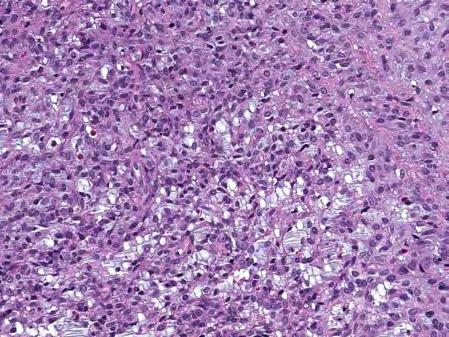
27 Diagnosis? Clinical: ESMC Extraskeletal myxoid chondrosarcoma (ESMC) Rare, 2.5% of all soft tissue sarcomas Painless mass of deep soft tissues of proximal extremities and trunk, but may be seen anywhere M>F, 4-7 th decades 50% rate of metastasis But long term survival characteristic Surgical excision 159 ESMC: Pathologic Features Uniform oval and spindled cells embedded in myxoid ground substance arranged in cords strands nest and sheets, with cells touching end to end Fibrous bands dividing tumor lobules Hypovascular Focal S100+ EWSR1-NR4A3 & RBP56-NR4A3 fusions


28 ESMC: Differential Cellular neurothekeoma NKIC3, PGP9.5, MiTF, SMA + Soft tissue chondroma Lacks myxoid matrix and atypia Osseous chondrosarcoma with soft tissue extension Imaging evidence Myoepithelioma/mixed tumor of soft tissue Keratin positive Ossifying fibromyxoid tumor of soft parts Shell of bone, stronger S NKIC-3 Cellular neurothekeoma 28
29 Cellular Neurothekeoma: IHC ST Chondroma Positive: NKIC3 MiTF PGP9.5 NSE S100A6 Vimentin 40-60% SMA Negative: S100 CD34 EMA HMB45 MelanA CK 170 OFMT OFMT OFMT OFMT 29


30 OFMT OFMT Myxoid Soft Tissue Lesions Tumor Cellularity Atypia Architecture Vessels Myxoma Low None Even cell Few distribution LGFMS Low None-low Abrupt myxoid collagen zones; swirling Low-grade MFS Low-mod Low-mod Perivascular cellularity Prominent in myxoid zones Curvilinear; thick Key points: Myxoid soft tissue lesions Despite that numerous benign & malignant soft tissue lesions may have a myxoid matrix, they can be separated from each other by systematic evaluation of certain perameters in conjuction with the clinical features including age, site & rate of growth There is little need for ancillary techniques, however molecular testing for translocations is becoming standard of care as molecular confirmation of diagnosis is increasingly important for targeted therapy initiation & enrollment in clinical trials. Myxoid LS Low Low Even cell distribution Plexiform, fine ESMC Low-mod Low Cords, chains Random,thick 30
Slide Seminar Spanish Society of Pathology
 Slide Seminar Spanish Society of Pathology John R. Goldblum, M.D. Chairman, Department of Anatomic Pathology Cleveland Clinic Professor of Pathology Cleveland Clinic Lerner College of Medicine 1921 Original
A 25 year old female with a palpable mass in the right lower quadrant of her abdomen
 May 2016 A 25 year old female with a palpable mass in the right lower quadrant of her abdomen Contributed by: Paul Ndekwe, MD, Resident Physician, Indiana University School of Department of Pathology and
Newer soft tissue entities
 Newer soft tissue entities Examples among fibroblastic tumors Turku, May 6, 2010 Markku Miettinen, M.D. AFIP, Washington, DC Fibroblastic neoplasms Solitary fibrous tumor /Hemangiopericytoma Low-grade
Selected Pseudomalignant Soft Tissue Tumors of the Skin and Subcutis
 Selected Pseudomalignant Soft Tissue Tumors of the Skin and Subcutis Andrew L. Folpe, M.D. Professor of Laboratory Medicine and Pathology Mayo Clinic, Rochester, MN folpe.andrew@mayo.edu 2016 MFMER slide-1
1/10/2018. Soft Tissue Tumors Showing Melanocytic Differentiation. Overview. Desmoplastic/ Spindle Cell Melanoma
 2016 MFMER slide-1 2016 MFMER slide-2 2016 MFMER slide-3 Soft Tissue Tumors Showing Melanocytic Differentiation Andrew L. Folpe, M.D. Professor of Laboratory Medicine and Pathology Mayo Clinic, Rochester,
Evening Specialty Conference Bone and Soft Tissue Pathology. Diagnostic pitfalls in bone and soft tissue pathology
 Evening Specialty Conference Bone and Soft Tissue Pathology. Case 1 Elizabeth G Demicco, MD, PhD Mount Sinai Hospital, New York Disclosure of Relevant Financial Relationships USCAP requires that all planners
Financial disclosures
 Mesenchymal Neoplasms with Melanocytic Differentiation By Konstantinos Linos MD, FCAP, FASDP Bone, Soft Tissue and Dermatopathology Assistant Professor of Pathology Dartmouth-Hitchcock Medical Center Geisel
Spindle Cell Lesions Of The Breast. Emad Rakha Professor of Breast Pathology and Consultant Pathologist
 Spindle Cell Lesions Of The Breast Emad Rakha Professor of Breast Pathology and Consultant Pathologist * SCLs comprise a wide spectrum of diseases, ranging from reactive processes to aggressive malignant
Special slide seminar
 Special slide seminar Tomáš Rozkoš The Fingerland Department of Pathology Charles University Medical Faculty and Faculty Hospital in Hradec Králové Czech Republic Case history, 33 years old resistance
Update On Lipomatous Tumors: Old Standbys and New Concepts
 Update On Lipomatous Tumors: Old Standbys and New Concepts John R. Goldblum, M.D. Chairman, Department of Anatomic Pathology Cleveland Clinic Professor of Pathology Cleveland Clinic Lerner College of Medicine
The Relevance of Cytologic Atypia in Cutaneous Neural Tumors
 The Relevance of Cytologic Atypia in Cutaneous Neural Tumors Recent Findings - New Developments New Problems Zsolt B. Argenyi, M.D. Professor of Pathology & Dermatology Director of Dermatopathology Department
SOFT TISSUE TUMOR PATHOLOGY: AN UPDATE
 SOFT TISSUE TUMOR PATHOLOGY: AN UPDATE Jason L. Hornick, MD, PhD July 18, 2013 Department of Pathology Brigham and Women s Hospital Harvard Medical School Boston, MA, USA I have no disclosures. New Soft
Cellular Neurothekeoma
 Cellular Neurothekeoma Scott W Binder, MD Pritzker Professor of Pathology & Dermatology Sr. Vice Chair Director, Pathology Clinical Services Chief, Dermatopathology Geffen/UCLA School of Medicine Clinical
Klinisch belang van chromosomale translocatie detectie in sarcomen
 Translocations in sarcomas Klinisch belang van chromosomale translocatie detectie in sarcomen Judith V.M.G. Bovée, M.D., Ph.D. Department of Pathology Leiden University Medical Center RNA binding DNA binding
57th Annual HSCP Spring Symposium 4/16/2016
 An Unusual Malignant Spindle Cell Lesion to Involve the Breast Erinn Downs-Kelly, D.O. Associate Professor of Pathology University of Utah & ARUP Laboratories No disclosures Case 39 y/o female with no
An Overview of Genital Stromal Tumors
 An Overview of Genital Stromal Tumors By Konstantinos Linos MD, FCAP, FASDP Bone, Soft Tissue and Dermatopathology Assistant Professor of Pathology Dartmouth-Hitchcock Medical Center Geisel School of Medicine
Myxo-inflammatory Fibroblastic sarcoma
 AKA Myxo-inflammatory Fibroblastic sarcoma Acral Myxoinflammatory fibroblastic sarcomaam.j.surg.path1998; 22; 911-924 Inflammatory myxoid tumour of soft parts with bizarre giant cells [Pathol.Res.Pract.
أملس عضلي غرن = Leiomyosarcoma. Leiomyosarcoma 1 / 5
 Leiomyosarcoma 1 / 5 EPIDEMIOLOGY Exact incidence is unknown, but older studies suggest that leiomyosarcomas comprise approximately 3 percent of soft-tissue sarcomas. Superficial leiomyosarcoma occurs
Malignant Peripheral Nerve Sheath Tumor
 C H A P T E R 120 Malignant Peripheral Nerve Sheath Tumor Currently, malignant peripheral nerve sheath tumor (MPNST) is the most commonly used generic name for the neoplasms known in the past as neurosarcoma,
Notice of Faculty Disclosure
 Mesenchymal Tumors of the Vulva: Old, New, Something(s) Different Napa Valley Conference Pathology Education Partners Inc May 15, 2018 Teri A. Longacre, M.D. longacre@stanford.edu Stanford University,
Fun with Fat. General Rules. Case
 Fun with Fat General Rules Imaging: location (deep vs. superficial) Superficial lesions are seldom liposarcomas Deep lesions may be benign or malignant Myxoid stroma is common in benign and malignant lesions
Contents Part I Introduction 1 General Description 2 Natural History: Importance of Size, Site, Histopathology
 Contents Part I Introduction 1 General Description... 3 1.1 Introduction... 3 1.2 Incidence and Prevalence... 5 1.3 Predisposing and Genetic Factors... 8 References... 16 2 Natural History: Importance
Desmoplastic Melanoma R/O BCC. Clinical Information. 74 y.o. man with lesion on left side of neck r/o BCC
 R/O BCC Sabine Kohler, M.D. Professor of Pathology and Dermatology Dermatopathology Service Stanford University School of Medicine Clinical Information 74 y.o. man with lesion on left side of neck r/o
Disclosures. An update on ancillary techniques in the diagnosis of soft tissue tumors. Ancillary techniques. Introduction
 Disclosures An update on ancillary techniques in the diagnosis of soft tissue tumors. I have nothing to disclose. Andrew Horvai, MD, PhD Clinical Professor, Pathology Introduction Ancillary techniques
Classification (1) Classification (3) Classification (2) Spindle cell lesions. Spindle cell lesions of bladder (Mills et al.
 Classification (3) Classification (2) Spindle cell lesions. Spindle cell lesions of bladder (Mills et al.") Non-epithelial tumours and nonepithelial tumour-like lesions of the bladder Dr Jonathan H Shanks The Christie NHS Foundation Trust, Manchester, UK Classification (1) Myofibroblastic proliferations and
Non-epithelial tumours and nonepithelial tumour-like lesions of the bladder Dr Jonathan H Shanks The Christie NHS Foundation Trust, Manchester, UK Classification (1) Myofibroblastic proliferations and
Diplomate of the American Board of Pathology in Anatomic and Clinical Pathology
 A 33-year-old male with a left lower leg mass. Contributed by Shaoxiong Chen, MD, PhD Assistant Professor Indiana University School of Medicine/ IU Health Partners Department of Pathology and Laboratory
21/07/2017. Hobnail endothelial cells are not the same as epithelioid endothelial cells
 UPDATE IN CUTANEOUS VASCULAR S DERMATOPATHOLOGY SESSION BELFAST PATHOLOGY JUNE 21/2017 Dr E Calonje St John s Institute of Dermatology, London, United Kingdom THE FAMILY OF VASCULAR S WITH EPITHELIOID
5/10. Pathology Soft tissue tumors. Farah Bhani. Mohammed Alorjani
 5/10 Pathology Soft tissue tumors Mohammed Alorjani Farah Bhani Slides are included in this sheet. Objectives: Soft tissue tumors 1. Describe soft tissue tumors. 2. Understand the classification of soft
Update on Cutaneous Mesenchymal Tumors. Thomas Brenn
 Update on Cutaneous Mesenchymal Tumors Thomas Brenn Cutaneous Mesenchymal Tumours Wide morphological and biological spectrum Myofibroblastic, smooth muscle, neural, vascular, apidocytic, undifferentiated;
Disclosures. An update on ancillary techniques in the diagnosis of soft tissue tumors. Ancillary techniques. Introduction
 Disclosures An update on ancillary techniques in the diagnosis of soft tissue tumors. I have nothing to disclose. Andrew Horvai, MD, PhD Clinical Professor, Pathology Introduction Ancillary techniques
Molecular pathology in soft tissue tumors. Sylvia Höller Pathologie
 Molecular pathology in soft tissue tumors Sylvia Höller Pathologie When do we perform molecular testing? Morphology and IHC are not clearly fitting with an entity some translocations are entity specific
Case 27 Male 42. Painless, static, well-circumscribed, subcutaneous nodule right lower leg,?lipoma. The best diagnosis is:
 Case 27 Male 42. Painless, static, well-circumscribed, subcutaneous nodule right lower leg,?lipoma. The best diagnosis is: A. Angiosarcoma B. Haemangiopericytoma C.Myopericytoma D.Myofibroma E. Angioleiomyoma
ACCME/Disclosures ALK FUSION-POSITIVE MESENCHYMAL TUMORS. Tumor types with ALK rearrangements. Anaplastic Lymphoma Kinase. Jason L.
 Companion Meeting of the International Society of Bone and Soft Tissue Pathology The Evolving Concept of Mesenchymal Tumors ALK FUSION-POSITIVE MESENCHYMAL TUMORS Jason L. Hornick, MD, PhD March 13, 2016
Post-test Self-assessment Cases
 Post-test Self-assessment Cases Ibrahim Khalifeh, M.D. Associate Professor Department of Pathology American University of Beirut Medical Center Beirut, Lebanon Case I History A 69 year old gentleman presenting
Diagnostic problems in uterine smooth muscle tumors
 Diagnostic problems in uterine smooth muscle tumors Marina Kos Ljudevit Jurak Clinical Department of Pathology, Clinical Hospital Center Sestre milosrdnice, Zagreb Institute of Pathology, University of
Pathology of Sarcoma ELEANOR CHEN, MD, PHD, ASSISTANT PROFESSOR DEPARTMENT OF PATHOLOGY UNIVERSITY OF WASHINGTON
 Pathology of Sarcoma ELEANOR CHEN, MD, PHD, ASSISTANT PROFESSOR DEPARTMENT OF PATHOLOGY UNIVERSITY OF WASHINGTON Presentation outline Background and epidemiology of sarcomas Sarcoma classification Sarcoma
3/27/2017. Disclosure of Relevant Financial Relationships
 Ophthalmic Pathology Evening Specialty Conference USCAP 2017 5 th March, 2017 Mukul K. Divatia, MD Assistant Professor Department of Pathology & Genomic Medicine Weill Cornell Medical College Houston Methodist
Enterprise Interest Nothing to declare
 Enterprise Interest Nothing to declare Diagnoses one would not like to miss in soft tissue pathology early in your career Marta Sbaraglia, MD Department of Pathology Hospital of Treviso University of Padua
Conceptual Evolution of Soft Tissue Tumors Classification
 Conceptual Evolution of Soft Tissue Tumors Classification Angelo P. Dei Tos M.D. Departments of Pathology & Oncology Treviso, Italy How WHO classification was reshaped Pathologists and Cytogeneticists
SMOOTH MUSCLE TUMOURS
 SMOOTH MUSCLE TUMOURS NORMAL SMOOTH MUSCLE Cytology Immunohistochemistry Ultrastructure Masson Trichrome Smooth Muscle Ultrastructure Many myofilaments running parallel to the long axis of the smooth
CASE REPORT Benign epithelioid peripheral nerve sheath tumour resembling schwannoma
 Malaysian J Pathol 2014; 36(3) : 217 221 CASE REPORT Benign epithelioid peripheral nerve sheath tumour resembling schwannoma Thejasvi KRISHNAMURTHY MD and SR NIVEDITHA MD, DNB Department of Pathology,
The Impact of Advances in Molecular Genetic Pathology on the. Classification, Diagnosis and Treatment of Selected Soft Tissue
 The Impact of Advances in Molecular Genetic Pathology on the Classification, Diagnosis and Treatment of Selected Soft Tissue Tumors of the Head and Neck Joaquín J. García MD and Andrew L. Folpe MD Department
Tumores de células pequeñas, redondas y azules: diagnóstico diferencial cuando el tiempo apremia
 Tumores de células pequeñas, redondas y azules: diagnóstico diferencial cuando el tiempo apremia Sílvia Bagué Servei de Patologia Hospital de Sant Pau Barcelona Soft tissue sarcomas Heterogeneous group
IN THE NAME OF GOD Dr. Kheirandish Oral and maxillofacial pathology
 IN THE NAME OF GOD Dr. Kheirandish Oral and maxillofacial pathology ORAL FOCAL MUCINOSIS Uncommon Tumorlike Cutaneous myxoid cyst Overproduction of hyaluronic acid by firoblasts Young adults Female Gingiva
ESS: Pathologic Insights
 GEIS XVI INTERNATIONAL SYMPOSIUM Seville 4th October 2018 ESS: Pathologic Insights Sílvia Bagué The Royal Marsden Hospital London (United Kingdom) I have no conflicts of interest Endometrial stromal sarcoma
Case 2. Dr. Sathima Natarajan M.D. Kaiser Permanente Medical Center Sunset
 Case 2 Dr. Sathima Natarajan M.D. Kaiser Permanente Medical Center Sunset History 24 year old male presented with a 3 day history of right flank pain, sharp in nature Denies fever, chills, hematuria or
Endometrial Stromal Tumors
 Endometrial Stromal Tumors WHO Categories: Endometrial Stromal Nodule (ESN) Endometrial Stromal Sarcoma, low grade (LGESS) Endometrial Stromal Sarcoma, high grade (HGESS) Undifferentiated Uterine Sarcoma
Diagnostic Approach to Soft Tissue Tumors
 SECTION 2 Diagnostic Approach to Soft Tissue Tumors Overview Biopsy and Resection of Soft Tissue Tumors 20 Clinical Approach Age- and Location-Based Approach to Diagnosis 24 Histologic Approach Pattern-Based
International Journal of Allied Medical Sciences
 International Journal of Allied Medical Sciences and Clinical Research (IJAMSCR) IJAMSCR Volume 2 Issue 4 Oct-Dec- 2014 Case report Medical research Myxofibrosarcoma of the male breast An infrequent neoplasm:
Lung Tumor Cases: Common Problems and Helpful Hints
 Lung Tumor Cases: Common Problems and Helpful Hints Brandon T. Larsen, MD, PhD Senior Associate Consultant Department of Laboratory Medicine and Pathology Mayo Clinic Arizona Arizona Society of Pathologists
ACCME/Disclosures. Everything is spindle - how far can we go with limited FNA material? Everything is spindle how far can we go? Everything is spindle
 ACCME/Disclosures The USCAP requires that anyone in a position to influence or control the content of CME disclose any relevant financial relationship WITH COMMERCIAL INTERESTS which they or their spouse/partner
From Morphology to Molecular Pathology: A Practical Approach for Cytopathologists Part 1-Cytomorphology. Songlin Zhang, MD, PhD LSUHSC-Shreveport
 From Morphology to Molecular Pathology: A Practical Approach for Cytopathologists Part 1-Cytomorphology Songlin Zhang, MD, PhD LSUHSC-Shreveport I have no Conflict of Interest. FNA on Lymphoproliferative
Mimics of Sarcoma. I have no conflict of interest to declare
 Maastricht 2018 Mimics of Sarcoma Cyril Fisher MA MD DSc FRCPath Consultant Pathologist, Royal Orthopaedic Hospital, Birmingham, UK Emeritus Professor of Tumour Pathology Institute of Cancer Research,
Tumors of Adipose Tissue Tumors Epidemiology Clinical Features. Morphology. Mature Adipocytes Separated by delicate fibrous septa
 Tumors of Adipose Tissue Lipoma Liposarcoma Most commonly happens in female The most common soft tissue tumor o Originates from matured Adipocytes Most commonly happes at the 4 th and 5 th decade of life
Morphologically Benign Lesions of Soft Tissue and Bone Which Metastasize - What Can We Do?
 Andrew L. Folpe, MD Mayo Clinic, Rochester, MN ISBSTP Handout 2010 Morphologically Benign Lesions of Soft Tissue and Bone Which Metastasize - What Can We Do? Introduction Over the past several decades
What is New in the 2015 WHO Lung Cancer Classification? Zhaolin Xu, MD, FRCPC, FCAP
 What is New in the 2015 WHO Lung Cancer Classification? Zhaolin Xu, MD, FRCPC, FCAP Professor, Dept of Pathology, Dalhousie University, Canada Pulmonary Pathologist and Cytopathologist, QEII HSC Senior
The Genetics of Myoepithelial Tumors: salivary glands, soft tissue and bone
 The Genetics of Myoepithelial Tumors: salivary glands, soft tissue and bone Cristina Antonescu, MD Memorial Sloan-Kettering Cancer Center, New York Nothing to declare Disclosure Spectrum of Myoepithelial
Mody. AIS vs. Invasive Adenocarcinoma of the Cervix
 Common Problems in Gynecologic Pathology Michael T. Deavers, M.D. Houston Methodist Hospital, Houston, Texas Common Problems in Gynecologic Pathology Adenocarcinoma in-situ (AIS) of the Cervix vs. Invasive
Nervous About Nerve Sheath Tumours?
 Goals Nervous About Nerve Sheath Tumours? Rajiv M. Patel, M.D. RCPA NZ ASM 2017 (3:30-4:15pm, Saturday, 23-09-17) Review spectrum of cutaneous nerve sheath tumors Vast majority are benign & include neurofibromas,
Adipocytic Tumours in children
 Università degli Studi di Padova Dipartimento di Medicina Sezione di Anatomia Patologica Generale e Citopatologia Adipocytic Tumours in children Rita Alaggio Basel Seminars in Pathology Paediatric Pathology
Inflammatory pseudotumor
 Inflammatory pseudotumor Inflammatory pseudotumor (IPT) Heterogeneous group of lesions of obscure etiology On physical and radiographic examination often confused with malignancy Synonyms Plasma cell granuloma
Mayo Medical Laboratories
 Mayo Medical Laboratories Virtual Lectures 2014 MFMER 2016 MFMER slide-1 Virtual Lectures Planning Committee Disclosure Summary As a provider accredited by ACCME, College of Medicine, Mayo Clinic (Mayo
2003 PIP-A A Cases. Paul K. Shitabata, M.D. APMG May 21, 2003
 2003 PIP-A A Cases Paul K. Shitabata, M.D. APMG May 21, 2003 Case 1 47F pelvic mass involving the right fallopian tube 5.5 cm intraluminal mass Fallopian Tube Adenocarcinoma Risk factors Breast, endometrial,
59 yo male with past medical history of prostate carcinoma, presented with upper abdominal pain
 December 2016 59 yo male with past medical history of prostate carcinoma, presented with upper abdominal pain Contributed by: Divya Sharma, MD. Fellow, Gastrointestinal Pathology, Department of Pathology
Fine Needle Aspiration Biopsy of a Myxoid Leiomyosarcoma with Epithelioid Features and It Metastasized to the Abdominal Wall - A Case Report -
 The Korean Journal of Pathology 2010; 44: 220-4 DOI: 10.4132/KoreanJPathol.2010.44.2.220 Fine Needle Aspiration Biopsy of a Myxoid Leiomyosarcoma with Epithelioid Features and It Metastasized to the Abdominal
Case Report Dendritic fibromyxolipoma in the latissimus dorsi: a case report and review of the literature
 Int J Clin Exp Pathol 2015;8(7):8650-8654 www.ijcep.com /ISSN:1936-2625/IJCEP0010580 Case Report Dendritic fibromyxolipoma in the latissimus dorsi: a case report and review of the literature Shuguang Liu
Cutaneous Mesenchymal Neoplasms with EWSR1 Rearrangement
 Cutaneous Mesenchymal Neoplasms with EWSR1 Rearrangement By Konstantinos Linos MD, FCAP, FASDP Bone, Soft Tissue and Dermatopathology Assistant Professor of Pathology Dartmouth-Hitchcock Medical Center
Surgical Pathology Evening Specialty Conference USCAP 2015
 Surgical Pathology Evening Specialty Conference USCAP 2015 John R. Goldblum, M.D. Chairman, Department of Pathology, Cleveland Clinic Professor of Pathology, Cleveland Clinic Lerner College of Medicine
Part 1. Slides 1-38, Rita Alaggio Soft tissue tumors Trondheim 14. mars 2013
 Part 1 Slides 1-38, Rita Alaggio Soft tissue tumors Trondheim 14. mars 2013 Pediatric Pathology Soft Tissue Tumors AN UPDATE Rita Alaggio Azienda Ospedaliera Università di Padova Soft Tissue Tumors More
Financial disclosures
 Cutaneous Mesenchymal Neoplasms with EWSR1 Rearrangement By Konstantinos Linos MD, FCAP, FASDP Bone, Soft Tissue and Dermatopathology Assistant Professor of Pathology Dartmouth-Hitchc Geisel School of
Benign and malignant epithelial lesions: Seborrheic keratosis: A common benign pigmented epidermal tumor occur in middle-aged or older persons more
 Benign and malignant epithelial lesions: Seborrheic keratosis: A common benign pigmented epidermal tumor occur in middle-aged or older persons more common on the trunk; but extremities, head and neck are
What really matters When and Why. Pathology of Uterine Mesenchymal Lesions. Nafisa Wilkinson London
 What really matters When and Why Pathology of Uterine Mesenchymal Lesions Nafisa Wilkinson London Patient centred approach immunohistochemistry Histological diagnosis Next generation sequencing Genetic
A case of giant cell tumour of soft parts in a horse Francesco Cian 1, Sarah Whiteoak 2, Jennifer Stewart 1
 A case of giant cell tumour of soft parts in a horse Francesco Cian 1, Sarah Whiteoak 2, Jennifer Stewart 1 1 Animal Health Trust, Newmarket, UK 2 608 Equine and Farm Vets, Rowington, UK Signalment: Horse,
CHAPTER 4. Smooth Muscle Tumours
 CHAPTER 4 Smooth Muscle Tumours Smooth muscle tumours arising at non-cutaneous, non-uterine locations have been the focus of a considerable conceptual shift in recent years and this is ongoing. Specifically,
Keywords solitary fibrous tumor, dedifferentiation, dedifferentiated solitary fibrous tumor, STAT6, GRIA2, cytokeratin, rhabdomyosarcomatous
 758452IJSXXX10.1177/1066896918758452International Journal of Surgical PathologyCreytens et al research-article2018 Pitfalls in Pathology Multifocal Cytokeratin Expression in a Dedifferentiated Solitary
Essential Dermatopathology. Jinah Kim, MD, PhD Department of Pathology and Dermatology Stanford University Medical Center
 Essential Dermatopathology Jinah Kim, MD, PhD Department of Pathology and Dermatology Stanford University Medical Center OBJECTIVES Review clinical, pathologic and molecular aspects of bone and fat tumors
ASCP Resident Review Mini-Course Series Session 2: Renal and Soft Tissue Pathology. Carole Vogler MD, FASCP Ema Dragoescu MD
 178 2011 ASCP Resident Review Mini-Course Series Session 2: Renal and Soft Tissue Pathology Carole Vogler MD, FASCP Ema Dragoescu MD 2011 Annual Meeting Las Vegas, NV AMERICAN SOCIETY FOR CLINICAL PATHOLOGY
SESSION 1: GENERAL (BASIC) PATHOLOGY CONCEPTS Thursday, October 16, :30am - 11:30am FACULTY COPY
 PATHOLOGY CONCEPTS Thursday, October 16, :30am - 11:30am FACULTY COPY") SESSION 1: GENERAL (BASIC) PATHOLOGY CONCEPTS Thursday, October 16, 2008 9:30am - 11:30am FACULTY COPY GOAL: Describe the basic morphologic (structural) changes which occur in various pathologic conditions.
SESSION 1: GENERAL (BASIC) PATHOLOGY CONCEPTS Thursday, October 16, 2008 9:30am - 11:30am FACULTY COPY GOAL: Describe the basic morphologic (structural) changes which occur in various pathologic conditions.
LAC + USC.
 Jeff McDavit,, M.D. LAC + USC mcdavit@usc.edu Clinical History 55 year old male with large, deep, non- tender left thigh mass. Seen at LAC+USC Med Ctr FNA clinic No h/o trauma or radiation Vimentin
Rhabdomyomas and Rhabdomyosarcomas (RMS) David M. Parham, MD Chief of Anatomic Pathology
 David M. Parham, MD Chief of Anatomic Pathology") Rhabdomyomas and Rhabdomyosarcomas (RMS) David M. Parham, MD Chief of Anatomic Pathology Tumors of skeletal muscle: Rhabdomyomas and rhabdomyosarcomas Embryonal muscle 2 3 4 5 6 7 8 Rhabdomyoma Benign
Rhabdomyomas and Rhabdomyosarcomas (RMS) David M. Parham, MD Chief of Anatomic Pathology Tumors of skeletal muscle: Rhabdomyomas and rhabdomyosarcomas Embryonal muscle 2 3 4 5 6 7 8 Rhabdomyoma Benign
Case Presentation. Maha Akkawi, MD, Fatima Obeidat, MD, Tariq Aladily, MD. Department of Pathology Jordan University Hospital Amman, Jordan
 Case Presentation Maha Akkawi, MD, Fatima Obeidat, MD, Tariq Aladily, MD Department of Pathology Jordan University Hospital Amman, Jordan The 25th Annual Congress of the ADIAP The 8/11/2013 1 5th International
Gastrointestinal stromal tumor
 Gastrointestinal stromal tumor 영남의대병리학교실 최준혁 Classification of gastrointestinal mesenchymal tumor Gastrointestinal stromal tumor(gist) Smooth muscle tumors : leiomyoma, leiomyosarcoma Neurogenic tumors
Case: The patient is a 24 year- old female who was found to have multiple mural nodules within the antrum. Solid and cystic components were noted on
 Case: The patient is a 24 year- old female who was found to have multiple mural nodules within the antrum. Solid and cystic components were noted on imaging. There is no significant past medical history.
No financial or other disclosures
 Case 2014-5 Esther N. Bit-Ivan, DO Northwestern University Jason Wang, MD Jason Park, MD Korgun Koral, MD Children s Medical Center Charles Timmons, MD Veena Rajaram, MD No financial or other disclosures
Disclosures. Giant Cell Rich Tumors of Bone. Outline. The osteoclast. Giant cell rich tumors 5/21/11
 Disclosures Giant Cell Rich Tumors of Bone Andrew Horvai, MD, PhD Associate Clinical Professor, Pathology This lecture discusses "off label" uses of a number of pharmaceutical agents. The speaker is describing
Normal endometrium: A, proliferative. B, secretory.
 Normal endometrium: A, proliferative. B, secretory. Nội mạc tử cung Nội mạc tử cung Cyclic changes in endometrium.. Approximate relationship of useful microscopic changes. Arias-Stella reaction in endometrial
S clerosing epithelioid fibrosarcoma is a rare tumour
 90 CASE REPORT Primary sclerosing epithelioid fibrosarcoma of the sacrum: a case report and review of the literature L T C Chow, Y H Lui, S M Kumta, P W Allen... Sclerosing epithelioid fibrosarcoma is
Atypical Palisaded Myofibroblastoma of Lymph Node: Report of a rare case.
 ISPUB.COM The Internet Journal of Pathology Volume 10 Number 1 Atypical Palisaded Myofibroblastoma of Lymph Node: Report of a rare case. V Kinnera, R Nandyala, M Yootla, K Mandyam Citation V Kinnera, R
Chondroid lipoma: A rare recently described benign lipomatous tumor
 Al-Malki et al. 7 case report peer Reviewed open OPEN ACCESS Chondroid lipoma: A rare recently described benign lipomatous tumor Salman T. Al-Malki, Abdullah S. Al-Khamiss Abstract Introduction: lipomatous
IMMUNOHISTOCHEMISTRY IN THE DIAGNOSIS OF SOFT TISSUE TUMORS
 IMMUNOHISTOCHEMISTRY IN THE DIAGNOSIS OF SOFT TISSUE TUMORS Nicolas de Saint Aubain Somerhausen Institut Jules Bordet / Hôpital Erasme nicolas.desaintaubain@synet.be ForPath 2005 1 I. Ancillary techniques
GUT-C 11/30/2017. Debasmita Das, M.D. PGY-1 Danbury Hospital
 GUT-C 11/30/2017 Debasmita Das, M.D. PGY-1 Danbury Hospital CLINICAL SUMMARY 8/2017 59 year old female Presented to the ED with 1 month history of general malaise, fever and weight loss PMH: Significant
05/07/2018. Types of challenges. Challenging cases in uterine pathology. Case 1 ` 65 year old female Post menopausal bleeding Uterine Polyp
 Types of challenges Challenging cases in uterine pathology Nafisa Wilkinson Gynaecological Pathologist UCLH London Lack of complete history often, NO clinical history at all! Cases from other centres often
Low-Grade Periductal Stromal of Breast: a case report
 Low-Grade Periductal Stromal of Breast: a case report Rosanna Nenna 1 Cosimo Damiano Inchingolo 1 Domenico Palmieri 2 Annalisa De Lucia 1 Giusy Elicio 1 Pina Miscioscia 1 ( 1 ) U.O.C. di Anatomia Patologica,
Fluorescence In Situ Hybridization in the Diagnosis of Soft Tissue Neoplasms: A Review. Munir R. Tanas, MD and John R.
 REVIEW ARTICLE Fluorescence In Situ Hybridization in the Diagnosis of Soft Tissue Neoplasms: A Review Munir R. Tanas, MD and John R. Goldblum, MD Abstract: This paper presents an overview of the role of
Slide seminar. Asist. Prof. Jože Pižem, MD, PhD Institute of Pathology Medical Faculty, University of Ljubljana
 Slide seminar Asist. Prof. Jože Pižem, MD, PhD Institute of Pathology Medical Faculty, University of Ljubljana Case 5 A 57-year-old man with a dermal/subcutaneous lesion on the scalp, which was interpreted
A case of fat-free pleomorphic lipoma occurring in the upper back and axilla simultaneously
 Wang et al. World Journal of Surgical Oncology 2013, 11:145 WORLD JOURNAL OF SURGICAL ONCOLOGY CASE REPORT Open Access A case of fat-free pleomorphic lipoma occurring in the upper back and axilla simultaneously
Index. J Juvenile hyaline fibromatosis, 27 Juvenile xanthogranuloma, 57 Juxta-articular myxoma, 152
 A Adenomatoid tumor, 76, 77 Adipose tissue tumors benign tumors angiolipoma, 6 chondroid lipoma, 9 fibrolipoma, 5 hibernoma, 8 lipoblastoma, 9 lipoma (see Lipoma) myelolipoma, 6 pleomorphic lipoma, 8 spindle
3/24/2017 DENDRITIC CELL NEOPLASMS: HISTOLOGY, IMMUNOHISTOCHEMISTRY, AND MOLECULAR GENETICS. Disclosure of Relevant Financial Relationships
 DENDRITIC CELL NEOPLASMS: HISTOLOGY, IMMUNOHISTOCHEMISTRY, AND MOLECULAR GENETICS Jason L. Hornick, M.D., Ph.D. Director of Surgical Pathology and Immunohistochemistry Brigham and Women s Hospital Professor
Dermatopathology. Dr. Rafael Botella Estrada. Hospital La Fe de Valencia
 Dermatopathology Dr. Rafael Botella Estrada. Hospital La Fe de Valencia Melanoma and mimics Dr. Martin Mihm Malignant lesions result from the accumulation of mutations Class I lesions (benign) Class II
Clear Cell Sarcoma of Right Knee with Bone Marrow Metastasis: A Case Report and review of Literature
 Case Report DOI: 10.21276/APALM.2017.1107 Clear Cell Sarcoma of Right Knee with Bone Marrow Metastasis: A Case Report and review of Literature Divya Shelly*, Shashank Mishra, Divya Gupta and Reena Bharadwaj