Other New entities in soft tissue tumors.
|
|
|
- Cornelius Glenn
- 5 years ago
- Views:
Transcription
1 Other New entities in soft tissue tumors. Angelo Paolo Dei Tos MD Departments of Pathology and Oncology General Hospital of Treviso, Italy Introduction During the past decade classification schemes have evolved continuously. The reappraisal of previously described entities have greatly contributed to major conceptual shifts. Pleomorphic malignant fibrous histiocytoma and hemangiopericytoma still represent the best examples, as their decline have allowed proper recognition of specific sarcoma subtypes such as pleomorphic rhabdomyosarcoma, myxofibrosarcoma, giant cell tumor of soft tissues or solitary fibrous tumor. By contrast, many lesions currently reported as new, with time has become part of spectrum of lesions. Examples are so called lipomatous hemangiopericytoma or giant cell angiofibroma (now both part of the morphologic continuum of solitary fibrous tumor) or hyalinizing spindle cell tumor with giant rosettes that has merged with low-grade fibromyxoid sarcoma to form a single tumor entity. Nonetheless, distinctive new lesions continue to be reported. This brief review will focus on a small group of recently reported mesenchymal lesions. Trendy entities like PEComa and myoepithelial lesions of soft tissue and bone are covered by distinct contributions. Myopericytoma Perivascular neoplasms comprise traditionally glomus tumor and so called hemangiopericytoma (HPC). Whereas glomus tumor represents a welldefined entity, the existence of HPC as a separate entity has been questioned. A number of neoplasms of different lines of differentiation are in fact characterized by a HPC-like vascular growth pattern, and the concept that most HPC represents examples of solitary fibrous tumors has eventually
2 reached a broad consensus. However, there exist lesions composed of cells expressing contractile proteins and organized in a perivascular growth pattern, that fulfills criteria set by Stout in his original description of hemangiopericytoma (1). These distinctive lesions have been collectively labeled as myopericytoma (2,3). Myopericytoma tends to occur in the superficial soft tissue of adults (peak incidence is in the fifth decade). Most frequently it arises in the lower limbs, followed by the upper limbs, trunk and head and neck. Microscopically, myopericytoma is composed of a spindle cell, myoid proliferation, organized in a distinctive perivascular pattern of growth, however a broad morphologic spectrum is observed. Immunohistochemically, most cases express smooth muscle actin and h-caldesmon. Desmin immunopositivity is rarely encountered. The clinical behavior is benign in most cases, however prominent cytologic atypia (which is rarely observed) correlates with potential of local recurrence and also metastatic spread (4). Epithelioid angiomatous nodule Cutaneous epithelioid vascular proliferations comprise a morphologic spectrum ranging from benign, reactive to frankly malignant conditions. A group of morphologically distinct cutaneous epithelioid vascular lesions that does not fit in any of the known entities was reported by Brenn in 2004 and labeled as epithelioid angiomatous nodule (EAN) (5). EAN usually arises in the trunk, extremities and, more rarely, on the face of adult patients with no gender predominance. Clinically, EAN presents as single (or multiple) erythematous to bluish nodules (or papules), of small size and short duration. Microscopically, EAN is composed of a circumscribed, unilobular, mostly solid proliferation of large polygonal epithelioid endothelial cells with vesicular nuclei and conspicuous nucleoli. Nuclear atypia is never observed. EAN is generally associated with a lymphoplasmacytic inflammatory infiltrate most prominent at the periphery of the lesion. Varying numbers of eosinophils are found scattered throughout the lesion. Immunohistochemically, neoplastic cells express common endothelial differentiation markers. Epithelioid angiomatous nodule is a benign, cutaneous vascular lesion, the relevance of
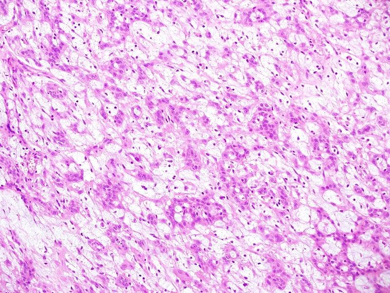
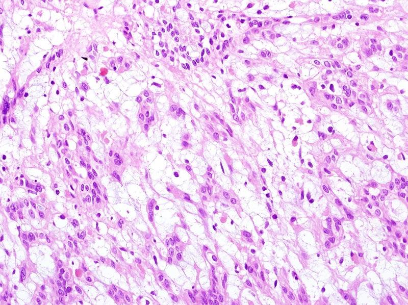
3 which resides in the fact it is not infrequently confused with epithelioid angiosarcoma. Reticular microcystic schwannoma Reticular microcystic schwannoma (RMS) is a rare variant of schwannoma with a distinctive predilection for viscera, especially the gastrointestinal tract (6), and mostly occurring in elderly patients (peak incidence is in the 6 th decade). A female predominance is observed. They are usually small, asymptomatic lesions most often measuring less than 3 cm in size. RMS usually occurs in the stomach, small bowel, and proximal large intestine. Microscopically it exhibits a striking microcystic and reticular lesional growth pattern with anastomosing and intersecting strands of spindle cells. Neoplastic cells tend to distribute around islands of myxoid or collagenous/hyalinized stroma. Mitotic activity is generally low and both atypia and necrosis are absent. RMS differs from usual schwannomas of the gastrointestinal tract also by lacking the characteristic peripheral cuff of lymphocytes. Immunohistochemically, strong nuclear and cytoplasmic positivity for S-100 and variably strong glial fibrillary acidic protein staining is generally observed. The histologic appearance of RMS raises a broad differential diagnosis, which includes gastrointestinal stromal tumor, perineurioma, and in more epithelioid examples, even an epithelial malignancy. Awareness of the entity and the use of an immunohistochemical panel of differentiation markers allows proper classification in most cases. RMS is an entirely benign neural neoplasm with no significant recurrent potential. Plexiform angiomyxoid tumor of the stomach Very recently, Takahashi and coworker have reported the occurrence in the gastric antrum of adult patients (peak incidence is in the 4 th decade) of a distinctive mesenchymal lesion characterized by a strikingly plexiform pattern of growth (7) The neoplasm can attain a large size and is composed of a
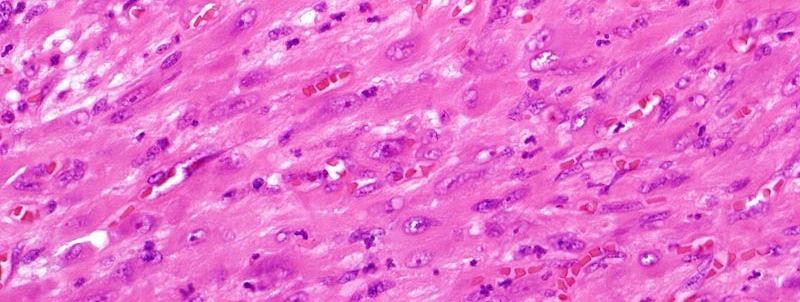
4 cytologically bland spindle cell proliferation, separated by abundant intercellular myxoid matrix, and associated with a rich capillary size vascular network. Cytologic atypia is generally absent and mitotic count very low. Immunohistochemically, tumor cells express alpha-smooth muscle actin and muscle actin, whereas are consistently negative for KIT, DOG1, CD34, and S- 100 protein. All cases analyzed so far have not shown mutations in the KIT or PDGFRA gene. As preliminary ultrastructural data seemed to point to myofibroblastic differentiation the authors proposed to label the lesion as plexiform angiomyxoid tumor of the stomach (PAMT). PAMT appears to be a benign entity, however, the frequently observed ulceration of the gastric mucosa may lead to potentially fatal gastrointestinal bleeding. A larger confirmatory series has been recently reported suggesting to modify the original label into plexiform fibromyxoma (8). Whatever the name, PAMT should be separated from GIST, nerve sheath tumors, and other fibromyxoid neoplasms Epithelioid myxofibrosarcoma Myxofibrosarcoma (MFS) is one of the most common soft tissue sarcomas of elderly patients and is exhibits a predilection for the superficial soft tissues of the limbs. Classic forms are represented by a multinodular, most often superficial proliferation composed of variably atypical neoplastic cells, mucinladen pseudolipoblasts, set in richly vascularized myxoid stroma. Recently, Nascimento and collaborators reported on a variant of myxofibrosarcoma that, while keeping most of the clinicopathologic features of the classic variant, is characterized by a striking epithelioid morphology, being therefore termed epithelioid myxofibrosarcoma (EMFS) (9). EMFS is typically characterized by a multinodular, infiltrating growth pattern with alternation of hypercellular and hypocellular myxoid areas. The presence of characteristic, prominent curvilinear vessels is almost often seen. Neoplastic cells tend to arrange singly and in small clusters, or sheets, wherein they show epithelioid morphology with round nuclei, vesicular chromatin, prominent nucleoli, and moderate amounts of eosinophilic cytoplasm. The epithelioid areas tend to be
5 multifocal with admixed areas of conventional MFS. Immunostains are most often negative for common differentiation markers. Differential diagnosis tends to be broad and include carcinoma, melanoma, and myoepithelial malignancies. EMFS tends to occur in the limbs of elderly patients (peak incidence is in the 6 th decade). EMFS in most cases is a high-grade sarcoma. A 70% local recurrence rate, associated with a 50% metastatic rate make EMFS a clinically more aggressive than conventional type myxofibrosarcoma. Pseudomyogenic hemangioendothelioma. Pseudomyogenic hemangioendothelioma (PMEHE) accounts as the most recently reported mesenchymal lesions (10). PMEHE represents a low-grade putative vascular malignancy most often occurring in the soft tissues of young adults. A male predominance is observed. Two third of the cases arise in the limbs followed by the trunk and the head and neck region. Multifocality has been reported in more than 60% of cases and half of cases were superficially located. Microscopically most tumor cells exhibit a spindled morphology, and are arranged in fascicles and sheets. A striking cytoplasmic eosinophilia reminiscent of rhabdomyoblastic differentiation is observed, along with vesicular nuclei harboring variably prominent nucleoli. Immunohistochemically, all tumors were positive for cytokeratin AE1/AE3 and FLI1, whereas approximately half of the cases variably expressed CD31. INI1 nuclear expression is preserved in all tumors tested so far. Clinically half of the patients experienced local recurrence within one year from diagnosis. Metastastic spread seems to be fairly rare as only one patient developed lymph node metastasis, and a second one developed widespread tumor dissemination. PMEHE may be closely related to the lesion previously reported under the name epithelioid sarcoma-like hemangioendothelioma (11). The recognition of this entity is crucial, in particular if the discrepancy between morphology and clinical behavior is considered.
6 References. 1. Stout AP, Murray MR. Hemangiopericytoma: a vascular tumor featuring Zimmermann s pericytes. Ann Surg 1942;116: Granter SR, Badizadegan K, Fletcher CD. Myofibromatosis in adults, glomangiopericytoma, and myopericytoma: a spectrum of tumors showing perivascular myoid differentiation. Am J Surg Pathol. 1998; 22: Mentzel T, Dei Tos AP, Sapi Z, Kutzner H. Myopericytoma of skin and soft tissues: clinicopathologic and immunohistochemical study of 54 cases. Am J Surg Pathol 2006; 30: Brenn T, Fletcher CD. Cutaneous epithelioid angiomatous nodule: a distinct lesion in the morphologic spectrum of epithelioid vascular tumors. Am J Dermatopathol 2004; 26: McMenamin ME, Fletcher CD. Malignant myopericytoma: expanding the spectrum of tumours with myopericytic differentiation. Histopathology 2002; 41: Liegl B, Bennett MW, Fletcher CD. Microcystic/reticular schwannoma: a distinct variant with predilection for visceral locations. Am J Surg Pathol 2008; 32: Takahashi Y, Shimizu S, Ishida T, Aita K, Toida S, Fukusato T, Mori S. Plexiform angiomyxoid myofibroblastic tumor of the stomach. Am J Surg Pathol 2007; 31:
7 8. Miettinen M, Makhlouf HR, Sobin LH, Lasota J. Plexiform fibromyxoma: a distinctive benign gastric antral neoplasm not to be confused with a myxoid GIST. Am J Surg Pathol 2009; 33: Nascimento AF, Bertoni F, Fletcher CD. Epithelioid variant of myxofibrosarcoma: expanding the clinicomorphologic spectrum of myxofibrosarcoma in a series of 17 cases. Am J Surg Pathol. 2007; 31: Hornick JL, Flecther CDM. Pseudomyogenic hemangioendothelioma: a distinctive often multicentric tumor with indolent behavior. Am J Surg Pathol 2011; in press. 11. Billings SD, Folpe AL, Weiss SW. Epithelioid sarcoma-like hemangioendothelioma. Am J Surg Pathol. 2003; 27:


8 Newly Recognized Soft Tissue Tumors Angelo Paolo Dei Tos M.D. Departments of Pathology & Oncology Treviso, ITALY
9 New Entity Reappraisal of lesions already described previously MFH, HPC Resuscitation of an entity that had been abandoned Pleomorphic RMS, GCT of Soft Tissues New clinicopathologic entities
10 Clinical History 50 year old male with a 2 cm, subcutaenous lump on thigh


11


12


13


14 SMA


15 H-Caldesmon
16 Diagnosis Myopericytoma
17 M > F Myopericytoma Peak incidence in 5th decade (age range: ) Lower limbs > upper limbs > head & neck > trunk Subcutaneous, solitary (rarely multiple), most often painless
18 Myopericytoma Oval spindle shaped myoid cell Perivascular growth pattern Glomus type features = glomangiopericytoma SMA + True hemangiopericytomas (?)
19 Classic HPC-like Angioleiomioma-likelike Myofibroma-like Cellular Atypical/Malignant Morphology


20 Classic type


21 HPC-like


22 Angioleiomyoma-likelike


23 Myofibroma-like


24 Hypcdllular


25 Cellular MP


26 Malignant/atypical myopericytoma
27 Hemangiopericytoma 1942, Stout and Murray Concept of perivascular (pericytic) neoplasm Pericyte Rouget (1873) Zimmermann (1923) Subsequent integration of lesions with HPC-like vascular pattern
28 HPC Diagnosis of exclusion Closely related (synonymous) with SFT Avoid creation of new monster Myopericytoma = true HPC
29 Hemangiopericytoma Spindle cells Perivascular pattern of growth Contractile properties SMA +/h-caldesmon +
30 Hemangiopericytoma = Myopericytoma
31 Clinical History 52 year old male with a 6 cm, mass located in the right thigh


32
33 Immunohistochemistry Vimentin + Scattered AE1/AE3 + ve cells
34 Diagnosis Epithelioid Myxofibrosarcoma
35
36 Myxoid Sarcomas Myxoid DFSP Myxoid Liposarcoma Myxofibrosarcoma Low Grade fibromyxoid sarcoma Myxoid Chondrosarcoma Myxoid Leyomiosarcoma Embryonal Rhabdomyosarcoma Acral myxoid fibrosarcoma
37 Myxofibrosarcoma Angervall, 1977 Myxoid MFH of Enzinger Spectrum of myxoid lesions High grade = myxoid MFH Histologic grade related to clinical outcome
38 Myxofibrosarcoma Elderly patients Lower limbs >upper limbs > limb girdles 2/3 subcutis, 1/3 deep seated IHC: vimentin, MSA and SMA (focal) Low grade no Mets High grade 30% metastatic rate Lungs > bone > mets Overall 5 years = 60%


39


40
41 Epithelioid Myxofibrosarcoma Elderly patients Limbs 50% metastatic rate Lungs > bone 73% recurrence rate Overall 5 years = 37% More aggressive then ordinary myxofibrosarcoma
42 Clinical History 52 year old male with a 6 cm, mass located in stomach Diagnosed as GIST


43 S-100
44 Diagnosis Microcystic-reticular reticular schwannoma
45
46 Microcystic-reticular reticular schwannoma Peak incidence in the sixth decades (any age) GI tract followed by superficial soft tissue of the extremities Striking microcystic and reticular lesional growth pattern S100 +; GFAP +/-


47


48


49
50 Microcystic-reticular reticular schwannoma Clinical behavior seems benign with no reported recurrences Differential diagnosis mainly with GIST KIT, DOG1 +
51 Clinical History 41 year old female with a 4 cm, ulcerated mass located in the stomach


52


53


54


55


56 SMA DES h-cal KIT
57 Diagnosis Plexiform angiomyxoid myofibroblastic tumor of the stomach
58


59
60 Plexiform angiomyxoid myofibroblastic tumor of the stomach Few cases reported Elderly patients with gastrointestinal hemorrhage due to mucosal ulceration GI tract Striking plexiform growth pattern Cytologically bland spindle cells set in intercellular myxoid matrix Rich capillary sized vascular network Variable expression of myogenic markers


61
62 Plexiform angiomyxoid myofibroblastic tumor of the stomach EM: myofibroblastic differentiation No mutations in the KIT and PDFGRA genes reported Clinical behavior seems to be benign Gastric bleeding potentially represents a life-threatening condition
63 Adults 12 cases Gastric antrum Benign clinicall behavior
64 Clinical History 18 year old male with a 3 cm, subcutaneous lump in the calf Basketball player


65


66


67


68 Cytokeratin AE1/AE3


69 CD31
70 IHC Cytokeratina AE1/AE3 + CD31 + CD34 INI1 + Smooth muscle actin focally +
71 Diagnosis Pseudomyogenic Hemangioendothelioma
72 Pseudomyogenic Hemangioendothelioma 50 cases (Fletcher & Hornick) Male predominance Peak incidence in 3rd decade 54% lower limb, 24% upper limb, 18% trunk, 4% head & neck Local recurrences in 58% LN mets in one case
73 Pseudomyogenic Hemangioendothelioma Plump spindle cells Brightly eosinophilic cytoplasm Immunohistochemical co-expression of epithelial and vascular markers Relationships with epithelioid sarcoma- like hemangioendothelioma


74 Stout AP Cancer Mar-Apr;15:400-9

75 Martin JF et al. Ann Anat Path :484
76 Acknowledgements Pathology Licia Laurino Sabrina Rossi Salvatore Romeo Cytogenetics Lucia Zanatta Laura Valori Eleonora Cappelletto Francesca Pol Molecular Pathology Luisa Toffolatti Marta Campo Dell Orto Giovanna Gallina Elisa Squizzato Irene Carraretto Serena Chinellato


77
Case 27 Male 42. Painless, static, well-circumscribed, subcutaneous nodule right lower leg,?lipoma. The best diagnosis is:
 Case 27 Male 42. Painless, static, well-circumscribed, subcutaneous nodule right lower leg,?lipoma. The best diagnosis is: A. Angiosarcoma B. Haemangiopericytoma C.Myopericytoma D.Myofibroma E. Angioleiomyoma
21/07/2017. Hobnail endothelial cells are not the same as epithelioid endothelial cells
 UPDATE IN CUTANEOUS VASCULAR S DERMATOPATHOLOGY SESSION BELFAST PATHOLOGY JUNE 21/2017 Dr E Calonje St John s Institute of Dermatology, London, United Kingdom THE FAMILY OF VASCULAR S WITH EPITHELIOID
Case: The patient is a 24 year- old female who was found to have multiple mural nodules within the antrum. Solid and cystic components were noted on
 Case: The patient is a 24 year- old female who was found to have multiple mural nodules within the antrum. Solid and cystic components were noted on imaging. There is no significant past medical history.
Newer soft tissue entities
 Newer soft tissue entities Examples among fibroblastic tumors Turku, May 6, 2010 Markku Miettinen, M.D. AFIP, Washington, DC Fibroblastic neoplasms Solitary fibrous tumor /Hemangiopericytoma Low-grade
Enterprise Interest Nothing to declare
 Enterprise Interest Nothing to declare Diagnoses one would not like to miss in soft tissue pathology early in your career Marta Sbaraglia, MD Department of Pathology Hospital of Treviso University of Padua
SOFT TISSUE TUMOR PATHOLOGY: AN UPDATE
 SOFT TISSUE TUMOR PATHOLOGY: AN UPDATE Jason L. Hornick, MD, PhD July 18, 2013 Department of Pathology Brigham and Women s Hospital Harvard Medical School Boston, MA, USA I have no disclosures. New Soft
Diplomate of the American Board of Pathology in Anatomic and Clinical Pathology
 A 33-year-old male with a left lower leg mass. Contributed by Shaoxiong Chen, MD, PhD Assistant Professor Indiana University School of Medicine/ IU Health Partners Department of Pathology and Laboratory
A 25 year old female with a palpable mass in the right lower quadrant of her abdomen
 May 2016 A 25 year old female with a palpable mass in the right lower quadrant of her abdomen Contributed by: Paul Ndekwe, MD, Resident Physician, Indiana University School of Department of Pathology and
Malignant Peripheral Nerve Sheath Tumor
 C H A P T E R 120 Malignant Peripheral Nerve Sheath Tumor Currently, malignant peripheral nerve sheath tumor (MPNST) is the most commonly used generic name for the neoplasms known in the past as neurosarcoma,
Conceptual Evolution of Soft Tissue Tumors Classification
 Conceptual Evolution of Soft Tissue Tumors Classification Angelo P. Dei Tos M.D. Departments of Pathology & Oncology Treviso, Italy How WHO classification was reshaped Pathologists and Cytogeneticists
Selected Pseudomalignant Soft Tissue Tumors of the Skin and Subcutis
 Selected Pseudomalignant Soft Tissue Tumors of the Skin and Subcutis Andrew L. Folpe, M.D. Professor of Laboratory Medicine and Pathology Mayo Clinic, Rochester, MN folpe.andrew@mayo.edu 2016 MFMER slide-1
The Relevance of Cytologic Atypia in Cutaneous Neural Tumors
 The Relevance of Cytologic Atypia in Cutaneous Neural Tumors Recent Findings - New Developments New Problems Zsolt B. Argenyi, M.D. Professor of Pathology & Dermatology Director of Dermatopathology Department
CASE REPORT Benign epithelioid peripheral nerve sheath tumour resembling schwannoma
 Malaysian J Pathol 2014; 36(3) : 217 221 CASE REPORT Benign epithelioid peripheral nerve sheath tumour resembling schwannoma Thejasvi KRISHNAMURTHY MD and SR NIVEDITHA MD, DNB Department of Pathology,
Atypical Palisaded Myofibroblastoma of Lymph Node: Report of a rare case.
 ISPUB.COM The Internet Journal of Pathology Volume 10 Number 1 Atypical Palisaded Myofibroblastoma of Lymph Node: Report of a rare case. V Kinnera, R Nandyala, M Yootla, K Mandyam Citation V Kinnera, R
Cellular Neurothekeoma
 Cellular Neurothekeoma Scott W Binder, MD Pritzker Professor of Pathology & Dermatology Sr. Vice Chair Director, Pathology Clinical Services Chief, Dermatopathology Geffen/UCLA School of Medicine Clinical
Financial disclosures
 Mesenchymal Neoplasms with Melanocytic Differentiation By Konstantinos Linos MD, FCAP, FASDP Bone, Soft Tissue and Dermatopathology Assistant Professor of Pathology Dartmouth-Hitchcock Medical Center Geisel
Evening Specialty Conference Bone and Soft Tissue Pathology. Diagnostic pitfalls in bone and soft tissue pathology
 Evening Specialty Conference Bone and Soft Tissue Pathology. Case 1 Elizabeth G Demicco, MD, PhD Mount Sinai Hospital, New York Disclosure of Relevant Financial Relationships USCAP requires that all planners
Update on Cutaneous Mesenchymal Tumors. Thomas Brenn
 Update on Cutaneous Mesenchymal Tumors Thomas Brenn Cutaneous Mesenchymal Tumours Wide morphological and biological spectrum Myofibroblastic, smooth muscle, neural, vascular, apidocytic, undifferentiated;
أملس عضلي غرن = Leiomyosarcoma. Leiomyosarcoma 1 / 5
 Leiomyosarcoma 1 / 5 EPIDEMIOLOGY Exact incidence is unknown, but older studies suggest that leiomyosarcomas comprise approximately 3 percent of soft-tissue sarcomas. Superficial leiomyosarcoma occurs
Dermatopathology. Dr. Rafael Botella Estrada. Hospital La Fe de Valencia
 Dermatopathology Dr. Rafael Botella Estrada. Hospital La Fe de Valencia DERMATOPATHOLOGY CASE CHALLENGE: RECOGNIZING MIMIS AND MASQUERADERS Rosalie Elenitsas. University of Pennsylvania Spectrum Lupus
ACCME/Disclosures ALK FUSION-POSITIVE MESENCHYMAL TUMORS. Tumor types with ALK rearrangements. Anaplastic Lymphoma Kinase. Jason L.
 Companion Meeting of the International Society of Bone and Soft Tissue Pathology The Evolving Concept of Mesenchymal Tumors ALK FUSION-POSITIVE MESENCHYMAL TUMORS Jason L. Hornick, MD, PhD March 13, 2016
3/27/2017. Disclosure of Relevant Financial Relationships
 Ophthalmic Pathology Evening Specialty Conference USCAP 2017 5 th March, 2017 Mukul K. Divatia, MD Assistant Professor Department of Pathology & Genomic Medicine Weill Cornell Medical College Houston Methodist
SMOOTH MUSCLE TUMOURS
 SMOOTH MUSCLE TUMOURS NORMAL SMOOTH MUSCLE Cytology Immunohistochemistry Ultrastructure Masson Trichrome Smooth Muscle Ultrastructure Many myofilaments running parallel to the long axis of the smooth
59 yo male with past medical history of prostate carcinoma, presented with upper abdominal pain
 December 2016 59 yo male with past medical history of prostate carcinoma, presented with upper abdominal pain Contributed by: Divya Sharma, MD. Fellow, Gastrointestinal Pathology, Department of Pathology
Contents Part I Introduction 1 General Description 2 Natural History: Importance of Size, Site, Histopathology
 Contents Part I Introduction 1 General Description... 3 1.1 Introduction... 3 1.2 Incidence and Prevalence... 5 1.3 Predisposing and Genetic Factors... 8 References... 16 2 Natural History: Importance
USCAP 2011: ASDP companion meeting. Steven D. Billings 1
 USCAP 2011: ASDP companion meeting. Steven D. Billings (billins@ccf.org) 1 Spindle cell tumors that make you say, Oh $*&%! This lecture will focus on examples of cutaneous tumors that present particular
Slide Seminar Spanish Society of Pathology
 Slide Seminar Spanish Society of Pathology John R. Goldblum, M.D. Chairman, Department of Anatomic Pathology Cleveland Clinic Professor of Pathology Cleveland Clinic Lerner College of Medicine 1921 Original
1/10/2018. Soft Tissue Tumors Showing Melanocytic Differentiation. Overview. Desmoplastic/ Spindle Cell Melanoma
 2016 MFMER slide-1 2016 MFMER slide-2 2016 MFMER slide-3 Soft Tissue Tumors Showing Melanocytic Differentiation Andrew L. Folpe, M.D. Professor of Laboratory Medicine and Pathology Mayo Clinic, Rochester,
Classification (1) Classification (3) Classification (2) Spindle cell lesions. Spindle cell lesions of bladder (Mills et al.
 Classification (3) Classification (2) Spindle cell lesions. Spindle cell lesions of bladder (Mills et al.") Non-epithelial tumours and nonepithelial tumour-like lesions of the bladder Dr Jonathan H Shanks The Christie NHS Foundation Trust, Manchester, UK Classification (1) Myofibroblastic proliferations and
Non-epithelial tumours and nonepithelial tumour-like lesions of the bladder Dr Jonathan H Shanks The Christie NHS Foundation Trust, Manchester, UK Classification (1) Myofibroblastic proliferations and
Desmoplastic Melanoma R/O BCC. Clinical Information. 74 y.o. man with lesion on left side of neck r/o BCC
 R/O BCC Sabine Kohler, M.D. Professor of Pathology and Dermatology Dermatopathology Service Stanford University School of Medicine Clinical Information 74 y.o. man with lesion on left side of neck r/o
Diagnostic Value of Immunohistochemistry in Soft Tissue Tumors
 Original Article DOI: 10.21276/APALM.1637 Diagnostic Value of Immunohistochemistry in Soft Tissue Tumors Sridevi. V*., Susruthan Muralitharan., and Thanka. J Dept of Pathology, SriMuthukumaran Medical
57th Annual HSCP Spring Symposium 4/16/2016
 An Unusual Malignant Spindle Cell Lesion to Involve the Breast Erinn Downs-Kelly, D.O. Associate Professor of Pathology University of Utah & ARUP Laboratories No disclosures Case 39 y/o female with no
A 58 year old man with multiple medical problems (ulcerative colitis, alcohol abuse, hypertension, cardiovascular disease) presented with dizziness,
 presented with dizziness,") A 58 year old man with multiple medical problems (ulcerative colitis, alcohol abuse, hypertension, cardiovascular disease) presented with dizziness, hematemesis and hematochezia. An EGD showed a 5.0 cm
A 58 year old man with multiple medical problems (ulcerative colitis, alcohol abuse, hypertension, cardiovascular disease) presented with dizziness, hematemesis and hematochezia. An EGD showed a 5.0 cm
Special slide seminar
 Special slide seminar Tomáš Rozkoš The Fingerland Department of Pathology Charles University Medical Faculty and Faculty Hospital in Hradec Králové Czech Republic Case history, 33 years old resistance
Spindle Cell Lesions Of The Breast. Emad Rakha Professor of Breast Pathology and Consultant Pathologist
 Spindle Cell Lesions Of The Breast Emad Rakha Professor of Breast Pathology and Consultant Pathologist * SCLs comprise a wide spectrum of diseases, ranging from reactive processes to aggressive malignant
An Overview of Cutaneous Vascular Neoplasms
 An Overview of Cutaneous Vascular Neoplasms By Konstantinos Linos MD, FCAP, FASDP Bone, Soft Tissue and Dermatopathology Assistant Professor of Pathology Dartmouth-Hitchcock Medical Center Geisel School
GUT-C 11/30/2017. Debasmita Das, M.D. PGY-1 Danbury Hospital
 GUT-C 11/30/2017 Debasmita Das, M.D. PGY-1 Danbury Hospital CLINICAL SUMMARY 8/2017 59 year old female Presented to the ED with 1 month history of general malaise, fever and weight loss PMH: Significant
Gastrointestinal stromal tumor
 Gastrointestinal stromal tumor 영남의대병리학교실 최준혁 Classification of gastrointestinal mesenchymal tumor Gastrointestinal stromal tumor(gist) Smooth muscle tumors : leiomyoma, leiomyosarcoma Neurogenic tumors
Update On Lipomatous Tumors: Old Standbys and New Concepts
 Update On Lipomatous Tumors: Old Standbys and New Concepts John R. Goldblum, M.D. Chairman, Department of Anatomic Pathology Cleveland Clinic Professor of Pathology Cleveland Clinic Lerner College of Medicine
Fun with Fat. General Rules. Case
 Fun with Fat General Rules Imaging: location (deep vs. superficial) Superficial lesions are seldom liposarcomas Deep lesions may be benign or malignant Myxoid stroma is common in benign and malignant lesions
3/24/2017 DENDRITIC CELL NEOPLASMS: HISTOLOGY, IMMUNOHISTOCHEMISTRY, AND MOLECULAR GENETICS. Disclosure of Relevant Financial Relationships
 DENDRITIC CELL NEOPLASMS: HISTOLOGY, IMMUNOHISTOCHEMISTRY, AND MOLECULAR GENETICS Jason L. Hornick, M.D., Ph.D. Director of Surgical Pathology and Immunohistochemistry Brigham and Women s Hospital Professor
Myxo-inflammatory Fibroblastic sarcoma
 AKA Myxo-inflammatory Fibroblastic sarcoma Acral Myxoinflammatory fibroblastic sarcomaam.j.surg.path1998; 22; 911-924 Inflammatory myxoid tumour of soft parts with bizarre giant cells [Pathol.Res.Pract.
Disclosures. An update on ancillary techniques in the diagnosis of soft tissue tumors. Ancillary techniques. Introduction
 Disclosures An update on ancillary techniques in the diagnosis of soft tissue tumors. I have nothing to disclose. Andrew Horvai, MD, PhD Clinical Professor, Pathology Introduction Ancillary techniques
IN THE NAME OF GOD Dr. Kheirandish Oral and maxillofacial pathology
 IN THE NAME OF GOD Dr. Kheirandish Oral and maxillofacial pathology ORAL FOCAL MUCINOSIS Uncommon Tumorlike Cutaneous myxoid cyst Overproduction of hyaluronic acid by firoblasts Young adults Female Gingiva
Primary Myoepithelioma of the Testis - A Case Report -
 The Korean Journal of Pathology 2011; 45(S1): S20-24 DOI: 10.4132/KoreanJPathol.2011.45.S1.S20 Primary Myoepithelioma of the Testis - A Case Report - Seong Muk Jeong 1 Jung Hee Lee 1 Won Young Park 1 Na
Part 1. Slides 1-38, Rita Alaggio Soft tissue tumors Trondheim 14. mars 2013
 Part 1 Slides 1-38, Rita Alaggio Soft tissue tumors Trondheim 14. mars 2013 Pediatric Pathology Soft Tissue Tumors AN UPDATE Rita Alaggio Azienda Ospedaliera Università di Padova Soft Tissue Tumors More
Pathology of Sarcoma ELEANOR CHEN, MD, PHD, ASSISTANT PROFESSOR DEPARTMENT OF PATHOLOGY UNIVERSITY OF WASHINGTON
 Pathology of Sarcoma ELEANOR CHEN, MD, PHD, ASSISTANT PROFESSOR DEPARTMENT OF PATHOLOGY UNIVERSITY OF WASHINGTON Presentation outline Background and epidemiology of sarcomas Sarcoma classification Sarcoma
Management of Multicentric Myopericytoma in the Maxillofacial Region: A Case Report
 Published online: July 4, 2013 1662 6575/13/0062 0350$38.00/0 This is an Open Access article licensed under the terms of the Creative Commons Attribution- NonCommercial-NoDerivs 3.0 License (www.karger.com/oa-license),
Morphologically Benign Lesions of Soft Tissue and Bone Which Metastasize - What Can We Do?
 Andrew L. Folpe, MD Mayo Clinic, Rochester, MN ISBSTP Handout 2010 Morphologically Benign Lesions of Soft Tissue and Bone Which Metastasize - What Can We Do? Introduction Over the past several decades
Fine Needle Aspiration Cytology of Gastric Glomus Tumor
 The Korean Journal of Pathology 2010; 44: 448-52 DOI: 10.4132/KoreanJPathol.2010.44.4.448 Fine Needle Aspiration Cytology of Gastric Glomus Tumor - A Case Report - Dong Geun Lee Kyu Yun Jang Myoung Ja
Soft Tissue Perineurioma
 The Korean Journal of Pathology 2009; 43: 266-70 DOI: 10.4132/KoreanJPathol.2009.43.3.266 Soft Tissue Perineurioma - A Case Report - Jun Mo Kim Joon Hyuk Choi Department of Pathology, Yeungnam University
Hemangioendothelioma with a Prominent Lymphoid Infiltrate Mimicking Follicular Dendritic Cell Tumor: Report of a Case
 Journal of Cancer Research Updates, 2013, 2, 135-139 135 Hemangioendothelioma with a Prominent Lymphoid Infiltrate Mimicking Follicular Dendritic Cell Tumor: Report of a Case Justin Kerstetter 1, Mia Perez
Note: The cause of testicular neoplasms remains unknown
 - In the 15- to 34-year-old age group, they are the most common tumors of men. - Tumors of the testis are a heterogeneous group of neoplasms that include: I. Germ cell tumors : 95%; all are malignant.
International Journal of Allied Medical Sciences
 International Journal of Allied Medical Sciences and Clinical Research (IJAMSCR) IJAMSCR Volume 2 Issue 4 Oct-Dec- 2014 Case report Medical research Myxofibrosarcoma of the male breast An infrequent neoplasm:
An Overview of Genital Stromal Tumors
 An Overview of Genital Stromal Tumors By Konstantinos Linos MD, FCAP, FASDP Bone, Soft Tissue and Dermatopathology Assistant Professor of Pathology Dartmouth-Hitchcock Medical Center Geisel School of Medicine
3/27/2017. Pulmonary Pathology Specialty Conference. Disclosure of Relevant Financial Relationships. Clinical History:
 Pulmonary Pathology Specialty Conference Saul Suster, M.D. Medical College of Wisconsin Disclosure of Relevant Financial Relationships USCAP requires that all planners (Education Committee) in a position
Notice of Faculty Disclosure
 Mesenchymal Tumors of the Vulva: Old, New, Something(s) Different Napa Valley Conference Pathology Education Partners Inc May 15, 2018 Teri A. Longacre, M.D. longacre@stanford.edu Stanford University,
Post-test Self-assessment Cases
 Post-test Self-assessment Cases Ibrahim Khalifeh, M.D. Associate Professor Department of Pathology American University of Beirut Medical Center Beirut, Lebanon Case I History A 69 year old gentleman presenting
Microcystic/Reticular Schwannoma: Morphological Features Causing Diagnostic Dilemma on Fine-Needle Aspiration Cytology
 ISSN 1941-5923 DOI: 10.12659/AJCR.892196 Received: 2014.08.08 Accepted: 2014.09.06 Published: 2014.12.04 Microcystic/Reticular Schwannoma: Morphological Features Causing Diagnostic Dilemma on Fine-Needle
A 9cm mass was excised from the jejunal wall and mesentery of a 33 year old woman.
 A Few Observations on Gastrointestinal Stromal Tumors and Their Differential Diagnosis E. Montgomery A 9cm mass was excised from the jejunal wall and mesentery of a 33 year old woman. 1 2 3 CD117/c-kit
Dermatopathology. Dr. Rafael Botella Estrada. Hospital La Fe de Valencia
 Dermatopathology Dr. Rafael Botella Estrada. Hospital La Fe de Valencia Melanoma and mimics Dr. Martin Mihm Malignant lesions result from the accumulation of mutations Class I lesions (benign) Class II
No financial or other disclosures
 Case 2014-5 Esther N. Bit-Ivan, DO Northwestern University Jason Wang, MD Jason Park, MD Korgun Koral, MD Children s Medical Center Charles Timmons, MD Veena Rajaram, MD No financial or other disclosures
The Genetics of Myoepithelial Tumors: salivary glands, soft tissue and bone
 The Genetics of Myoepithelial Tumors: salivary glands, soft tissue and bone Cristina Antonescu, MD Memorial Sloan-Kettering Cancer Center, New York Nothing to declare Disclosure Spectrum of Myoepithelial
Immunohistochemical Staining for Claudin-1 Can Help Distinguish Meningiomas From Histologic Mimics
 Anatomic Pathology / CLAUDIN-1 IN MENINGIOMAS Immunohistochemical Staining for Claudin-1 Can Help Distinguish Meningiomas From Histologic Mimics Hejin P. Hahn, MD, PhD, Elizabeth A. Bundock, MD, PhD, and
Case 2. Dr. Sathima Natarajan M.D. Kaiser Permanente Medical Center Sunset
 Case 2 Dr. Sathima Natarajan M.D. Kaiser Permanente Medical Center Sunset History 24 year old male presented with a 3 day history of right flank pain, sharp in nature Denies fever, chills, hematuria or
Case Presentation. Maha Akkawi, MD, Fatima Obeidat, MD, Tariq Aladily, MD. Department of Pathology Jordan University Hospital Amman, Jordan
 Case Presentation Maha Akkawi, MD, Fatima Obeidat, MD, Tariq Aladily, MD Department of Pathology Jordan University Hospital Amman, Jordan The 25th Annual Congress of the ADIAP The 8/11/2013 1 5th International
A case of giant cell tumour of soft parts in a horse Francesco Cian 1, Sarah Whiteoak 2, Jennifer Stewart 1
 A case of giant cell tumour of soft parts in a horse Francesco Cian 1, Sarah Whiteoak 2, Jennifer Stewart 1 1 Animal Health Trust, Newmarket, UK 2 608 Equine and Farm Vets, Rowington, UK Signalment: Horse,
Mesenchymal neoplasms of the gastrointestinal tract what s new? Newton ACS Wong Department of Histopathology Bristol Royal Infirmary
 Mesenchymal neoplasms of the gastrointestinal tract what s new? Newton ACS Wong Department of Histopathology Bristol Royal Infirmary Talk plan Summary from 2010 talk. What s happened since 2010. GISTs
University Journal of Pre and Para Clinical Sciences
 ISSN 2455 2879 Volume 2 Issue 1 2016 Metaplastic carcinoma breast a rare case report Abstract : Metaplastic carcinoma of the breast is a rare malignancy with two distinct cell lines described as a breast
Disclosures. An update on ancillary techniques in the diagnosis of soft tissue tumors. Ancillary techniques. Introduction
 Disclosures An update on ancillary techniques in the diagnosis of soft tissue tumors. I have nothing to disclose. Andrew Horvai, MD, PhD Clinical Professor, Pathology Introduction Ancillary techniques
Mody. AIS vs. Invasive Adenocarcinoma of the Cervix
 Common Problems in Gynecologic Pathology Michael T. Deavers, M.D. Houston Methodist Hospital, Houston, Texas Common Problems in Gynecologic Pathology Adenocarcinoma in-situ (AIS) of the Cervix vs. Invasive
Financial disclosures
 Cutaneous Mesenchymal Neoplasms with EWSR1 Rearrangement By Konstantinos Linos MD, FCAP, FASDP Bone, Soft Tissue and Dermatopathology Assistant Professor of Pathology Dartmouth-Hitchc Geisel School of
Intussuception due to gastrointestinal stromal tumor with neural differentiation in a patient with. Von Recklinghausen Neurofibromatosis,
 Turkish Journal of Cancer Vol 31/ No.4 /2001 Intussuception due to gastrointestinal stromal tumor with neural differentiation in a patient with Von Recklinghausen Neurofibromatosis (NF-1): A case report
Leiomyosarcoma Of The Intestine With Osseous Differentiation- A Rare Presentation
 International Journal Of Medical Science And Clinical Inventions Volume 2 issue 04 2015 page no. 866-871 ISSN: 2348-991X Available Online At: http://valleyinternational.net/index.php/our-jou/ijmsci Leiomyosarcoma
Low-Grade Periductal Stromal of Breast: a case report
 Low-Grade Periductal Stromal of Breast: a case report Rosanna Nenna 1 Cosimo Damiano Inchingolo 1 Domenico Palmieri 2 Annalisa De Lucia 1 Giusy Elicio 1 Pina Miscioscia 1 ( 1 ) U.O.C. di Anatomia Patologica,
CASE REPORT PLEOMORPHIC LIPOSARCOMA OF PECTORALIS MAJOR MUSCLE IN ELDERLY MAN- CASE REPORT & REVIEW OF LITERATURE.
 PLEOMORPHIC LIPOSARCOMA OF PECTORALIS MAJOR MUSCLE IN ELDERLY MAN- CASE REPORT & REVIEW OF LITERATURE. M. Madan 1, K. Nischal 2, Sharan Basavaraj. C. J 3. HOW TO CITE THIS ARTICLE: M. Madan, K. Nischal,
Diagnostic Approach to Soft Tissue Tumors
 SECTION 2 Diagnostic Approach to Soft Tissue Tumors Overview Biopsy and Resection of Soft Tissue Tumors 20 Clinical Approach Age- and Location-Based Approach to Diagnosis 24 Histologic Approach Pattern-Based
Imaging Features and Sonographically Guided Core-Needle Biopsy of Facial Myopericytoma: A Case Report 1 안면부근혈관주위세포종의영상소견및초음파유도하핵생검 : 증례보고 1
 Case Report pissn 1738-2637 Imaging Features and Sonographically Guided Core-Needle Biopsy of Facial Myopericytoma: A Case Report 1 안면부근혈관주위세포종의영상소견및초음파유도하핵생검 : 증례보고 1 Sang Kwon Lee, MD 1, Sun Young Kwon,
Endometrial Stromal Tumors
 Endometrial Stromal Tumors WHO Categories: Endometrial Stromal Nodule (ESN) Endometrial Stromal Sarcoma, low grade (LGESS) Endometrial Stromal Sarcoma, high grade (HGESS) Undifferentiated Uterine Sarcoma
Case: The patient is a 62 year old woman with a history of renal cell carcinoma that was removed years ago. A 2.4 cm liver mass was found on CT
 Case: The patient is a 62 year old woman with a history of renal cell carcinoma that was removed years ago. A 2.4 cm liver mass was found on CT during follow- up. ALT, AST, Alk Phos and bilirubin were
Cutaneous Mesenchymal Neoplasms with EWSR1 Rearrangement
 Cutaneous Mesenchymal Neoplasms with EWSR1 Rearrangement By Konstantinos Linos MD, FCAP, FASDP Bone, Soft Tissue and Dermatopathology Assistant Professor of Pathology Dartmouth-Hitchcock Medical Center
Evaluating and Reporting Gastrointestinal Stromal Tumors after Imatinib Mesylate Treatment
 The Open Pathology Journal, 2009, 3, 53-57 53 Open Access Evaluating and Reporting Gastrointestinal Stromal Tumors after Imatinib Mesylate Treatment Katie L. Dennis * and Ivan Damjanov Department of Pathology
From Morphology to Molecular Pathology: A Practical Approach for Cytopathologists Part 1-Cytomorphology. Songlin Zhang, MD, PhD LSUHSC-Shreveport
 From Morphology to Molecular Pathology: A Practical Approach for Cytopathologists Part 1-Cytomorphology Songlin Zhang, MD, PhD LSUHSC-Shreveport I have no Conflict of Interest. FNA on Lymphoproliferative
Disclosure of Relevant Financial Relationships
 Surgical Pathology Companion Meeting Case 5: Locally Recurrent Chest wall Mass Cristina Antonescu, MD Department of Pathology, Memorial Sloan Kettering Cancer Center Disclosure of Relevant Financial Relationships
Pleomorphic Liposarcoma: A Clinicopathologic Analysis Of 19 Cases
 Pleomorphic Liposarcoma: A Clinicopathologic Analysis Of 19 Cases Katharine A. Downes, M.D., John R. Goldblum, M.D., Elizabeth A. Montgomery, M.D., Cyril Fisher, M.D., F.R.C.Path. Departments of Anatomic
Solitary Fibrous Tumor of the Kidney with Massive Retroperitoneal Recurrence. A Case Presentation
 246) Prague Medical Report / Vol. 113 (2012) No. 3, p. 246 250 Solitary Fibrous Tumor of the Kidney with Massive Retroperitoneal Recurrence. A Case Presentation Sfoungaristos S., Papatheodorou M., Kavouras
Normal endometrium: A, proliferative. B, secretory.
 Normal endometrium: A, proliferative. B, secretory. Nội mạc tử cung Nội mạc tử cung Cyclic changes in endometrium.. Approximate relationship of useful microscopic changes. Arias-Stella reaction in endometrial
Case of Pleomorphic Dermal Sarcoma of the Eyelid Treated with Micrographic Surgery and Secondary Intention Healing
 JI Kim, et al pissn 1013-9087ㆍeISSN 2005-3894 Ann Dermatol Vol. 28, No. 5, 2016 http://dx.doi.org/10.5021/ad.2016.28.5.632 CASE REPORT Case of Pleomorphic Dermal Sarcoma of the Eyelid Treated with Micrographic
IMMUNOHISTOCHEMISTRY IN THE DIAGNOSIS OF SOFT TISSUE TUMORS
 IMMUNOHISTOCHEMISTRY IN THE DIAGNOSIS OF SOFT TISSUE TUMORS Nicolas de Saint Aubain Somerhausen Institut Jules Bordet / Hôpital Erasme nicolas.desaintaubain@synet.be ForPath 2005 1 I. Ancillary techniques
Case Report A SMARCB1-deficient vulvar neoplasm with prominent myxoid stroma: report of a case showing ERG and FLI1 expression
 Int J Clin Exp Pathol 2015;8(6):7526-7532 www.ijcep.com /ISSN:1936-2625/IJCEP0008225 Case Report A SMARCB1-deficient vulvar neoplasm with prominent myxoid stroma: report of a case showing ERG and FLI1
ESS: Pathologic Insights
 GEIS XVI INTERNATIONAL SYMPOSIUM Seville 4th October 2018 ESS: Pathologic Insights Sílvia Bagué The Royal Marsden Hospital London (United Kingdom) I have no conflicts of interest Endometrial stromal sarcoma
Angiomatoid Fibrous Histiocytoma : A Case Report
 영남의대학술지제24권제2호 Yeungnam Univ. J. of Med. Vol.24 No.2 p315-321, Dec. 2007 증례 Angiomatoid Fibrous Histiocytoma : A Case Report Joon Hyuk Choi, Woo Jung Sung, Nam Hyuk Lee* Department of Pathology, and *Department
Microcystic/Reticular Schwannoma of the Proximal Sigmoid Colon. Case Report With Review of Literature. Anshu Trivedi, MD; Saverio Ligato, MD
 Microcystic/Reticular Schwannoma of the Proximal Sigmoid Colon N We report a case of microcystic/reticular schwannoma of the proximal sigmoid in a 61-year-old man. A 12- mm polyp was detected while the
Tumores de células pequeñas, redondas y azules: diagnóstico diferencial cuando el tiempo apremia
 Tumores de células pequeñas, redondas y azules: diagnóstico diferencial cuando el tiempo apremia Sílvia Bagué Servei de Patologia Hospital de Sant Pau Barcelona Soft tissue sarcomas Heterogeneous group
Keywords solitary fibrous tumor, dedifferentiation, dedifferentiated solitary fibrous tumor, STAT6, GRIA2, cytokeratin, rhabdomyosarcomatous
 758452IJSXXX10.1177/1066896918758452International Journal of Surgical PathologyCreytens et al research-article2018 Pitfalls in Pathology Multifocal Cytokeratin Expression in a Dedifferentiated Solitary
International Journal of Health Sciences and Research ISSN:
 International Journal of Health Sciences and Research www.ijhsr.org ISSN: 2249-9571 Case Report Malignant Gastrointestinal Stromal tumor of the Sigmoid Colon with Perforation and Peritonitis - an Unusual
PROBLEMS OF PROGNOSTICATION IN SOFT TISSUE TUMOURS. Christopher D.M. Fletcher Brigham and Women s Hospital and Harvard Medical School Boston, MA
 PROBLEMS OF PROGNOSTICATION IN SOFT TISSUE TUMOURS Christopher D.M. Fletcher Brigham and Women s Hospital and Harvard Medical School Boston, MA Dr. Fletcher has no conflict of interest or disclosures to
case report Oman Medical Journal [2016], Vol. 31, No. 1: 60 64
![case report Oman Medical Journal [2016], Vol. 31, No. 1: 60 64](/thumbs/90/102852192.jpg "case report Oman Medical Journal [2016], Vol. 31, No. 1: 60 64") case report Oman Medical Journal [2016], Vol. 31, No. 1: 60 64 Malignant Gastric Glomus Tumor: A Case Report and Literature Review of a Rare Entity Shaesta Zaidi * and Maha Arafah Department of Histopathology,
case report Oman Medical Journal [2016], Vol. 31, No. 1: 60 64 Malignant Gastric Glomus Tumor: A Case Report and Literature Review of a Rare Entity Shaesta Zaidi * and Maha Arafah Department of Histopathology,
BSD 2015 Case 19. Female 21. Nodule on forehead. The best diagnosis is:
 BSD 2015 Case 19 Female 21. Nodule on forehead. The best diagnosis is: A. mixed tumour of skin B. porocarcinoma C. nodular hidradenoma D. metastatic adenocarcinoma BSD 2015 Case 19 Female 21 Nodule on
Case 1 10/2/17. Myxoid Soft Tissue Tumors & Tumor-like Lesions. Myxofibro- or Fibromyxo-?: Myxoid Soft Tissue Tumours We Are All Mixed Up About
 Myxoid Soft Tissue Tumors & Tumor-like Lesions Myxofibro- or Fibromyxo-?: Myxoid Soft Tissue Tumours We Are All Mixed Up About Rajiv M. Patel, M.D. RCPA NZ ASM 2017 (4:15-5:00pm, Saturday, 23-09-17) Heterogenous