Contrast Enhanced Spectral Mammography (CESM) in Cancer Staging
|
|
|
- Kathlyn Rose
- 5 years ago
- Views:
Transcription
1 Contrast Enhanced Spectral Mammography (CESM) in Cancer Staging Carrie Rochman,MD Assistant Professor,Breast Imaging UVA Dept of Radiology and Medical Imaging
2 Disclosures Research Agreement and Funding; Theraclion, Inc.
3 Objectives Imaging technique for CESM Appearance of breast cancer on CESM Role of CESM in cancer staging Primary detection Ipsilateral Extent of disease Detection of contralateral disease Role of CESM in cancer follow up Limitations of CESM
4 Objectives Imaging technique for CESM Appearance of breast cancer on CESM Role of CESM in cancer staging Primary detection Ipsilateral Extent of disease Detection of contralateral disease Role of CESM in cancer follow up Limitations of CESM

5 What is CESM? IV injection of low osmolar agents similar to those used for CT Dual energy mammogram performed after administration of IV iodinated contrast Two exposures at low and high energy Result = 2 mammographic images in each view Total imaging time 6-10 minutes
 image Similar to standard")

6 What is CESM? Low Energy (LE) image Similar to standard FFDM Provides morphologic information Recombined image Provides physiologic information about areas of increased blood flow

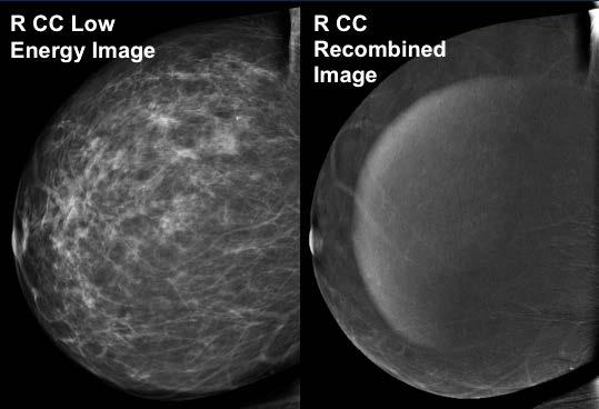
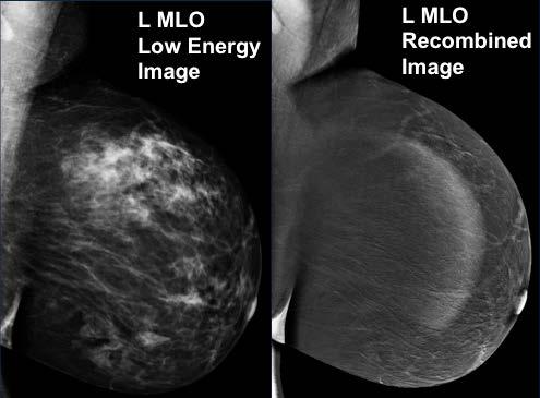
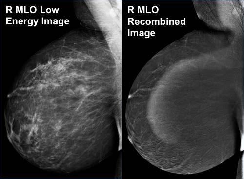

7 Normal CESM
8 Objectives Imaging technique for CESM Appearance of breast cancer on CESM Role of CESM in cancer staging Primary detection Ipsilateral Extent of disease Detection of contralateral disease Role of CESM in cancer follow up Limitations of CESM


9 Appearance of Breast Cancer on CESM - Mass L CC L CC L MLO L MLO Path = IDC


10 Cancer on CESM Non Mass Enhancement Path = DCIS

11 CESM vs. Digital Mammography Several studies comparing CESM to standard FFDM Improved sensitivity and specificity of CESM in the detection of breast cancer, especially in pre-menopausal women and women with dense breasts.
12 Objectives Imaging technique for CESM Appearance of breast cancer on CESM Role of CESM in cancer staging Primary detection Screening Recalls and Symptomatic Patients Ipsilateral Extent of disease Detection of contralateral disease Role of CESM in cancer follow up Limitations of CESM
 Lobbes, M. B. I., Lalji, U., Houwers, J., Nijssen, E. C.")
13 CESM for Screening Recalls CESM (Black bar) improved sensitivity, specificity, PPV, and NPV when compared to FFDM (grey bar) CESM was especially useful in downgrading mammographic lesions based on false-positive recalls, as well as decreasing the need for 6-12 month follow up imaging for inconclusive findings (i.e., BI-RADS 3 findings) Lobbes, M. B. I., Lalji, U., Houwers, J., Nijssen, E. C., Nelemans, P. J., van Roozendaal, L., et al. (2014). Contrastenhanced spectral mammography in patients referred from the breast cancer screening programme. European Radiology, 24(7),
14 CESM for Screening Recalls CESM increased diagnostic accuracy with improved sensitivity 96.9 % (+3.9 %), specificity 69.7 % (+33.8 %) and area under the ROC curve (+0.188). Lalji, U. C., Houben, I. P. L., Prevos, R., Gommers, S., van Goethem, M., Vanwetswinkel, S., et al. (2016). Contrastenhanced spectral mammography in recalls from the dutch breast cancer screening program: Validation of results in a large multireader, multicase study. European Radiology, 26(12),

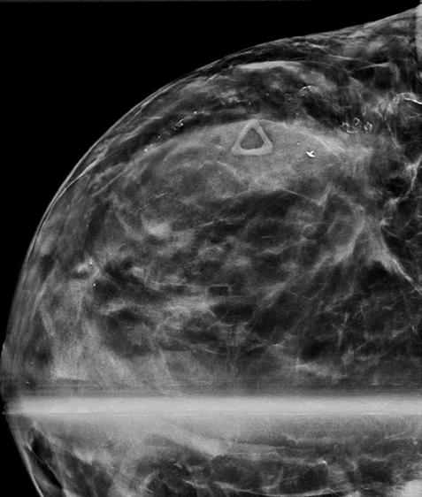
15 70 y.o. Baseline Exam


16 Initial Imaging for Screening Recall CESM
17 CESM vs. FFDM for Symptomatic Patients Retrospective reader studies comparing CESM over standard mammography in symptomatic patients with suspicious palpable abnormalities ROC analysis performance of CESM over FFDM, with area under the curve of 0.93 versus 0.83 (p<0.025). Increased Sensitivity (95% versus 84%, p<0.025) Increased Specificity (81% versus 63%, p<0.025) Tumour size estimation at CESM was significantly more accurate than FFDM alone, the latter tending to undersize lesions. S.L. Tennant, J.J. James, E.J. Cornford, et al.contrast-enhanced spectral mammography improves diagnostic accuracy in the symptomatic setting Clin Radiol, 71 (11) (2016), pp
18 Objectives Imaging technique for CESM Appearance of breast cancer on CESM Role of CESM in cancer staging Primary detection Ipsilateral Extent of disease Detection of contralateral disease Role of CESM in cancer follow up Limitations of CESM
19 Tumor Size: Comparison of CESM to FFDM and US Comparison of mean tumor size estimation on CESM, FFDM, and US vs. pathology gold standard Improved assessment of tumor size compared to FFDM and US C. Dromain, F. Thibault, S. Muller, et al.dual-energy contrast-enhanced digital mammography: initial clinical results Eur Radiol, 21 (2011), pp K.S. Blum, C. Rubbert, B. Mathys, et al.use of contrast-enhanced spectral mammography for intramammary cancer staging: preliminary results Acad Radiol, 21 (2014), pp
20 Tumor size: Comparison of CESM to MRI Prospective study 80 cases (61 IDC, 13 ILC, 6 pure DCIS) Breast cancer was visible in 66/80 MG, 80/80 CESM, and 77/79 MRI examinations No significant difference was found between lesion size measurement on MRI and CESM compared with histopathology. EM Fallenberg, C Dromain, F Diekmann, et al.contrast-enhanced spectral mammography versus MRI: Initial results in the detection of breast cancer and assessment of tumour size Eur Radiol, 24 (2014), pp


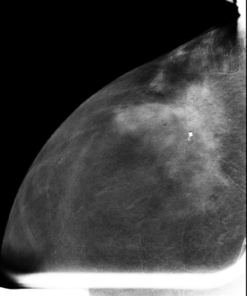
21 Tumor Size: Ill Defined Palpable Breast Mass Pt is on Dialysis: Unable to Have MRI

22 Tumor Size
23 Multifocal Breast Cancer R CC Low Energy Image R CC Recombined Image R MLO Low Energy Image R MLO Recombined Image


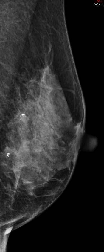
24 Surgical Planning L CC FFDM Image 72 y/o female with a screening detected 19 mm high density, irregular mass with indistinct margins Biopsy at an outside facility demonstrated invasive ductal carcinoma.

25 Case Extent 3 ( of Disease: Surgical Planning L CC Low Energy Image L CC Recombined Image CESM was peformed to better assess tumor size. Biopsy was subsequently performed, demonstrating multifocal IDC. CESM was able to better determine extent of disease and directed the location for staging biopsy. The patient was able to undergo breast conserving therapy.
26 Objectives Imaging technique for CESM Appearance of breast cancer on CESM Role of CESM in cancer staging Primary detection Ipsilateral Extent of disease Detection of contralateral disease Role of CESM in cancer follow up Limitations of CESM
27 Detection of Contralateral Disease Synchronous breast cancer occurs in 1-3% of newly diagnosed breast cancers Lobular cancer in the index breast, family history of breast cancer, young age at diagnosis of first cancer and BRCA mutations are important risk factors that are known to be at increased risk for BBC Padmanabhan N, Subramanyan A, Radhakrishna S. Synchronous Bilateral Breast Cancers. J Clin Diagn Res. 2015;9(9):XC05-XC08. McCaul KA. Bilateral breast cancer incidence and survival. [PhD thesis]. North Terrace, ADELAIDE SA 5005: University of Adelaide; Available from: University of Adelaide, School of Population Health and Clinical Practice, Library E-Reserve

28 Contralateral Malignancy R CC Low Energy Image R CC Recombined Image R MLO Low Energy Image R MLO Recombined Image


29 Prior Normal Left Mammogram with DBT Pathology showed ILC
30 Objectives Imaging technique for CESM Appearance of breast cancer on CESM Role of CESM in cancer staging Primary detection Ipsilateral Extent of disease Detection of contralateral disease Role of CESM in cancer follow up Limitations of CESM
31 Role of CESM in Cancer Follow Up Response to Neoadjuvant Therapy Morphology of tumor with LE image Physiology of contrast enhancement CESM and MRI lesion size were highly correlated CESM at least as reliable as MRI in assessing the response to NAC Iotti V1, et al. Contrast-enhanced spectral mammography in neoadjuvant chemotherapy monitoring: a comparison with breast magnetic resonance imaging. Breast Cancer Res Sep 11;19(1):106. doi: /s


32 Monitoring Response to Neoadjuvant Therapy R MLO Recombined CESM R MLO Low Energy CESM R MLO Recombined CESM R MLO Low Energy CESM Prior Current


33 Follow Up after BCT Dense or heterogeneously dense High risk patients when MRI is no longer covered by insurance Previous malignancy mammographically occult R CC CESM R CC
34 Objectives Imaging technique for CESM Appearance of breast cancer on CESM Role of CESM in cancer staging Primary detection Ipsilateral Extent of disease Detection of contralateral disease Role of CESM in cancer follow up Limitations of CESM
35 Limitations of CESM Contraindications for CESM No biopsy ability Allergy to intravenous iodinated contrast Acute kidney injury (increase in serum Cr by >0.3 mg/dl within 48 hours or increase of >1.5 times baseline within prior 7 days) Chronic renal failure (GFR <30), unless patient is on regular hemodialysis Breast Implants False Positives False Negatives


36 Limitations of CESM False Positives Benign Lesions Fibroadenomas are the most common benign lesion that demonstrate enhancement on CESM Papillomas, hamartomas, intra-mammary lymph nodes, fat necrosis, and complicated cysts can also enhance


37 Bilateral Breast Cancer with Incidental False Positive Fibroadenoma R MLO L MLO R MLO L MLO
38 False Positive Papilloma


39 False Negatvie CESM Screening detected calcs. On CESM no enhancement is seen in the area of the calcifications. Path = intermediate grade DCIS. The absence of enhancement does not deter biopsy of suspicious imaging findings seen on the low energy CESM images.


40 R CC Recombined Image R MLO Recombined Image False Negative CESM Path = Well-differentiated IDC with lobular features. R CC Low Energy Image R MLO Low Energy Image


41 CESM False Negative Likely due to masking effect of marked background enhancement
42 Objectives Imaging technique for CESM Appearance of breast cancer on CESM Role of CESM in cancer staging Primary detection Ipsilateral Extent of disease Detection of contralateral disease Role of CESM in cancer follow up Limitations of CESM
43 References Ali-Mucheru, M., Pockaj, B., Patel, B., Pizzitola, V., Wasif, N., Stucky, C. C., et al. (2016). Contrast-enhanced digital mammography in the surgical management of breast cancer. Annals of Surgical Oncology, 23(Suppl 5), Bhimani, C., Matta, D., Roth, R. G., Liao, L., Tinney, E., Brill, K., et al. (2017). Contrast-enhanced spectral mammography: Technique, indications, and clinical applications. Academic Radiology, 24(1), Cheung, Y., Lin, Y., Wan, Y., Yeow, K., Huang, P., Lo, Y., et al. (2014). Diagnostic performance of dual-energy contrast-enhanced subtracted mammography in dense breasts compared to mammography alone: Interobserver blind-reading analysis. European Radiology, 24(10), Covington, M. F., Pizzitola, V. J., Lorans, R., Pockaj, B. A., Northfelt, D. W., Appleton, C. M., et al. (2018). The future of contrast-enhanced mammography. AJR.American Journal of Roentgenology, 210(2), Dromain, C., Thibault, F., Muller, S., Rimareix, F., Delaloge, S., Tardivon, A., et al. (2011). Dual-energy contrast-enhanced digital mammography: Initial clinical results. European Radiology, 21(3), Fallenberg, E. M., Dromain, C., Diekmann, F., Engelken, F., Krohn, M., Singh, J. M., et al. (2014). Contrast-enhanced spectral mammography versus MRI: Initial results in the detection of breast cancer and assessment of tumour size.european Radiology, 24(1), Helal, M. H., Mansour, S. M., Zaglol, M., Salaleldin, L. A., Nada, O. M., & Haggag, M. A. (2017). Staging of breast cancer and the advanced applications of digital mammogram: What the physician needs to know? The British Journal of Radiology, 90(1071), Iotti, V., Ravaioli, S., Vacondio, R., Coriani, C., Caffarri, S., Sghedoni, R., et al. (2017). Contrast-enhanced spectral mammography in neoadjuvant chemotherapy monitoring: A comparison with breast magnetic resonance imaging.breast Cancer Research : BCR, 19(1), James, J. J., & Tennant, S. L. (2018). Contrast-enhanced spectral mammography (CESM) doi: Jochelson, M. S., Dershaw, D. D., Sung, J. S., Heerdt, A. S., Thornton, C., Moskowitz, C. S., et al. (2013). Bilateral contrast-enhanced dual-energy digital mammography: Feasibility and comparison with conventional digital mammography and MR imaging in women with known breast carcinoma. Radiology, 266(3), Lalji, U. C., Houben, I. P. L., Prevos, R., Gommers, S., van Goethem, M., Vanwetswinkel, S., et al. (2016). Contrast-enhanced spectral mammography in recalls from the dutch breast cancer screening program: Validation of results in a large multireader, multicase study. European Radiology, 26(12), Lee-Felker, S. A., Tekchandani, L., Thomas, M., Gupta, E., Andrews-Tang, D., Roth, A., et al. (2017). Newly diagnosed breast cancer: Comparison of contrast-enhanced spectral mammography and breast MR imaging in the evaluation of extent of disease. Radiology, 285(2), Lewin, J. M., Isaacs, P. K., Vance, V., & Larke, F. J. (2003). Dual-energy contrast-enhanced digital subtraction mammography: Feasibility. Radiology, 229(1), Li, L., Roth, R., Germaine, P., Ren, S., Lee, M., Hunter, K., et al. (2017). Contrast-enhanced spectral mammography (CESM) versus breast magnetic resonance imaging (MRI): A retrospective comparison in 66 breast lesions.diagnostic and Interventional Imaging, 98(2), Lobbes, M. B. I., Smidt, M. L., Houwers, J., Tjan-Heijnen, V. C., & Wildberger, J. E. (2013). Contrast enhanced mammography: Techniques, current results, and potential indications doi: " Lobbes, M. B. I., Lalji, U., Houwers, J., Nijssen, E. C., Nelemans, P. J., van Roozendaal, L., et al. (2014). Contrast-enhanced spectral mammography in patients referred from the breast cancer screening programme. European Radiology, 24(7), Luczynska, E., Heinze-Paluchowska, S., Dyczek, S., Blecharz, P., Rys, J., & Reinfuss, M. (2014). Contrast-enhanced spectral mammography: Comparison with conventional mammography and histopathology in 152 women. Korean Journal of Radiology, 15(6), Patel, B. K., Garza, S. A., Eversman, S., Lopez-Alvarez, Y., Kosiorek, H., & Pockaj, B. A. (2017). Assessing tumor extent on contrast-enhanced spectral mammography versus fullfield digital mammography and ultrasound. Clinical Imaging, 46, Patel, B. K., Lobbes, M. B. I., & Lewin, J. (2018). Contrast enhanced spectral mammography: A review. Seminars in Ultrasound, CT, and MR, 39(1), Patel, B. K., Hilal, T., Covington, M., Zhang, N., Kosiorek, H. E., Lobbes, M., et al. (2018). Contrast-enhanced spectral mammography is comparable to MRI in the assessment of residual breast cancer following neoadjuvant systemic therapy. Annals of Surgical Oncology, 25(5), Tennant, S. L., James, J. J., Cornford, E. J., Chen, Y., Burrell, H. C., Hamilton, L. J., et al. (2016). Contrast-enhanced spectral mammography improves diagnostic accuracy in the symptomatic setting doi:

44 Thank You!
Emerging Techniques in Breast Imaging: Contrast-Enhanced Mammography and Fast MRI
 Emerging Techniques in Breast Imaging: Contrast-Enhanced Mammography and Fast MRI Lilian Wang, M.D. Breast Imaging Section Department of Radiology Northwestern Medicine Overview Rationale for new imaging
Monitoring neo-adjuvant chemotherapy: comparison of contrast-enhanced spectral mammography (CESM) and MRI versus breast cancer characteristics
 and MRI versus breast cancer characteristics") Monitoring neo-adjuvant chemotherapy: comparison of contrast-enhanced spectral mammography (CESM) and MRI versus breast cancer characteristics Poster No.: B-1062 Congress: ECR 2016 Type: Scientific Paper
Monitoring neo-adjuvant chemotherapy: comparison of contrast-enhanced spectral mammography (CESM) and MRI versus breast cancer characteristics Poster No.: B-1062 Congress: ECR 2016 Type: Scientific Paper
Contrast-enhanced spectral mammography (CESM) in a large scale breast cancer screening program. Preliminary clinical experience.
 in a large scale breast cancer screening program. Preliminary clinical experience.") Contrast-enhanced spectral mammography (CESM) in a large scale breast cancer screening program. Preliminary clinical experience. Poster No.: C-1651 Congress: ECR 2012 Type: Educational Exhibit Authors:
Contrast-enhanced spectral mammography (CESM) in a large scale breast cancer screening program. Preliminary clinical experience. Poster No.: C-1651 Congress: ECR 2012 Type: Educational Exhibit Authors:
Contrast-Enhanced Digital Mammography
 2015 ARRS Breast Symposium Contrast-Enhanced Digital Mammography John Lewin, M.D. Diversified Radiology of Colorado CEDM - Outline History Technique Literature Review / Cases Clinical Status Inexpensive,
Mammographic imaging of nonpalpable breast lesions. Malai Muttarak, MD Department of Radiology Chiang Mai University Chiang Mai, Thailand
 Mammographic imaging of nonpalpable breast lesions Malai Muttarak, MD Department of Radiology Chiang Mai University Chiang Mai, Thailand Introduction Contents Mammographic signs of nonpalpable breast cancer
Pitfalls and Limitations of Breast MRI. Susan Orel Roth, MD Professor of Radiology University of Pennsylvania
 Pitfalls and Limitations of Breast MRI Susan Orel Roth, MD Professor of Radiology University of Pennsylvania Objectives Review the etiologies of false negative breast MRI examinations Discuss the limitations
Contrast Enhanced Spectral Mammography (CESM) Updates
 Updates") Contrast Enhanced Spectral Mammography (CESM) Updates Georgeta Mihai, PhD, DABR Medical Physicist, BIDMC, Boston Assistant Professor, Harvard Medical School, Boston Disclosures None Acknowledgments: Da
Contrast Enhanced Spectral Mammography (CESM) Updates Georgeta Mihai, PhD, DABR Medical Physicist, BIDMC, Boston Assistant Professor, Harvard Medical School, Boston Disclosures None Acknowledgments: Da
Contrast enhanced spectral mammography: A literature review
 Contrast enhanced spectral mammography: A literature review Poster No.: R-0172 Congress: RANZCR-AOCR 2012 Type: Authors: Keywords: DOI: Educational Exhibit S. Buzynski, D. Taylor; Perth/AU Breast, Digital
What s New in Breast Imaging. Jennifer A. Harvey, M.D., FACR Professor of Radiology University of Virginia
 What s New in Breast Imaging Jennifer A. Harvey, M.D., FACR Professor of Radiology University of Virginia Disclosure Hologic, Inc. Shareholder and research agreement. Volpara Solutions, Ltd. Shareholder
Contrast-enhanced spectral mammography: CESM descriptive analysis with pathologic correlation
 Contrast-enhanced spectral mammography: CESM descriptive analysis with pathologic correlation Poster No.: C-0930 Congress: ECR 2015 Type: Scientific Exhibit Authors: F. RUBIO, P. Escobar, C. Jurado; Sevilla/ES
OPEN ACCESS DIAGNOSTIC PERSPECTIVES
 OPEN ACCESS DIAGNOSTIC PERSPECTIVES Contrast-Enhanced Spectral Mammography: A Radiologic-Pathologic Perspective of a Novel Functional Imaging Modality for Breast Cancer Ma. Theresa Buenaflor 1 and Francis
BREAST. Keywords Breast cancer. Mammography. Quality control. CESM. CEDM
 Eur Radiol (2015) 25:2813 2820 DOI 10.1007/s00330-015-3695-2 BREAST Evaluation of low-energy contrast-enhanced spectral mammography images by comparing them to full-field digital mammography using EUREF
Contrast Enhanced Mammography (CE2D/CEDM) Selected Bibliography
 Selected Bibliography") Contrast Enhanced Mammography (CE2D/CEDM) Selected Bibliography Updated December 2017 Date Diagnostic MRI comparison Dose Outcomes Economics Cancer Detection Manufacturer The Future of Contrast-Enhanced
Contrast Enhanced Mammography (CE2D/CEDM) Selected Bibliography Updated December 2017 Date Diagnostic MRI comparison Dose Outcomes Economics Cancer Detection Manufacturer The Future of Contrast-Enhanced
BREAST MRI. Elizabeth A. Rafferty, M.D. Avon Comprehensive Breast Center Massachusetts General Hospital Harvard Medical School
 BREAST MRI Elizabeth A. Rafferty, M.D. Avon Comprehensive Breast Center Massachusetts General Hospital Harvard Medical School BREAST MRI Any assessment of the breast parenchyma requires the administration
Contrast Enhanced Spectral Mammography (CESM) Initial UK Experience. Dr Sarah L Tennant BMedSci, BMBS, MRCP, FRCR
 Initial UK Experience. Dr Sarah L Tennant BMedSci, BMBS, MRCP, FRCR") Contrast Enhanced Spectral Mammography (CESM) Initial UK Experience Dr Sarah L Tennant BMedSci, BMBS, MRCP, FRCR Vote Now Your experience of CESM 1. No experience of CESM 44% 2. I ve seen some cases in
Contrast Enhanced Spectral Mammography (CESM) Initial UK Experience Dr Sarah L Tennant BMedSci, BMBS, MRCP, FRCR Vote Now Your experience of CESM 1. No experience of CESM 44% 2. I ve seen some cases in
ROLE OF MRI IN SCREENING, DIAGNOSIS AND MANAGEMENT OF BREAST CANCER. B.Zandi Professor of Radiology
 ROLE OF MRI IN SCREENING, DIAGNOSIS AND MANAGEMENT OF BREAST CANCER B.Zandi Professor of Radiology Introduction In the USA, Breast Cancer is : The Most Common Non-Skin Cancer The Second Leading cause of
Oct 18. Sep 18. Jul 18. Aug 18 X X
 Comparative Dose of Contrast Enhanced Spectral Mammography (CESM), Digital Mammography, and Digital Breast Tomosynthesis. Phillips J, Mihai G, Hassonjee SE, Raj SD, Palmer MR, Brook A, Zhang D. AJR Am
MR sin plass i brystkreftdiagnostikk, dagens anbefalinger og fremtidsperspektiver
 MR sin plass i brystkreftdiagnostikk, dagens anbefalinger og fremtidsperspektiver Kathinka Kurz, MD, PhD, seksjonsoverlege SUS, kathinka.dehli.kurz@sus.no Technique - Subtraction Without contrast agent
Imaging in breast cancer. Mammography and Ultrasound Donya Farrokh.MD Radiologist Mashhad University of Medical Since
 Imaging in breast cancer Mammography and Ultrasound Donya Farrokh.MD Radiologist Mashhad University of Medical Since A mammogram report is a key component of the breast cancer diagnostic process. A mammogram
Initial Experience with Contrast Enhanced Digital Mammography (SenoBright) In a Comprehensive Clinical Breast Center
 In a Comprehensive Clinical Breast Center") Journal of Cancer Therapy, 2017, 8, 146-154 http://www.scirp.org/journal/jct ISSN Online: 2151-1942 ISSN Print: 2151-1934 Initial Experience with Contrast Enhanced Digital Mammography (SenoBright) In a
Journal of Cancer Therapy, 2017, 8, 146-154 http://www.scirp.org/journal/jct ISSN Online: 2151-1942 ISSN Print: 2151-1934 Initial Experience with Contrast Enhanced Digital Mammography (SenoBright) In a
BREAST MRI. Elizabeth A. Rafferty, M.D. Avon Comprehensive Breast Center Massachusetts General Hospital Harvard Medical School
 BREAST MRI Elizabeth A. Rafferty, M.D. Avon Comprehensive Breast Center Massachusetts General Hospital Harvard Medical School BREAST MRI Any assessment of the breast parenchyma requires the administration
Contrast-Enhanced Spectral Mammography
 Contrast-Enhanced Spectral Mammography Illuminating Breast Cancer Detection SenoBright HD TM gehealthcare.com/senobright Mammography is the most reliable imaging technique for breasts, but limitations
Breast Imaging! Ravi Adhikary, MD!
 Breast Imaging! Ravi Adhikary, MD! ACS Estimated Cancers Statistics 2014! Breast! New Cases in Women! 232,670 (+67,570 in situ)! Deaths in Women! 40,000! Colon! 48,380! 24,040! Cervical! 12,360! 4,020!
Standard Breast Imaging Modalities. Lilian Wang, M.D. Breast Imaging Section Department of Radiology Northwestern Medicine
 Standard Breast Imaging Modalities Lilian Wang, M.D. Breast Imaging Section Department of Radiology Northwestern Medicine Overview Standard breast imaging modalities Mammography Ultrasound MRI Imaging
EARLY DETECTION: MAMMOGRAPHY AND SONOGRAPHY
 EARLY DETECTION: MAMMOGRAPHY AND SONOGRAPHY Elizabeth A. Rafferty, M.D. Avon Comprehensive Breast Center Massachusetts General Hospital Harvard Medical School Breast Cancer Screening Early detection of
Amammography report is a key component of the breast
 Review Article Writing a Mammography Report Amammography report is a key component of the breast cancer diagnostic process. Although mammographic findings were not clearly differentiated between benign
BREAST MRI. VASILIKI FILIPPI RADIOLOGIST CT MRI & PET/CT Departments Hygeia Hospital, Athens, Greece
 BREAST MRI VASILIKI FILIPPI RADIOLOGIST CT MRI & PET/CT Departments Hygeia Hospital, Athens, Greece Breast ΜR Imaging (MRM) Breast MR imaging is an extremely powerful diagnostic tool, that when used in
Since its introduction in 2000, digital mammography has become
 Review Article Smith A, PhD email : Andrew.smith@hologic.com Since its introduction in 2000, digital mammography has become an accepted standard of care in breast cancer screening and has paved the way
Breast MRI Update. Jeffrey C. Weinreb, MD, FACR Yale University School of Medicine
 Breast MRI Update Jeffrey C. Weinreb, MD, FACR jeffrey.weinreb@yale.edu Yale University School of Medicine I disclose the following financial relationships with relevant commercial interests: Bracco Bayer
BREAST IMAGING and NEW IMAGING MODALITIES- A Surgeons view
 BREAST IMAGING and NEW IMAGING MODALITIES- A Surgeons view DR CHANTEL THORNTON SPECIALIST BREAST CANCER SURGEON BMSc (hons) MBBS (hons) FRACS Epworth Hospital, Richmond- Agora Centre for Women s Health
Financial Disclosures
 Financial Disclosures 3D Mammography: The Latest Developments in the Breast Imaging Arena I have no financial disclosures Dr. Katharine Lampen-Sachar Breast and Body Radiologist Radiology Associates of
Throughout this policy, bracketed numbers link topics across multiple sections according to the indication numbers in the following list.
 Subject: Magnetic Resonance Imaging of the Breast Page: 1 of 33 Last Review Status/Date: September 2015 Magnetic Resonance Imaging of the Breast Description Magnetic resonance imaging (MRI) of the breast
Assessment of extent of disease: digital breast tomosynthesis (DBT) versus full-field digital mammography (FFDM)
 versus full-field digital mammography (FFDM)") Assessment of extent of disease: digital breast tomosynthesis (DBT) versus full-field digital mammography (FFDM) Poster No.: C-1237 Congress: ECR 2012 Type: Scientific Paper Authors: N. Seo 1, H. H. Kim
Assessment of extent of disease: digital breast tomosynthesis (DBT) versus full-field digital mammography (FFDM) Poster No.: C-1237 Congress: ECR 2012 Type: Scientific Paper Authors: N. Seo 1, H. H. Kim
Angela Gilliam, MD University of Colorado Surgical Grand Rounds November 3, 2008
 Angela Gilliam, MD University of Colorado Surgical Grand Rounds November 3, 2008 Breast Cancer Most common cancer in American women 180,000 new cases per year Second most common cause of cancer death 44,000
Breast Cancer. Most common cancer among women in the US. 2nd leading cause of death in women. Mortality rates though have declined
 Breast Cancer Most common cancer among women in the US 2nd leading cause of death in women Mortality rates though have declined 1 in 8 women will develop breast cancer Breast Cancer Breast cancer increases
Does elastography change the indication to biopsy? IBDC
 Does elastography change the indication to biopsy? A LEXANDRA A THANASIOU, M D DEPARTMENT OF RADIOLOGY CURIE INSTITUTE PARIS, FRANCE IBDC Ultrasound Detected Cancers Physician-performed ultrasound increases
Breast Cancer. Saima Saeed MD
 Breast Cancer Saima Saeed MD Breast Cancer Most common cancer among women in the US 2nd leading cause of death in women 1 in 8 women will develop breast cancer Incidence/mortality rates have declined Breast
Contrast-Enhanced Spectral Mammography helps to improve breast cancer diagnostics
 GE Healthcare Contrast-Enhanced Spectral Mammography helps to improve breast cancer diagnostics SenoBright TM mammography, Midwest U.S. hospital Simple test benefits clinicians and patients A hospital
National Diagnostic Imaging Symposium 2013 SAM - Breast MRI 1
 National Diagnostic Imaging Symposium 2013 December 8-12, 2013 Disney s Yacht Club Resort Lake Buena Vista, Florida Self Assessment Module Questions, Answers and References Day SAM Title - Each SAM title
EARLY DETECTION: MAMMOGRAPHY AND SONOGRAPHY
 EARLY DETECTION: MAMMOGRAPHY AND SONOGRAPHY Elizabeth A. Rafferty, M.D. Avon Comprehensive Breast Center Massachusetts General Hospital Harvard Medical School Breast Cancer Screening Early detection of
Breast Imaging Lexicon
 9//201 200 BI RADS th Edition 201 BI RADS th Edition Breast Imaging Lexicon Mammographic Pathology and Assessment Categories Deborah Thames, R.T.(R)(M)(QM) The Advanced Health Education Center Nonmember:
UW Radiology Review Course Breast Calcifications. BI-RADS 5 th Edition
 UW Radiology Review Course Breast Calcifications Grace Kalish, MD Vantage Radiology BI-RADS 5 th Edition Benign Skin Vascular Large rod like Coarse popcorn Suspicious Amorphous Coarse heterogenous Fine
Contrast-enhanced Breast MRI RSSA 2013
 Contrast-enhanced Breast MRI RSSA 2013 Prof. dr. Maurice van den Bosch University Medical Center Utrecht, the Netherlands Index 1) Breast cancer 2) Why MRI of the breast 3) Technique 4) Interpretation
Armed Forces Institute of Pathology.
 Armed Forces Institute of Pathology www.radpath.com Armed Forces Institute of Pathology Breast Disease www.radpath.org Armed Forces Institute of Pathology Interpretation of Breast MRI Leonard M. Glassman
RSNA, /radiol Appendix E1. Methods
 RSNA, 2016 10.1148/radiol.2016151097 Appendix E1 Methods US and Near-infrared Data Acquisition Four optical wavelengths (740 nm, 780 nm, 808 nm, and 830 nm) were used to sequentially deliver the light
Here are examples of bilateral analog mammograms from the same patient including CC and MLO projections.
 Good afternoon. It s my pleasure to be discussing Diagnostic Breast Imaging over the next half hour. I m Wei Yang, Professor of Diagnostic Radiology and Chief, the Section of Breast Imaging as well as
MP Magnetic Resonance Imaging for Detection and Diagnosis of Breast Cancer
 Medical Policy MP 6.01.29 BCBSA Ref. Policy: 6.01.29 Last Review: 09/19/2018 Effective Date: 09/19/2018 Section: Radiology Related Policies 6.01.45 Computer-Aided Evaluation of Malignancy With Magnetic
Breast Cancer Imaging
 Breast Cancer Imaging I. Policy University Health Alliance (UHA) will cover breast imaging when such services meet the medical criteria guidelines (subject to limitations and exclusions) indicated below.
Triple-negative breast cancer: which typical features can we identify on conventional and MRI imaging?
 Triple-negative breast cancer: which typical features can we identify on conventional and MRI imaging? Poster No.: C-1862 Congress: ECR 2013 Type: Educational Exhibit Authors: V. Bertani 1, A. Gualano
MANAGEMENT OF DENSE BREASTS. Nichole K Ingalls, MD, MPH NW Surgical Specialists September 25, 2015
 MANAGEMENT OF DENSE BREASTS Nichole K Ingalls, MD, MPH NW Surgical Specialists September 25, 2015 No financial disclosures National Cancer Institute National Cancer Institute Increased Cancer Risk... DENSITY
Non-mass Enhancement on Breast MRI. Aditi A. Desai, MD Margaret Ann Mays, MD
 Non-mass Enhancement on Breast MRI Aditi A. Desai, MD Margaret Ann Mays, MD Breast MRI Important screening and diagnostic tool, given its high sensitivity for breast cancer detection Breast MRI - Indications
Detailed Program of the second BREAST IMAGING AND INTERVENTIONS PROGRAM am am : Clinician s requirements from breast imaging
 Detailed Program of the second BREAST IMAGING AND INTERVENTIONS PROGRAM 2012 Day one, 2 nd November BREAST IMAGING AND INTERVENTIONS PROGRAM 2012 9.00 AM 9.10 am Introduction 9.10 am - 9.30 am : Clinician
SenoBright Contrast Enhanced Spectral Mammography Technology. Ann-Katherine Carton Sylvie Saab-Puong Matt Suminski
 SenoBright Contrast Enhanced Spectral Mammography Technology Ann-Katherine Carton Sylvie Saab-Puong Matt Suminski White Paper October 2012 SenoBright Contrast Enhanced Spectral Mammography Technology Ann-Katherine
Ruud Pijnappel Professor of Radiology, UMC Utrecht. Chair Dutch Expert Centre for Screening Board EUSOBI
 Ruud Pijnappel Professor of Radiology, UMC Utrecht Best practice in Breast Imaging: what s new and what women need to know and Update on the Second Implementation Report of the 2003 Council Recommendation
Dual-energy contrast-enhanced digital mammography: initial clinical results
 Eur Radiol (2011) 21:565 574 DOI 10.1007/s00330-010-1944-y BREAST Dual-energy contrast-enhanced digital mammography: initial clinical results Clarisse Dromain & Fabienne Thibault & Serge Muller & Françoise
High Risk Screening: A Multimodality Approach
 High Risk Screening: A Multimodality Approach John Lewin, M.D., FACR, FSBI The Women s Imaging Center Denver, Colorado Disclosures Consultant to Hologic Previously received research funds from Hologic
Successful Breast MRI Program : The ingredients
 Successful Breast MRI Program : The ingredients Dr. Smriti Hari Associate Professor Deptt. Of Radiology All India Institute of Medical Sciences New Delhi How to perform Breast MRI Breast MRI descriptors
Radiologic and pathologic correlation of non-mass like breast lesions on US and MRI: Benign, high risk, versus malignant
 Radiologic and pathologic correlation of non-mass like breast lesions on US and MRI: Benign, high risk, versus malignant Poster No.: C-1161 Congress: ECR 2013 Type: Educational Exhibit Authors: J. Kwak,
Tomosynthesis and breast imaging update. Dr Michael J Michell Consultant Radiologist King's College Hospital NHS Foundation Trust
 Tomosynthesis and breast imaging update Dr Michael J Michell Consultant Radiologist King's College Hospital NHS Foundation Trust Breast imaging new technology BREAST CANCER FLT PET shows different grades
The role of magnetic resonance imaging in the diagnosis of breast cancer
 Review article NOWOTWORY Journal of Oncology 2017, volume 67, number 3, 185 192 DOI: 10.5603/NJO.2017.0031 Polskie Towarzystwo Onkologiczne ISSN 0029 540X www.nowotwory.edu.pl The role of magnetic resonance
Evaluation of BI-RADS 3 lesions in women with a high risk of hereditary breast cancer.
 Evaluation of BI-RADS 3 lesions in women with a high risk of hereditary breast cancer. Poster No.: C-0346 Congress: ECR 2014 Type: Scientific Exhibit Authors: A. Thomas 1, R. Dominguez Oronoz 1, S. Roche
Breast Imaging: Multidisciplinary Approach. Madelene Lewis, MD Assistant Professor Associate Program Director Medical University of South Carolina
 Breast Imaging: Multidisciplinary Approach Madelene Lewis, MD Assistant Professor Associate Program Director Medical University of South Carolina No Disclosures Objectives Discuss a multidisciplinary breast
Is Probably Benign Really Just Benign? Peter R Eby, MD, FSBI Virginia Mason Medical Center Seattle, WA
 Is Probably Benign Really Just Benign? Peter R Eby, MD, FSBI Virginia Mason Medical Center Seattle, WA Disclosures: CONSULTANT FOR DEVICOR MEDICAL ARS Question 1 Is probably benign really just benign?
BI-RADS Categorization As a Predictor of Malignancy 1
 Susan G. Orel, MD Nicole Kay, BA Carol Reynolds, MD Daniel C. Sullivan, MD BI-RADS Categorization As a Predictor of Malignancy 1 Index terms: Breast, biopsy, 00.1261 Breast neoplasms, localization, 00.125,
Improving Reading Time of Digital Breast Tomosynthesis with Concurrent Computer Aided Detection
 White Paper Improving Reading Time of Digital Breast Tomosynthesis with Concurrent Computer Aided Detection WHITE PAPER 2 3 Abstract PowerLook Tomo Detection, a concurrent computer-aided detection (CAD)
Outline. Digital Breast Tomosynthesis: Update and Pearls for Implementation. Tomosynthesis Dataset: 2D/3D (Hologic Combo Acquisition)
") Outline Digital Breast Tomosynthesis (DBT) the new standard of care Digital Breast Tomosynthesis: Update and Pearls for Implementation Emily F. Conant, M.D. Professor, Chief of Breast Imaging Department
Outline Digital Breast Tomosynthesis (DBT) the new standard of care Digital Breast Tomosynthesis: Update and Pearls for Implementation Emily F. Conant, M.D. Professor, Chief of Breast Imaging Department
Ana Sofia Preto 19/06/2013
 Ana Sofia Preto 19/06/2013 Understanding the underlying pathophysiologic processes leading to the various types of calcifications Description and illustration of the several types of calcifications, according
Breast MRI: Friend or Foe?
 Breast : Friend or Foe? APPLEGATE HAS DOUBLE MASTECTOMY IN CANCER SCARE DIAGNOSED WITH CANCER IN ONE BREAST Comments: 0 ASSOCIATED PRESS 8/19/2008 UCSF Postgraduate Course March 19, 2009 E. Shelley Hwang
Screening Options in Dense Breasts. Donna Plecha, M.D. Co-Director UHCMC Breast Centers Associate Professor of Radiology Director of Breast Imaging
 Screening Options in Dense Breasts Donna Plecha, M.D. Co-Director UHCMC Breast Centers Associate Professor of Radiology Director of Breast Imaging Dense Breasted Women Decreased sensitivity of mammography
BI-RADS and Breast MRI. Kathy Borovicka, M.D. Thursday February 15, 2018
 BI-RADS and Breast MRI Kathy Borovicka, M.D. Thursday February 15, 2018 Learning Objectives Be familiar with the Breast Imaging Reporting and Data System (BI-RADS) Understand the components of a breast
Breast MRI: Friend or Foe?
 Breast MRI: Friend or Foe? UCSF Postgraduate Course May 18, 2013 Cheryl Ewing, MD Clinical Professor of Surgery UCSF Department of Surgery APPLEGATE HAS DOUBLE MASTECTOMY IN CANCER SCARE DIAGNOSED WITH
Leonard M. Glassman MD
 BI-RADS The New BI-RADS Leonard M. Glassman MD FACR Former Chief of Breast Imaging American Institute for Radiologic Pathology Washington Radiology Associates, PC Breast Imaging Reporting and Data System
Diagnostic Dilemmas of Breast Imaging
 Diagnostic Dilemmas of Breast Imaging Common Causes of Error in Breast Cancer Detection By: Jason Cord, M.D. Mammography: Initial Imaging The standard for detection of breast cancer Screening mammography
PAAF vs Core Biopsy en Lesiones Mamarias Case #1
 5/19/2014 PAAF vs Core Biopsy en Lesiones Mamarias Case #1 Fine Needle Aspiration Cytology of Breast: Correlation with Needle Core Biopsy 64-year-old woman Mass in breast Syed Hoda, MD CD31 Post-Radiation
Digital breast tomosynthesis (DBT) occult breast cancers: clinical, radiological and histopathological features.
 occult breast cancers: clinical, radiological and histopathological features.") Digital breast tomosynthesis (DBT) occult breast cancers: clinical, radiological and histopathological features. Poster No.: C-1707 Congress: ECR 2015 Type: Scientific Exhibit Authors: V. Vinci 1, A. Iqbal
Digital breast tomosynthesis (DBT) occult breast cancers: clinical, radiological and histopathological features. Poster No.: C-1707 Congress: ECR 2015 Type: Scientific Exhibit Authors: V. Vinci 1, A. Iqbal
Disclosures. Breast Cancer. Breast Imaging Modalities. Breast Cancer Screening. Breast Cancer 6/4/2014
 : Information for the Primary Care Physician Disclosures No financial relationships with commercial entities producing health care products/services. Roxsann Roberts, MD Section Chief, MRI Erlanger/EmCare
Tips and Tricks to performing Magnetic Resonance Imaging Guided Breast Interventional Procedures Habib Rahbar, MD, FSBI October 23, 2018, 7:00pm ET
 Tips and Tricks to performing Magnetic Resonance Imaging Guided Breast Interventional Procedures Habib Rahbar, MD, FSBI October 23, 2018, 7:00pm ET SAM Questions/Answers/Rationales/References 1. Below
S. Murgo, MD. Chr St-Joseph, Mons Erasme Hospital, Brussels
 S. Murgo, MD Chr St-Joseph, Mons Erasme Hospital, Brussels? Introduction Mammography reports are sometimes ambiguous and indecisive. ACR has developped the BIRADS. BIRADS consists of a lexicon in order
Can Digital Breast Tomosynthesis(DBT) Perform Better than Standard Digital Mammography Workup in a Breast Cancer Assessment Clinic?
 Perform Better than Standard Digital Mammography Workup in a Breast Cancer Assessment Clinic?") Can Digital Breast Tomosynthesis(DBT) Perform Better than Standard Digital Mammography Workup in a Breast Cancer Assessment Clinic? Accepted for publication in European Radiology Authors: S Mall, J Noakes,
Can Digital Breast Tomosynthesis(DBT) Perform Better than Standard Digital Mammography Workup in a Breast Cancer Assessment Clinic? Accepted for publication in European Radiology Authors: S Mall, J Noakes,
Breast cancer tumor size: Correlation between MRI and histopathology
 Breast cancer tumor size: Correlation between MRI and histopathology Poster No.: C-0409 Congress: ECR 2010 Type: Topic: Scientific Exhibit Breast Authors: H. Khan, M. Hoosein, M. Alattar, S. Tenant, L.
Management of Palpable Abnormalities in the Breast Katerina Dodelzon, MD July 31, 2018, 7:00pm ET
 Management of Palpable Abnormalities in the Breast Katerina Dodelzon, MD July 31, 2018, 7:00pm ET SAM Questions 1. 21 year old female presenting with left breast palpable mass, what is the most appropriate
Breast Cancer Screening and Diagnosis
 Breast Cancer Screening and Diagnosis Priya Thomas, MD Assistant Professor Clinical Cancer Prevention and Breast Medical Oncology University of Texas MD Anderson Cancer Center Disclosures Dr. Thomas has
AB MR Interpretation Overview
 AB MR Interpretation Overview Goal of AB MR interpretation is to maintain high sensitivity and specificity In order to minimize false positives and short term follow ups, it is fundamental to focus only
Medical Policy An independent licensee of the Blue Cross Blue Shield Association
 Magnetic Resonance Imaging (MRI) of the Breast Page 1 of 51 Medical Policy An independent licensee of the Blue Cross Blue Shield Association Title: Magnetic Resonance Imaging (MRI) of the Breast Professional
Correlation between lesion type and the additional value of digital breast tomosynthesis
 Correlation between lesion type and the additional value of digital breast tomosynthesis Poster No.: C-1604 Congress: ECR 2011 Type: Scientific Exhibit Authors: C. Van Ongeval, L. Cockmartin, A. Van Steen,
Lesion Imaging Characteristics Mass, Favoring Benign Circumscribed Margins Intramammary Lymph Node
 Lesion Imaging Characteristics Mass, Favoring Benign Circumscribed Margins Intramammary Lymph Node Oil Cyst Mass, Intermediate Concern Microlobulated Margins Obscured Margins Mass, Favoring Malignant Indistinct
Emerging Technologies in Breast Imaging
 June 30, 2016 Emerging Technologies in Breast Imaging Jay A. Baker, M.D. Division of Breast Imaging New Technologies in Breast Imaging Full Field Digital Mammography (FFDM) X-ray/Mammography Computer-Aided
BREAST DENSITY WHAT IS IT? WHY IS IT IMPORTANT? & What IOWA SF250 Means to Patients and Providers
 BREAST DENSITY WHAT IS IT? WHY IS IT IMPORTANT? & What IOWA SF250 Means to Patients and Providers Arnold Honick, MD Radiology Consultants of Iowa, PLC ahonick@rciowa.com BREAST DENSITY LEGISLATION Nancy
MEDICAL IMAGING AND BREAST DISEASE HOW CAN WE HELP YOU?
 MEDICAL IMAGING AND BREAST DISEASE HOW CAN WE HELP YOU? Barbara M. Preston, M.D. SCREENING MAMMOGRAPHY AVERAGE RISK PATIENTS KAISER RECOMMENDATION: ALL WOMEN (INCLUDING TRANSGENDER FEMALES) Every 1-21
Diagnostic benefits of ultrasound-guided. CNB) versus mammograph-guided biopsy for suspicious microcalcifications. without definite breast mass
 versus mammograph-guided biopsy for suspicious microcalcifications. without definite breast mass") Volume 118 No. 19 2018, 531-543 ISSN: 1311-8080 (printed version); ISSN: 1314-3395 (on-line version) url: http://www.ijpam.eu ijpam.eu Diagnostic benefits of ultrasound-guided biopsy versus mammography-guided
Volume 118 No. 19 2018, 531-543 ISSN: 1311-8080 (printed version); ISSN: 1314-3395 (on-line version) url: http://www.ijpam.eu ijpam.eu Diagnostic benefits of ultrasound-guided biopsy versus mammography-guided
Can magnetic resonance imaging obviate the need for biopsy for microcalcifications?
 Original Article Can magnetic resonance imaging obviate the need for biopsy for microcalcifications? Shinya Yamamoto, Takashi Chishima Department of Breast Surgery, Yokohama Rosai Hospital, Yokohama 222-0036,
DR AISHA A UMAR CHIEF CONSULTANT RADIOLOGIST NATIONAL HOSPITAL ABUJA.
 DR AISHA A UMAR CHIEF CONSULTANT RADIOLOGIST NATIONAL HOSPITAL ABUJA. OUTLINE WHY DO WE IMAGE WHOM TO IMAGE WHEN TO IMAGE HOW TO IMAGE WHAT TO IMAGE WITH PERSONAL EXPERIENCE CONCLUSION/RECOMMENDATIONS
Comparison between the role of contrast enhanced mammography and dynamic contrast enhanced MRI in assessment of breast cancer recurrence
 The Egyptian Journal of Hospital Medicine (October 2018) Vol. 73(1), Page 5875-5885 Comparison between the role of contrast enhanced mammography and dynamic contrast enhanced MRI in assessment of breast
MRI in breast cancer: diagnosis and intervention. Dr Sue Barter Addenbrookes Hospital, Cambridge UK
 MRI in breast cancer: diagnosis and intervention Dr Sue Barter Addenbrookes Hospital, Cambridge UK Intervention will be discussed in High Risk Screening! Indications UK and Europe: Breast MRI is well established
Invasive lobular carcinoma of the breast; spectrum of imaging findings.
 Invasive lobular carcinoma of the breast; spectrum of imaging findings. Poster No.: C-0847 Congress: ECR 2014 Type: Educational Exhibit Authors: D. Mandich, T. Diaz de Bustamante, L. Koren, M. Arroyo,
Current Status of Supplementary Screening With Breast Ultrasound
 Current Status of Supplementary Screening With Breast Ultrasound Stephen A. Feig, M.D., FACR Fong and Jean Tsai Professor of Women s Imaging Department of Radiologic Sciences University of California,
Combined Screening With Ultrasound and Mammography vs Mammography Alone in Women at Elevated Risk of Breast Cancer JAMA. 2008;299(18):
:") ORIGINAL CONTRIBUTION Combined Screening With Ultrasound and vs Alone in Women at Elevated Risk of Breast Cancer Wendie A. Berg, MD, PhD Jeffrey D. Blume, PhD Jean B. Cormack, PhD Ellen B. Mendelson, MD
ORIGINAL CONTRIBUTION Combined Screening With Ultrasound and vs Alone in Women at Elevated Risk of Breast Cancer Wendie A. Berg, MD, PhD Jeffrey D. Blume, PhD Jean B. Cormack, PhD Ellen B. Mendelson, MD
Compressive Re-Sampling for Speckle Reduction in Medical Ultrasound
 Compressive Re-Sampling for Speckle Reduction in Medical Ultrasound Professor Richard Mammone Rutgers University Email Phone Number Christine Podilchuk, Lev Barinov, Ajit Jairaj and William Hulbert ClearView
A comparison of the accuracy of film-screen mammography, full-field digital mammography, and digital breast tomosynthesis
 Clinical Radiology xxx (2012) 1e6 Contents lists available at SciVerse ScienceDirect Clinical Radiology journal homepage: www.clinicalradiologyonline.net A comparison of the accuracy of film-screen mammography,