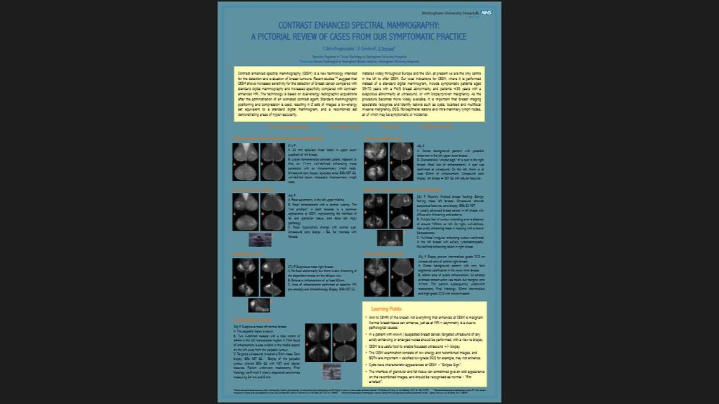
Contrast Enhanced Spectral Mammography (CESM) Initial UK Experience. Dr Sarah L Tennant BMedSci, BMBS, MRCP, FRCR
|
|
|
- Joanna Cross
- 5 years ago
- Views:
Transcription

1 Contrast Enhanced Spectral Mammography (CESM) Initial UK Experience Dr Sarah L Tennant BMedSci, BMBS, MRCP, FRCR
2 Vote Now Your experience of CESM 1. No experience of CESM 44% 2. I ve seen some cases in journals/at conferences 54% 3. I ve had to review CESM performed elsewhere in my routine practice 1% 4. I use CESM in my routine practice 1% 10
3 Which opinion most closely matches your views on CESM? 1. It s just poor man s MRI 11% 2. We don t have GE equipment, so it s not relevant to me 0% 3. I can see its potential 42% 4. I don t know enough about it to have an opinion Vote Now 47% 10
4 What is CESM? A relatively new technology Nottingham Breast Institute (NBI) is the first UK centre Essentially, a mammogram with the aid of contrast
5
6 CESM - Physics Standard digital mammography might use Rh/Rh target/filter combination at 29 kvp K edge of iodine is 33.2keV A typical clinical concentration of iodine in the breast results in a low signal intensity, and is hardly distinguishable from the background
7 CESM - Physics CESM is based on dual-energy acquisitions 2 images acquired using distinct low-energy (LE standard mammography KV and filtration) and high-energy (HE higher KV with strong filtration) X-ray spectra The differences between X-ray attenuation of iodine and breast tissues at these two energy levels are exploited to suppress the background breast tissue
8 Typical LE and HE spectra used with Senobright and attenuation of breast tissue and iodine as a function of x-ray energy
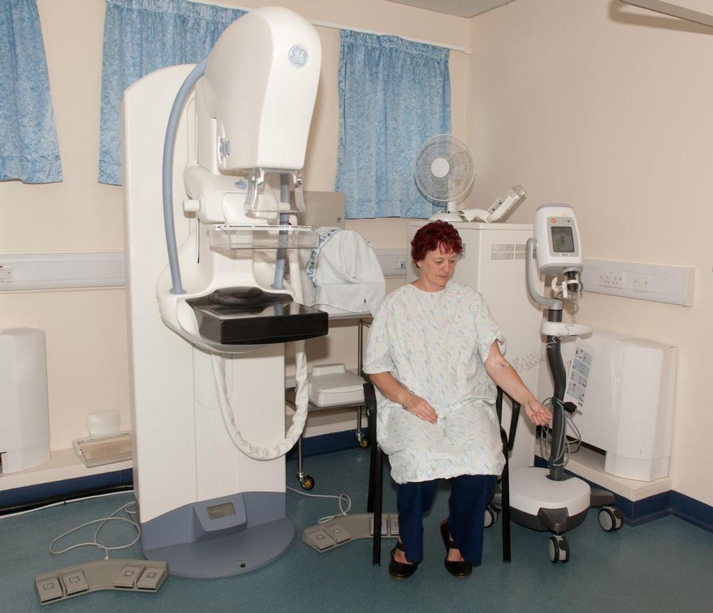
9 CESM at NBI Introduction of a New Technology Local R&D team advice Classified as Service Evaluation providing written into departmental protocols Registered with local Audit and New Technologies committees Patient Information Sheet Consent Form
10 CESM at NBI Current Indications First-line imaging (instead of a standard digital mammogram) in symptomatic patients with a breast abnormality classified clinically as malignant in patients >35 years Second-line imaging (instead of a standard digital mammogram) in symptomatic patients <35 years with a malignant-appearing abnormality at ultrasound
11 CESM at NBI Practicalities Majority of patients will not have recent renal function/egfr Drew up specific criteria to exclude those patients at significant risk of contrastnephropathy
12 CESM at NBI Contra-indications Pregnancy Lactation Iodine allergy Renal failure Diabetic +/- on Metformin (unless recent, normal renal function available) Inability to give informed consent Inability to tolerate mammography Age >70 (specifically to reduce risk of contrast nephropathy)

13
14 CESM at NBI Practicalities Warming cabinet Resus and anaphylaxis training Iopamidol 300, 100mls, through a pump injector at 3mls/sec No CRIS code we use XMCGB (old code for a breast pneumocystogram)
15

16 Typical CESM Imaging Procedure for a bilateral breast examination with 2 views per breast
17 CESM at NBI So far so good We did our first case on 26 th November 2013 As of 7 th November 2014, we have performed 114 CESM examinations 1 contrast reaction No documented nephropathies 1 technical failure (wrong paddle) Audit ongoing but initial results promising excellent correlation with MRI and histology, and low false-positives

18 Case 1 30 Y F presents with a benign-feeling mass in the left breast (P3) Ultrasound performed as first-line imaging due to patient age This showed a solid lesion and 14g corebiopsy was obtained
19 Case 1 Core biopsy showed invasive carcinoma of no special type (NST), grade 2 MDT decision to offer CESM instead of standard digital mammogram for staging

20 Low Energy

21 Recombined Imaging The recently biopsied tumour in the left upper midline is seen as an enhancing mass measuring 21 mm. There is a second enhancing mass inferior to this in the left central breast, which measures 12mm. Total area to include both lesions is 40 mm. The right breast is within normal limits.

22 Case 1 Focussed ultrasound showed the second lesion, and ultrasoundguided core biopsy confirmed NST grade 2 tumour
23 Case 1 Mastectomy confirmed 2 well-defined tumour foci 19 and 13mm Associated multiple tumour foci with lobular features and vascular invasion Whole tumour size 35mm
24 Case 2 65 Y F Suspicious thickening right upper outer breast

25 Case 2 - Low energy MLOs

26 Case 2 - Low energy CCs

27 Case 2 Recombined MLOs

28 34mm avidly enhancing mass Case 2 Recombined CCs
29 Case 2 - Ultrasound
30 Case 3 Ultrasound-guided core biopsy - Grade 2 tumour of No Special Type (NST) Patient opted for WLE Invasive Carcinoma Size: 24 mm Whole tumour (DCIS + invasive carcinoma) size: 34 mm Localised, Grade 3, NST with lymphocyte rich stroma Had contralateral reduction too - all benign
31 Case 3 67 Y F Suspicious mass right breast centrally

32 Case 3 - Low energy MLOs

33 Case 3 - Low energy CCs

34 Case 3 Recombined MLOs

35 Case 3 Recombined CCs

36 A - 22mm enhancing mass B - 5mm enhancing mass Case 3 - Zoomed Case 3 Zoomed CC


37 Case 3 - Ultrasound Main lesion Second lesion
38 Case 3 - Ultrasound core biopsy Main lesion = Grade 2 tumour with lobular features Second lesion = NST, grade 1 Patient opted for mastectomy
39 Case 3 - Final histology - Mastectomy 2 distinct tumours A: pure special type lobular, Grade 2, 30mm B: NST, grade 1, 4mm
40 Case 4 25 Y Previously attended with benign-feeling change right breast Ultrasound-guided core of a vague area of reduced echo change came back as intermediate grade DCIS Mammogram required for accurate staging

41 Case 4 LE MLOs

42 Case 4 LE CCs

43 Case 4 LE MLO, zoomed and windowed

44 Case 4 Recombined MLOs

45 Case 4 Recombined CCs 48mm enhancement corresponding to faint calcification
46 Case 4 Attempt at WLE ultrasound wire guided 50mm HGDCIS with foci of micro invasion - margin <1mm Mastectomy and SNB performed no residual disease
47 Case 5 45 Y Suspicious mass right breast

48 Case 5 LE MLOs

49 Case 5 LE CCs

50 Case 5 Recombined MLOs

51 Case 5 Recombined CCs Eclipse sign of cyst, and faint nodular background enhancement in both breasts

52 Case 5 Ultrasound showed a simple cyst which was aspirated to dryness Patient reassured and discharged
53 Case 6 61 Y F Large ill defined mass right breast with skin tether and dimpling

54 Case 6 LE MLOs

55 Case 6 LE CCs

56 Case 6 Recombined MLOs

57 Case 6 Recombined CCs LE images show a spiculate mass in the central right breast but enhancement on the recombined images is more extensive - approx. 60mm
58 Case 6 MDT decision neoadjuvant chemotherapy Baseline MRI performed, as is current protocol

59 Case 6 CEMRI Axial Reformat
60 Case 6 CEMRI Sagittal Reformat The total size of abnormality at MRI is 55mm

61 Case 6 -Right recombined MLO vs Sagittal CEMRI Reformat
62
63 The Pros Instant access performed in clinic Quick to perform and report Hangs well on PACS can scroll between low and recombined images Excellent correlation with MRI and pathological size increases radiologists confidence in accurately assessing local stage
64 The Cons Slows clinic down a little (mainly consenting process) Can t see as far back as on an MRI (chest wall lesions may be missed)
65 The Future? High-risk screening (dense breasts) Surveillance/follow-up Problem-solving/screening assessment Neoadjuvant response
66 Which opinion most closely matches your views on CESM now? 1. It s just poor man s MRI 10.8% 2. We don t have GE equipment, so it s not relevant to me 0.0% 3. I can see its potential 4. I don t know enough about it to have an opinion 2.0% Vote Now 87.3% 10


67 @drsarahtennant
Contrast-Enhanced Spectral Mammography helps to improve breast cancer diagnostics
 GE Healthcare Contrast-Enhanced Spectral Mammography helps to improve breast cancer diagnostics SenoBright TM mammography, Midwest U.S. hospital Simple test benefits clinicians and patients A hospital
Contrast-Enhanced Spectral Mammography
 Contrast-Enhanced Spectral Mammography Illuminating Breast Cancer Detection SenoBright HD TM gehealthcare.com/senobright Mammography is the most reliable imaging technique for breasts, but limitations
SenoBright Contrast Enhanced Spectral Mammography Technology. Ann-Katherine Carton Sylvie Saab-Puong Matt Suminski
 SenoBright Contrast Enhanced Spectral Mammography Technology Ann-Katherine Carton Sylvie Saab-Puong Matt Suminski White Paper October 2012 SenoBright Contrast Enhanced Spectral Mammography Technology Ann-Katherine
BREAST IMAGING and NEW IMAGING MODALITIES- A Surgeons view
 BREAST IMAGING and NEW IMAGING MODALITIES- A Surgeons view DR CHANTEL THORNTON SPECIALIST BREAST CANCER SURGEON BMSc (hons) MBBS (hons) FRACS Epworth Hospital, Richmond- Agora Centre for Women s Health
Contrast-Enhanced Digital Mammography
 2015 ARRS Breast Symposium Contrast-Enhanced Digital Mammography John Lewin, M.D. Diversified Radiology of Colorado CEDM - Outline History Technique Literature Review / Cases Clinical Status Inexpensive,
Contrast Enhanced Spectral Mammography (CESM) Updates
 Updates") Contrast Enhanced Spectral Mammography (CESM) Updates Georgeta Mihai, PhD, DABR Medical Physicist, BIDMC, Boston Assistant Professor, Harvard Medical School, Boston Disclosures None Acknowledgments: Da
Contrast Enhanced Spectral Mammography (CESM) Updates Georgeta Mihai, PhD, DABR Medical Physicist, BIDMC, Boston Assistant Professor, Harvard Medical School, Boston Disclosures None Acknowledgments: Da
Here are examples of bilateral analog mammograms from the same patient including CC and MLO projections.
 Good afternoon. It s my pleasure to be discussing Diagnostic Breast Imaging over the next half hour. I m Wei Yang, Professor of Diagnostic Radiology and Chief, the Section of Breast Imaging as well as
Imaging in breast cancer. Mammography and Ultrasound Donya Farrokh.MD Radiologist Mashhad University of Medical Since
 Imaging in breast cancer Mammography and Ultrasound Donya Farrokh.MD Radiologist Mashhad University of Medical Since A mammogram report is a key component of the breast cancer diagnostic process. A mammogram
Breast Health and Imaging Glossary
 Contact: Lorna Vaughan HerSpace Breast Imaging & Biopsy Associates 300 State Route 35 South W. Long Branch, NJ 07764 732-571-9100, ext. 104 lorna@breast-imaging.com Breast Health and Imaging Glossary Women
Armed Forces Institute of Pathology.
 Armed Forces Institute of Pathology www.radpath.com Armed Forces Institute of Pathology Breast Disease www.radpath.org Armed Forces Institute of Pathology Interpretation of Breast MRI Leonard M. Glassman
Case Scenario 1 History and Physical 3/15/13 Imaging Pathology
 Case Scenario 1 History and Physical 3/15/13 The patient is an 84 year old white female who presented with an abnormal mammogram. The patient has a five year history of refractory anemia with ringed sideroblasts
Standard Breast Imaging Modalities. Lilian Wang, M.D. Breast Imaging Section Department of Radiology Northwestern Medicine
 Standard Breast Imaging Modalities Lilian Wang, M.D. Breast Imaging Section Department of Radiology Northwestern Medicine Overview Standard breast imaging modalities Mammography Ultrasound MRI Imaging
Mammography and Other Screening Tests. for Breast Problems
 301.681.3400 OBGYNCWC.COM Mammography and Other Screening Tests What is a screening test? for Breast Problems A screening test is used to find diseases, such as cancer, in people who do not have signs
Case study 1. Rie Horii, M.D., Ph.D. Division of Pathology Cancer Institute Hospital, Japanese Foundation for Cancer Research
 NCCN/JCCNB Seminar in Japan April 15, 2012 Case study 1 Rie Horii, M.D., Ph.D. Division of Pathology Cancer Institute Hospital, Japanese Foundation for Cancer Research Present illness: A 50y.o.premenopausal
Breast Imaging! Ravi Adhikary, MD!
 Breast Imaging! Ravi Adhikary, MD! ACS Estimated Cancers Statistics 2014! Breast! New Cases in Women! 232,670 (+67,570 in situ)! Deaths in Women! 40,000! Colon! 48,380! 24,040! Cervical! 12,360! 4,020!
Angela Gilliam, MD University of Colorado Surgical Grand Rounds November 3, 2008
 Angela Gilliam, MD University of Colorado Surgical Grand Rounds November 3, 2008 Breast Cancer Most common cancer in American women 180,000 new cases per year Second most common cause of cancer death 44,000
Breast Cancer Screening and Treatment Mrs Belinda Scott Breast Surgeon Breast Associates Auckland
 Breast Cancer Screening and Treatment 2009 Mrs Belinda Scott Breast Surgeon Breast Associates Auckland BREAST CANCER THE PROBLEM 1.1 million women per year 410,000 deaths each year Increasing incidence
Breast Cancer Imaging
 Breast Cancer Imaging I. Policy University Health Alliance (UHA) will cover breast imaging when such services meet the medical criteria guidelines (subject to limitations and exclusions) indicated below.
Detailed Program of the second BREAST IMAGING AND INTERVENTIONS PROGRAM am am : Clinician s requirements from breast imaging
 Detailed Program of the second BREAST IMAGING AND INTERVENTIONS PROGRAM 2012 Day one, 2 nd November BREAST IMAGING AND INTERVENTIONS PROGRAM 2012 9.00 AM 9.10 am Introduction 9.10 am - 9.30 am : Clinician
Lesion Imaging Characteristics Mass, Favoring Benign Circumscribed Margins Intramammary Lymph Node
 Lesion Imaging Characteristics Mass, Favoring Benign Circumscribed Margins Intramammary Lymph Node Oil Cyst Mass, Intermediate Concern Microlobulated Margins Obscured Margins Mass, Favoring Malignant Indistinct
Diagnostic Dilemmas of Breast Imaging
 Diagnostic Dilemmas of Breast Imaging Common Causes of Error in Breast Cancer Detection By: Jason Cord, M.D. Mammography: Initial Imaging The standard for detection of breast cancer Screening mammography
Chief Investigator Adele Francis University of Birmingham UK. Prof MWR Reed (CoI) University of Sheffield
 University of Sheffield") The LORIS Trial: A multicentre, randomised phase III trial of standard surgery versus active monitoring in women with newly diagnosed low risk ductal carcinoma in situ. Chief Investigator Adele Francis
The LORIS Trial: A multicentre, randomised phase III trial of standard surgery versus active monitoring in women with newly diagnosed low risk ductal carcinoma in situ. Chief Investigator Adele Francis
CURRENT METHODS IN IMAGE GUIDED BREAST BIOPSY
 CURRENT METHODS IN IMAGE GUIDED BREAST BIOPSY Stuart Silver April 24, 2004 OBJECTIVES Review development of current techniques Discuss stereotactic breast biopsy Discuss US guided breast biopsy 1 OBJECTIVES
ROLE OF MRI IN SCREENING, DIAGNOSIS AND MANAGEMENT OF BREAST CANCER. B.Zandi Professor of Radiology
 ROLE OF MRI IN SCREENING, DIAGNOSIS AND MANAGEMENT OF BREAST CANCER B.Zandi Professor of Radiology Introduction In the USA, Breast Cancer is : The Most Common Non-Skin Cancer The Second Leading cause of
Pitfalls and Limitations of Breast MRI. Susan Orel Roth, MD Professor of Radiology University of Pennsylvania
 Pitfalls and Limitations of Breast MRI Susan Orel Roth, MD Professor of Radiology University of Pennsylvania Objectives Review the etiologies of false negative breast MRI examinations Discuss the limitations
Melissa Hartman, DO Women s Health Orlando VA Medical Center
 Melissa Hartman, DO Women s Health Orlando VA Medical Center Most common non-skin cancer and Second deadliest cancer in women Majority are diagnosed by abnormal screening study An approach to breast cancer
Presented by: Lillian Erdahl, MD
 Presented by: Lillian Erdahl, MD Learning Objectives What is Breast Cancer Types of Breast Cancer Risk Factors Warning Signs Diagnosis Treatment Options Prognosis What is Breast Cancer? A disease that
Contrast-enhanced spectral mammography (CESM) in a large scale breast cancer screening program. Preliminary clinical experience.
 in a large scale breast cancer screening program. Preliminary clinical experience.") Contrast-enhanced spectral mammography (CESM) in a large scale breast cancer screening program. Preliminary clinical experience. Poster No.: C-1651 Congress: ECR 2012 Type: Educational Exhibit Authors:
Contrast-enhanced spectral mammography (CESM) in a large scale breast cancer screening program. Preliminary clinical experience. Poster No.: C-1651 Congress: ECR 2012 Type: Educational Exhibit Authors:
Emerging Techniques in Breast Imaging: Contrast-Enhanced Mammography and Fast MRI
 Emerging Techniques in Breast Imaging: Contrast-Enhanced Mammography and Fast MRI Lilian Wang, M.D. Breast Imaging Section Department of Radiology Northwestern Medicine Overview Rationale for new imaging
Breast Cancer Diagnosis, Treatment and Follow-up
 Breast Cancer Diagnosis, Treatment and Follow-up What is breast cancer? Each of the body s organs, including the breast, is made up of many types of cells. Normally, healthy cells grow and divide to produce
Imaging Guidelines for Breast Cancer Screening
 Imaging Guidelines for Breast Cancer Screening Sarah Colwick, MD Dr. Sarah Colwick was born and raised in Sikeston, MO. She attended college and medical school at the University of Missouri-Kansas City
Mammography. What is Mammography? What are some common uses of the procedure?
 Mammography What is Mammography? Mammography is a specific type of imaging that uses a low-dose x-ray system to examine breasts. A mammography exam, called a mammogram, is used to aid in the early detection
Breast MRI Update. Jeffrey C. Weinreb, MD, FACR Yale University School of Medicine
 Breast MRI Update Jeffrey C. Weinreb, MD, FACR jeffrey.weinreb@yale.edu Yale University School of Medicine I disclose the following financial relationships with relevant commercial interests: Bracco Bayer
Contrast enhanced spectral mammography: A literature review
 Contrast enhanced spectral mammography: A literature review Poster No.: R-0172 Congress: RANZCR-AOCR 2012 Type: Authors: Keywords: DOI: Educational Exhibit S. Buzynski, D. Taylor; Perth/AU Breast, Digital
CHAPTER 2 MAMMOGRAMS AND COMPUTER AIDED DETECTION
 9 CHAPTER 2 MAMMOGRAMS AND COMPUTER AIDED DETECTION 2.1 INTRODUCTION This chapter provides an introduction to mammogram and a description of the computer aided detection methods of mammography. This discussion
Screening Mammograms: Questions and Answers
 CANCER FACTS N a t i o n a l C a n c e r I n s t i t u t e N a t i o n a l I n s t i t u t e s o f H e a l t h D e p a r t m e n t o f H e a l t h a n d H u m a n S e r v i c e s Screening Mammograms:
Mammography is a most effective imaging modality in early breast cancer detection. The radiographs are searched for signs of abnormality by expert
 Abstract Methodologies for early detection of breast cancer still remain an open problem in the Research community. Breast cancer continues to be a significant problem in the contemporary world. Nearly
EARLY DETECTION: MAMMOGRAPHY AND SONOGRAPHY
 EARLY DETECTION: MAMMOGRAPHY AND SONOGRAPHY Elizabeth A. Rafferty, M.D. Avon Comprehensive Breast Center Massachusetts General Hospital Harvard Medical School Breast Cancer Screening Early detection of
Mammography. What is Mammography?
 Scan for mobile link. Mammography Mammography is a specific type of breast imaging that uses low-dose x-rays to detect cancer early before women experience symptoms when it is most treatable. Tell your
EARLY DETECTION: MAMMOGRAPHY AND SONOGRAPHY
 EARLY DETECTION: MAMMOGRAPHY AND SONOGRAPHY Elizabeth A. Rafferty, M.D. Avon Comprehensive Breast Center Massachusetts General Hospital Harvard Medical School Breast Cancer Screening Early detection of
B3 lesions: A Practical Approach. Dr Nisha Sharma
 B3 lesions: A Practical Approach Dr Nisha Sharma B3 lesions NHS BSP 6.7% (3.3%-12.6%) of diagnostic core biopsies will be categorised as B3 El Sayed et al (2008) 8 centres in UK and B3 ranged from 2.3
Monitoring neo-adjuvant chemotherapy: comparison of contrast-enhanced spectral mammography (CESM) and MRI versus breast cancer characteristics
 and MRI versus breast cancer characteristics") Monitoring neo-adjuvant chemotherapy: comparison of contrast-enhanced spectral mammography (CESM) and MRI versus breast cancer characteristics Poster No.: B-1062 Congress: ECR 2016 Type: Scientific Paper
Monitoring neo-adjuvant chemotherapy: comparison of contrast-enhanced spectral mammography (CESM) and MRI versus breast cancer characteristics Poster No.: B-1062 Congress: ECR 2016 Type: Scientific Paper
Correlation between lesion type and the additional value of digital breast tomosynthesis
 Correlation between lesion type and the additional value of digital breast tomosynthesis Poster No.: C-1604 Congress: ECR 2011 Type: Scientific Exhibit Authors: C. Van Ongeval, L. Cockmartin, A. Van Steen,
Epworth Healthcare Benign Breast Disease Symposium. Sat Nov 12 th 2016
 Epworth Healthcare Benign Breast Disease Symposium Breast cancer is common Sat Nov 12 th 2016 Benign breast disease is commoner, and anxiety about breast disease commoner still Breast Care Campaign UK
ACRIN 6666 IM Additional Evaluation: Additional Views/Targeted US
 Additional Evaluation: Additional Views/Targeted US For revised or corrected form check box and fax to 215-717-0936. Instructions: The form is completed based on recommendations (from ID form) for additional
Contrast-enhanced Breast MRI RSSA 2013
 Contrast-enhanced Breast MRI RSSA 2013 Prof. dr. Maurice van den Bosch University Medical Center Utrecht, the Netherlands Index 1) Breast cancer 2) Why MRI of the breast 3) Technique 4) Interpretation
Ana Sofia Preto 19/06/2013
 Ana Sofia Preto 19/06/2013 Understanding the underlying pathophysiologic processes leading to the various types of calcifications Description and illustration of the several types of calcifications, according
Case Scenario 1. 2/15/2011 The patient received IMRT 45 Gy at 1.8 Gy per fraction for 25 fractions.
 Case Scenario 1 1/3/11 A 57 year old white female presents for her annual mammogram and is found to have a suspicious area of calcification, spread out over at least 4 centimeters. She is scheduled to
What s New in Breast Imaging. Jennifer A. Harvey, M.D., FACR Professor of Radiology University of Virginia
 What s New in Breast Imaging Jennifer A. Harvey, M.D., FACR Professor of Radiology University of Virginia Disclosure Hologic, Inc. Shareholder and research agreement. Volpara Solutions, Ltd. Shareholder
3-marker technique for the localisation and delineation of residual tumour bed following neoadjuvant chemotherapy in patients within the I-SPY 2 trial
 3-marker technique for the localisation and delineation of residual tumour bed following neoadjuvant chemotherapy in patients within the I-SPY 2 trial Poster No.: C-2125 Congress: ECR 2016 Type: Authors:
Contrast Enhanced Spectral Mammography (CESM) in Cancer Staging
 in Cancer Staging") Contrast Enhanced Spectral Mammography (CESM) in Cancer Staging Carrie Rochman,MD Assistant Professor,Breast Imaging UVA Dept of Radiology and Medical Imaging Disclosures Research Agreement and Funding;
Contrast Enhanced Spectral Mammography (CESM) in Cancer Staging Carrie Rochman,MD Assistant Professor,Breast Imaging UVA Dept of Radiology and Medical Imaging Disclosures Research Agreement and Funding;
Breast Cancer. Most common cancer among women in the US. 2nd leading cause of death in women. Mortality rates though have declined
 Breast Cancer Most common cancer among women in the US 2nd leading cause of death in women Mortality rates though have declined 1 in 8 women will develop breast cancer Breast Cancer Breast cancer increases
Imaging the Symptomatic Patient. Avice M.O Connell MD,FACR,FSBI Professor of Imaging Sciences Director, Women s Imaging University of Rochester
 Imaging the Symptomatic Patient Avice M.O Connell MD,FACR,FSBI Professor of Imaging Sciences Director, Women s Imaging University of Rochester The four most common symptoms Mass Pain Discharge Infection
A GP S APPROACH TO BREAST LUMPS AND SYMPTOMS DR KK CHEUNG GPGC WORKSHOP
 A GP S APPROACH TO BREAST LUMPS AND SYMPTOMS DR KK CHEUNG GPGC WORKSHOP 18.08.18 HAVE A SYSTEM HISTORY EXAMINATION INVESTIGATION FOLLOW UP BREAST SYMPTOMS HISTORY DON T FORGET SKIN CHANGES AND NIPPLE CHANGES
BREAST MRI. Elizabeth A. Rafferty, M.D. Avon Comprehensive Breast Center Massachusetts General Hospital Harvard Medical School
 BREAST MRI Elizabeth A. Rafferty, M.D. Avon Comprehensive Breast Center Massachusetts General Hospital Harvard Medical School BREAST MRI Any assessment of the breast parenchyma requires the administration
Breast and Ovarian Cancer
 Patient Education Breast and Ovarian Cancer Screening and detection The goal of screening for cancer is to find it as early as possible, when it is easiest to cure. This handout describes the symptoms
PAAF vs Core Biopsy en Lesiones Mamarias Case #1
 5/19/2014 PAAF vs Core Biopsy en Lesiones Mamarias Case #1 Fine Needle Aspiration Cytology of Breast: Correlation with Needle Core Biopsy 64-year-old woman Mass in breast Syed Hoda, MD CD31 Post-Radiation
Breast Cancer. Saima Saeed MD
 Breast Cancer Saima Saeed MD Breast Cancer Most common cancer among women in the US 2nd leading cause of death in women 1 in 8 women will develop breast cancer Incidence/mortality rates have declined Breast
Digital Breast Tomosynthesis from a first idea to clinical routine
 International Master Programm Biomedical Engineering Digital Breast Tomosynthesis from a first idea to clinical routine Historical background 2D imaging of 3D objects has important limitations Jörg Barkhausen
BREAST MRI. VASILIKI FILIPPI RADIOLOGIST CT MRI & PET/CT Departments Hygeia Hospital, Athens, Greece
 BREAST MRI VASILIKI FILIPPI RADIOLOGIST CT MRI & PET/CT Departments Hygeia Hospital, Athens, Greece Breast ΜR Imaging (MRM) Breast MR imaging is an extremely powerful diagnostic tool, that when used in
Diagnostic benefits of ultrasound-guided. CNB) versus mammograph-guided biopsy for suspicious microcalcifications. without definite breast mass
 versus mammograph-guided biopsy for suspicious microcalcifications. without definite breast mass") Volume 118 No. 19 2018, 531-543 ISSN: 1311-8080 (printed version); ISSN: 1314-3395 (on-line version) url: http://www.ijpam.eu ijpam.eu Diagnostic benefits of ultrasound-guided biopsy versus mammography-guided
Volume 118 No. 19 2018, 531-543 ISSN: 1311-8080 (printed version); ISSN: 1314-3395 (on-line version) url: http://www.ijpam.eu ijpam.eu Diagnostic benefits of ultrasound-guided biopsy versus mammography-guided
PATIENT INFORMATION. about BREAST CANCER
 PATIENT INFORMATION about BREAST CANCER What is Breast Cancer? The female breast is made up mainly of: Lobules (milk-producing glands) Ducts (tiny tubes that carry the milk from the lobules to the nipple)
WHICH INDICATION FOR BREAST MRI?
 WHICH INDICATION FOR BREAST MRI? Dr. P. De Visschere, Prof. Dr. G. Villeirs Genitourinary Radiology and Mammography University Hospital Gent Symposium Belgian Menopause Society 13/03/2010 Which Indication
Case #1: 75 y/o Male (treated and followed by prostate cancer oncology specialist ).
.") SOLID TUMORS WORKSHOP Cases for review Prostate Cancer Case #1: 75 y/o Male (treated and followed by prostate cancer oncology specialist ). January 2009 PSA 4.4, 20% free; August 2009 PSA 5.2; Sept 2009
SOLID TUMORS WORKSHOP Cases for review Prostate Cancer Case #1: 75 y/o Male (treated and followed by prostate cancer oncology specialist ). January 2009 PSA 4.4, 20% free; August 2009 PSA 5.2; Sept 2009
Table of contents. Page 2 of 40
 Page 1 of 40 Table of contents Introduction... 4 1. Background Information... 6 1a: Referral source for the New Zealand episodes... 6 1b. Invasive and DCIS episodes by referral source... 7 1d. Age of the
Case Scenario 1: This case has been slightly modified from the case presented during the live session to add clarity.
 Case Scenario 1: This case has been slightly modified from the case presented during the live session to add clarity. Background: 46 year old married premenopausal female with dense breasts has noticed
Breast Imaging Update: Old Dog New Tricks
 Breast Imaging Update: Old Dog New Tricks Claire McKay, DO M&S Imaging Assoc. San Antonio, TX cmckayhart@juno.com Goals Describe modalities available, old and new Provide understanding of pros and cons
ANNEX 1 OBJECTIVES. At the completion of the training period, the fellow should be able to:
 1 ANNEX 1 OBJECTIVES At the completion of the training period, the fellow should be able to: 1. Breast Surgery Evaluate and manage common benign and malignant breast conditions. Assess the indications
Updates in Mammography. Dr. Yang Faridah A. Aziz Department of Biomedical Imaging University Malaya Medical Centre
 Updates in Mammography Dr. Yang Faridah A. Aziz Department of Biomedical Imaging University Malaya Medical Centre Updates in Mammography Breast Imaging Dr. Yang Faridah A. Aziz Department of Biomedical
BREAST MRI. Elizabeth A. Rafferty, M.D. Avon Comprehensive Breast Center Massachusetts General Hospital Harvard Medical School
 BREAST MRI Elizabeth A. Rafferty, M.D. Avon Comprehensive Breast Center Massachusetts General Hospital Harvard Medical School BREAST MRI Any assessment of the breast parenchyma requires the administration
Guidance on the management of B3 lesions
 Guidance on the management of B3 lesions Lesion diagnosed on 14g or vacuumassisted biopsy (VAB) Risk of upgrade Recommended investigation Suggested approach for follow-up if no malignancy on VAE awaiting
Initial Experience with Contrast Enhanced Digital Mammography (SenoBright) In a Comprehensive Clinical Breast Center
 In a Comprehensive Clinical Breast Center") Journal of Cancer Therapy, 2017, 8, 146-154 http://www.scirp.org/journal/jct ISSN Online: 2151-1942 ISSN Print: 2151-1934 Initial Experience with Contrast Enhanced Digital Mammography (SenoBright) In a
Journal of Cancer Therapy, 2017, 8, 146-154 http://www.scirp.org/journal/jct ISSN Online: 2151-1942 ISSN Print: 2151-1934 Initial Experience with Contrast Enhanced Digital Mammography (SenoBright) In a
Breast Cancer Screening
 Scan for mobile link. Breast Cancer Screening What is breast cancer screening? Screening examinations are tests performed to find disease before symptoms begin. The goal of screening is to detect disease
Intracystic papillary carcinoma of the breast
 Intracystic papillary carcinoma of the breast Poster No.: C-1932 Congress: ECR 2011 Type: Educational Exhibit Authors: V. Dimarelos, F. TZIKOS, N. Kotziamani, G. Rodokalakis, 1 2 3 1 1 1 2 T. MALKOTSI
Disclaimer no conflict of interest
 Disclaimer no conflict of interest Benign Breast Disease Alison Hayes FRACS Content Clinical assessment of the breast Triple assessment Focal nodularity Breast pain Cysts Infection Nipple discharge Gynaecomastia
The latest developments - Automated Breast Volume Scanning. Dr. med. M. Golatta
 The latest developments - Automated Breast Volume Scanning Dr. med. M. Golatta Automated Breast Volume US: Why? o Mammography is limited in dense breasts: high false negative rate o Many of these tumors
Maria João Cardoso, MD, PhD
 Locally Advanced Breast Cancer Specific Issues in LocorregionalTreatment Surgery, MD, PhD Head Breast Surgeon Breast Unit, Champalimaud Foundation Lisbon, Portugal 1 Conflict of Interest Disclosure No
The early detection programme for breast cancer
 Making a wellinformed decision The early detection programme for breast cancer Why are women offered a mammogram through the quality controlled screening programme? Women between 50 and 69 years of age
Breast health and screening
 Breast health and screening Dr Kathy Wong MBBS (HK), FRCR (UK), FHKCR FHKAM (Radiology) Clinical Radiologist Department of Diagnostic and Interventional Radiology Kwong Wah Hospital Questions regarding
Breast Cancer Task Force of the Greater Miami Valley A collaborative effort of health care professionals and breast cancer survivors in the Greater
 Breast Cancer Task Force of the Greater Miami Valley A collaborative effort of health care professionals and breast cancer survivors in the Greater Dayton Area Last Updated Fall 2014 TABLE OF CONTENTS
Since its introduction in 2000, digital mammography has become
 Review Article Smith A, PhD email : Andrew.smith@hologic.com Since its introduction in 2000, digital mammography has become an accepted standard of care in breast cancer screening and has paved the way
One or Two Clusters of Crushed Stone like Calcifications on the Mammogram Produced by Malignancy
 66 One or Two Clusters of Crushed Stone like Calcifications on the Mammogram Produced by Malignancy Example 2.13 A 36-year-old woman who recentlyfelt a small hard lump in the upper-outer quadrant of her
Emerging Technologies in Breast Imaging
 June 30, 2016 Emerging Technologies in Breast Imaging Jay A. Baker, M.D. Division of Breast Imaging New Technologies in Breast Imaging Full Field Digital Mammography (FFDM) X-ray/Mammography Computer-Aided
Large health system benefits from multiple scanners for breast MRI
 Publication for the Philips MRI Community Issue 42 DECEMBER 2010 Large health system benefits from multiple scanners for breast MRI Breast MR and MR-guided biopsy are daily practice at WellStar Health
TRIAL SYNOPSIS LORIS. The Low Risk DCIS Trial. Chief Investigator. Miss Adele Francis
 TRIAL SYNOPSIS LORIS Chief Investigator The Low Risk DCIS Trial Miss Adele Francis ISRCTN No. 27544579 Sponsor University of Birmingham, United Kingdom Trial Design Objectives of Feasibility Study A multi-centre,
Digital breast tomosynthesis (DBT) occult breast cancers: clinical, radiological and histopathological features.
 occult breast cancers: clinical, radiological and histopathological features.") Digital breast tomosynthesis (DBT) occult breast cancers: clinical, radiological and histopathological features. Poster No.: C-1707 Congress: ECR 2015 Type: Scientific Exhibit Authors: V. Vinci 1, A. Iqbal
Digital breast tomosynthesis (DBT) occult breast cancers: clinical, radiological and histopathological features. Poster No.: C-1707 Congress: ECR 2015 Type: Scientific Exhibit Authors: V. Vinci 1, A. Iqbal
Mammographic imaging of nonpalpable breast lesions. Malai Muttarak, MD Department of Radiology Chiang Mai University Chiang Mai, Thailand
 Mammographic imaging of nonpalpable breast lesions Malai Muttarak, MD Department of Radiology Chiang Mai University Chiang Mai, Thailand Introduction Contents Mammographic signs of nonpalpable breast cancer
MEDICAL IMAGING AND BREAST DISEASE HOW CAN WE HELP YOU?
 MEDICAL IMAGING AND BREAST DISEASE HOW CAN WE HELP YOU? Barbara M. Preston, M.D. SCREENING MAMMOGRAPHY AVERAGE RISK PATIENTS KAISER RECOMMENDATION: ALL WOMEN (INCLUDING TRANSGENDER FEMALES) Every 1-21
Breast Evaluation & Management Guidelines
 Breast Evaluation & Management Guidelines Pamela L. Kurtzhals, M.D. F.A.C.S. Head, Dept. of General Surgery Scripps Clinic, La Jolla Objective Review screening & diagnostic guidelines Focused patient complaints
Breast imaging in general practice
 Breast series CLINICAL PRACTICE Breast imaging in general practice Nehmat Houssami, MBBS, FAFPHM, FASBP, PhD, is Associate Clinical Director, NSW Breast Cancer Institute, Westmead Hospital, Honorary Senior
New Imaging Modalities for better Screening and Diagnosis
 New Imaging Modalities for better Screening and Diagnosis Miri Sklair-Levy, MD Department of Diagnostic Imaging Sheba Medical Center, Sackler School of Medicine, Tel Aviv University Department of Diagnostic
Ductal carcinoma in situ: ultrasound, mammography and MRI features with pathologic correlation
 Ductal carcinoma in situ: ultrasound, mammography and MRI features with pathologic correlation Poster No.: C-2252 Congress: ECR 2013 Type: Educational Exhibit Authors: L. Fernandes, H. A. M. R. Tinto,
The exact cause of breast cancer remains unknown, yet certain factors are linked to the chance of getting the disease. They are as below:
 Published on: 9 Feb 2013 Breast Cancer What Is Cancer? The body is made up of cells that grow and die in a controlled way. Sometimes, cells keep dividing and growing without normal controls, causing an
BI-RADS and Breast MRI. Kathy Borovicka, M.D. Thursday February 15, 2018
 BI-RADS and Breast MRI Kathy Borovicka, M.D. Thursday February 15, 2018 Learning Objectives Be familiar with the Breast Imaging Reporting and Data System (BI-RADS) Understand the components of a breast
Breast Cancer. Common kinds of breast cancer are
 Breast Cancer A breast is made up of three main parts: glands, ducts, and connective tissue. The glands produce milk. The ducts are passages that carry milk to the nipple. The connective tissue (which
Case Scenario 1: This case has been slightly modified from the case presented during the live session to add clarity.
 Case Scenario 1: This case has been slightly modified from the case presented during the live session to add clarity. Background: 46 year old married premenopausal female with dense breasts has noticed
BREAST. Keywords Breast cancer. Mammography. Quality control. CESM. CEDM
 Eur Radiol (2015) 25:2813 2820 DOI 10.1007/s00330-015-3695-2 BREAST Evaluation of low-energy contrast-enhanced spectral mammography images by comparing them to full-field digital mammography using EUREF
Cairo/EG, Khartoum/SD, London/UK Biological effects, Diagnostic procedure, Ultrasound, Mammography, Breast /ecr2015/C-0107
 Role of sono-mammography in the evaluation of clinically palapble breast masses during pregnancy & lactation with differentaition between true patholgical & false physiological lobular hyperlpasia.sudanese
THE DIAGNOSTIC WORKUP: THE TEAM APPROACH
 X-Ray Associates of New Mexico, P.C. THE DIAGNOSTIC WORKUP: THE TEAM APPROACH MICHAEL N. LINVER, MD, FACR DAWN DERENBURGER, RTRM Disclosure There are no conflicts of interest or relevant financial interests
DR AISHA A UMAR CHIEF CONSULTANT RADIOLOGIST NATIONAL HOSPITAL ABUJA.
 DR AISHA A UMAR CHIEF CONSULTANT RADIOLOGIST NATIONAL HOSPITAL ABUJA. OUTLINE WHY DO WE IMAGE WHOM TO IMAGE WHEN TO IMAGE HOW TO IMAGE WHAT TO IMAGE WITH PERSONAL EXPERIENCE CONCLUSION/RECOMMENDATIONS