Applications of Flow Cytometry in Diagnostic Cytology of Body Cavity Fluids
|
|
|
- Leslie Blake
- 6 years ago
- Views:
Transcription


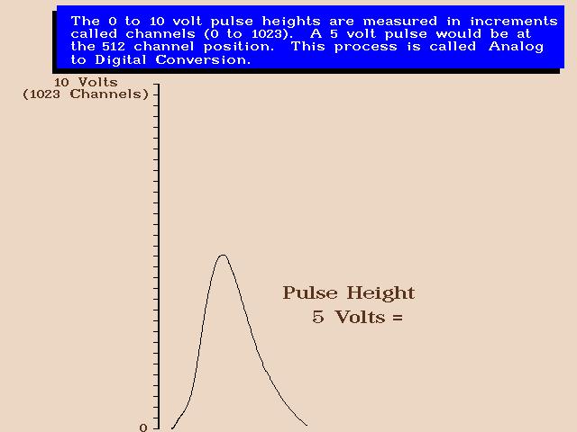
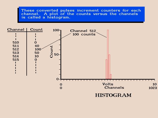
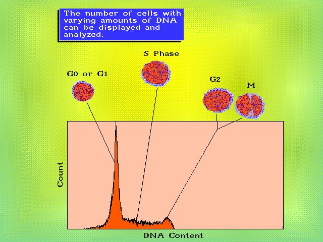
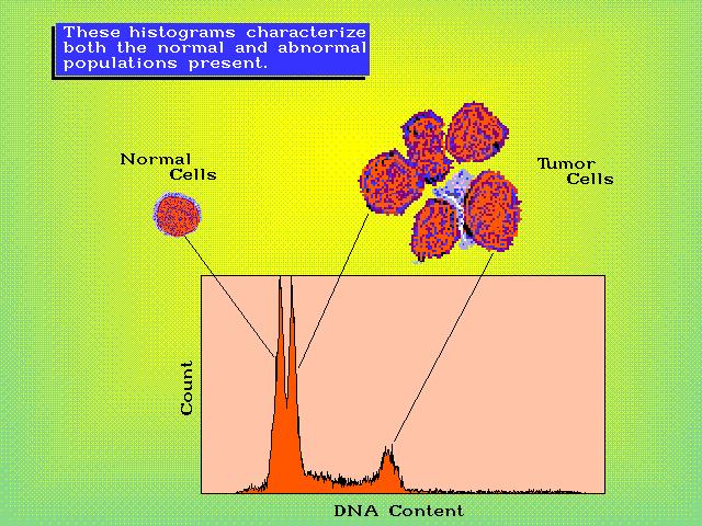
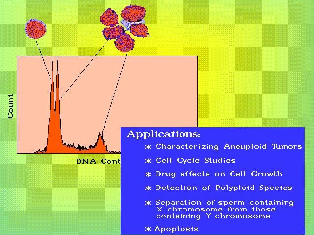
1 Applications of Flow Cytometry in Diagnostic Cytology of Body Cavity Fluids Awtar Krishan, PhD. Professor, Department of Pathology University of Miami School of Medicine Beckman Coulter Symposium, Hong Kong, April Jackson Memorial Medical Center Univ. of Miami Miller School of Medicine 2


2 UMH VAMC JMH-E RMSB BPEI RT 3 JACKSON MEMORIAL MEDICAL CENTER UNIVERSITY OF MIAMI MILLER SCHOOL OF MEDICINE JMH DTC 4
3 Diagnostic Cytology of Cells in Body Cavity Fluids Pleural or peritoneal fluids are often present in patients with lung, breast and ovarian tumors. AT UM/JMH more than 27,000 body cavity fluid specimens are processed annually % of body cavity fluids from patients with a proven malignancy are false negative as diagnostic cytology can not find tumor cells. Motherby et al., Diag.Cytopath.20: 350, 1999 Ganjei et al., Acta Cytologica, 48: 653, False Negative in Diagnostic Cytology of Body Cavity Fluids Tumor cells may not be present in peritoneal or pleural fluid. Enough tumor cells may not be present for visual examination under a microscope. Tumor cells may be morphologically indistinguishable from normal epithelial and mesenchymal cells. 6

4 Diagnostic Cytology of Body Cavity Fluids Cellular patterns and morphological characteristics of the individual cells. In samples with atypical cells, immunocytochemistry may be used to identify tumor cells and suggest the possible site of origin. 7 8

5 Tight cluster of malignant cells in pleural fluid of a breast ca. Cluster of tumor cells Reactive mesothelial cells Ganjei, Jorda & Krishan 9 Effusion Cytology, Demoss Medical Ber-EP4/EMA Epithelial Membrane Antigen. Expressed in: % of carcinomas, 4% of mesotheliomas 0% of benign mesothelial proliferation. Comin CE, et el. Amer. J. Surg. Path. 31: , Davidson B, et al., Diagn Cytopathol. 35: ,

6 EMA positive cells in peritoneal fluid of a gastric ca. EMA Ganjei, Jorda & Krishan Effusion Cytology, Demoss Medical 11 Thyroid Transcription Factor-1 A nuclear receptor found in 90% small-cell lung adenocarcinomas, ~23% of endometrial and endocervical ca. with negligible expression in squamous cell carcinomas. Siami, K., et al. Am J Surg Pathol. 31: , Kalhor, N., et el. Mod. Path., 19: , Ordonez, NG, Mod Path. 19: ,


7 TTF-1 Positive cells in pleural fluid of an adenocarcinoma TTF-1 Ganjei, Jorda & Krishan Effusion Cytology, Demoss Medical 13 ER positive cells in pleural fluid of a Breast CA ER Ganjei, Jorda & Krishan Effusion Cytology, Demoss Medical 14

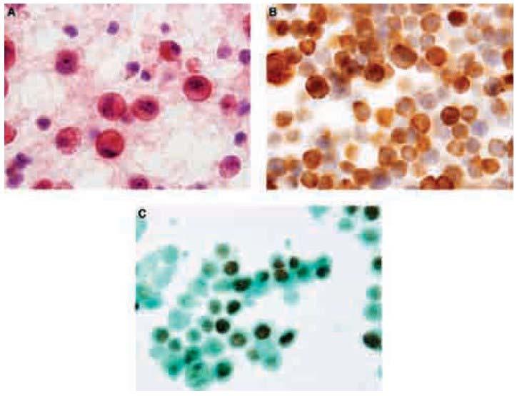
8 ER positive cells in pleural fluid of a breast ca. ER Ganjei, Jorda & Krishan Effusion Cytology, Demoss Medical 15 EMA and ER positive cells in pleural fluid of a breast ca. EMA ER Ganjei, Jorda & Krishan Effusion Cytology, Demoss Medical 16

9 Marker Expression of TTF-1, ER & Calretinin 17 18


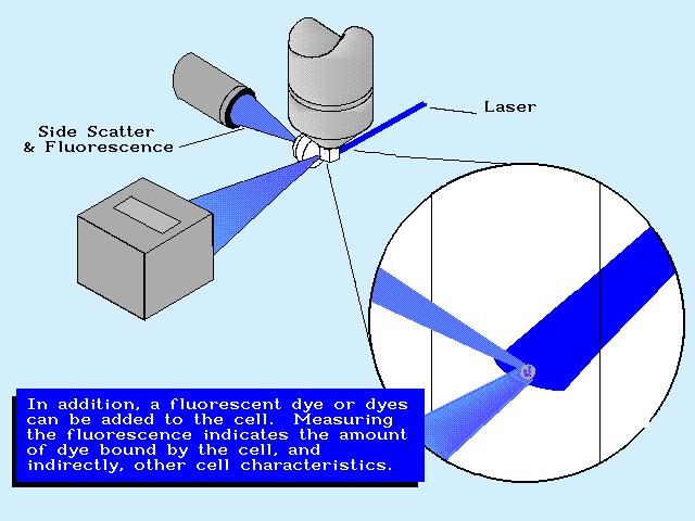
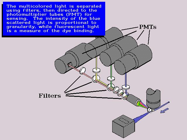
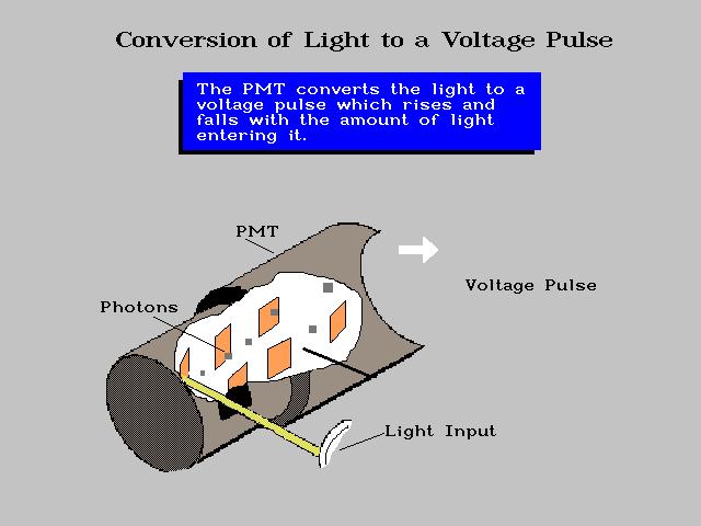
10 BASICS OF A FLOW CYTOMETER 19 20
11 21 22
12 23 24
13 25 26
14 27 28
15 Flow cytometric Analysis of Body Cavity Fluids Flow cytometry was extensively used in 70 s for the detection of malignant cells with aneuploid DNA content in peritoneal, pleural, and cerebrospinal fluids. In several studies, flow analysis detected cells with aneuploid DNA content in fluids with negative cytology. On re-examination these samples were found to contain tumor cells thus reducing the false-negative rate from 21.8% to 4.7%. Lovecchio, et al.,obstet.gynecol.67: 675, Nuclear Volume/protein content vs. DNA Content of Human Tumor Cells As tumor cells and nuclei are often larger in size than normal cells, flow cytometric analysis of nuclear volume/protein content vs. DNA content could be used to differentiate between normal and tumor cells. Expression of secondary markers could then be studied in tumor nuclei to suggest the site of their origin. 30
16 Normal Nuclei Malignant Nuclei NPE Analysis NPE Analysis NPE>16.0 NPE= Nuclear Volume vs. DNA Content Prostate Cancer Volume DNA Krishan, et al. Cytometry 43,
17 Nuclear Volume vs. DNA Content Normal Breast Primary Breast Cancer Krishan, et al. Cytometry 43, Nuclear Volume vs. DNA Content Primary Breast Cancer L.N. Metastasis 3% Tumor Metastasis Krishan, et al. Cytometry 43,
18 Nuclear Volume vs. DNA Content Normal Colon Primary Colon Cancer Krishan, et al. Cytometry 43, Nuclear Volume vs. DNA Content Primary Colon Cancer Colon Metastasis 5% Tumor Metastasis Krishan, et al. Cytometry 43,
19 Nuclear Volume vs. DNA Content Gastric Cancer T R B C Volume DNA 37 Nuclear Volume vs. DNA Content Ovarian Cancer Volume T R B C DNA 38


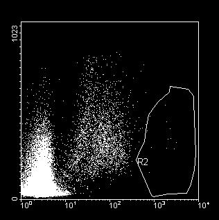
20 Nuclear Volume vs. DNA Content Non-Small Cell Lung Carcinoma 13% Tumor Load Aneuploid Diploid 39 FORWARD SCATTER VS. DNA CONTENT F O R W A R D S C AT T E R SIDE SCATTER DNA CONTENT 40


21 Aneuploid cells in a false negative peritoneal fluid DNA Content DNA vs. Protein Krishan et al. Diag. Cytopath. 34; , Aneuploid cells in a false negative peritoneal fluid DAPI DNA Content DNA vs. Protein Krishan et al. Diag. Cytopath. 34; ,
22 DNA Flow Analysis of limited value? Some studies reported that DNA flow cytometry was in general less sensitive than cytology for the detection of malignant cells and a higher percentage of false positives were seen by flow analysis. Based on these reports, it was generalized that DNA flow analysis did not offer any advantages over cytomorphology for the detection of malignant cells in body cavity fluids. Hedley, et al., Eur.J. Clin. Onc. 20: 749, False Positive aneuploid cells in peritoneal fluid 44
23 False Positives in peritoneal fluid In some of the patients with liver cirrhosis or end stage liver disease, cells with large volume and greater DNA fluorescence are seen. These populations do not form a distinct peak in DNA histograms and may be caused by changes in chromatin density and fluorochrome binding rather than by the presence of true aneuploidy. Krishan et al. Diagnostic Cytopathology, DNA Cytometry and Immunocytology 130 body cavity fluids were examined for DNA aneuploidy and for expression of Epithelial Membrane Antigen by immunocytology (EMA-ICC). Sensitivity for detection of tumor cells was: DNA aneuploidy alone = 38% Cytology alone = 58.8% Cytology and DNA aneuploidy = 73.5% Cytology and EMA-ICC = 79.4% A combination of cytology and DNA aneuploidy had higher sensitivity than DNA aneuploidy alone (73% versus 38%). Krishan, et al., Diagnostic Cytopathology, 34:528,
24 High Resolution DNA Cytometry, Conventional Diagnostic Cytology and EMA immunocytochemistry Out of 22 cytology positive samples, 20 were confirmed to be malignant on follow up. 4/8 suspicious samples with normal diploid DNA content had malignant cells. 7/15 samples with aneuploid cells were malignant and 8/15 were false positive. Most of the false positive aneuploid samples were ascites of patients with cirrhosis and liver disease. High resolution flow cytometry in combination with EMA immunocytochemistry reduced the false negatives from 41.2% to 14.7 %; an absolute reduction of 26.5% and relative reduction of 64.3%. Krishan A, et al. Diagn. Cytopathol. 34: , Flow Cytometry in Diagnostic Cytology Diagnostic Cytology Conventional Diagnostic cytology has a false 50%. Combination of diagnostic cytology with IHC can increase detection of tumor cells. Observer bias and small sample size can lead to artifacts hr are needed for results to be reported. Flow Cytometry DNA Flow Cytometry has a false 58%. DNA Analysis in combination with IHC marker detection can increase sensitivity from 58 to 100%. Data is based on a large sample size and lack of observer bias. Data can be obtained in 2-4 hrs. 48
25 Ber-EP4 Epithelial antigen. Expressed in: % of carcinomas, 4% of mesotheliomas 0% of benign mesothelial proliferation. Comin CE, et el. Amer. J. Surg. Path. 31: , Davidson B, et al., Diagn Cytopathol. 35: , DNA Aneuploidy and Ber-EP4 Expression 1.00 A B Cell Count 1.18 Forward Scatter 11.44% DNA Content Ber-EP4-FITC 1.00 C D Cell Count 1.86 Forward Scatter 63.08% BCF112, DNA Content Ber-EP4-FITC BCF123 50
26 Ber-Ep4 Expression in triploid cells from a peritoneal fluid A B GAM IgG FITC Ber EP-4-FITC 0.03% 2.45% DNA Content C D Cell Count Diploid cells Cell Count Aneuploid cells Ber-EP4-FITC ISOTYPE vs. Ber-Ep4-Mob 51 Thyroid Transcription Factor-1 A nuclear receptor found in 90% small-cell lung adenocarcinomas, ~23% of endometrial and endocervical ca. with negligible expression in squamous cell carcinomas. Siami, K., et al. Am J Surg Pathol. 31: , Kalhor, N., et el. Mod. Path., 19: , Ordonez, NG, Mod Path. 19: ,

27 TTF1 positive nuclei in peritoneal fluid of a lung adenocarcinoma TTF-1 Ganjei, Jorda & Krishan Effusion Cytology, Demoss Medical 53 TTF-1 Expression in Aneuploid Cells from a Pleural Fluid T T F Isotype A TTF-1 Mab B E X P R E S S I O N DNA DNA CONTENT Content DNA content vs. TTF expression in a human pleural fluid specimen stained with either the isotype or anti-ttf monoclonal antibody. Note the high expression of TTF-1 reactivity in cells with aneuploid DNA content. Krishan et al. Diag. Cytopath. 34; ,
28 Flow Cytometric Monitoring of Marker Expression in Cells from Body Cavity Fluids Fluids with aneuploid cells: 48/226 (21%) TTF-1 Expression: 45/150 (30%) Progesterone Expression : 40/66 (60%) Ber-Ep4 Expression: 9/20 (45%) 55 DNA Aneuploidy and Marker Expression Seventy-nine BCF were analyzed by flow cytometry for detection of aneuploidy and expression of Ber-EP4, progesterone, MUC4 or thyroid transcription factor-1. DNA index of equal to or greater than 1.2 was seen in 33/79 (41.7%) of the samples. By combining data on positive marker expression with that of DNA aneuploidy, the sensitivity for detection of malignant samples was increased from 58.5 to 100%. Krishan, et al., Cytometry Part A 77A: ,
29 Flow Immunocytology Flow analysis can be used for rapid detection of the following diagnostic markers in cells from body cavity fluids: Mucins (MUC1, MUC4, MUC16) Epithelial Antigens ( EMA, Ber-Ep4) Calretinin (mesotheliomas) Cytokeratins (CK7, 20) Hormone receptors ( ER, PR, VDR) TTF-1 (adeno.ca of lung) P53, P63 ( Squamous vs. adenoca of lung) Stem Cell Markers: CD34, CD90, CD117, CD133, CXCR4 ALDH1, CD44+/CD24- phenotype 57 Flow Cytometric Analysis of Cells in Body Cavity Fluids Conclusions High resolution flow cytometry can be used for rapid identification of cells with aneuploid DNA content. Nuclear volume and protein content can be used to differentiate between normal and tumor cells with diploid DNA content. Specific marker expression ( e.g., ER, EMA, TTF-1) can be used to suggest a possible site of origin. Multiparametric flow analysis may be able to reduce the false negatives in body cavity fluid cytology. 58
30 Tumor Stem Cell Marker Expression in Cells from Body Cavity Fluids Ber-EP4 TTF-1 ALDH-1 CD44+ CD24-59 Tumor Stem Cell Markers ALDH1 is expressed in both hematopoietic and tumor stem cells. CD44+/CD24-/CD133+ phenotype is characteristic of breast cancer stem cells. Wright, MH et al. Breast Can. Res. 10: R10, 2008 Ginestier, C et al., Cell Stem Cell. 1: , 2007 Sheridan, C., et al., Breast Can. Res. 8: R59,


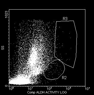
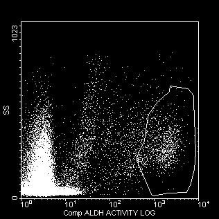
31 ALDH + Over-expression in Cells with Small side scatter BAAA-DA+DEAB BAAA-DA S I D E S C A T T E R ALDH br /SSC low BAAA-DA BAAA-DA = BodipyTM-aminoacetaldehyde diethyl acetal {ALDEFLUOR} DEAB = Diethylamino-benzaldehyde 61 ALDH1, CD44 and CD24 Expression in cells from body cavity fluids 2.24% D 0.25% E 0.04% F 1.3% 4.1% 0.1% C D % ALDEFLUOR ALDEFLUOR+ DEAB CD44 62
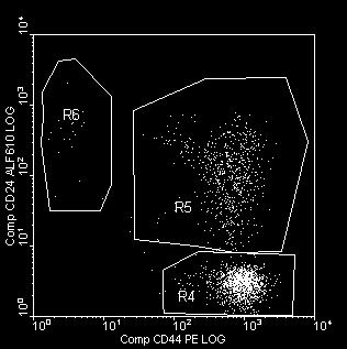
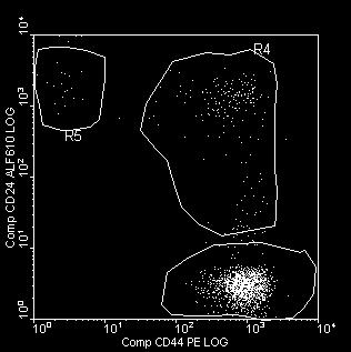
32 Tumor Stem Cell Analysis in Body Cavity Fluids CD34/CD45 cells CD90, CD117, CD133 CXCR4 Expression Electronic Cell Volume of Stem Cells Side Population (SP cells) Hoechst Vybrant DyeCycle Violet V35003 ALDH1 positive Cells CD44+/CD24- Cytometry A 73: , 2008 Cytometry B: April, ALDH1, CD44 and CD24 Expression in body cavity fluids with negative cytology C D 2 4 ALDEFLUOR ALDEFLUOR+ DEAB CD44 64

33 CD44+ and CD24 Expression in Ber EP4+, cells from a benign and a malignant body cavity fluid. A 11.54% 73.15% B Forward Scatter Ber-EP4-FITC 11.44% C CD24-PE-Alexa Fluor % CD44-PE 14.24% 44.90% F 63.08% 6.15% Ber-EP4-FITC 3.06% CD44-PE BCF 112, 123 fig 1 BCF2 65 ALDH1, CD44 and CD24 Expression In peritoneal and pleural fluids of patients containing malignant cells, ALDH bright cells with SSC low and SSC high and CD44 + /CD24 - expression are seen. However, similar cells were also seen in body cavity fluids of some of the patients who had negative cytology and did not have a proven malignancy. The presence of ALDH bright cells with CD44 + /CD24 - expression in inflammatory and benign body fluids needs further evaluation. 66
34 Acknowledgements I. Sabe, MD, PhD William Tellford ( NCI) A. Redkar, PhD S. Adiga, PhD S. Rao, Ph.D P. Arya, PhD Raquel Cabana ( NPE) Richard Thomas ( NPE) A. Oppeneheimer P. Dandekar R. Hamelik Parvin Ganjei, MD Jorda Merce, MD Mehrdad Nadji, MD Supported by NIH CA 09733, DAMD and Fl.DOH Grants 67
Ascitic Fluid and Use of Immunocytochemistry. Mercè Jordà, University of Miami
 Ascitic Fluid and Use of Immunocytochemistry Mercè Jordà, University of Miami Is It Malignant? Yes? No Ascitic Fluid Cytomorphologic Useful Findings Tight clusters with smooth borders Cellular and nuclear
ACCURACY OF IMMUNOHISTOCHEMISTRY IN EVALUATION
 POL J PATHOL 2011; 2: 95-100 ACCURACY OF IMMUNOHISTOCHEMISTRY IN EVALUATION OF MALIGNANT PLEURAL AND PERITONEAL EFFUSIONS FERESHTEH ENSANI, FARNAZ NEMATIZADEH, GITI IRVANLOU Department of Cytology, Cancer
ACCME/Disclosures. Diagnosing Mesothelioma in Limited Tissue Samples. Papanicolaou Society of Cytopathology Companion Meeting March 12 th, 2016
 Diagnosing Mesothelioma in Limited Tissue Samples Papanicolaou Society of Cytopathology Companion Meeting March 12 th, 2016 Sanja Dacic, MD, PhD University of Pittsburgh ACCME/Disclosures GENERAL RULES
ACCME/Disclosures. Case 4 USCAP Pulmonary Panel Case 4 History
 Case 4 USCAP Pulmonary Panel 2016 Andrew Churg, MD Department of Pathology Vancouver General Hospital & University of British Columbia Vancouver, BC achurg@mail.ubc.ca. ACCME/Disclosures The USCAP requires
Urinary Bladder: WHO Classification and AJCC Staging Update 2017
 Urinary Bladder: WHO Classification and AJCC Staging Update 2017 Houston Society of Clinical Pathologists 58 th Annual Spring Symposium Houston, TX April 8, 2017 Jesse K. McKenney, MD Classification
Value of antimesothelioma HBME 1 in the diagnosis of inflammatory and malignant pleural effusions
 Romanian Journal of Morphology and Embryology 2006, 47(4):351 355 ORIGINAL PAPER Value of antimesothelioma HBME 1 in the diagnosis of inflammatory and malignant pleural effusions LILIANA MOCANU 1), ANCA
How to Recognize Gynecologic Cancer Cells from Pelvic Washing and Ascetic Specimens
 How to Recognize Gynecologic Cancer Cells from Pelvic Washing and Ascetic Specimens Wenxin Zheng, M.D. Professor of Pathology and Gynecology University of Arizona zhengw@email.arizona.edu http://www.zheng.gynpath.medicine.arizona.edu/index.html
Effusion Cytology: Diagnostic Challenges
 Effusion Cytology: Diagnostic Challenges Tarik M. Elsheikh, MD Professor and Medical Director, Anatomic Pathology Cleveland Clinic Outside Consult Case 45 year old woman, presented with nausea, dyspnea,
Immunohistochemistry on Fluid Specimens: Technical Considerations
 Immunohistochemistry on Fluid Specimens: Technical Considerations Blake Gilks Dept of Pathology University of British Columbia, Vancouver, BC, Canada Disclosures None Learning Objectives At the end of
Presentation material is for education purposes only. All rights reserved URMC Radiology Page 1 of 98
 Presentation material is for education purposes only. All rights reserved. 2011 URMC Radiology Page 1 of 98 Radiology / Pathology Conference February 2011 Brooke Koltz, Cytopathology Resident Presentation
Serous Effusions. Spasenija Savic Prince, MD Pathology, University Hospital Basel, Switzerland
 Serous Effusions Spasenija Savic Prince, MD Pathology, University Hospital Basel, Switzerland Serous membrane Body cavities: Pleural Pericardial Peritoneal Effusion = Excess of fluid 80% Benign 20% Malignant
Serous effusion Objectives. Cytology of Serous Effusions From basics to challenges
 Cytology of Serous Effusions From basics to challenges Cytology of Serous Effusions From basics to challenges Pınar Fırat, MD, MIAC Department of Pathology, İstanbul University, İstanbul Faculty of Medicine,
SELECTED DILEMMAS IN RESPIRATORY CYTOPATHOLOGY (2 CASES)
") SELECTED DILEMMAS IN RESPIRATORY CYTOPATHOLOGY (2 CASES) Dr. Mariamma Joseph Professor of Pathology Division Head Cytopathology Department of Pathology and Laboratory Medicine LHSC and Western University
SELECTED DILEMMAS IN RESPIRATORY CYTOPATHOLOGY (2 CASES) Dr. Mariamma Joseph Professor of Pathology Division Head Cytopathology Department of Pathology and Laboratory Medicine LHSC and Western University
4/12/2018. MUSC Pathology Symposium Kiawah Island April 18, Jesse K. McKenney, MD
 MUSC Pathology Symposium Kiawah Island April 18, 2018 Jesse K. McKenney, MD 1 Urothelial Carcinoma with Alternative Differentiation 2 Urothelial Carcinoma with Alternative Differentiation Recognition as
WT1, Estrogen Receptor, and Progesterone Receptor as Markers for Breast or Ovarian Primary Sites in Metastatic Adenocarcinoma to Body Fluids
 Anatomic Pathology / WT1, ESTROGEN RECEPTOR, AND PROGESTERONE RECEPTOR IN CYTOLOGY OF BODY FLUIDS WT1, Estrogen Receptor, and Progesterone Receptor as Markers for Breast or Ovarian Primary Sites in Metastatic
Mesothelioma: diagnostic challenges from a pathological perspective. Naseema Vorajee August 2016
 Mesothelioma: diagnostic challenges from a pathological perspective Naseema Vorajee August 2016 Naseema.vorajee@nhls.ac.za Pleural diseases (whether neoplastic, reactive or infective) may have similar
Cytological Sub-classification of Lung Cancer: Morphologic and Molecular Characteristics. Mercè Jordà, University of Miami
 Cytological Sub-classification of Lung Cancer: Morphologic and Molecular Characteristics Mercè Jordà, University of Miami Mortality Lung cancer is the most frequent cause of cancer incidence and mortality
Follow up of the Guidelines for Cytopathologic Diagnosis of Malignant Mesothelioma
 Follow up of the Guidelines for Cytopathologic Diagnosis of Malignant Mesothelioma Assoc. Prof. Katalin Dobra, Senior Lecturer in Molecular Pathology Karolinska University Hospital Stockholm, Sweden Disclosure
Supplementary Figure 1. Identification of tumorous sphere-forming CSCs and CAF feeder cells. The LEAP (Laser-Enabled Analysis and Processing)
") Supplementary Figure 1. Identification of tumorous sphere-forming CSCs and CAF feeder cells. The LEAP (Laser-Enabled Analysis and Processing) platform with laser manipulation to efficiently purify lung
Supplementary Figure 1. Identification of tumorous sphere-forming CSCs and CAF feeder cells. The LEAP (Laser-Enabled Analysis and Processing) platform with laser manipulation to efficiently purify lung
Journal of Cytology & Histology
 ISSN: 2157-7099 ojournal of Cytology & Hist logy Journal of Cytology & Histology Sen, et al., 2015, 6:2 DOI: 10.4172/2157-7099.1000314 Research Article Article Open Open Access Morphometric Analysis and
The role of immunohistochemistry in surgical pathology of the uterine corpus and cervix
 The role of immunohistochemistry in surgical pathology of the uterine corpus and cervix Prof. Ben Davidson, MD PhD Department of Pathology, Norwegian Radium Hospital, Oslo University Hospital, Oslo, Norway
Cancers of unknown primary : Knowing the unknown. Prof. Ahmed Hossain Professor of Medicine SSMC
 Cancers of unknown primary : Knowing the unknown Prof. Ahmed Hossain Professor of Medicine SSMC Definition Cancers of unknown primary site (CUPs) Represent a heterogeneous group of metastatic tumours,
GATA3: A Promising Marker for Metastatic Breast Carcinoma in Serous Effusion Specimens
 GATA3: A Promising Marker for Metastatic Breast Carcinoma in Serous Effusion Specimens Paul W. Shield, PhD, FFSc(RCPA) 1,2 ; David J. Papadimos, MBBS, FRCPA 1,3 ; and Michael D. Walsh, PhD 3 BACKGROUND:
Case 1. Slide 1 History: 65 year old male presents with bilateral pleural effusions, a 40 pack year smoking history and peripheral and hilar lung
 Case 1. Slide 1 History: 65 year old male presents with bilateral pleural effusions, a 40 pack year smoking history and peripheral and hilar lung masses. Specimen shown is from a tap of the pleural effusion.
Applications of IHC. Determination of the primary site in metastatic tumors of unknown origin
 Applications of IHC Determination of the primary site in metastatic tumors of unknown origin Classification of tumors that appear 'undifferentiated' by standard light microscopy Precise classification
Cytological evaluation of effusion fluid with cell block technique and cytology smears among Sudanese patients
 EUROPEAN ACADEMIC RESEARCH Vol. IV, Issue 3/ June 2016 ISSN 2286-4822 www.euacademic.org Impact Factor: 3.4546 (UIF) DRJI Value: 5.9 (B+) Cytological evaluation of effusion fluid with cell block technique
Immunohistochemical classification of lung carcinomas and mesotheliomas. Prof. Mogens Vyberg NordiQC Institute of Pathology Aalborg, Denmark
 Immunohistochemical classification of lung carcinomas and mesotheliomas Prof. Mogens Vyberg NordiQC Institute of Pathology Aalborg, Denmark Endobronchial ultrasound guided transbronchial needle biopsy
Key Words: effusion; carcinoma; immunocytochemistry; direct smear; cytology
 The Application of Immunocytochemistry to Direct Smears in the Diagnosis of Effusions Stewart M. Knoepp, M.D., Ph.D., { Jeremiah Placido, M.D., { Kristina L. Fields, B.S., Dafydd Thomas, M.D., Ph.D., and
Diagnostic IHC in lung and pleura pathology
 Diagnostic IHC in lung and pleura pathology Mogens Vyberg Professor of Clinical Pathology Director of NordiQC Aalborg University Hospital, Aalborg, Denmark WHO 2004 and Web Malignant mesothelioma Epithelioid
Hyperchromatic Crowded Groups: What is Your Diagnosis? Session 3000
 Hyperchromatic Crowded Groups: What is Your Diagnosis? Session 3000 Thomas A. Bonfiglio, M.D. Professor Emeritus, Pathology and Laboratory Medicine University of Rochester Disclosures In the past 12 months,
Tissue-based Immunohistochemical Biomarker Expression in Malignant Glandular Lesions of the Uterine Cervix: a Systematic Review
 Tissue-based Immunohistochemical Biomarker Expression in Malignant Glandular Lesions of the Uterine Cervix: a Systematic Review Sandra Lee MD, FRCPC 1 *, Vikrant V. Sahasrabuddhe, MBBS, DrPH 2 *, Diana
INTRODUCTION TO PATHOLOGICAL TECHNIQUES. 1. Types of routine biopsy procedures 2. Special exams (IHC, FISH)
") INTRODUCTION TO PATHOLOGICAL TECHNIQUES 1. Types of routine biopsy procedures 2. Special exams (IHC, FISH) Biopsy-Indications Diffuse/multifocal lesions (neoplastic, inflammatory, etc) Etiology of the
INTRODUCTION TO PATHOLOGICAL TECHNIQUES 1. Types of routine biopsy procedures 2. Special exams (IHC, FISH) Biopsy-Indications Diffuse/multifocal lesions (neoplastic, inflammatory, etc) Etiology of the
Fellowship in Cytopathology Department of Pathology. All India Institute of Medical Sciences (AIIMS) Jodhpur, Rajasthan, India
 Jodhpur, Rajasthan, India") Fellowship in Cytopathology Department of Pathology All India Institute of Medical Sciences (AIIMS) Jodhpur, Rajasthan, India Syllabus for Fellowship in Cytopathology: FNAC Direct, Guided, EUS Exfoliative
Fellowship in Cytopathology Department of Pathology All India Institute of Medical Sciences (AIIMS) Jodhpur, Rajasthan, India Syllabus for Fellowship in Cytopathology: FNAC Direct, Guided, EUS Exfoliative
Biopsy Interpretation of Spindle cell proliferations of the Serosa
 Biopsy Interpretation of Spindle cell proliferations of the Serosa Richard Attanoos, Cardiff. U.K. Disclosure of Relevant Financial Relationships USCAP requires that all planners (Education Committee)
Editorial Process: Submission:11/02/2017 Acceptance:05/19/2018
 RESEARCH ARTICLE Editorial Process: Submission:11/02/2017 Acceptance:05/19/2018 Comparative Analysis of Modified Liquid-Based Cytology and CytoRich Red Preparation in Assessment of Serous Effusion for
Update on Thyroid FNA The Bethesda System. Shikha Bose M.D. Associate Professor Cedars Sinai Medical Center
 Update on Thyroid FNA The Bethesda System Shikha Bose M.D. Associate Professor Cedars Sinai Medical Center Thyroid Nodules Frequent occurrence Palpable: 4-7% of adults Ultrasound: 10-31% Majority benign
BOSNIAN-TURKISH CYTOPATHOLOGY SCHOOL June 18-19, 2016 Sarajevo. Case Discussions. 60 year old woman Routine gynecologic control LBC
 BOSNIAN-TURKISH CYTOPATHOLOGY SCHOOL June 18-19, 2016 Sarajevo Case Discussions Prof Dr Sıtkı Tuzlalı Tuzlalı Pathology Laboratory 60 year old woman Routine gynecologic control LBC 1 2 Endometrial thickening
Schedule of Accreditation issued by United Kingdom Accreditation Service 2 Pine Trees, Chertsey Lane, Staines-upon-Thames, TW18 3HR, UK
 Schedule of ccreditation United Kingdom ccreditation Service 2 Pine Trees, Chertsey Lane, Staines-upon-Thames, TW18 3HR, UK External Quality ssessment Services for Cancer Diagnostics CIC Issue No: 005
International Journal of Health Sciences and Research ISSN:
 International Journal of Health Sciences and Research www.ijhsr.org ISSN: 2249-9571 Original Research Article Utility of Modified Cell Block Technique in Cases of Pleural Effusion Suspected of Malignancy
Cytyc Corporation - Case Presentation Archive - October 2001
 ThinPrep Pap Test History: 82 Year Old Female Specimen Type: Peritoneal Washings Case provided by Dr. Berle Stratton, Southwest Washington Medical Center, Vancouver, Washington. *The images, analysis and
Breast cancer: IHC classification. Mogens Vyberg Professor of Clinical Pathology Director of NordiQC Aalborg University Hospital, Aalborg, Denmark
 Breast cancer: IHC classification Mogens Vyberg Professor of Clinical Pathology Director of NordiQC Aalborg University Hospital, Aalborg, Denmark http://upload.wikimedia.org/wikipedia/commons/1/1a/breast.svg
Cytyc Corporation - Case Presentation Archive - July 2002
 ThinPrep Pap Test History: 34 Year Old Female LMP: Day 20 Specimen Type: Cervical/Vaginal Case provided by Mark Tulecke, M.D. and Gabrielle Trawinski CT (ASCP), Mount Auburn Hospital, Cambridge, Massachusetts.
COMPARATIVE STUDY OF CELL - BLOCKS & ROUTINE CYTOLOGICAL SMEARS OF PLEURAL & PERITONEAL FLUIDS IN SUSPECTED CASES OF MALIGNANCY
 ORIGINAL RESEARCH COMPARATIVE STUDY OF CELL - BLOCKS & ROUTINE CYTOLOGICAL SMEARS OF PLEURAL & PERITONEAL FLUIDS IN SUSPECTED CASES OF MALIGNANCY Geethu G Nair,*, Anupama Achyuthan Manjula 2 Assistant
Case 3 - GYN. History: 66 year old, routine Pap test. Dr. Stelow
 Case 3 - GYN History: 66 year old, routine Pap test Dr. Stelow Case 3 66 year year old woman Routine Pap Test Cytologic Features 3 dimensional clusters of cells with small to moderate amount of
Hepatocyte Nuclear Factor-1b Is Not a Specific Marker of Clear Cell Carcinoma in Serous Effusions
 Hepatocyte Nuclear Factor-1b Is Not a Specific Marker of Clear Cell Carcinoma in Serous Effusions Ben Davidson, MD, PhD 1,2 BACKGROUND: The transcription factor hepatocyte nuclear factor-1b (HNF1b) has
Immunocytochemistry and DNA-image cytometry in diagnostic effusion cytology. II. Diagnostic accuracy in equivocal smears
 59 Immunocytochemistry and DNA-image cytometry in diagnostic effusion cytology. II. Diagnostic accuracy in equivocal smears Helma Motherby a,, Nicolaus Friedrichs a, Mary Kube a, Bahram Nadjari a, Kristiane
Atypical Hyperplasia/EIN
 EIN Atypical Hyperplasia/EIN Based on scientific and diagnostic advances, in 2014 the WHO moved that the precursor lesion for endometrioid carcinoma be atypical hyperplasia/ein, rather than what was previously
Expression of Cytokeratin 5/6 in Epithelial Neoplasms: An Immunohistochemical Study of 509 Cases
 Expression of Cytokeratin 5/6 in Epithelial Neoplasms: An Immunohistochemical Study of 509 Peiguo G. Chu, M.D., Ph.D., Lawrence M. Weiss, M.D. Department of Pathology, City of Hope National Medical Center,
Claudin-4 Expression in Triple Negative Breast Cancer: Correlation with Androgen Receptors and Ki-67 Expression
 Claudin-4 Expression in Triple Negative Breast Cancer: Correlation with Androgen Receptors and Ki-67 Expression Mona A. Abd-Elazeem, Marwa A. Abd- Elazeem Pathology department, Faculty of Medicine, Tanta
SREBP-2 promotes stem cell-like properties and metastasis by transcriptional activation of c-myc in prostate cancer
 SREBP-2 promotes stem cell-like properties and metastasis by transcriptional activation of c-myc in prostate cancer Supplementary Material Supplementary Methods Supplementary References Supplementary Figure
New Developments in Immunohistochemistry for Gynecologic Pathology
 New Developments in Immunohistochemistry for Gynecologic Pathology Michael T. Deavers, M.D. Professor, Departments of Pathology and Gynecologic Oncology Immunohistochemistry in Gynecologic Pathology Majority
Nordic Immunohistochemical Quality Control
 Nordic Immunohistochemical Quality Control Immunohistochemistry in the classifiation of neoplasias of the alimentary tract & External Quality Assurance of Immunohistochemistry for GI cancer markers Mogens
Single and Multiplex Immunohistochemistry
 Single and Multiplex Immunohistochemistry Steve Westra, BS Reagent Product Specialist Leica Biosystems IHC Theory Polyclonal vs Monoclonal Polyclonal reagents Detect a multitude of epitopes Batch to batch
Overview of Indeterminate Cytology
 83 rd Annual Meeting American Thyroid Association Overview of Indeterminate Cytology Scott Boerner MD FRCPC Head Cytopathology, University Health Network University of Toronto DISCLOSURE Nothing to disclose
Supplementary Figure 1. Double-staining immunofluorescence analysis of invasive colon and breast cancers. Specimens from invasive ductal breast
 Supplementary Figure 1. Double-staining immunofluorescence analysis of invasive colon and breast cancers. Specimens from invasive ductal breast carcinoma (a) and colon adenocarcinoma (b) were staining
Cutaneous metastases. Thaddeus Mully. University of California, San Francisco Professor, Departments of Pathology and Dermatology
 Cutaneous metastases Thaddeus Mully University of California, San Francisco Professor, Departments of Pathology and Dermatology DISCLOSURE OF RELATIONSHIPS WITH INDUSTRY Thaddeus Mully Course C005 Essential
Atypical And Suspicious Categories In Fine Needle Aspiration Cytology Of The Breast
 IOSR Journal of Dental and Medical Sciences (IOSR-JDMS) e-issn: 2279-853, p-issn: 2279-861.Volume 15, Issue 1 Ver. III (October. 216), PP 57-61 www.iosrjournals.org Atypical And Suspicious Categories in
EBUS-TBNA Diagnosis and Staging of Lung Cancer
 EBUS-TBNA Diagnosis and Staging of Lung Cancer Nirag Jhala MD, MIAC Professor of Pathology and Lab Med. Director of Anatomic Pathology and Cytopathology Lewis Katz School of Medicine@ Temple University
NEW IHC A n t i b o d i e s
 NEW IHC Antibodies TABLE OF CONTENTS NEW IHC ANTIBODIES from Cell Marque CITED1 (5H6).... 1 Claudin 7 (5D10F3).... 1 GATA1 (4F5).... 1 Transgelin (2A10C2).... 1 NEW IHC ANTIBODIES using RabMAb Technology
Primary enteric adenocarcinoma with predominantly signet ring features of the lung: A case report with clinicopathological and molecular findings
 CASE REPORT Primary enteric adenocarcinoma with predominantly signet ring features of the lung: A case report with clinicopathological and molecular findings Makoto Nagashima 1, Ayako Moriyama 1, Yasuo
Award Top Quizzes For Residents
 Award Top Quizzes For Residents Giovanni Negri, MD, Bolzano (Italy) Eva M. Wojcik MD, Department of Pathology, Loyola University, Chicago (USA) Esther D. Rossi MD, PhD, MIAC, Division of Anatomic Pathology
ROLE OF TTF-1, CK20, AND CK7 IMMUNOHISTOCHEMISTRY FOR DIAGNOSIS OF PRIMARY
 Y.C. Su, Y.C. Hsu, and C.Y. Chai ROLE OF TTF-1, CK20, AND CK7 IMMUNOHISTOCHEMISTRY FOR DIAGNOSIS OF PRIMARY AND SECONDARY LUNG ADENOCARCINOMA Yue-Chiu Su 1, Yu-Chang Hsu 2, and Chee-Yin Chai 1,3 Departments
Well-differentiated Papillary Mesothelioma of the Pleura Diagnosed by Video-Assisted Thoracic Surgical Pleural Biopsy : A Case Report
 Showa Univ J Med Sci 25 1, 67 72, March 2013 Case Report Well-differentiated Papillary Mesothelioma of the Pleura Diagnosed by Video-Assisted Thoracic Surgical Pleural Biopsy : A Case Report Yuri TOMITA
Molecular classification of breast cancer implications for pathologists. Sarah E Pinder
 Molecular classification of breast cancer implications for pathologists Sarah E Pinder Courtesy of CW Elston Histological types Breast Cancer Special Types 17 morphological special types 25-30% of all
Ph.D. THESIS ENDOMETRIAL HYPERPLASIAS IN PERIMENOPAUSE SUMMARY
 UNIVERSITY OF MEDICINE AND PHARMACY OF CRAIOVA FACULTY OF MEDICINE Ph.D. THESIS ENDOMETRIAL HYPERPLASIAS IN PERIMENOPAUSE SUMMARY SCIENTIFIC COORDINATOR: PROF. DR. MIHAI B. BRĂILA, Ph.D. Ph.D. Graduand:
Introduction. 23 rd Annual Seminar in Pathology. FLUIDS, Part 1. Pittsburgh, PA Gladwyn Leiman UVMMC, VT
 23 rd Annual Seminar in Pathology Pittsburgh, PA Gladwyn Leiman UVMMC, VT FLUIDS, Part 1 "Blue walls", Claudia Hansen, 2009 Introduction o Challenging to everyone o Almost any benign or malignant process
Palpable Breast Lesions Cytomorphological Analysis and Scoring System with Histopatholgical Correlation
 IOSR Journal of Dental and Medical Sciences (IOSR-JDMS) e-issn: 2279-0853, p-issn: 2279-0861.Volume 15, Issue 10 Ver. III (October. 2016), PP 25-29 www.iosrjournals.org Palpable Breast Lesions Cytomorphological
Immunohistochemical Expression of Cytokeratin 5/6 in Gynaecological Tumors.
 ISPUB.COM The Internet Journal of Pathology Volume 13 Number 2 Immunohistochemical Expression of Cytokeratin 5/6 in Gynaecological Tumors. A Baghla, S Choudhry, A Kataria Citation A Baghla, S Choudhry,
Diagnosis of a granular cell tumour at the abdominal wall using fine needle aspiration cytology and histology: Case report
 Case Report Diagnosis of a granular cell tumour at the abdominal wall using fine needle aspiration cytology and histology: Case report Journal of International Medical Research 2015, Vol. 43(4) 592 596!
Successful flow cytometric immunophenotyping of body fluid specimens
 Successful flow cytometric immunophenotyping of body fluid specimens Fiona E. Craig, MD Division of Hematopathology Mayo Clinic Arizona 2017 MFMER slide-1 Financial disclosure No conflicts 2017 MFMER slide-2
performed to help sway the clinician in what the appropriate diagnosis is, which can substantially alter the treatment of management.
 Hello, I am Maura Polansky at the University of Texas MD Anderson Cancer Center. I am a Physician Assistant in the Department of Gastrointestinal Medical Oncology and the Program Director for Physician
Lung Cytology: Lessons Learned from Errors in Practice
 Lung Cytology: Lessons Learned from Errors in Practice Stephen S. Raab, M.D. Department of Laboratory Medicine Eastern Health and Memorial University of Newfoundland, St. John s, NL and University of Washington,
Exfoliative cytology of diffuse mesothelioma
 Exfoliative cytology of diffuse mesothelioma G. HEFIN ROBERTS AND G. M. CAMPBELL From the Pathology Department, Southern General Hospital, Glasgow J. clin. Path., 1972, 25, 577-582 SYNOPSIS The exfoliative
Neuroendocrine neoplasms of the lung
 Neuroendocrine neoplasms of the lung M Papotti, L Righi, & M Volante University of Turin at San Luigi Hospital TORINO NETs OF THE LUNG Menu - Spectrum of NE lung tumors - CARCINOID TUMORS - SCLC /LCNEC
Routine DNA cytometry of benign and malignant pleural evusions by means of the remote quantitation server Euroquant: a prospective study
 760 J Clin Pathol 2000;53:760 764 Routine DNA cytometry of benign and malignant pleural evusions by means of the remote quantitation server Euroquant: a prospective study K Kayser, S Blum, M Beyer, G Haroske,
CYTOMORPHOLOGY MODULE 28.1 INTRODUCTION OBJECTIVES 28.2 GENERAL GUIDELINES. Notes
 28 CYTOMORPHOLOGY 28.1 INTRODUCTION Light microscopic examination of stained cells in smears is the method of choice of diagnostic cytology. It allows classification of most normal cells as to type and
Novel Biomarkers (Kallikreins) for Prognosis and Therapy Response in Ovarian cancer
 for Prognosis and Therapy Response in Ovarian cancer") Novel Biomarkers (Kallikreins) for Prognosis and Therapy Response in Ovarian cancer Eleftherios P. Diamandis, M.D., Ph.D., FRCP(C) EORTC-NCI-ASCO Meeting,November 16, 2007 Yousef GM, Diamandis EP. Endocr.
Novel Biomarkers (Kallikreins) for Prognosis and Therapy Response in Ovarian cancer Eleftherios P. Diamandis, M.D., Ph.D., FRCP(C) EORTC-NCI-ASCO Meeting,November 16, 2007 Yousef GM, Diamandis EP. Endocr.
Mody. AIS vs. Invasive Adenocarcinoma of the Cervix
 Common Problems in Gynecologic Pathology Michael T. Deavers, M.D. Houston Methodist Hospital, Houston, Texas Common Problems in Gynecologic Pathology Adenocarcinoma in-situ (AIS) of the Cervix vs. Invasive
Application of Urovision FISH testing for diagnosis of bladder cancer
 Application of Urovision FISH testing for diagnosis of bladder cancer Eva M. Wojcik, MD Chair and Professor of Pathology and Urology Loyola University, Chicago, USA Bladder cancer - current status ~ 76,900
HOW TO GET THE MOST INFORMATION FROM A TUMOR BIOPSY
 HOW TO GET THE MOST INFORMATION FROM A TUMOR BIOPSY 7 TH Annual New York Lung Cancer Symposium Saturday, November 10, 2012 William D. Travis, M.D. Attending Thoracic Pathologist Memorial Sloan Kettering
04/09/2018. Salivary Gland Pathology in the Molecular Era Old Friends, Old Foes, & New Acquaintances
 Salivary Gland Pathology in the Molecular Era Old Friends, Old Foes, & New Acquaintances Jennifer L. Hunt, MD, MEd Aubrey J. Hough Jr, MD, Endowed Professor of Pathology Chair of Pathology and Laboratory
EVALUATION OF THE CHANGES RESULTING FROM TAMOXIFEN ADMINISTRATION. A COMBINED DNA FLOWCYTOMETRIC AND HISTOPATHOLOGICAL STUDY
 EVALUATION OF THE CHANGES RESULTING FROM TAMOXIFEN ADMINISTRATION. A COMBINED DNA FLOWCYTOMETRIC AND HISTOPATHOLOGICAL STUDY Tamoxifen Tamoxifen is a non steroidal tiphenylethylene y first synthesized
TUMOR,NEOPLASM. Pathology Department, Zhejiang University School of Medicine,
 TUMOR,NEOPLASM Pathology Department, Zhejiang University School of Medicine, 马丽琴,maliqin198@zju.edu.cn The points in this chapter What is a neoplasm (conception) Morphology of neoplasm Macroscopy of Neoplasm
Cytology and the Investigation of Carcinoma of Unknown Primary (CUP) Dr Anna Green ST5, St Thomas Hospital London, UK
 Dr Anna Green ST5, St Thomas Hospital London, UK") Cytology and the Investigation of Carcinoma of Unknown Primary (CUP) Dr Anna Green ST5, St Thomas Hospital London, UK Objectives Introduction to CUP Our experience of cytology and CUP Role of Cytology
Cytology and the Investigation of Carcinoma of Unknown Primary (CUP) Dr Anna Green ST5, St Thomas Hospital London, UK Objectives Introduction to CUP Our experience of cytology and CUP Role of Cytology
Objectives. Atypical Glandular Cells. Atypical Endocervical Cells. Reactive Endocervical Cells
 2013 California Society of Pathologists 66 th Annual Meeting San Francisco, CA Atypical Glandular Cells to Early Invasive Adenocarcinoma: Cervical Cytology and Histology Christina S. Kong, MD Associate
From Morphology to Molecular Pathology: A Practical Approach for Cytopathologists Part 1-Cytomorphology. Songlin Zhang, MD, PhD LSUHSC-Shreveport
 From Morphology to Molecular Pathology: A Practical Approach for Cytopathologists Part 1-Cytomorphology Songlin Zhang, MD, PhD LSUHSC-Shreveport I have no Conflict of Interest. FNA on Lymphoproliferative
of 20 to 80 and subsequently declines [2].
![of 20 to 80 and subsequently declines [2].](/thumbs/80/81450506.jpg "of 20 to 80 and subsequently declines [2].") - - According to the 2014 World Health Organization (WHO) classification and tumor morphology, primary ovarian tumors are subdivided into three categories: epithelial (60%), germ cell (30%), and sex-cord
- - According to the 2014 World Health Organization (WHO) classification and tumor morphology, primary ovarian tumors are subdivided into three categories: epithelial (60%), germ cell (30%), and sex-cord
Key Words: PAX8; PAX2; Müllerian; carcinoma; effusion
 Diagnostic Utility of PAX8 and PAX2 Immunohistochemistry in the Identification of Metastatic Müllerian Carcinoma in Effusions William Wiseman, D.O., Claire W. Michael, M.D., and Michael H. Roh, M.D., Ph.D.*
Do Your Flow Cytometric LDTs. Validation Guidelines? Fiona E. Craig, MD University of Pittsburgh School of Medicine
 Do Your Flow Cytometric LDTs Conform to the ICSH ICCS Validation Guidelines? Fiona E. Craig, MD University of Pittsburgh School of Medicine How should LDTs be validated? Accuracy Specificity Sensitivity
Pancreatic Cytopathology: The Solid Neoplasms
 Pancreatic Cytopathology: The Solid Neoplasms Syed Z. Ali, M.D. Professor of Pathology and Radiology Director of Cytopathology The Johns Hopkins Hospital Baltimore, Maryland Pancreatic Cytopathology: Past,
Recent advances in breast cancers
 Recent advances in breast cancers Breast cancer is a hetrogenous disease due to distinct genetic alterations. Similar morphological subtypes show variation in clinical behaviour especially in response
Cellometer Image Cytometry for Cell Cycle Analysis
 Cellometer Cytometry for Cell Cycle Analysis Importance of Cell Cycle Research Oncology: Since cancer cells often undergo abnormal cell division and proliferation, it is important to understand the cell
Improved Detection of Cervical Cancer and High Grade Neoplastic Lesions by a Combination of Conventional Cytology and DNA Automated Image Cytometer
 Journal of Cancer Therapy, 2010, 1, 47-51 doi:10.4236/jct.2010.12008 Published Online June 2010 (http://www.scirp.org/journal/jct) 47 Improved Detection of Cervical Cancer and High Grade Neoplastic Lesions
Workshop for O& G trainees and paramedics 17 Dec 2011 Cytological Interpretation
 Workshop for O& G trainees and paramedics 17 Dec 2011 Cytological Interpretation May Yu Director of Cytology Laboratory Service Department of Anatomical & Cellular Pathology Prince of Wales Hospital Cervical
ISSN X (Print) Original Research Article. DOI: /sjams
 Original Research Article. DOI: /sjams") DOI: 10.21276/sjams.2016.4.7.33 Scholars Journal of Applied Medical Sciences (SJAMS) Sch. J. App. Med. Sci., 2016; 4(7C):2468-2473 Scholars Academic and Scientific Publisher (An International Publisher
DOI: 10.21276/sjams.2016.4.7.33 Scholars Journal of Applied Medical Sciences (SJAMS) Sch. J. App. Med. Sci., 2016; 4(7C):2468-2473 Scholars Academic and Scientific Publisher (An International Publisher
A Useful Antibody Panel for Differential Diagnosis Between Peritoneal Mesothelioma and Ovarian Serous Carcinoma in Japanese Cases
 Anatomic Pathology / Peritoneal Mesothelioma A Useful Antibody Panel for Differential Diagnosis Between Peritoneal Mesothelioma and Ovarian Serous Carcinoma in Japanese Cases Yukio Takeshima, MD, Vishwa
Histopathological diagnosis of CUP
 Histopathological diagnosis of CUP Dr Karin Oien karin.oien@glasgow.ac.uk Disclosure slide Dr Karin Oien has no financial interests in any company mentioned in this presentation. Dr Karin Oien is conducting
HANDOUT. Bile Duct Brushing Cytology: A Morphologic and Molecular Approach
 HANDOUT Bile Duct Brushing Cytology: A Morphologic and Molecular Approach Lester J. Layfield, M.D. Professor and Chair Department of Pathology & Anatomical Sciences University of Missouri Introduction
Morphologic and Immunocytochemical Performances of Effusion Cell Blocks Prepared Using 3 Different Methods
 Anatomic Pathology / Performance of Different Cell Block Preparation Techniques Morphologic and Immunocytochemical Performances of Effusion Cell Blocks Prepared Using 3 Different Methods Xin Jing, MD,
Impact of immunostaining of pulmonary and mediastinal cytology
 Impact of immunostaining of pulmonary and mediastinal cytology Harman Sekhon MD, PhD Director of Cytopathology Head of Ottawa-site Ontario Tumour Bank June 20, 2014 Disclaimer Pfizer: Honorarium-Advisory