Pediatric Computed Tomography Dose Optimization Strategies: A Literature Review
|
|
|
- Percival Reynolds
- 5 years ago
- Views:
Transcription
1 Pediatric Computed Tomography Dose Optimization Strategies: A Literature Review Abstract Introduction Computed tomography (CT) dose optimization is an important issue in radiography as CT is the largest contributor to medical radiation dose and its use is increasing. However, CT dose optimization for pediatric patients could be more challenging than the adult counterpart. The purpose of this literature review was to identify and discuss the current pediatric CT dose saving techniques. Optimized pediatric protocols were also proposed. Methods A comprehensive literature search was conducted using the Medline, ProQuest Health and Medical Complete, PubMed, ScienceDirect, Scopus, Springer Link and Web of Science databases, and by using the keywords: CT, pediatric, optimization, protocol and radiation dose to identify articles focusing on pediatric CT dose optimization strategies published between 2004 and Results and Summary Seventy-seven articles were identified in the literature search. Strategies for optimizing a range of scan parameters and technical considerations including tube voltage and current, iterative reconstruction, diagnostic reference levels, bowtie filters, scout view, pitch, scan 1
2 collimation and time, overscanning and overbeaming for pediatric patients with different ages and body sizes and compositions were discussed. An example of optimized pediatric protocols specific to age and body size for the 64-slice CT scanners was devised. It is expected this example could provide more ideas to medical radiation technologists, radiologists and medical physicists on the way to optimize their pediatric protocols. 2
3 Introduction Computed tomography (CT) dose optimization is an important issue in radiography as CT is the largest contributor to medical radiation dose and its use is increasing [1,2]. However, CT dose optimization for pediatric patients could be more challenging than the adult counterpart because: 1. children are more radiosensitive to radiation [3]; 2. they have longer lifetime allowing potential radiation effects to manifest [4]; and 3. there are large variations of body size and composition (percentages of fat, muscle and bone) within each age group and across different groups [5-7]. Some clinical centers have implemented age and child-size specific CT scan protocols, and its effect on dose reduction has been recognized [5,8,9]. However, variations of these age and child-size specific protocols exist which makes other departments difficult to follow [5-12]. Also, there is a lack of standardization with respect to the definition of pediatric patients. One of the common definitions is those aged 0-15 years are considered as pediatric patients [13-15]. Such definition is used in this paper. The purpose of this literature review was to identify and discuss the current pediatric CT dose saving techniques. Optimized pediatric protocols and further study directions were also proposed. It is expected this review could potentially increase the awareness of medical radiation technologists on the range of dose saving techniques available in the literature and encourage them to optimize their protocols reducing the risk of pediatric CT examinations. Methods A comprehensive literature search was conducted using the Medline, ProQuest Health and Medical Complete, PubMed, ScienceDirect, Scopus, Springer Link and Web of Science 3
4 databases, and by using the keywords: CT, pediatric, optimization, protocol and radiation dose to identify articles focusing on pediatric CT dose optimization strategies. The article inclusion criteria were: 1. published between 2004 and 2014; 2. original research paper; 3. peer-reviewed; and 4. written in English. Articles were excluded if they belong to conference abstract and commentary. Figure 1 illustrates the literature search process. Insert Figure 1 about here Results and Discussion Seventy-seven articles were identified in the literature search and covered a range of areas including pediatric CT protocols specific to age, body size and composition, scan parameters that influence radiation dose and image quality, and other technical considerations (Figure 1). They were discussed in the following sections. Protocols Specific to Age, Body Size and Composition According to the International Atomic Energy Agency (IAEA) survey of pediatric CT practices in 40 countries published in 2013 [13], more than half of the clinical centers relied on pre-programmed scan protocols provided by manufacturers. In most of these scan protocols, specific techniques are suggested for each patient age group because it is assumed that body sizes and compositions (percentages of fat, muscle and bone) of patients within the same age group should be similar. However, recent studies indicated even in the same age group, there are great variations of body size and composition due to factors such as obesity [5-21]. 4
5 Since the last decade, some manufacturers and clinical centers have started to optimize their protocols based on not only patient age but also their body size in terms of weight. This is known as the color-coded system [9]. The scan settings such as tube potential and current and exposure time are tailored to specific patient conditions leading to lower dose and better image outcome. However, this approach requires medical physicists possessing sound knowledge of scanner specifications with support from radiologists and medical radiation technologists to develop these optimized protocols which is not feasible in many health institutions [13,22]. Patient size parameters such as weight and body mass index (BMI) are commonly used for developing size specific protocols for pediatric and adult patients. Recently, effective diameter and cross-sectional dimension have been considered as more accurate indicators of body size and habitus [5-7]. Also, awareness on variation of organ radiosensitivity across the children age range as well as between genders has been increased [2,4,23-26]. The following sections discuss individual scan parameters and other technical considerations for developing protocols specific to age, body size and composition. Tube Voltage Both phantom and clinical studies showed a lower tube voltage (with other settings such as tube current unchanged) could reduce radiation dose and improve image contrast for smallsized patients [27-30]. For example, a tube voltage as low as 60 kv could be used in pediatric CT examinations for structures with high subject contrast such as chest and bone [31]. Also, in (iodinated) contrast studies, a low tube voltage closer to the k-absorption edge 5
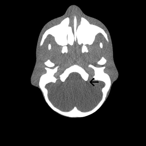
6 of iodine, 33 kev would increase the probability of photoelectric interaction improving the conspicuity of hyper- or hypo- vascular structures [29-35]. The amount of contrast medium could also be reduced in this case minimizing the chance of contrast-induced nephropathy [30,34-37]. However, an inappropriate, low tube voltage could cause the beam hardening artefact and decrease the image contrast due to the increase of image noise (Figure 2) [29,31]. If the image noise is excessive when using a lower tube voltage, adjustment of other parameters such as tube current will become essential [7,27]. The impact of image contrast reduction on pediatric abdominal examinations tends to be more significant when comparing with those for adults because less fatty tissue would normally be present between visceral organs of children representing a lower subject contrast [21,38,39]. In contrast enhanced soft tissue procedures, for example, to assess the small lesions in abdomen, the use of higher tube voltage would be necessary [27,40]. Although patient weight, BMI, effective diameter or cross-sectional dimension of body part can be used to indicate the body size for optimizing tube voltage in pediatric examinations, this information may not always be available [8,9,11-15,27]. Insert Figure 2 about here Automatic tube potential selection with tube current modulation (APSCM) is a relatively new function available in some latest CT scanners. It can assess the attenuation property of body part (i.e. body composition) which is difficult to measure in traditional settings and select the optimal tube voltage (in the range of kv) for achieving a balance between radiation dose and image quality automatically [27, 36-41]. Although its dose reduction capability was demonstrated in studies focusing on adult patients [37-44], its potential for pediatric CT dose optimization still needs to be confirmed. In a recent study of APSCM in pediatric imaging, 6
7 94% of the chest and abdomen examinations of this study had the tube voltage reduced to 70, 80 or 100 kv resulting in dose decrease up to 27% when comparing with the situation of the standard setting at 120 kv [27]. Tube Current Similar to tube voltage, a decrease in tube current with other parameters unchanged reduces radiation dose but it could potentially increase image noise. Phantom studies showed that the tube current could be halved for every cm reduction of diameter of body part, and if image noise is a concern, the ma could be decreased by 50% when there is a 4-6 cm reduction in diameter [31]. However, this rule of thumb does not apply to pediatric head CT because the tube current selection mainly depends on the skull bone composition (amount of calcium content) rather than the diameter of body part. The skull bone composition is more related to age [45]. Tube-current modulation (TCM) is again a recent CT system development. It only requires medical radiation technologists to provide image quality reference inputs. Automatic tube current adjustment takes place subsequently based on the size and attenuation property of body part (i.e. body composition) in the x-y plane (angular modulation), along the z-axis (zaxis modulation) or combined modulation. Studies reported the extent of dose reduction would be in the range of 26-50%. They highly recommended medical radiation technologists to apply TCM to their clinical practices for dose reduction while maintaining the image quality [9,26,46]. When it is used with a low tube voltage, the dose reduction effect will become more prominent [7]. 7
8 However, the challenge associated with this function in pediatric imaging is the lack of established guidelines of image quality reference values as they vary across manufacturers. The reference values depend on specific detector configuration, scanning geometry and beam filtration of different scanner models and manufacturers [47]. Table 1 shows the latest modulation software details of various manufacturers [31,47-51]. Although TCM is developed for dose reduction, in some situations such as the use of thin slice, low pitch and short scan time, the dose might increase unexpectedly for maintaining the target image quality (offsetting the negative effect on quality induced by these settings) [21,31,37,45,52]. Insert Table 1 about here Iterative Reconstruction (IR) Filtered back projection (FBP) is the standard image processing method for CT image formation. Recently, some CT manufacturers have provided the option to use iterative algorithms to reconstruct images and it is known as IR. This technique could reduce image noise and hence address the potential problems of tube voltage and current reduction for dose optimization [53,54]. The details of the latest IR products of different manufacturers are provided in Table 2 [53-57]. Insert Table 2 about here Recently, successful use of IR has been reported in pediatric CT studies such as cardiac, chest and abdomen with possible dose reduction up to 50% depending on diagnostic requirement and patient condition [55-58]. Further reduction up to 90% could be achieved in CT follow- 8
9 up procedures [56]. Nevertheless, IR in pediatric routine CT examinations is a controversial area because various factors such as detector efficiency and TCM can affect the reconstruction performance [55-61]. Also, the individual preferences of practicing radiologists should be considered as some described IR images as plastic or fogy in appearance [53-61]. A standard protocol for the appropriate use of IR should be established based on specific pathology, body size and selection of reconstruction kernel [58]. Diagnostic Reference Levels (DRLs) DRLs are percentile points (commonly 75 th percentile) of radiological examination dose distributions [62,63]. It provides clinical centers references of examination doses for protocol optimization [18-22,64-68]. However, the majority of published DRLs for pediatric CT imaging are just age specific and its inadequacy was noted [63-66]. Also, accurate CT dose measurements are not always readily available which makes it difficult to compare the examination doses with the DRLs [8,22]. The established CT DRLs are normally expressed in the quantities of volume CT dose index (CTDIvol), dose-length product (DLP) and effective dose (E) [67]. Although readings of CTDIvol and DLP are provided by modern scanners, their accuracies are questionable. For example, they determine the CTDIvol and DLP just based on 16 and / or 32 cm phantom(s) rather than specific to individual patient sizes. If 32 cm phantom is used, the dose reading provided would underestimate the actual dose received [8,68-69]. Further inaccuracies would be introduced if the effective dose is obtained through converting the DLP to E based on age and region specific conversion coefficients [22]. 9
10 Bowtie Filters CT scanners are normally equipped with bowtie filters to shape the X-ray beam for uniform photon distribution leading to optimal radiation dose and image quality [67]. However, cautions should be taken for pediatric cases because of their smaller body sizes. If patients are not placed to the gantry center, increases of dose to the peripheral and noise to the center will be expected [37,52,70-72]. The situation may be even worse when low dose or TCM technique is used inappropriately because it could be another potential source of noise as discussed previously [52]. Optimized Scanogarm or Scout View The optimization of scanogram or scout view is commonly neglected by medical radiation technologists. However, studies reported the arrangement of placing the X-ray tube under the table for scout view could reduce the dose to one third of the original [21,73]. Although this arrangement is only available in newer scanners, the length of the scout view should always be optimized to just cover the region of interest in any case. This is especially important when considering the smaller body sizes of pediatric patients. Optimized Pitch and Scan Collimation Higher pitch or thinner collimation is important in pediatric cases for obtaining adequate image spatial resolution along the z-axis. However, this would increase the image noise and hence decrease contrast-to-noise ratio [45,53]. This is a potential problem in pediatric imaging due to their lower subject contrast. In order to address this issue, some CT scanners 10
11 would increase the tube current automatically to compensate for the loss in image quality which may go against the concept of dose optimization [45]. A better approach could be to use special post-processing reconstruction software such as algorithm filters and IR to optimize the image outcome [39,74]. Scan Time A short scan time is preferable in pediatric imaging for motion unsharpness reduction and hence the higher temporal resolution. This also minimizes the need of the use of sedation. However, the potential issue associated with its use is the introduction of image noise because the number of profiles for image reconstruction would reduce and an increase of radiation dose might be needed to suppress this [31]. Although modern scanners provide a range of scan time selections from 0.28 to 0.5 s, a rotation time of 0.5 s should be used to achieve a balance between temporal and contrast resolutions and radiation dose [53]. Overscanning (Overranging) and Overbeaming Overscanning (overranging) refers to scanning body part greater than the one planned for obtaining adequate data for image reconstruction [75]. Its effect is greater in pediatric patients than adults because of smaller body sizes in children [73-78]. Generally, the extent of overranging mainly depends on detector collimation and pitch as they affect the dose profile. When they increase, the dose profile will expand and cover a greater area leading to a higher overrange dose (dose deposited outside the imaged volume) [75,77,78]. However, the modern scanners (64 slices or above) provide adaptive pre-patient collimators to reduce the excessive radiation at the start and end of the scan range [75]. 11
12 The use of newer systems also brings a positive effect on minimizing overbeaming. Overbeaming refers to radiation dose falling outside the active detector area in a gantry rotation due to the focal spot penumbra. When a scanner (such as 32 slices or above) provides a greater active detector area, the effect of overbeaming reduces. Its effect is inversely proportional to the number of detectors and collimation width. Its impact is not significant for the latest scanners [31,75-78]. Optimized Pediatric Protocols and Further Study Directions In this literature review, the needs and challenges of optimizing pediatric protocols specific to age, body size and composition were discussed. Through exploring individual scan parameters and technical considerations, basic strategies for pediatric CT dose optimization were identified. However, cautions should be taken when applying these techniques into clinical centers because the specifications of various scanners would be different. Table 3 presents optimized pediatric protocols specific to age and body size for the 64-slice CT systems devised based on phantom and clinical studies included in this review with reference to the scanner user manuals [8,15,31,53,62,66,75,79-81]. Their effectiveness still needs to be confirmed which can be considered as one of the further study directions. It is expected that this example would provide medical radiation technologists, radiologists and medical physicists more ideas on developing protocols specific to age and body size, and encourage them to review and optimize the pediatric protocols in their departments. Although there is limited discussion on protocols specific to gender due to insufficient information provided by the literature, this could be considered as another future study direction. 12
13 Insert Table 3 about here Summary Since the last decade, manufacturers and clinical centers have started to develop pediatric scanning protocols specific to age, body size and composition for better dose optimization. However, the recent IAEA survey suggested many departments still rely on default protocols provided by manufacturers without further optimization. This literature review identified a range of scan parameters and technical considerations that should be optimized for pediatric patients with different ages and body sizes including tube voltage and current, IR, DRLs, bowtie filters, scout view, pitch, scan collimation and time, overscanning and overbeaming. Although the discussion is not comprehensive, basic strategies to optimize these factors were suggested. An example of optimized pediatric protocols specific to age and body size for the 64-slice CT scanners was devised. It is expected this example could provide more ideas to medical radiation technologists, radiologists and medical physicists on the way to optimize their pediatric protocols. 13
14 References [1] Westar, S. J., Li, X., & Gulati, K., et al. (2013). Entrance skin dosimetry and sizespecific dose estimate from pediatric chest CTA. J Cardiovasc Comput Tomogr 8(2), [2] Miglioretti, D. L., Johnson, E., & Williams, A., et al. (2013). The use of computed tomography in pediatrics and the associated radiation exposure and estimated cancer risk. JAMA Pediatr 167(8), [3] Feng, S. T., Law, M. W., & Huang, B., et al. (2010). Radiation dose and cancer risk from pediatric CT examinations on 64-slice CT: a phantom study. Eur J Radiol 76(2), e [4] Mathews, J. D., Forsythe, A. V., Brady, Z., et al. (2013). Cancer risk in 680,000 people exposed to computed tomography scans in childhood or adolescence: data linkage study of 11 million Australians. BMJ 346, [5] Hopkins, K. L., Pettersson, D. R., & Koudelka, C.W., et al. (2013). Size-appropriate radiation doses in pediatric body CT: a study of regional community adoption in the United States. Pediatr Radiol 43(9), [6] Reid, J., Gamberoni, J., Dong, F., & Davros, W. (2010). Optimization of kvp and mas for pediatric low-dose simulated abdominal CT: is it best to base parameter selection on object circumference? AJR Am J Roentgenol 195(4), [7] Dong, F., Davros, W., Pozzuto, J., & Reid, J. (2012). Optimization of kilovoltage and tube current-exposure time product based on abdominal circumference: an oval phantom study for pediatric abdominal CT. AJR Am J Roentgenol 199(3),
15 [8] Watson, D. J., & Coakley, K. S. (2010). Paediatric CT reference doses based on weight and CT dosimetry phantom size: local experience using a 64-slice CT scanner. Pediatr Radiol 40(5), [9] Singh, S., Kalra, M. K., &Moore, M. A., et al. (2009). Dose reduction and compliance with pediatric CT protocols adapted to patient size, clinical indication, and number of prior studies. Radiology 252(1), [10] Cheng, P. M., Vachon, L. A., & Duddalwar, V. A. (2013). Automated pediatric abdominal effective diameter measurements versus age-predicted body size for normalization of CT dose. J Digit Imaging 26(6), [11] Brady, Z., Ramanauskas, F., Cain, T. M., & Johnston, P.N. (2012). Assessment of paediatric CT dose indicators for the purpose of optimisation. Br J Radiol 85(1019), [12] Griffiths, C., Gately, P., Marchant, P. R., & Cooke, C. B. (2013). A five year longitudinal study investigating the prevalence of childhood obesity: comparison of BMI and waist circumference. Public Health 127(12), [13] Vassileva, J., Rehani, M. M., & Applegate, K., et al. (2013). IAEA survey of paediatric computed tomography practice in 40 countries in Asia, Europe, Latin America and Africa: procedures and protocols. Eur Radiol 23(3), [14] Santos, J., Foley, S., & Paulo, G., et al. (2014). The establishment of computed tomography diagnostic reference levels in Portugal. Radiat Prot Dosimetry 158(3), [15] Verdun, F. R., Gutierrez, D., & Vader, J. P., et al. (2008). CT radiation dose in children: a survey to establish age-based diagnostic reference levels in Switzerland. Eur Radiol 18(9),
16 [16] Brady, S., & Kaufman, R. (2012). Investigation of American Association of Physicists in Medicine Report 204 size-specific dose estimates for pediatric CT implementation. Radiology 265(3), [17] Kritsaneepaiboon, S., Trinavarat, P., & Visrutaratna, P. (2012). Survey of pediatric MDCT radiation dose from university hospitals in Thailand: a preliminary for national dose survey. Acta Radiol 53(7), [18] Jarvinen, H., Merimaa, K., & Seuri, R., et al. (2011). Patient doses in paediatric CT: feasibility of setting diagnostic reference levels. Radiat Prot Dosimetry 147(1-2), [19] Lyu, Y., Ouyang, F., & Ye, X. Y., et al. (2013). Trends in overweight and obesity among rural preschool children in southeast China from 1998 to Public Health 127(12), [20] Miqueleiz, E., Lostao, L., & Ortega, P., et al. (2014). Trends in the prevalence of childhood overweight and obesity according to socioeconomic status: Spain, Eur J Clin Nutr 68(2), [21] Sorantin, E., Weissensteiner, S., Hasenburger, G., & Riccabona, M. (2013). CT in children - dose protection and general considerations when planning a CT in a child. Eur J Radiol 82(7), [22] Thomas, K. E., & Wang, B. (2008). Age-specific effective doses for pediatric MSCT examinations at a large children's hospital using DLP conversion coefficients: a simple estimation method. Pediatr Radiol 38(6), [23] Lobo, L., & Antunes, D. (2013). Chest CT in infants and children. Eur J Radiol 82(7),
17 [24] Journy, N., Ancelet, S., & Rehel, J. L., et al. (2014). Predicted cancer risks induced by computed tomography examinations during childhood, by a quantitative risk assessment approach. Radiat Environ Biophys 53(1), [25] Angel, E., Yaghmai, N., & Jude, C. M., et al. (2009). Dose to radiosensitive organs during routine chest CT: effects of tube current modulation. AJR Am J Roentgenol 193(5), [26] Duan, X., Wang, J., & Christner, J. A., et al. (2011). Dose reduction to anterior surfaces with organ-based tube-current modulation: evaluation of performance in a phantom study. AJR Am J Roentgenol 197(3), [27] Siegel, M. J., Hildebolt, C., & Bradley, D. (2013). Effects of automated kilovoltage selection technology on contrast-enhanced pediatric CT and CT angiography. Radiology 289(2), [28] Staniszewska, M. A., Obrzut, M., & Rybka, K. (2005). Phantom studies for possible dose reduction in CT head procedures. Radiat Prot Dosimetry 114(1 3), [29] Yu, L. F., Bruesewitz, M. R., & Thomas, K. B., et al. (2011). Optimal tube potential for radiation dose reduction in pediatric CT: principles, clinical implementations, and pitfalls. Radiographics 31(3), [30] Nakaura, T., Awai, K., & Oda, S., et al. (2011). Low-kilovoltage, high-tube-current MDCT of liver in thin adults: pilot study evaluating radiation dose, image quality, and display settings. AJR Am J Roentgenol 196(6), [31] Nievelstein, R. A., van Dam, I. M., & van der Molen, A. J. (2010). Multidetector CT in children: current concepts and dose reduction strategies. Pediatr Radiol 40(8), [32] Goo, H. G. (2012). CT radiation dose optimization and estimation: an update for radiologists. Korean J Radiol 13(1),
18 [33] Macari, M., Spieler, B., & Kim, D., et al. (2010). Dual-source dual energy MDCT of pancreatic adenocarcinoma: initial observations with data generated at 80 kvp and at simulated weighted-average 120 kvp. AJR Am J Roentgenol 194 (1), [34] Huda, W., & Vance, A. (2007). Patient radiation doses from adult and pediatric CT. AJR Am J Roentgenol 188(2), [35] Schindera, S. T., Winklehner, A., & Alkadhi, H., et al. (2013). Effect of automatic tube voltage selection on image quality and radiation dose in abdominal CT angiography of various body sizes: A phantom study. Clin Radiol 68(2), [36] Sodickson, A. (2012). Strategies for reducing radiation exposure in multi-detector row CT. Radiol Clin North Am 50(1), [37] Yu, L., Fletcher, J. G., & Grant, K. L., et al. (2013). Automatic selection of tube potential for radiation dose reduction in vascular and contrast-enhanced abdominopelvic CT. AJR Am J Roentgenol 201(2), [38] Strauss, K. J., Goske, M. J., & Kaste, S. C., et al. (2010). Image gently: ten steps you can take to optimize image quality and lower CT dose for pediatric patients. AJR Am J Roentgenol 194(4), [39] Rao, P., Bekhit, E., & Ramanauskas, F., et al. (2013). CT head in children. Eur J Radiol 82, [40] Brisse, H. J., Brenot, J., & Pierrat, N., et al. (2009). The relevance of image quality indices for dose optimization in abdominal multi-detector row CT in children: experimental assessment with pediatric phantoms. Phys Med Biol 54(7), [41] Eller, A., Wuest, W., & Scharf, M., et al. (2013). Attenuation-based automatic kilovolt (kv)-selection in computed tomography of the chest: effects on radiation exposure and image quality. Eur J Radiol 82(12),
19 [42] Hough, D. M., Fletcher, J. G., & Grant, K. L., et al. (2012). Lowering kilovoltage to reduce radiation dose in contrast-enhanced abdominal CT: initial assessment of a prototype automated kilovoltage selection tool. AJR Am J Roentgenol 199(5), [43] Winklehner, A., Goetti, R., & Baumueller, S., et al. (2011). Automated attenuationbased tube potential selection for thoracoabdominal computed tomography angiography: improved dose effectiveness. Invest Radiol 46(12), [44] Park, Y. J., Kim, Y. J., & Lee, J. W., et al. (2012). Automatic tube potential selection with tube current modulation (APSCM) in coronary CT angiography: comparison of image quality and radiation dose with conventional body mass index-based protocol. J Cardiovasc Comput Tomogr 6(3), [45] McCollough, C. H., Primak, A. N., Braun, N., Kofler, J., & Yu, L., Christner, J. (2009). Strategies for reducing radiation dose in CT. Radiol Clin North Am 47(1), [46] Greess, H., Lutze, J., & Nomayr, A., et al. (2004). Dose reduction in subsecond multislice spiral CT examination of children by online tube current modulation. Eur Radiol 14(6), [47] Soderberg, M., & Gunnarsson, M. (2010). Automatic exposure control in computed tomography - an evaluation of systems from different manufacturers. Acta Radiol 51(6), [48] Singh, S., Kalra, MK., & Ali Khawaja, et al. (2014). Radiation dose optimization and thoracic computed tomography. Radiol Clin North Am 52(1),
20 [49] Solomon, J. B., Li, X., & Samei, E. (2013). Relating noise to image quality indicators in CT examinations with tube current modulation. AJR Am J Roentgenol 200(3), [50] Paterson, A., & Frush, D. P. (2007). Dose reduction in paediatric MDCT: general principles. Clin Radiol 62(6), [51] Sookpeng, S., Martin C.J., & Gentle, D.J., et al (2014). Relationships between patient size, dose and image noise under automatic tube current modulation systems. J Radiol Prot 34(1), [52] Li, J., Udayasankar, U. K., & Toth, T. L., et al. (2007). Automatic patient centering for MDCT: effect on radiation dose. AJR Am J Roentgenol 188(2), [53] MacDougall, R. D., Strauss, K. J., & Lee, E. Y. (2013). Managing radiation dose from thoracic multidetector computed tomography in pediatric patients: background, current issues, and recommendations. Radiol Clin North Am 51(4), [54] Beister, M., Kolditz, D., & Kalender, W. A. (2012). Iterative reconstruction methods in X-ray CT. Phys Med 28(2), [55] Vorona, G. A., Ceschin, R. C., & Clayton, B. L., et al. (2011). Reducing abdominal CT radiation dose with the adaptive statistical iterative reconstruction technique in children: a feasibility study. Pediatr Radiol 41(9), [56] Mieville, F. A., Berteloot, L., & Grandjean, A., et al. (2013). Model-based iterative reconstruction in pediatric chest CT: assessment of image quality in a prospective study of children with cystic fibrosis. Pediatr Radiol 43(5), [57] Kalra, M. K., Woisetschla, M., & Dahlstro, N., et al. (2012). Radiation dose reduction with sinogram affirmed iterative reconstruction technique for abdominal computed tomography. J Comput Assist Tomogr 36(3),
21 [58] Lee, S. H., Kim, M. J., Yoon, C. S., & Lee, M. J. (2012). Radiation dose reduction with the adaptive statistical iterative reconstruction (ASIR) technique for chest CT in children: an intra-individual comparison. Eur J Radiol 81(9), [59] Prakash, P., Kalra, M. K., & Digumarthy, S. R., et al. (2010). Radiation dose reduction with chest computed tomography using adaptive statistical iterative reconstruction technique: initial experience. J Comput Assist Tomogr, 34(1), [60] Kilic, K., Erbas, G., & Guryildirim, M., et al. (2013). Quantitative and qualitative comparison of standard-dose and low-dose pediatric head computed tomography: a retrospective study assessing the effect of adaptive statistical iterative reconstruction. J Comput Assist Tomogr 37(3), [61] Lee, Y., Jin, K. N., & Lee, N. K. (2012). Low-dose computed tomography of the chest using iterative reconstruction versus filtered back projection: comparison of image quality. J Comput Assist Tomogr 36(5), [62] Fukushima, Y., Tsushima, Y., & Takei, H., et al. (2012). Diagnostic reference level of computed tomography (CT) in Japan. Radiat Prot Dosimetry 151(1), [63] Goske, M. J., Strauss, K. P., & Coombs, L. P., et al. (2013). Diagnostic reference ranges for pediatric abdominal CT. Radiology 268(1), [64] Shrimpton, P. C., Hillier, M. C., & Lewis, M. A., et al. (2006). National survey of doses from CT in the UK: Br J Radiol 79(948), [65] Yakoumakis, E., Karlatira, M., & Gialousis, G., et al. (2009). Effective dose variation in paediatric computed tomography: dose reference levels in Greece. Health Phys 97(6), [66] Galanski, M., Nagel, H. D., & Stamn, G. (2006). Pediatric CT exposure practice in the federal republic of Germany: results of a nationwide survey in Medizinische Hochschule Hannover. 21
22 hannover.de/fileadmin/kliniken/diagnostische_radiologie/download/report_german_p aed-ct-survey_2005_06.pdf. [67] Christner, J. A., Kofler, J. M., & McCollough, C. H. (2010). Estimating effective dose for CT using dose-length product compared with using organ doses: consequences of adopting International Commission on Radiological Protection publication 103 or dual-energy scanning. AJR Am J Roentgenol 194(4), [68] McCollough, C. H., Leng, S., & Yu, L., et al. (2011). CT dose index and patient dose: they are not the same thing. Radiology 259(2), [69] Goo, H. W. (2011). Individualized volume CT dose index determined by crosssectional area and mean density of the body to achieve uniform image noise of contrast-enhanced pediatric chest CT obtained at variable kv levels and with combined tube current modulation. Pediatr Radiol 41(7), [70] Lai, N. K., Chen, T. R., & Tyan, Y. S., et al. (2013). Off-centre effect on dose reduction to anterior surfaces with organ-based tube-current modulation. Radiation Measurements 59, [71] Kalra, M. K., Maher, M. M., & Kamath, R. S., et al. (2004). Sixteen-detector row CT of abdomen and pelvis: study for optimization of z-axis modulation technique performed in 153 patients. Radiology 233(1), [72] Brisse, H. J., Robilliard, M., & Savignoni, A., et al. (2009). Assessment of organ absorbed dose and estimation of effective doses from pediatric anthropomorphic phantom measurements for MDCT with and without automatic exposure control. Health Phys 97(4),
23 [73] Lambert, J., MacKenzie, J. D., & Cody, D. D., et al. (2014). Techniques and tactics for optimizing CT dose in adults and children: state of the art and future advances. J Am Coll Radiol 11(3), [74] van der Molen, A. J., & Geleijns, J. (2007). Overranging in multisection CT: quantification and relative contribution to dose-comparison of four 16-section CT scanners. Radiology 242(1), [75] Tsalafoutas, I. A. (2011). The impact of overscan on patient dose with first generation multislice CT scanners. Phys Med 27(2), [76] Schilham, A., van der Molen, A. J., & Prokop M., & de Jong, H. W. (2010). Overranging at multisection CT: an underestimated source of excess radiation exposure. Radiographics 30(4), [77] Irwan, R., de Vries, H. B., & Sijens, P. E. (2008).The impact of scan length on the exposure levels in 16- and 64-row multidetector computed tomography: a phantom study. Acad Radiol 15(9), [78] Deak, P. D., Dipl Ing, O. L., & Lell, M., et al. (2009). Effects of adaptive section collimation on patient radiation dose in multisection spiral CT. Radiology 252(1), [79] Santos, J., Batista Mdo, C., & Foley, S., et al. (2014). Paediatric CT optimisation utilising Catphan 600 and age-specific anthropomorphic phantoms. Radiat Prot Dosimetry 162(4), [80] Ledenius, K., Stalhammar, F., & Wiklund, L. M., et al. (2010). Evaluation of imageenhanced paediatric computed tomography brain examinations. Radiat Prot Dosimetry 139(1-3),
24 [81] Bonne, J. M., Strauss, K. J., & Cody, D. D., et al. (2011). American Association of Physicists in Medicine (AAPM) report no size-specific dose estimates (SSDE) in pediatric and adult body CT examinations. Maryland: AAPM. 24
25 Figure Captions Figure 1. Flowchart illustrating the article identification and selection process. Figure 2. Siemens Somatom Flash 128 computed tomography images of a paediatric anthropomorphic phantom using two different tube voltage and current settings. A & C, acceptable image quality was obtained when 100 kv and 120 mas were selected. The volume computed tomography dose index (CTDIvol) was 11.6 mgy. B & D, images obtained with 80 kv and 100 mas show excessive image noise and beam hardening artefact (arrows) but more than 50% reduction in dose was achieved (CTDIvol: 5.0 mgy). 25
26 Table 1 Tube-current Modulation Product Details of Different Manufacturers [31,47-51] Manufacturer Angular Modulation Z-axis Modulation Combined Modulation Siemens CARE Dose ZEC CARE Dose 4D Image Quality Reference Parameter The medical radiation technologist (MRT) is required to select the image quality reference effective mas (QRM) value and adaptation strength. The tube current is adjusted automatically based on the patient size and body composition. Philips D-DOM Z-DOM - A reference image concept is used by the system. The mas is normalised after the scout view to maintain the image quality close to that of the reference image. The MRT can accept the recommended tube current setting or make any necessary change. GE Smart ma Auto ma Auto ma 3D The MRT is required to select the reference noise index and mas range. The tube current is adjusted automatically to acquire images with noise level matching the selected noise index regardless of the patient size. Toshiba - SURE Exposure SURE Exposure 3D The MRT is required to select the reference noise index and mas range. The tube current is adjusted automatically based on the scout view of the patient, X-ray intensities reaching the detector and reference parameter inputs. 26
27 Table 2 Latest Iterative Reconstruction Product Details of Different Manufacturers [53-57] Manufacturer Product Name Product Detail GE VEO VEO implements model-based iterative reconstruction technique based on focal spot and detector sizes to replace adaptive statistical iterative reconstruction (ASIR). Only a single iterative reconstruction strength is available. Siemens Sinogram affirmed iterative reconstruction (SAFIR) SAFIR has succeeded the image reconstruction in space (IRIS). It is designed to work in both projection space and image domains. Five iterative reconstruction strengths are available. Philips idose4 idose4 is Philips fourth generation IR product and also one of the components of their DoseRight package. It is designed to work in both projection space and image domains. Seven iterative reconstruction strengths are available. Toshiba Adaptive iterative dose reduction (AIDR) The iterative processing of AIDR 3D is performed in both raw data and reconstruction domains while AIDR only focuses on the reconstruction domain. 27
28 Table 3 Optimized Pediatric Protocols Specific to Age and Body Size for the 64-slice Computed Tomography Systems [8,15,31,53,62,66, 75,79-81] Age [year] Weight [kg] Body Size Acquisition Parameter DRLs Reconstruction Parameter Effective kv mas Time Pitch FOV TCM Scan Scan CTDIvol DLP Algorithm Diameter [s] [mm] Length Mode [mgy] [mgycm] Filter [cm] [mm] Beam Collimation [mm] BRAIN x5.0 Axial SF 5 SF C30s 0.5 Small Off Restricted x0.6 Helical B 3 B B80f x5.0 Axial SF 5 SF C30s 0.5 Small Off Restricted x0.6 Helical B 3 B B80f x5.0 Axial SF 5 SF C30s 1 Small Off Restricted x0.6 Helical B 3 B B80f x5.0 Axial SF 5 SF C30s 1 Medium Off Restricted x0.6 Helical B 3 B B80f CHEST <15 90(80 a ) x1.25 Small Off Restricted 3 Helical B30f/B80f (80 a ) x1.25 Small Off Restricted 3 Helical B30f/B80f (80 a ) x1.25 Medium 80 b Restricted 3 Helical B30f/B80f > (90 a ) x1.25 Medium 100 b Restricted 3 Helical >150 - B30f/B80f ABDOMEN < x1.25 Small Off Restricted 2-5 Helical B30f/B80f x1.25 Small Off Restricted 2-5 Helical B30f/B80f (80 a ) x1.25 Medium 125 b Restricted 2-5 Helical B30f/B80f > (80 a ) x1.25 Medium 125 b Restricted 2-5 Helical >200 - B30f/B80f B, bone; CTDIvol, volume computed tomography dose index; DLP, dose-length product; DRLs, dose reference levels; FOV, field of view; kv, kilovoltage; mas, milliampere seconds; SF, soft tissue; TCM, tube-current modulation. ᵃ For contrast studies ᵇ Reference mas for TCM Slice Thickness [mm] Slice Thickness [mm] 28
29

30
31
32
33
Doses from Cervical Spine Computed Tomography (CT) examinations in the UK. John Holroyd and Sue Edyvean
 examinations in the UK. John Holroyd and Sue Edyvean") Doses from Cervical Spine Computed Tomography (CT) examinations in the UK John Holroyd and Sue Edyvean Why a new dose survey? Number of enquires received concerning the current NDRL Concern that could
Doses from Cervical Spine Computed Tomography (CT) examinations in the UK John Holroyd and Sue Edyvean Why a new dose survey? Number of enquires received concerning the current NDRL Concern that could
ESTABLISHING DRLs in PEDIATRIC CT. Keith Strauss, MSc, FAAPM, FACR Cincinnati Children s Hospital University of Cincinnati College of Medicine
 ESTABLISHING DRLs in PEDIATRIC CT Keith Strauss, MSc, FAAPM, FACR Cincinnati Children s Hospital University of Cincinnati College of Medicine CT Dose Indices CTDI INTRODUCTION CTDI 100, CTDI w, CTDI vol
Managing Radiation Risk in Pediatric CT Imaging
 Managing Radiation Risk in Pediatric CT Imaging Mahadevappa Mahesh, MS, PhD, FAAPM, FACR, FACMP, FSCCT. Professor of Radiology and Cardiology Johns Hopkins University School of Medicine Chief Physicist
Doses from pediatric CT examinations in Norway Are pediatric scan protocols developed and in daily use?
 Doses from pediatric CT examinations in Norway Are pediatric scan protocols developed and in daily use? Eva Godske Friberg * Norwegian Radiation Protection Authority, P.O. Box, Østerås, Norway Abstract.
The Optimisation of Routine Paediatric CT Scanning Protocols
 Faculty of Science and Engineering Department of Medical Radiation Sciences The Optimisation of Routine Paediatric CT Scanning Protocols Khalid Mohammed Salim Al-Mahrooqi This thesis is presented for the
Survey of patients CT radiation dose in Jiangsu Province
 Original Article Page 1 of 6 Survey of patients CT radiation dose in Jiangsu Province Yuanyuan Zhou 1, Chunyong Yang 1, Xingjiang Cao 1, Xiang Du 1, Ningle Yu 1, Xianfeng Zhou 2, Baoli Zhu 1, Jin Wang
Acknowledgments. A Specific Diagnostic Task: Lung Nodule Detection. A Specific Diagnostic Task: Chest CT Protocols. Chest CT Protocols
 Personalization of Pediatric Imaging in Terms of Needed Indication-Based Quality Per Dose Acknowledgments Duke University Medical Center Ehsan Samei, PhD Donald Frush, MD Xiang Li PhD DABR Cleveland Clinic
Reducing Radiation Dose in Body CT: A Primer on Dose Metrics and Key CT Technical Parameters
 Medical Physics and Informatics Review Maldjian and Goldman Reducing Radiation Dose in Body CT Medical Physics and Informatics Review FOCUS ON: Pierre D. Maldjian 1 Alice R. Goldman Maldjian PD, Goldman
CURRENT CT DOSE METRICS: MAKING CTDI SIZE-SPECIFIC
 CURRENT CT DOSE METRICS: MAKING CTDI SIZE-SPECIFIC Keith Strauss, MSc, FAAPM, FACR Cincinnati Children s Hospital University of Cincinnati College of Medicine Acknowledgments John Boone, PhD Michael McNitt-Grey,
Translating Protocols Across Patient Size: Babies to Bariatric
 Translating Protocols Across Patient Size: Babies to Bariatric Cynthia H. McCollough, PhD, FACR, FAAPM Professor of Radiologic Physics Director, CT Clinical Innovation Center Department of Radiology Mayo
SPECIFIC PRINCIPLES FOR DOSE REDUCTION IN HEAD CT IMAGING. Rajiv Gupta, MD, PhD Neuroradiology, Massachusetts General Hospital Harvard Medical School
 SPECIFIC PRINCIPLES FOR DOSE REDUCTION IN HEAD CT IMAGING Rajiv Gupta, MD, PhD Neuroradiology, Massachusetts General Hospital Harvard Medical School OUTLINE 1 st Presentation: Dose optimization strategies
Optimizing radiation dose by varying age at pediatric temporal bone CT
 JOURNAL OF APPLIED CLINICAL MEDICAL PHYSICS, VOLUME 16, NUMBER 1, 2015 Optimizing radiation dose by varying age at pediatric temporal bone CT Daichi Noto, 1 Yoshinori Funama, 2a Mika Kitajima, 3 Daisuke
To Shield or Not to Shield? Lincoln L. Berland, M.D.
 To Shield or Not to Shield? Lincoln L. Berland, M.D. Disclosures Consultant to: Nuance, Inc. Page 2 Breast Radiation on CT Use of chest CT has increased in women vulnerable to cancer induction by radiation.
CT Optimisation for Paediatric SPECT/CT Examinations. Sarah Bell
 CT Optimisation for Paediatric SPECT/CT Examinations Sarah Bell Sarah.bell14@nhs.net Outline 1. Introduction 2. Aims and Objectives 3. Methods 4. Results 5. Discussion 6. Conclusions 7. References Introduction
State of the art and future development for standardized estimation of organ doses in CT
 State of the art and future development for standardized estimation of organ doses in CT March 2015 William J. O Connel, Dr. Ph, Senior Medical Physicist Imagination at work. Agenda Introduction Duke Florida
Thoracic examinations with 16, 64, 128 and 256 slices CT: comparison of exposure doses measured with an anthropomorphic phantom and TLD dosimeters
 Thoracic examinations with 16, 64, 128 and 256 slices CT: comparison of exposure doses measured with an anthropomorphic phantom and TLD dosimeters Poster No.: C-2584 Congress: ECR 2015 Type: Scientific
How to Develop CT Protocols for Children
 How to Develop CT Protocols for Children Introduction Prior to 2001 the vast majority of CT imaging of children was conducted using the same or similar techniques used for adult imaging. In 2001, several
Chief Radiographer TEI Clinical Associate 2016
 MDCT Principles i and Applications Ε ΑGADAKOS MSc Ε. ΑGADAKOS MSc Chief Radiographer TEI Clinical Associate 2016 Aim To understand d recent technological advances in MSCT and how they can be effectively
A more accurate method to estimate patient dose during body CT examinations with tube current modulation
 A more accurate method to estimate patient dose during body CT examinations with tube current modulation Poster No.: C-0738 Congress: ECR 2014 Type: Scientific Exhibit Authors: A. Kawaguchi 1, Y. Matsunaga
Toshiba Aquillion 64 CT Scanner. Phantom Center Periphery Center Periphery Center Periphery
 Comparison of radiation dose and imaging performance for the standard Varian x-ray tube and the Richardson Healthcare ALTA750 replacement tube for the Toshiba Aquillion CT scanners. by Robert L. Dixon,
8/18/2011. Acknowledgements. Managing Pediatric CT Patient Doses INTRODUCTION
 Managing Pediatric CT Patient Doses Keith J. Strauss, MSc, FAAPM, FACR President X-Ray Computations, Inc. Boston, Massachusetts Acknowledgements Marilyn Goske, MD John Boone, PhD Cynthia McCollough, PhD
Automatic Patient Centering for MDCT: Effect on Radiation Dose
 Patient Centering for MDCT CT Imaging Original Research Jianhai Li 1 Unni K. Udayasankar 1 Thomas L. Toth 2 John Seamans 2 William C. Small 1 Mannudeep K. Kalra 1,3 Li J, Udayasankar UK, Toth TL, Seamans
Radiation Dose Reduction Strategies in Coronary CT Angiography
 Radiation Dose Reduction Strategies in Coronary CT Angiography Noor Diyana Osman, PhD noordiyana@usm.my Contents: Introduction Radiation dosimetry in CT Radiation risk associated with coronary CT angiography
Why is CT Dose of Interest?
 Why is CT Dose of Interest? CT usage has increased rapidly in the past decade Compared to other medical imaging CT produces a larger radiation dose. There is direct epidemiological evidence for a an increase
Ultralow Dose Chest CT with MBIR
 Ultralow Dose Chest CT with MBIR Ella A. Kazerooni, M.D. Professor & Director Cardiothoracic Radiology Associate Chair for Clinical Affairs University of Michigan Disclosures Consultant: GE Healthcare
Patient / Organ Dose in CT
 Patient / Organ Dose in CT Patient specific and organ dose estimation H.D. Nagel Dr. HD Nagel, Science & Technology for Radiology Buchholz / Germany www.sascrad.com 1 Topics CTDI & patient dose SSDE Organ
Computed tomography Acceptance testing and dose measurements
 Computed tomography Acceptance testing and dose measurements Jonas Andersson Medical Physicist, Ph.D. Department of Radiation Sciences University Hospital of Norrland, Umeå Sweden Contents The Computed
Original Article Thoracic Imaging
 Original Article Thoracic Imaging https://doi.org/10.3348/kjr.2018.19.6.1179 pissn 1229-6929 eissn 2005-8330 Korean J Radiol 2018;19(6):1179-1186 Size-Specific Dose Estimation In the Korean Lung Cancer
Application of CARE kv and SAFIRE in Contrast-Enhanced CT Examination on Thorax
 Application of CARE kv and SAFIRE in Contrast-Enhanced CT Examination on Thorax Poster No.: C-0013 Congress: ECR 2013 Type: Authors: Scientific Exhibit Z. Dejian 1, X. Zhuodong 2, J. Hui 2, L. Xiao 3 ;
Radiography/Radiology
 Radiography/Radiology Activity for 2017 Activity No: A1(17) Topic CT radiation Article CT radiation: key concepts for gentle and wise use Approved for (3) Clinical Continuing Educational Units (CEU s)
Low Dose Era in Cardiac CT
 Low Dose Era in Cardiac CT DIANA E. LITMANOVICH, MD Department of Radiology Beth Israel Deaconess Medical Center Harvard Medical School Disclosures Neither I nor my immediate family members have a financial
Dianna Cody, PhD, DABR, FAAPM Professor & Clinical Operations Director Imaging Physics U.T. M.D. Anderson Cancer Center Houston, TX
 Dianna Cody, PhD, DABR, FAAPM Professor & Clinical Operations Director Imaging Physics U.T. M.D. Anderson Cancer Center Houston, TX Learning Objectives: Limitations for estimating patient dose for CT Methods
Pediatric CT: Strategies to Lower Radiation Dose
 Pediatric Imaging Review Zacharias et al. Strategies to Lower Pediatric CT Radiation Dose Pediatric Imaging Review FOCUS ON: Claudia Zacharias 1 Adam M. Alessio 2 Randolph K. Otto 3 Ramesh S. Iyer 3 Grace
Accounting for Imaging Dose
 Accounting for Imaging Dose High Profile Over-exposures Lead to Growing Concern FDA issues warning in October 2009-209 patients exposed to 8 times typical dose for CT brain perfusion scan (3-4 Gy) - Some
X-Ray & CT Physics / Clinical CT
 Computed Tomography-Basic Principles and Good Practice X-Ray & CT Physics / Clinical CT INSTRUCTORS: Dane Franklin, MBA, RT (R) (CT) Office hours will be Tuesdays from 5pm to 6pm CLASSROOM: TIME: REQUIRED
Dual-Energy CT: The Technological Approaches
 Dual-Energy CT: The Technological Approaches Dushyant Sahani, M.D Director of CT Associate Professor of Radiology Massachusetts General Hospital Harvard Medical School Email-dsahani@partners.org Disclosure
Automated CT Protocol Design Advantages and Pitfalls of Algorithm-Based Technique Selection in Pediatrics. Disclosures 7/22/2014. Learning Objectives
 Automated CT Protocol Design Advantages and Pitfalls of Algorithm-Based Technique Selection in Pediatrics Robert MacDougall, M.Sc. Department of Radiology Boston Children s Hospital Disclosures 2 Learning
Radiation Dose To Pediatric Patients in Computed Tomography in Sudan
 Radiation Dose To Pediatric Patients in Computed Tomography in Sudan Omer Osman,Saeed Medical Physics Department, ALNeelain University, Sudan Presentation outlines Introduction Objectives Materials and
Debra Pennington, MD Director of Imaging Dell Children s Medical Center
 Debra Pennington, MD Director of Imaging Dell Children s Medical Center 1 Gray (Gy) is 1 J of radiation energy/ 1 kg matter (physical quantity absorbed dose) Diagnostic imaging doses in mgy (.001 Gy)
SOMATOM Drive System Owner Manual Dosimetry and imaging performance report
 www.siemens.com/healthcare SOMATOM Drive System Owner Manual Dosimetry and imaging performance report Table of contents 1 Dosimetry and imaging performance report 5 1.1 Dose information 5 1.1.1 General
Pediatric chest HRCT using the idose 4 Hybrid Iterative Reconstruction Algorithm: Which idose level to choose?
 Journal of Physics: Conference Series PAPER OPEN ACCESS Pediatric chest HRCT using the idose 4 Hybrid Iterative Reconstruction Algorithm: Which idose level to choose? To cite this article: M Smarda et
Ask EuroSafe Imaging. Tips & Tricks. CT Working Group. Optimization of scan length to reduce CT radiation dose
 Ask EuroSafe Imaging Tips & Tricks CT Working Group Optimization of scan length to reduce CT radiation dose Alban Gervaise (Centre Hospitalier Universitaire Nancy, FR) Mika Kortesniemi (HUS Medical Imaging
Quality Control and Patient Dosimetry on line for Computed Tomography
 Quality Control and Patient Dosimetry on line for Computed Tomography Jose I. Ten 1,2, Eliseo Vano 2,3, Jose M. Fernandez-Soto 2,3, Roberto Sanchez 3, Juan Arrazola 1,2 1 Diagnostic Radiology Service and
RADIATION PROTECTION IN DIAGNOSTIC AND INTERVENTIONAL RADIOLOGY. L19: Optimization of Protection in Mammography
 IAEA Training Material on Radiation Protection in Diagnostic and Interventional Radiology RADIATION PROTECTION IN DIAGNOSTIC AND INTERVENTIONAL RADIOLOGY L19: Optimization of Protection in Mammography
Radiation Dose Optimization in Cardiac CT: A Technical Review. CHENG Wai Kwong. 17 May Contents
 Radiation Dose Optimization in Cardiac CT: A Technical Review CHENG Wai Kwong 17 May 2014 Contents 1. Introduction and background 2. Cardiac CT synchronization techniques 3. 4. Conclusion 1 Introduction
Radiology Rounds A Newsletter for Referring Physicians Massachusetts General Hospital Department of Radiology
 Radiology Rounds A Newsletter for Referring Physicians Massachusetts General Hospital Department of Radiology Minimizing CT Radiation Dose CT examinations improve health care and are an essential part
Combined Anatomical and Functional Imaging with Revolution * CT
 GE Healthcare Case studies Combined Anatomical and Functional Imaging with Revolution * CT Jean-Louis Sablayrolles, M.D. Centre Cardiologique du Nord, Saint-Denis, France Case 1 Whole Brain Perfusion and
Assessment of effective dose in paediatric CT examinations
 Assessment of effective dose in paediatric CT examinations E. Dougeni 1,2 CL. Chapple 1, J. Willis 1, G. Panayiotakis 2 1 Regional Medical Physics Department, Freeman Hospital, Freeman Road, Newcastle
Ask EuroSafe Imaging. Tips & Tricks. Paediatric Imaging Working Group. Shielding in pediatric CT
 Ask EuroSafe Imaging Tips & Tricks Paediatric Imaging Working Group Shielding in pediatric CT Claudio Granata (IRCCS Istituto Giannina Gaslini, IT) Joana Santos (ESTeSC-Coimbra Health School, PT) Elina
Dose to Radiosensitive Organs During Routine Chest CT: Effects of Tube Current Modulation
 Medical Physics and Informatics Original Research Angel et al. Radiation Dose During Chest CT Medical Physics and Informatics Original Research Erin Angel 1,2 Nazanin Yaghmai 1 Cecilia Matilda Jude 1 John
With increasing use of computed tomography (CT) in modern medicine, concerns have arisen regarding increasing radiation dose to the community from med
 in modern medicine, concerns have arisen regarding increasing radiation dose to the community from med") Note: This copy is for your personal non-commercial use only. To order presentation-ready copies for distribution to your colleagues or clients, contact us at www.rsna.org/rsnarights. ORIGINAL RESEARCH
Note: This copy is for your personal non-commercial use only. To order presentation-ready copies for distribution to your colleagues or clients, contact us at www.rsna.org/rsnarights. ORIGINAL RESEARCH
Measurement of organ dose in abdomen-pelvis CT exam as a function of ma, KV and scanner type by Monte Carlo method
 Iran. J. Radiat. Res., 2004; 1(4): 187-194 Measurement of organ dose in abdomen-pelvis CT exam as a function of ma, KV and scanner type by Monte Carlo method M.R. Ay 1, M. Shahriari 2, S. Sarkar 3, P.
CT Dose Estimation. John M. Boone, Ph.D., FAAPM, FSBI, FACR Professor and Vice Chair of Radiology. University of California Davis Medical Center
 CT Dose Estimation John M. Boone, Ph.D., FAAPM, FSBI, FACR Professor and Vice Chair of Radiology 1 University of California Davis Medical Center CT Dose Estimation Introduction The CTDI Family of Metrics
Measurements of Air Kerma Index in Computed Tomography: A comparison among methodologies
 Measurements of Air Kerma Index in Computed Tomography: A comparison among methodologies Thêssa C. Alonso 1, 2, Arnaldo P. Mourão 1, 3, Teógenes A. Da Silva 1, 2 1 Program of Nuclear Science and Techniques
Medical Physics and Informatics Original Research
 Medical Physics and Informatics Original Research Christner et al. Estimating Effective Dose for CT Medical Physics and Informatics Original Research FOCUS ON: Jodie A. Christner 1 James M. Kofler Cynthia
Triple Rule-out using 320-row-detector volume MDCT: A comparison of the wide volume and helical modes
 Triple Rule-out using 320-row-detector volume MDCT: A comparison of the wide volume and helical modes Poster No.: C-0488 Congress: ECR 2012 Type: Authors: Keywords: DOI: Scientific Exhibit E.-J. Kang,
Original Article. Yohei Marukawa a,b*, Shuhei Sato b, Takashi Tanaka b, Akihiro Tada b, Yuichiro Kanie c, and Susumu Kanazawa b
 Original Article http ://escholarship.lib.okayama-u.ac.jp/amo/ Evaluating Low-kV Dual-source CT Angiography by High-pitch Spiral Acquisition and Iterative Reconstruction in Pediatric Congenital Heart Disease
Studies in both the United States and Europe have revealed that computed tomographic (CT) examinations account for only up to 15% of all imaging exami
 examinations account for only up to 15% of all imaging exami") Note: This copy is for your personal non-commercial use only. To order presentation-ready copies for distribution to your colleagues or clients, contact us at www.rsna.org/rsnarights. Alice B. Smith, MD
Note: This copy is for your personal non-commercial use only. To order presentation-ready copies for distribution to your colleagues or clients, contact us at www.rsna.org/rsnarights. Alice B. Smith, MD
Pediatric Imaging Original Research
 Pediatric Imaging Original Research Podberesky et al. Dose Estimates of Pediatric Cardiac CT Angiography Using 320-MDCT Scanner Pediatric Imaging Original Research Daniel J. Podberesky 1 Erin Angel 2 Terry
Cardiac CT Techniques in Neonates (and infants)
") Cardiac CT Techniques in Neonates (and infants) Siddharth P. Jadhav, MD Director, Body CT and MRI Edward B. Singleton Department of Pediatric Radiology Texas Children s Hospital Disclosures None Objectives
Cardiac CT Techniques in Neonates (and infants) Siddharth P. Jadhav, MD Director, Body CT and MRI Edward B. Singleton Department of Pediatric Radiology Texas Children s Hospital Disclosures None Objectives
Radiation dose reduction in computed tomography: techniques and future perspective
 REVIEW Radiation dose reduction in computed tomography: techniques and future perspective Despite universal consensus that computed tomography (CT) overwhelmingly benefits patients when used for appropriate
Dual-Energy 101: Principles, Methods and Dose
 Dual-Energy 101: Principles, Methods and Dose Juan Carlos Ramirez-Giraldo, Ph.D Staff Scien2st, Collabora2ons Manager SE Region ISCT San Francisco, 2017 Siemens Medical Solu2ons USA, Inc., 2017 Page 1
Low-dose CT coronary angiography: Role of adaptive statistical iterative reconstruction (ASIR)
") Low-dose CT coronary angiography: Role of adaptive statistical iterative reconstruction (ASIR) Poster No.: 456 Congress: ESCR 2012 Type: Authors: Keywords: DOI: Scientific Exhibit M. A. Glazkova, I. Arkhipova,
Low-dose CT coronary angiography: Role of adaptive statistical iterative reconstruction (ASIR) Poster No.: 456 Congress: ESCR 2012 Type: Authors: Keywords: DOI: Scientific Exhibit M. A. Glazkova, I. Arkhipova,
CT Dosimetry in the Clinical Environment: Methods and Analysis
 CT Dosimetry in the Clinical Environment: Methods and Analysis Manuel Arreola, Ph.D. DABR Associate Chair of Radiology Director, Medical Physics Graduate Program Department of Radiology University of Florida
CT Dose Optimization for Whole- Body PET/CT Examinations
 Nuclear Medicine and Molecular Imaging Original Research Tonkopi et al. CT Dose Optimization for PET/CT Nuclear Medicine and Molecular Imaging Original Research FOCUS ON: Elena Tonkopi 1,2 Andrew A. Ross
Estimating Iodine Concentration from CT Number Enhancement
 Estimating Iodine Concentration from CT Number Enhancement Rosemary Eaton, Andrew Shah, Jane Shekhdar Medical Physics, Mount Vernon Hospital CT Users Group 4 th October 212, Edinburgh Summary Background
CT Low Dose Lung Cancer Screening. Part I. Journey to LDCT LCS Program
 CT Low Dose Lung Cancer Screening Part I Journey to LDCT LCS Program Paul Johnson, M.S., DABHP, DABR Cleveland Clinic September 26, 2015 Lung Caner is No. 1 In Cancer Related Death In The United States
A Snapshot on Nuclear Cardiac Imaging
 Editorial A Snapshot on Nuclear Cardiac Imaging Khalil, M. Department of Physics, Faculty of Science, Helwan University. There is no doubt that nuclear medicine scanning devices are essential tool in the
Patient Doses in Chest CT Examinations: Comparison of Various CT Scanners
 SERBIAN JOURNAL OF ELECTRICAL ENGINEERING Vol. 10, No. 1, February 2013, 31-36 UDK: 621.386.8:616.24-073 DOI: 10.2298/SJEE1301031B Patient Doses in Chest CT Examinations: Comparison of Various CT Scanners
CT Radiation Risks and Dose Reduction
 CT Radiation Risks and Dose Reduction Walter L. Robinson, M.S. D.A.B.S.N.M., D.A.B.M.P., D.A.B.R. Consultant Certified Medical Radiation Health & Diagnostic Imaging Physicist Medical Radiation and Children
True Dual Energy. Dr. Stefan Ulzheimer, Siemens Healthcare GmbH. DEfinitely Siemens
 DEfinitely Siemens True Dual Energy Dr. Stefan Ulzheimer, Siemens Healthcare GmbH International version. Not for distribution in the US. Unrestricted Siemens AG 2015 All rights reserved. The products/features
Ultrasound. Computed tomography. Case studies. Utility of IQon Spectral CT in. cardiac imaging
 Ultrasound Computed tomography Case studies Utility of IQon Spectral CT in cardiac imaging Cardiac imaging is a challenging procedure where it is necessary to image a motion-free heart. This requires a
CT examination is a high-radiation-dose imaging technique
 ORIGINAL RESEARCH J.S.P. Tan K.-L. Tan J.C.L. Lee C.-M. Wan J.-L. Leong L.-L. Chan Comparison of Eye Lens Dose on Neuroimaging Protocols between 16- and 64-Section Multidetector CT: Achieving the Lowest
8/1/2017. Financial Disclosures. Dose Tracking at MGH. How Dose Tracking Affected Protocol Optimization in a Tertiary Quaternary Healthcare Center
 How Dose Tracking Affected Protocol Optimization in a Tertiary Quaternary Healthcare Center Mannudeep K. Kalra, MD Webster Center for Quality and Safety Massachusetts General Hospital Harvard Medical School
Original Article INTRODUCTION
 Original Article http://dx.doi.org/10.3348/kjr.2012.13.6.720 pissn 1229-6929 eissn 2005-8330 Korean J Radiol 2012;13(6):720-727 Radiation Dose Reduction of Chest CT with Iterative Reconstruction in Image
Optimization of kvp and mas for Pediatric Low-Dose Simulated Abdominal CT: Is It Best to Base Parameter Selection on Object Circumference?
 Pediatric Imaging Original Research Reid et al. Parameter Selection for Pediatric Abdominal CT Downloaded from www.ajronline.org by 46.3.3.24 on 2/3/18 from IP address 46.3.3.24. Copyright ARRS. For personal
Simon Nepveu 1, Irina Boldeanu 1, Yves Provost 1, Jean Chalaoui 1, Louis-Mathieu Stevens 2,3, Nicolas Noiseux 2,3, Carl Chartrand-Lefebvre 1,3
 Coronary Artery Bypass Graft Imaging with CT Angiography and Iterative Reconstruction: Quantitave Evaluation of Radiation Dose Reduction and Image Quality Simon Nepveu 1, Irina Boldeanu 1, Yves Provost
Cardiac CT - Coronary Calcium Basics Workshop II (Basic)
") Cardiac CT - Coronary Calcium Basics Workshop II (Basic) J. Jeffrey Carr, MD, MSCE Dept. of Radiology & Public Health Sciences Wake Forest University School of Medicine Winston-Salem, NC USA No significant
Cardiac CT - Coronary Calcium Basics Workshop II (Basic) J. Jeffrey Carr, MD, MSCE Dept. of Radiology & Public Health Sciences Wake Forest University School of Medicine Winston-Salem, NC USA No significant
Implementation & optimization of a lung cancer screening CT program. Presented by Izabella Barreto at the 2016 Florida AAPM Chapter Meeting
 Implementation & optimization of a lung cancer screening CT program Presented by Izabella Barreto at the 2016 Florida AAPM Chapter Meeting Izabella Barreto, Nathan Quails, Catherine Carranza, Nathalie
Managing Patient Dose in Computed Tomography (CT) INTERNATIONAL COMMISSION ON RADIOLOGICAL PROTECTION
 INTERNATIONAL COMMISSION ON RADIOLOGICAL PROTECTION") Managing Patient Dose in Computed Tomography (CT) International Commission on Radiological Protection Information abstracted from ICRP Publication 87 Available at www.icrp.org Task Group: M.M. Rehani,
Managing Patient Dose in Computed Tomography (CT) International Commission on Radiological Protection Information abstracted from ICRP Publication 87 Available at www.icrp.org Task Group: M.M. Rehani,
128-slice dual-source CT coronary angiography using highpitch scan protocols in 102 patients
 128-slice dual-source CT coronary angiography using highpitch scan protocols in 102 patients Poster No.: C-0634 Congress: ECR 2010 Type: Scientific Exhibit Topic: Cardiac Authors: Y. H. Choe, J. W. Lee,
Scientific Exhibit. Authors: D. Takenaka, Y. Ohno, Y. Onishi, K. Matsumoto, T.
 The feasibility of biphasic contrast-media-injection-protocol for chest imaging on 320-slice volume MDCT: Direct comparison of biphasic and bolus contrast-media injection protocols on 320-slice volume
CT Guided Procedures And Interesting Cases. Stephen Kim, MD Diagnostic and Interventional Radiology
 CT Guided Procedures And Interesting Cases Stephen Kim, MD Diagnostic and Interventional Radiology CT guided procedure benefits Precise lesion targeting Clear image guidance for needle placement Immediate
A comparison of radiation doses from modern multi-slice Computed Tomography angiography and conventional diagnostic Angiography:
 A comparison of radiation doses from modern multi-slice Computed Tomography angiography and conventional diagnostic Angiography: Rob Loader Oliver Gosling Introduction Approached by Dr Oliver Gosling (Research
Cardiac CT Lowering the Dose Dramatically
 Cardiac CT Lowering the Dose Dramatically U. Joseph Schoepf, MD, FAHA, FSCBT MR, FSCCT Professor of Radiology, Medicine, and Pediatrics Director of Cardiovascular Imaging Disclosures Consultant for / research
Wong ET, MacGregor K, Li I, Chen YA, Concepcion L, Dowdell T, Gray BG
 Systematic Review of the Adult Weight-Based Computed Tomography Protocol Literature for Clinical Implementation, Radiation Dose Optimization and Knowledge Translation: Preliminary Results Wong ET, MacGregor
An Update of VirtualDose Software Used for Assessing Patient Organ Doses from CT Examinations
 An Update of VirtualDose Software Used for Assessing Patient Organ Doses from CT Examinations Aiping Ding, X. George Xu Rensselaer Polytechnic Institute Troy, NY USA http://rrmdg.rpi.edu 1 Acknowledgements
Artifact reduction strategies for prosthetic heart valve CT imaging
 Int J Cardiovasc Imaging (2012) 28:2099 2108 DOI 10.1007/s10554-012-0041-5 ORIGINAL PAPER Artifact reduction strategies for prosthetic heart valve CT imaging Jesse Habets Petr Symersky Tim Leiner Bas A.
Radiation Dose Reduction: Should You Use a Bismuth Breast Shield?
 Radiation Dose Reduction: Should You Use a Bismuth Breast Shield? Lincoln L. Berland, M.D., F.A.C.R. Michael V. Yester, Ph.D. University of Alabama at Birmingham Breast Radiation on CT Use of chest CT
Fundamentals, Techniques, Pitfalls, and Limitations of MDCT Interpretation and Measurement
 Fundamentals, Techniques, Pitfalls, and Limitations of MDCT Interpretation and Measurement 3 rd Annual Imaging & Physiology Summit November 20-21, 21, 2009 Seoul, Korea Wm. Guy Weigold, MD, FACC Cardiovascular
B. CT protocols for the spine
 B. CT protocols for the spine Poster No.: A-003 Congress: ECR 2010 Type: Invited Speaker Topic: Neuro Authors: B. Tins; Oswestry/UK Keywords: CT, spine, diagnostic imaging protocol DOI: 10.1594/ecr2010/A-003
Article. Reference. Comparison of organ doses and image quality between CT and flat panel XperCT scans in wrist and inner ear examinations
 Article Comparison of organ doses and image quality between CT and flat panel XperCT scans in wrist and inner ear examinations DAMET, J, et al. Abstract The aim of this study was to evaluate and compare
How do the Parameters affect Image Quality and Dose for Abdominal CT? Image Review
 How do the Parameters affect Image Quality and Dose for Abdominal CT? Image Review Mannudeep K. Kalra, MD, DNB Massachusetts General Hospital Harvard Medical School Financial Disclosure This presentation
Effect of radiation dose reduction on image quality in adult head CT with noise-suppressing reconstruction system with a 256 slice MDCT
 JOURNAL OF APPLIED CLINICAL MEDICAL PHYSICS, VOLUME 16, NUMBER 3, 2015 Effect of radiation dose reduction on image quality in adult head CT with noise-suppressing reconstruction system with a 256 slice
Patient Radiation Doses from Adult and Pediatric CT
 Radiation Doses from CT CT Imaging Original Research Walter Huda 1 Awais Vance 1,2 Huda W, Vance A Keywords: MDCT, pediatric CT, physics of radiology, radiation dose DOI:.2214/AJR.06.01 Received January
Estimating Patient Radiation Dose from Computed Tomography
 Estimating Patient Radiation Dose from Computed Tomography C. Cagnon, J. DeMarco, E. Angel, M. McNitt-Gray UCLA David Geffen School of Medicine 1 Patient Dose from CT Advances in Technology... Helical
CT NUMBER ACCURACY ANALYSIS FOR RADIOTHERAPY TREATMENT PLANNING IMAGING
 CT NUMBER ACCURACY ANALYSIS FOR RADIOTHERAPY TREATMENT PLANNING IMAGING Julian Liu a, Keisha Robinson a, DhanaJayan Kothandan a and Joshua Luis b (a) Cancer Centre London (b) University College London
Dose reduction in oncological staging multidetector CT: effect of iterative reconstruction
 Cite this article as: Karpitschka M, Augart D, Becker H-C, Reiser M, Graser A. Dose reduction in oncological staging multidetector CT: effect of iterative reconstruction. Br J Radiol 2013;86:20120224.
Radiation Dose Estimations to the Thorax Using Organ-Based Dose Modulation
 Cardiopulmonary Imaging Original Research Lungren et al. Organ-Based Dose Modulation Cardiopulmonary Imaging Original Research Matthew P. Lungren 1 Terry T. Yoshizumi 1 Samuel M. Brady 1 Greta Toncheva