Estimating Iodine Concentration from CT Number Enhancement
|
|
|
- Stewart Jenkins
- 6 years ago
- Views:
Transcription
1 Estimating Iodine Concentration from CT Number Enhancement Rosemary Eaton, Andrew Shah, Jane Shekhdar Medical Physics, Mount Vernon Hospital CT Users Group 4 th October 212, Edinburgh
2 Summary Background to the project Phantom measurements: How is CT number related to iodine concentration? Anthropomorphic phantom measurements Conclusions
3 Background - SPUtNIk SPUtNIk project: Accuracy and Cost-Effectiveness of Dynamic Contrast Enhanced Computed Tomography in the Characterisation of Solitary Pulmonary Nodules Single Pulmonary Nodule Investigation
4 Background - SPUtNIk SPUtNIk project: Accuracy and Cost-Effectiveness of Dynamic Contrast Enhanced Computed Tomography in the Characterisation of Solitary Pulmonary Nodules Single pulmonary nodules -?malignant Dynamic contrast enhanced CT to assess vascularity Imaging pre-contrast and every 6s for 4 min ROI drawn (manually) over nodule at widest part in each series CT number plotted as a function of time Maximum enhancement over baseline Previous work: > 15HU (at 12 kv) positive for malignancy Swensen et al, Lung nodule enhancement at CT: multicentre study, Radiology 2; 214:73-8
5 SPUtNIk our concerns Can we use a lower kv to increase enhancement and improve ENR (enhancement to noise ratio)? Kalender et al (Med Phys 29; 36:993-17) show that CNRD is optimised at <6 kv for thorax imaging using iodine contrast, at a range of patient sizes Can we achieve adequate noise statistics at low kv, given the technical limitations on mas? Does the size of the patient affect measured CT number? Does the location of the ROI (eg in lung, next to a rib, near the heart) affect CT number?
6 How is CT number related to iodine concentration?
7 CT number vs [I] Iodine Optiray 35 contrast agent mixed with water Concentration determined by measuring mass Six iodine concentrations ( mg/ml) and water Expected concentration: 9 mg/ml in blood stream after complete mixing; threshold.6 mg/ml in nodule Scanner Siemens Somatom Definition 8, 1, 12 kv CT number of vials in air and surrounding Siemens QA phantom Vials scanned individually and adjacent to high iodine concentrations
8 Iodine in air 12 Mean enhancement in CT number over water (HU) Iodine concentration (mg/ml) 8 kv, one vial at a time 8 kv, all vials together 12 kv, one vial at a time 12 kv, all vials together Linear (12 kv, one vial at a time) Linear (8 kv, all vials together) Linear (8 kv, one vial at a time) Linear (12 kv, all vials together)
9 Iodine next to water phantom 1 Mean enhancement in CT number over water (HU) Graphs.. 8 kv 1 kv 12 kv Linear (12 kv) Linear (1 kv) Linear (8 kv) Iodine concentration (mg/ml)
10 CT number vs [I] Gradient of CT number vs iodine concentration changes depending on what else is in the field of view Beam hardening effects More attenuation in the useful beam -> higher mean energy -> more penetrating -> material is less attenuating -> CT number is lower
11 Mean enhancement in CT number over water (HU) kv, one vial at a time 8 kv, all vials together 12 kv, one vial at a time 12 kv, all vials together 8 kv, near water 1 kv, near water 12 kv, near water Iodine concentration (mg/ml)
12 CT number vs [I] Our results compared to previous measurements Miles (27) found a significant variation between scanners AND over time Attributed to anode pitting and changes in effective filtration over time kv Medium CT no. to iodine ratio / HU (mg/ml) -1 This work Miles (27) 8 air water air water Miles (21) 12 air chest water air water 17.4 Miles, Young, Chica & Esser, Quantitative contrast-enhanced computed tomography: is there a need for system calibration?, Eur Radiol 27; 17: Miles, Griffiths & Fuentes, Standardised perfusion value: universal CT contrast enhancement scale that correlates with FDG PET in lung nodules, Radiology 21; 22:
13 CT number vs [I] Our results compared to previous measurements Miles (27) found a significant variation between scanners AND over time Attributed to anode pitting and changes in effective filtration over time We will measure each scanner individually Lung-equivalent phantom with I inserts Radiographers will be provided with a water phantom with I inserts for consistency measurements over the timescale of the trial New tubes? kv Medium CT no. to iodine ratio / HU (mg/ml) -1 This work Miles (27) 8 air water air water Miles (21) 12 air chest water air water 17.4
14 Anthropomorphic phantom measurements
15 Phantom measurements Does the CT number depend on where in the lung the iodine is located? Does it depend on the size of the patient? Do these effects change with kv?
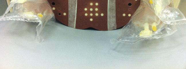
16 Method ART anthropomorphic phantom Thorax section Plugs removed in 3 locations: central lung; near rib; near heart 1 ml syringe filled with iodine solution and inserted into channels Two iodine solutions and water considered: 1. mg/ml,.6 mg/ml,. mg/ml Scanned at 8 kv, 1 kv, 12 kv Reduced FOV (15 cm) used in accordance with trial protocol Three different patient sizes : 5 kg, 8 kg, 95 kg
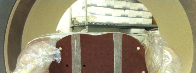
17
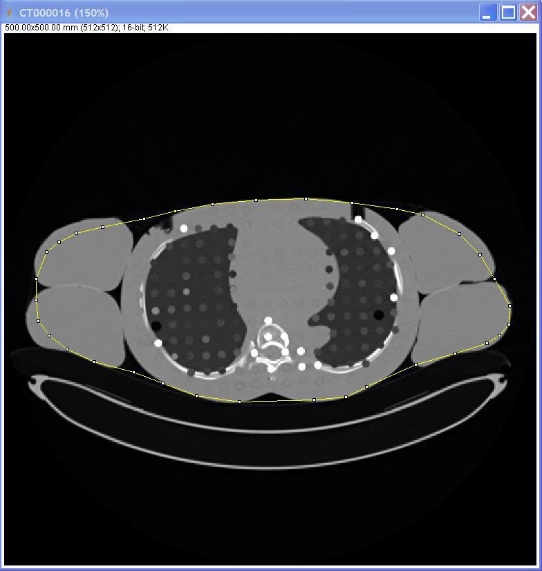
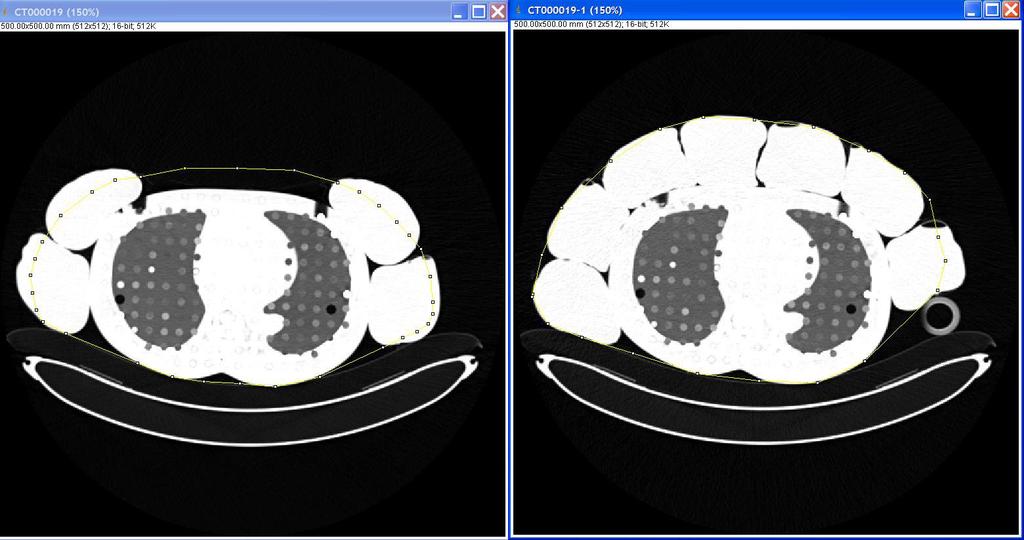
18 Method Images exported to CD and read with ImageJ ROI placed over central part of iodine solution ROI had to be moved manually as the phantom was not entirely straight and the ROI was small Inter-slice variation measurements made at 7 slices, ignoring gaps between phantom slabs Mean and SD in CT number recorded for each ROI nearly 2 measurements in total
19 Results 8 kv
20 Method Considered CT number enhancement, ie CT#(iodine) CT#(water) in same location. The CT number of water varied significantly depending on location in the phantom, phantom size and kv 7 Water CT number at 1 kv in small phantom 6 5 CT number (HU) rib lung heart Slice number
21 Method Considered CT number enhancement, ie CT#(iodine) CT#(water) in same location. The CT number of water varied significantly depending on location in the phantom, phantom size and kv Measuring enhancement more closely mimics the clinical situation where we are interested in increase in CT number compared to pre-contrast scan Excluded: slices between slabs of phantom air bubbles at ends of syringes slices at base of lung where diaphragm begins to appear Take mean of CT number enhancement in all remaining slices Students T-test (2-talied) to assess significance of differences in enhancement between different locations in phantom and different patient sizes (p<.5)
22 Results CT number varied with all parameters: kv Iodine concentration Following results are for 1. mg/ml Location within phantom Phantom size
23 kv 3 Results 8 kv
24 6 5 1 kv Results 8 kv

25 kv
26 Analysis Enhancement is a bit erratic Generally: Enhancement slightly depressed near ribs beam hardening artefacts Enhancement near heart is variable beam hardening effects vs beam hardening correction? Enhancement in lung tissue near expected, except at 8 kv (high) Remember, considering enhancement not simply CT# Which kv should we use for the trial?
27 8 7 8 kv 6 Results 8 kv Decided not to use 8 kv Statistically significant variation in enhancement, depending on location within phantom and phantom size At 8 kv there is a significant difference in beam quality at different positions in the phantom Difficulties in obtaining high enough mas to keep noise low and ENR high for large patients
28 kv Results 8 kv Decided against 12 kv too: Enhancement in the large phantom varied significantly between positions Since enhancement values are lowest, a high dose needs to be delivered to obtain reasonable ENR (> 1.5?)* *Miles et al Current status and guidelines for the assessment of tumour vascular support with dynamic contrast-enhanced computed tomography (Eur Radiol. 212; 22: ), suggests ENR > 1.5.


 < 6 2 2.1 8.6 6 9 35 1.6 15.")
29 6 5 1 kv Results 8 kv We will use 1 kv in the SPUtNIk trial Least variation between phantoms and positions ENR > 1.5 at reasonable CTDI Threshold enhancement of 2 HU Mass (kg) mas ENR CTDI (mgy) < >
30 Analysis Limitations: Hard to know what patient mass our phantoms are ROI very small for 1ml syringes (area 5.9mm 2 / diameter 2.7mm). Nodules in trial will be >8mm, ROI drawn on widest axial slice at 7% nodule diameter on lung window Uncertainty in [I] and/or non-uniform distribution along syringe. Minimised by shaking our stock vial before drawing into the syringe Position of heart ROI in the heart itself for some slices, in the lung for others Since reduced FOV used, not sure what beam hardening correction is being applied. We will use a patient-size phantom with reduced FOV for the scanner assessments
31 Conclusions
32 Conclusions CT number does increase with increasing iodine concentration, but Need to use enhancementover baseline, rather than raw CT number Be aware of the limitationsand uncertaintyin enhancement measurements Think about what phantomto use to check the CT# / [I] for your scanner. Air? Water? Lung? The kvyou choose will depend on the application and required ENR All of these things will vary between scanners and recon kernels

33 Acknowledgements Maria Lewis (Guy s & St Thomas ) SPUtNIk trial group University of Southampton Clinical Trials Unit Project ref. 9/22/117
34 SPUtNIk Single Pulmonary Nodule Investigation Sites taking part in the trial: Aberdeen Brighton & Sussex Glasgow Mount Vernon Oxford Papworth Southampton UCH London
35 Thank you for listening Rosemary Eaton, Andrew Shah, Jane Shekhdar Medical Physics, Mount Vernon Hospital CT Users Group 4 th October 212, Edinburgh
CT NUMBER ACCURACY ANALYSIS FOR RADIOTHERAPY TREATMENT PLANNING IMAGING
 CT NUMBER ACCURACY ANALYSIS FOR RADIOTHERAPY TREATMENT PLANNING IMAGING Julian Liu a, Keisha Robinson a, DhanaJayan Kothandan a and Joshua Luis b (a) Cancer Centre London (b) University College London
CT Optimisation for Paediatric SPECT/CT Examinations. Sarah Bell
 CT Optimisation for Paediatric SPECT/CT Examinations Sarah Bell Sarah.bell14@nhs.net Outline 1. Introduction 2. Aims and Objectives 3. Methods 4. Results 5. Discussion 6. Conclusions 7. References Introduction
To Shield or Not to Shield? Lincoln L. Berland, M.D.
 To Shield or Not to Shield? Lincoln L. Berland, M.D. Disclosures Consultant to: Nuance, Inc. Page 2 Breast Radiation on CT Use of chest CT has increased in women vulnerable to cancer induction by radiation.
ESTABLISHING DRLs in PEDIATRIC CT. Keith Strauss, MSc, FAAPM, FACR Cincinnati Children s Hospital University of Cincinnati College of Medicine
 ESTABLISHING DRLs in PEDIATRIC CT Keith Strauss, MSc, FAAPM, FACR Cincinnati Children s Hospital University of Cincinnati College of Medicine CT Dose Indices CTDI INTRODUCTION CTDI 100, CTDI w, CTDI vol
Acknowledgments. A Specific Diagnostic Task: Lung Nodule Detection. A Specific Diagnostic Task: Chest CT Protocols. Chest CT Protocols
 Personalization of Pediatric Imaging in Terms of Needed Indication-Based Quality Per Dose Acknowledgments Duke University Medical Center Ehsan Samei, PhD Donald Frush, MD Xiang Li PhD DABR Cleveland Clinic
CURRENT CT DOSE METRICS: MAKING CTDI SIZE-SPECIFIC
 CURRENT CT DOSE METRICS: MAKING CTDI SIZE-SPECIFIC Keith Strauss, MSc, FAAPM, FACR Cincinnati Children s Hospital University of Cincinnati College of Medicine Acknowledgments John Boone, PhD Michael McNitt-Grey,
SOMATOM Drive System Owner Manual Dosimetry and imaging performance report
 www.siemens.com/healthcare SOMATOM Drive System Owner Manual Dosimetry and imaging performance report Table of contents 1 Dosimetry and imaging performance report 5 1.1 Dose information 5 1.1.1 General
Patient dose assessment of CT perfusion scanning at the RSCH
 Patient dose assessment of CT perfusion scanning at the RSCH Lesley Leavesley, Emma Whitehead, Matthew Pryor, Debbie Peet Regional Radiation Protection Service Royal Surrey County Hospital, Guildford Overview
Dual-Energy CT: The Technological Approaches
 Dual-Energy CT: The Technological Approaches Dushyant Sahani, M.D Director of CT Associate Professor of Radiology Massachusetts General Hospital Harvard Medical School Email-dsahani@partners.org Disclosure
Doses from Cervical Spine Computed Tomography (CT) examinations in the UK. John Holroyd and Sue Edyvean
 examinations in the UK. John Holroyd and Sue Edyvean") Doses from Cervical Spine Computed Tomography (CT) examinations in the UK John Holroyd and Sue Edyvean Why a new dose survey? Number of enquires received concerning the current NDRL Concern that could
Doses from Cervical Spine Computed Tomography (CT) examinations in the UK John Holroyd and Sue Edyvean Why a new dose survey? Number of enquires received concerning the current NDRL Concern that could
Dental ConeBeam Computed Tomography (CBCT) X-ray Systems
 X-ray Systems") Dental ConeBeam Computed Tomography (CBCT) X-ray Systems PROPOSED REVISIONS TO 4732.XXXX, 1.0 4732.#### DENTAL CONEBEAM COMPUTED TOMOGRAPHY (CBCT) X-RAY SYSTEMS; STATIONARY AND MOBILE. Subpart 1. Applicability.
Dental ConeBeam Computed Tomography (CBCT) X-ray Systems PROPOSED REVISIONS TO 4732.XXXX, 1.0 4732.#### DENTAL CONEBEAM COMPUTED TOMOGRAPHY (CBCT) X-RAY SYSTEMS; STATIONARY AND MOBILE. Subpart 1. Applicability.
Paediatric Dose Reduction and Image Quality
 Paediatric Dose Reduction and Image Quality Alan Whiteside The majority of this work was undertaken as part of MSc Thesis of Helen Dixon. Introduction Paediatric CT protocols result in a higher effective
Doses from pediatric CT examinations in Norway Are pediatric scan protocols developed and in daily use?
 Doses from pediatric CT examinations in Norway Are pediatric scan protocols developed and in daily use? Eva Godske Friberg * Norwegian Radiation Protection Authority, P.O. Box, Østerås, Norway Abstract.
Implementation of the 2012 ACR CT QC Manual in a Community Hospital Setting BRUCE E. HASSELQUIST, PH.D., DABR, DABSNM ASPIRUS WAUSAU HOSPITAL
 Implementation of the 2012 ACR CT QC Manual in a Community Hospital Setting BRUCE E. HASSELQUIST, PH.D., DABR, DABSNM ASPIRUS WAUSAU HOSPITAL Conflict of Interest Disclaimer Employee of Aspirus Wausau
Dual-Energy Imaging of Bone Marrow Edema on a Dedicated Multi-Source Cone-Beam CT System for the Extremities
 Dual-Energy Imaging of Bone Edema on a Dedicated Multi-Source Cone-Beam CT System for the Extremities W Zbijewski, 1 A Sisniega, 1 JW Stayman, 1 N Packard, 2 J Yorkston, 2 G Thawait, 3 S Demehri, 3 J Fritz,
Varian Edge Experience. Jinkoo Kim, Ph.D Henry Ford Health System
 Varian Edge Experience Jinkoo Kim, Ph.D Henry Ford Health System Disclosures I participate in research funded by Varian Medical Systems. Outline of Presentation Review advanced imaging in Varian Edge Linear
Toshiba Aquillion 64 CT Scanner. Phantom Center Periphery Center Periphery Center Periphery
 Comparison of radiation dose and imaging performance for the standard Varian x-ray tube and the Richardson Healthcare ALTA750 replacement tube for the Toshiba Aquillion CT scanners. by Robert L. Dixon,
Gender differences in CT calcium scoring: A phantom study
 Gender differences in CT calcium scoring: A phantom study Nicholas Petrick, Qin Li, Benjamin Berman, Marios A Gavrielides, Rongping Zeng, Berkman Sahiner CDRH/OSEL/DIDSR U.S. Food and Drug Administration
Chapter 6. Hester Gietema Cornelia Schaefer-Prokop Willem Mali Gerard Groenewegen Mathias Prokop. Accepted for publication in Radiology
 Chapter 6 Interscan variability of semiautomated volume measurements in intraparenchymal pulmonary nodules using multidetector-row computed tomography: Influence of inspirational level, nodule size and
Breast Cancer PET/CT Imaging Protocol
 Breast Cancer PET/CT Imaging Protocol Scanning Protocol: Patients are scanned from the top of the neck through the pelvis. Arms-up position is used to avoid beam-hardening artifact in the chest and abdomen.
Computed tomography Acceptance testing and dose measurements
 Computed tomography Acceptance testing and dose measurements Jonas Andersson Medical Physicist, Ph.D. Department of Radiation Sciences University Hospital of Norrland, Umeå Sweden Contents The Computed
Dual Energy CT of Pulmonary Embolism
 Dual Energy CT of Pulmonary Embolism U. Joseph Schoepf, MD, FAHA, FSCBT MR, FSCCT Professor of Radiology, Medicine, and Pediatrics Director of Cardiovascular Imaging Disclosures Consultant for / research
X-Ray & CT Physics / Clinical CT
 Computed Tomography-Basic Principles and Good Practice X-Ray & CT Physics / Clinical CT INSTRUCTORS: Dane Franklin, MBA, RT (R) (CT) Office hours will be Tuesdays from 5pm to 6pm CLASSROOM: TIME: REQUIRED
Translating Protocols Across Patient Size: Babies to Bariatric
 Translating Protocols Across Patient Size: Babies to Bariatric Cynthia H. McCollough, PhD, FACR, FAAPM Professor of Radiologic Physics Director, CT Clinical Innovation Center Department of Radiology Mayo
Biases affecting tumor uptake measurements in FDG-PET
 Biases affecting tumor uptake measurements in FDG-PET M. Soret, C. Riddell, S. Hapdey, and I. Buvat Abstract-- The influence of tumor diameter, tumor-tobackground activity ratio, attenuation, spatial resolution,
Accounting for Imaging Dose
 Accounting for Imaging Dose High Profile Over-exposures Lead to Growing Concern FDA issues warning in October 2009-209 patients exposed to 8 times typical dose for CT brain perfusion scan (3-4 Gy) - Some
Patient / Organ Dose in CT
 Patient / Organ Dose in CT Patient specific and organ dose estimation H.D. Nagel Dr. HD Nagel, Science & Technology for Radiology Buchholz / Germany www.sascrad.com 1 Topics CTDI & patient dose SSDE Organ
True Dual Energy. Dr. Stefan Ulzheimer, Siemens Healthcare GmbH. DEfinitely Siemens
 DEfinitely Siemens True Dual Energy Dr. Stefan Ulzheimer, Siemens Healthcare GmbH International version. Not for distribution in the US. Unrestricted Siemens AG 2015 All rights reserved. The products/features
Alfred Health's CT quality control program
 Alfred Health's CT quality control program Poster No.: R-0037 Congress: Type: Authors: 2014 CSM Scientific Exhibit A. Perdomo, Z. Brady, N. Tran, L. Hudson, K. Provis; PRAHRAN/ AU Keywords: Quality assurance,
8/18/2011. Acknowledgements. Managing Pediatric CT Patient Doses INTRODUCTION
 Managing Pediatric CT Patient Doses Keith J. Strauss, MSc, FAAPM, FACR President X-Ray Computations, Inc. Boston, Massachusetts Acknowledgements Marilyn Goske, MD John Boone, PhD Cynthia McCollough, PhD
Why is CT Dose of Interest?
 Why is CT Dose of Interest? CT usage has increased rapidly in the past decade Compared to other medical imaging CT produces a larger radiation dose. There is direct epidemiological evidence for a an increase
Automated CT Protocol Design Advantages and Pitfalls of Algorithm-Based Technique Selection in Pediatrics. Disclosures 7/22/2014. Learning Objectives
 Automated CT Protocol Design Advantages and Pitfalls of Algorithm-Based Technique Selection in Pediatrics Robert MacDougall, M.Sc. Department of Radiology Boston Children s Hospital Disclosures 2 Learning
SPECIFIC PRINCIPLES FOR DOSE REDUCTION IN HEAD CT IMAGING. Rajiv Gupta, MD, PhD Neuroradiology, Massachusetts General Hospital Harvard Medical School
 SPECIFIC PRINCIPLES FOR DOSE REDUCTION IN HEAD CT IMAGING Rajiv Gupta, MD, PhD Neuroradiology, Massachusetts General Hospital Harvard Medical School OUTLINE 1 st Presentation: Dose optimization strategies
Typical PET Image. Elevated uptake of FDG (related to metabolism) Lung cancer example: But where exactly is it located?
 Lung cancer example: But where exactly is it located?") Typical PET Image Elevated uptake of FDG (related to metabolism) Lung cancer example: But where exactly is it located? PET/CT Oncology Imaging Anatometabolic fusion images are useful in the management
Typical PET Image Elevated uptake of FDG (related to metabolism) Lung cancer example: But where exactly is it located? PET/CT Oncology Imaging Anatometabolic fusion images are useful in the management
Optimized PET/CT protocols: how much CT is needed? Increasing use of PET-CT. Imaging in Lymphoma
 BVS/ABR Workshop 2011 on dose related to multimodality imaging Optimized PET/CT protocols: how much CT is needed? 2008 Mean medical radiation exposure/head Belgium= 2.42 msv The Netherlands= 0.8 msv Belgium
Experimental measurement with an anthropomorphic phantom of the proton dose distribution in the presence of metal implants
 E13195: Slightly better series/treatment with PRONE combination with vertical field. - This field is only possible in case the tumor is not too caudal (problem with energy). - It will increase the integral
State of the art and future development for standardized estimation of organ doses in CT
 State of the art and future development for standardized estimation of organ doses in CT March 2015 William J. O Connel, Dr. Ph, Senior Medical Physicist Imagination at work. Agenda Introduction Duke Florida
Outcomes in the NLST. Health system infrastructure needs to implement screening
 Outcomes in the NLST Health system infrastructure needs to implement screening Denise R. Aberle, MD Professor of Radiology and Bioengineering David Geffen School of Medicine at UCLA 1 Disclosures I have
Conventional and spiral CT dose indices in Yazd general hospitals, Iran
 Iran. J. Radiat. Res., 2006; 3 (4): 183-189 Conventional and spiral CT dose indices in Yazd general hospitals, Iran F. Bouzarjomehri 1*,M.H.Zare 2, D. Shahbazi 2 1 Department of Medical Physics, Shahid
Thoracic examinations with 16, 64, 128 and 256 slices CT: comparison of exposure doses measured with an anthropomorphic phantom and TLD dosimeters
 Thoracic examinations with 16, 64, 128 and 256 slices CT: comparison of exposure doses measured with an anthropomorphic phantom and TLD dosimeters Poster No.: C-2584 Congress: ECR 2015 Type: Scientific
Radiation Dose Reduction: Should You Use a Bismuth Breast Shield?
 Radiation Dose Reduction: Should You Use a Bismuth Breast Shield? Lincoln L. Berland, M.D., F.A.C.R. Michael V. Yester, Ph.D. University of Alabama at Birmingham Breast Radiation on CT Use of chest CT
CT Radiation Risks and Dose Reduction
 CT Radiation Risks and Dose Reduction Walter L. Robinson, M.S. D.A.B.S.N.M., D.A.B.M.P., D.A.B.R. Consultant Certified Medical Radiation Health & Diagnostic Imaging Physicist Medical Radiation and Children
Ultralow Dose Chest CT with MBIR
 Ultralow Dose Chest CT with MBIR Ella A. Kazerooni, M.D. Professor & Director Cardiothoracic Radiology Associate Chair for Clinical Affairs University of Michigan Disclosures Consultant: GE Healthcare
E. Senéterre*, F. Paganin**, J.M. Bruel*, F.B. Michel**, J. Bousquet**
 Eur Respir J, 1994, 7, 596 6 DOI: 1.1183/931936.94.73596 Printed in UK - all rights reserved Copyright ERS Journals Ltd European Respiratory Journal ISSN 93-1936 TECHNICAL NOTE Measurement of the internal
FDG-18 PET/CT - radiation dose and dose-reduction strategy
 FDG-18 PET/CT - radiation dose and dose-reduction strategy Poster No.: C-1856 Congress: ECR 2014 Type: Authors: Keywords: DOI: Scientific Exhibit P. Nicholson, S. McSweeney, K. O'Regan; Cork/IE Radiation
Cone Beam CT Protocol Optimisation for Prostate Imaging with the Varian Radiotherapy OBI imaging system. Dr Craig Moore & Dr Tim Wood
 Cone Beam CT Protocol Optimisation for Prostate Imaging with the Varian Radiotherapy OBI imaging system Dr Craig Moore & Dr Tim Wood Background With the increasing use of CBCT imaging alongside complex
Joint ICTP/IAEA Advanced School on Dosimetry in Diagnostic Radiology and its Clinical Implementation May 2009
 2033-4 Joint ICTP/ Advanced School on Dosimetry in Diagnostic Radiology and its Clinical Implementation 11-15 May 2009 Dosimetry for General Radiology and Clinical Uncertainty Peter Homolka EFOMP Training
Managing Radiation Risk in Pediatric CT Imaging
 Managing Radiation Risk in Pediatric CT Imaging Mahadevappa Mahesh, MS, PhD, FAAPM, FACR, FACMP, FSCCT. Professor of Radiology and Cardiology Johns Hopkins University School of Medicine Chief Physicist
Cardiac CTA Prospective Gating Broad Beam
 Cardiac CTA Prospective Gating Broad Beam ACQUISITION- Broad Beam Gating: Prospective Non Contrast Scan-Calcium Score Patient Position Supine Feet First into Gantry Heart Isocenter Scanogram AP and Lateral
National Cancer Institute
 National Cancer Institute A Dosimetry Summary of CT Participants in the National Lung Screening Trial (NLST) U.S. DEPARTMENT OF HEALTH AND HUMAN SERVICES National Institutes of Health AAPM 2015 Anaheim,
Measurement of organ dose in abdomen-pelvis CT exam as a function of ma, KV and scanner type by Monte Carlo method
 Iran. J. Radiat. Res., 2004; 1(4): 187-194 Measurement of organ dose in abdomen-pelvis CT exam as a function of ma, KV and scanner type by Monte Carlo method M.R. Ay 1, M. Shahriari 2, S. Sarkar 3, P.
Dual-Energy 101: Principles, Methods and Dose
 Dual-Energy 101: Principles, Methods and Dose Juan Carlos Ramirez-Giraldo, Ph.D Staff Scien2st, Collabora2ons Manager SE Region ISCT San Francisco, 2017 Siemens Medical Solu2ons USA, Inc., 2017 Page 1
An audit of radiation dose of 4D CT in a radiotherapy department
 An audit of radiation dose of 4D CT in a radiotherapy department Poster No.: R-0097 Congress: Type: Authors: Keywords: DOI: 2014 CSM Scientific Exhibit T. Hubbard, J. Callahan, J. Cramb, R. Budd, T. Kron;
Quantitative and Qualitative Assessment of Thorax Cone Beam CT Image Quality across Multiple Imaging Systems
 Quantitative and Qualitative Assessment of Thorax Cone Beam CT Image Quality across Multiple Imaging Systems Matthew Williams Pre-registration Clinical Scientist Velindre NHS Trust, Cardiff Computed Tomography
CT angiography of pulmonary arteries to detect pulmonary embolism with low kv settings
 CT angiography of pulmonary arteries to detect pulmonary embolism with low kv settings Poster No.: C-3289 Congress: ECR 2010 Type: Scientific Exhibit Topic: Chest - Your latest results Authors: M. K. Gill,
Low Dose CT Lung Screening: What is Technically Required?
 Low Dose CT Lung Screening: What is Technically Required? COMP/CCPM Annual Scientific Meeting Ottawa, Ontario July 15 th, 2017 Yogesh Thakur, PhD, MCCPM Medical Physicist Lead and Regional RSO (X-Ray)
Survey of patients CT radiation dose in Jiangsu Province
 Original Article Page 1 of 6 Survey of patients CT radiation dose in Jiangsu Province Yuanyuan Zhou 1, Chunyong Yang 1, Xingjiang Cao 1, Xiang Du 1, Ningle Yu 1, Xianfeng Zhou 2, Baoli Zhu 1, Jin Wang
Improved image quality of low-dose thoracic CT examinations with a new postprocessing software*
 JOURNAL OF APPLIED CLINICAL MEDICAL PHYSICS, VOLUME 11, NUMBER 3, Summer 2010 Improved image quality of low-dose thoracic CT examinations with a new postprocessing software* Anne Catrine Traegde Martinsen,
Calculation of Effective Doses for Radiotherapy Cone-Beam CT and Nuclear Medicine Hawkeye CT Laura Sawyer
 Calculation of Effective Doses for Radiotherapy Cone-Beam CT and Nuclear Medicine Hawkeye CT Laura Sawyer Department of Medical Physics and Bioengineering, Royal United Hospital, Bath Overview Varian Acuity
Measurement error of spiral CT Volumetry:
 Measurement error of spiral CT Volumetry: Influence of Low Dose CT Technique 1 Tae Gyu Lee, M.D. 2, Myung Jin Chung, M.D., Sung Bum Cho, M.D. 2, Jae Min Cho, M.D., Seog Joon Kim, M.D. 2, Sang Hyun Baik,
Managing the imaging dose during Image-guided Radiotherapy. Martin J Murphy PhD Department of Radiation Oncology Virginia Commonwealth University
 Managing the imaging dose during Image-guided Radiotherapy Martin J Murphy PhD Department of Radiation Oncology Virginia Commonwealth University Radiographic image guidance has emerged as the new paradigm
Crowd-Sourcing Quality in Imaging
 Crowd-Sourcing Quality in Imaging Ricardo S. Avila rick.avila@accumetra.com April 20, 2017 2017 Dialog For Action on Cancer Screening and Prevention Image Quality For Lung Cancer Screening Since 2015:
A multicentric study on patient dose in multislice CT
 A multicentric study on patient dose in multislice CT A.Stratis 1, M.Molfetas 1, S.Kottou 2, A.Louizi 2 1. Medical Physics department, Evangelismos General hospital of Athens, Athens, Greece 2. Medical
Guidelines for the Management of Pulmonary Nodules Detected by Low-dose CT Lung Cancer Screening
 Guidelines for the Management of Pulmonary Nodules Detected by Low-dose CT Lung Cancer Screening 1. Introduction In January 2005, the Committee for Preparation of Clinical Practice Guidelines for the Management
Fused monochromatic imaging acquired by single source dual energy CT in hepatocellular carcinoma during arterial phase: an initial experience
 Original Article Fused monochromatic imaging acquired by single source dual energy CT in hepatocellular carcinoma during arterial phase: an initial experience Shun-Yu Gao, Xiao-Peng Zhang, Yong Cui, Ying-Shi
CT examination is a high-radiation-dose imaging technique
 ORIGINAL RESEARCH J.S.P. Tan K.-L. Tan J.C.L. Lee C.-M. Wan J.-L. Leong L.-L. Chan Comparison of Eye Lens Dose on Neuroimaging Protocols between 16- and 64-Section Multidetector CT: Achieving the Lowest
Loren Ketai, MD; Mathurn Malby, BS; Kirk Jordan, MD; Andrew Meholic, MD; and Julie Locken, MD
 Small Nodules Detected on Chest Radiography* Does Size Predict Calcification? Loren Ketai, MD; Mathurn Malby, BS; Kirk Jordan, MD; Andrew Meholic, MD; and Julie Locken, MD Study objectives: To determine
Whole Body CT Protocol Update 2018
 Whole Body CT Protocol Update 2018 10 th Nordic Course in Trauma Radiology Gothenburg, Sweden K.SHANMUGANATHAN M.D. Disclosure of Commercial Interest Neither I nor my immediate family members have a financial
Measurements of Air Kerma Index in Computed Tomography: A comparison among methodologies
 Measurements of Air Kerma Index in Computed Tomography: A comparison among methodologies Thêssa C. Alonso 1, 2, Arnaldo P. Mourão 1, 3, Teógenes A. Da Silva 1, 2 1 Program of Nuclear Science and Techniques
Chapter 5. Pulmonary nodules detected at lung cancer screening: Interobserver variability of semiautomated volume measurements
 Chapter 5 Pulmonary nodules detected at lung cancer screening: Interobserver variability of semiautomated volume measurements Hester Gietema Ying Wang Dongming Xu Rob van Klaveren Harry de Koning Ernst
4D PET: promises and limitations
 4D PET: promises and limitations Tinsu Pan, Ph.D. M.D. Anderson Cancer Center The University of Texas Background Outlines Gating techniques: Deep inspiration breath hold 4D PET/CT Non-gating techniques
Hi RES Extremity - (04/18/2011) CTDI: ~13 mgy per acquisition Used for evaluation of: Ankle Elbow Hand Wrist Foot /Calcaneous Toes Fingers
 CTDI: ~13 mgy per acquisition Used for evaluation of: Ankle Elbow Hand Wrist Foot /Calcaneous Toes Fingers") P a g e 1 Hi RES Extremity - (04/18/2011) CTDI: ~13 mgy per acquisition Used for evaluation of: Ankle Elbow Hand Wrist Foot /Calcaneous Toes Fingers Billing: 1. CT Upper/Lower Extremity of concern without
P a g e 1 Hi RES Extremity - (04/18/2011) CTDI: ~13 mgy per acquisition Used for evaluation of: Ankle Elbow Hand Wrist Foot /Calcaneous Toes Fingers Billing: 1. CT Upper/Lower Extremity of concern without
Cardiopulmonary Imaging Original Research
 Cardiopulmonary Imaging Original Research Ebner et al. MIP and CAD Algorithms in CT for Lung Cancer Screening Cardiopulmonary Imaging Original Research Lukas Ebner 1 Justus E. Roos 1 Jared D. Christensen
Uozu city/jp, Minatoku, Tokyo/JP Bones, Extremities, CT, Surgery, Physics, Artifacts, Image verification /ecr2014/C-0462
 Metal Artifact Reduction Algorithm enablesreduce metal artifacts and improvement of diagnosis in the postoperative Pedicle screws implant for Spinal Fusion: A Phantom Study Poster No.: C-0462 Congress:
HI-Res Extremity Sensation 16
 Page 1 Routine Extremity - (2/14/2013) CTDI: ~20 mgy per acquisition Used for evaluation of: Humerus Forearm Femur Knee Tib/Fib Billing: 1. CT Upper/Lower Extremity of concern without contrast, with contrast,
CT angiography techniques. Boot camp
 CT angiography techniques Boot camp Overview Basic concepts Contrast administration arterial opacification Time scan acquisition during the arterial phase Protocol examples Helical non-gated CTA Pulmonary
PET CT for Staging Lung Cancer
 PET CT for Staging Lung Cancer Rohit Kochhar Consultant Radiologist Disclosures Neither I nor my immediate family members have financial relationships with commercial organizations that may have a direct
Ask EuroSafe Imaging. Tips & Tricks. CT Working Group. Optimization of scan length to reduce CT radiation dose
 Ask EuroSafe Imaging Tips & Tricks CT Working Group Optimization of scan length to reduce CT radiation dose Alban Gervaise (Centre Hospitalier Universitaire Nancy, FR) Mika Kortesniemi (HUS Medical Imaging
8/1/2017. Financial Disclosures. Dose Tracking at MGH. How Dose Tracking Affected Protocol Optimization in a Tertiary Quaternary Healthcare Center
 How Dose Tracking Affected Protocol Optimization in a Tertiary Quaternary Healthcare Center Mannudeep K. Kalra, MD Webster Center for Quality and Safety Massachusetts General Hospital Harvard Medical School
THE SAAD CENTRE FOR RADIOGRAPHY OPEN FOR BUSINESS. The University for business and the professions
 THE SAAD CENTRE FOR RADIOGRAPHY OPEN FOR BUSINESS The University for business and the professions THE SAAD CENTRE FOR RADIOGRAPHY OPEN FOR BUSINESS The Department of Radiography at City University London
Optimizing radiation dose by varying age at pediatric temporal bone CT
 JOURNAL OF APPLIED CLINICAL MEDICAL PHYSICS, VOLUME 16, NUMBER 1, 2015 Optimizing radiation dose by varying age at pediatric temporal bone CT Daichi Noto, 1 Yoshinori Funama, 2a Mika Kitajima, 3 Daisuke
CT Quality Control Manual FAQs
 CT Quality Control Manual FAQs General Question: How often will the QC Manual be updated and how will those updates be communicated? Answer: The ACR CT Physics Subcommittee will review any comments, issues
Breast CT and Dosimetry
 2013 ICTP/IAEA Training Course on Radiation Protection of Patients Trieste Breast CT and Dosimetry John M. Boone, Ph.D., FAAPM, FSBI, FACR Professor and Vice Chair (Research) of Radiology Professor of
Introduction and Background
 CT Lung Cancer Screening and the Medical Physicist: Background, Findings and Participant Dosimetry Summary of the National Lung Screening Trial (NLST) Randell Kruger, PhD, DABR Medical Physics Section
Radiation Exposure in Pregnancy. John R. Mayo UNIVERSITY OF BRITISH COLUMBIA
 Radiation Exposure in Pregnancy John R. Mayo UNIVERSITY OF BRITISH COLUMBIA Illustrative Clinical Scenario 32 year old female 34 weeks pregnant with recent onset shortness of breath and central chest pain
354 Korean J Radiol 9(4), August 2008
, August 2008") Review of Failed CT Phantom Image Evaluations in 2005 and 2006 by the CT Accreditation Program of the Korean Institute for Accreditation of Medical Image Hye Jung Park, MD 1 Seung Eun Jung, MD 1, 2 Young
Review of Failed CT Phantom Image Evaluations in 2005 and 2006 by the CT Accreditation Program of the Korean Institute for Accreditation of Medical Image Hye Jung Park, MD 1 Seung Eun Jung, MD 1, 2 Young
IGRT1 technologies. Paweł Kukołowicz Warsaw, Poland
 IGRT1 technologies Paweł Kukołowicz Warsaw, Poland Minimal prerequisite for good, efficient radiotherapy ICTP 2015 Paweł Kukołowicz 2/29 Minimal prerequisite for good, efficient radiotherapy Well trained
Handzettel 1. CT Contrast Media. Agenda. Contrast Media Definition. Agenda. Why we need contrast media? Agenda
 Agenda CT Contrast Media Weena Swatdiswanee Factorinvolvein contrast enchancement Senior Application Specialist, CT Regional Headquarter Asia Australia weena.swat@siemens.com Page 1 Page 2 Agenda Contrast
SpiderX. Portable Residual Stress X-Ray Diffractometer.
 Portable Residual Stress X-Ray Diffractometer www.gnr.it About SpiderX GNR Analytical Instrument offers equipment based on X-Ray Diffraction to measure residual stress state and retained austenite content.
Assessment of effective dose in paediatric CT examinations
 Assessment of effective dose in paediatric CT examinations E. Dougeni 1,2 CL. Chapple 1, J. Willis 1, G. Panayiotakis 2 1 Regional Medical Physics Department, Freeman Hospital, Freeman Road, Newcastle
Metal Artefact Reduction in CT
 Metal Artefact Reduction in CT DANIEL MARRINER Metal Artefact Reduction in CT Metal Artefact Clinical Indications for MAR SEMAR and How It Works Technical Considerations Case Studies utilising SEMAR Metal
Dianna Cody, PhD, DABR, FAAPM Professor & Clinical Operations Director Imaging Physics U.T. M.D. Anderson Cancer Center Houston, TX
 Dianna Cody, PhD, DABR, FAAPM Professor & Clinical Operations Director Imaging Physics U.T. M.D. Anderson Cancer Center Houston, TX Learning Objectives: Limitations for estimating patient dose for CT Methods
Recommendations for cross-sectional imaging in cancer management, Second edition
 www.rcr.ac.uk Recommendations for cross-sectional imaging in cancer management, Second edition Carcinoma of unknown primary origin (CUP) Faculty of Clinical Radiology www.rcr.ac.uk Contents Carcinoma of
Modelling the effect of lead and other materials for shielding of the fetus in CT pulmonary angiography
 The British Journal of Radiology, 81 (2008), 499 503 SHORT COMMUNICATION Modelling the effect of lead and other materials for shielding of the fetus in CT pulmonary angiography 1 G R IBALL, MSc, BSc, 2
Triple Rule-out using 320-row-detector volume MDCT: A comparison of the wide volume and helical modes
 Triple Rule-out using 320-row-detector volume MDCT: A comparison of the wide volume and helical modes Poster No.: C-0488 Congress: ECR 2012 Type: Authors: Keywords: DOI: Scientific Exhibit E.-J. Kang,
CT Dose Estimation. John M. Boone, Ph.D., FAAPM, FSBI, FACR Professor and Vice Chair of Radiology. University of California Davis Medical Center
 CT Dose Estimation John M. Boone, Ph.D., FAAPM, FSBI, FACR Professor and Vice Chair of Radiology 1 University of California Davis Medical Center CT Dose Estimation Introduction The CTDI Family of Metrics
Electron Beam CT versus 16-slice Spiral CT: How Accurately Can We Measure. Coronary Artery Calcium Volume?
 Electron Beam CT versus 16-slice Spiral CT: How Accurately Can We Measure Coronary Artery Calcium Volume? 1 Objective: The purpose of this study is to investigate how accurately we can measure CAC volume
CT SCAN PROTOCOL. Shoulder
 CT SCAN PROTOCOL Shoulder Purpose and Summary CT images made with this protocol are used to provide the orthopedic surgeon with a detailed 3D anatomical reconstruction of the patient s scapula and proximal
Managing Patient Dose in Computed Tomography (CT) INTERNATIONAL COMMISSION ON RADIOLOGICAL PROTECTION
 INTERNATIONAL COMMISSION ON RADIOLOGICAL PROTECTION") Managing Patient Dose in Computed Tomography (CT) International Commission on Radiological Protection Information abstracted from ICRP Publication 87 Available at www.icrp.org Task Group: M.M. Rehani,
Managing Patient Dose in Computed Tomography (CT) International Commission on Radiological Protection Information abstracted from ICRP Publication 87 Available at www.icrp.org Task Group: M.M. Rehani,