Dental Assisting COURSE ONE
|
|
|
- Damian Moody
- 5 years ago
- Views:
Transcription
1 Dental Assisting COURSE ONE 1
2 History of Dentistry False teeth used to be made of wood or ivory Often painful dental treatments were done by "guzzling some vodka" Wilhelmina Conrad Roentgen discovered radiographs, X-rays in 1895 C.Edmund Kells - New Orleans dentist was the first to employ a dental assistant in the Certifying Board of American Dental Assistants Association was established year programs for dental assisting education 2
3 Professional Dental Assistant Professionalism is an attitude that is apparent in everything we do and say Must demonstrate patience and compassion when dealing with patients and other team members The dental assistant has many roles - chair side, expanded functions, administrative, etc. After completing the necessary requirements in an institution - clinical and didactics, the certified dental assistant must write and be successful in the dental assisting national board exam 3
4 The Dental Healthcare Team Dentist - has received an undergraduate degree, successfully completed dental school and may it may not specialize (such as Endodontist, Orthodontist, etc.). Licensed with their association Dental hygienist - successfully completed a clinical and didactics course, taken the national dental hygiene exam and performed the clinical exam (if graduated from a non-accredited school). Needs to be registered with an association Dental receptionist - may not may not have completed a specialized dental reception course Dental laboratory technician - usually does not work in the dental office but in a laboratory setting. Performs tasks given by a dentist. They would have passed their written exam and be apart of their association 4
5 Dental Ethics REVIEW THE CODE OF ETHICS AT THE FOLLOWING LINK: de_of_ethics_public.pdf 5
6 Ethical Dilemmas Identify alternatives - what are the likely outcomes of each choice? Determine the professional implications - what should and should not be done professionally with each alternative Rank the alternatives - choose the best alternative Choose a course of action - make a judgment call after following all steps and you should feel confident in your decision 6
7 Dentistry and the Law Criminal law - crimes against society Civil law - involves crimes against individuals Felony - major crime Misdemeanor - lesser offense that may result in fines or suspension of license Infraction - minor offense (not paying registration fees on time) 7
8 Types of Supervision Direct - the dentist must be physically present in the office, he or she will review the work delegated to the dental assistant before and after completion General - (indirect) the dentist has authorized and delegated specific procedures to a qualified dental assistant. An example would be exposing radiographs that have been prescribed 8
9 Duty of Care/Standard of Care Legal concept that provides boundaries Examples include - a patient with HIV CANNOT be refused treatment Abandonment - the dentist must give the patient written notification to discontinue treatment to allow the patient to find another dentist in good time 9
10 Acts of Omission and Commission Malpractice - is professional negligence and the two types are listed: 1. Act of omission - is failure for a professional to perform an act that would have been performed, such as failure to diagnose bone loss because radiographs are not taken 2. Act of commission - an act that a professional would not perform, such as administrating too much anesthetic to a child that would result in an overdose 10
11 Informed Consent The patient must understand and be capable to understand informed consent Implied consent - the patient is agreeing to treatments by sitting in the dental chair no knowing what their appointment is for Written consent - BEST form of consent! Informed refusal - the patient has this right and can refuse all or a portion of treatment at any time but the dentist may require written refusal for documentation (refusing radiogram as an example) Minors - the parent(s) or guardian must give consent for minors 11
12 Patient Records Financial records ARE NOT included in the patients chart It's better to document "too much" Entries must be made in ink and anything that needs to be crossed out, has to be done using a single line through the word(s), dated, and initialed The dentist owns all records and charts but the patient has the right to access this information at all times 12
13 Child Abuse The dental assistant has the professional responsibility to report any suspected acts of child abuse to a child protection agency in your area or to the police. The dental assistant CANNOT be sued if suspected child abuse is indeed false, this is called 'immunity' 13
14 General Anatomy Anatomical position - the body stands erect with face forward, feet together, arms at the sides, and palms forward Midsagittal plane - median/midline plane, vertical plane that divides the body into equal left and right halves Horizontal plane - transverse plane, divides the body into superior (upper) and inferior (lower) portions Frontal plane - coronal plane, any vertical plane at right angles to the midsagittal plane that divides the body into anterior (front) and posterior (back) portions Distal - furthest from the point and mesial is closest to the point Dorsal - on the back and ventral is on the front 14
15 General Physiology Periosteum - first layer of the bone 'surrounds the bone'. Responsible for the life of the bone and very necessary for nutrition, repair, etc. This is anchored to the bone by Sharpey's Fibers Osteoblasts - 'building bone' responsible in bone formation Osteoclasts - 'cancels bone' destroys the bone Compact bone - cortical bone, hard and strong. This forms the outer layer Cancellous bone - spongy bone, found inside the bone. Trabeculae are found inside that form a honeycomb pattern where the spaces are filled with bone marrow Bone marrow - produces white blood cells to fight infection, red blood cells or carry oxygen, or platelets to help stop bleeding 15
16 Blood Vessels Arteries - large blood vessels that carry blood away from the heart to all regions Capillaries - microscopic vessels that connect the arterial and venous system Veins - low pressure connecting system to return waste filled blood to the heart. Veins have thinner walls and are less elastic 16
17 Blood and Blood Cells Plasma - transports nutrients, hormones and waste. Plasma is 91% water and the rest consists of proteins RBC - erythrocytes, contain the protein hemoglobin and transports oxygen. When erythrocytes are no longer useful they are destroyed by macrophages WBC - leukocytes, fighting disease. The five major groups of leukocytes are: 1. Basophils - have many functions 2. Eosinophils - increase with allergic reactions 3. Lymphocytes - immune process 4. Monocytes act as macrophages and dispose of dead and dying cells as well as debris 5. Neutrophils - fight disease by engulfing germs 6. Thrombocytes also known as platelets, are very small. Clots the blood 17
18 Rh Factor Rh factor is an additional antigen present on the surface of the red blood cells of some individuals. Rh positive - contains the factor Rh negative - does not contain the factor Anti-Rh antibodies are not naturally found but do develop if exposed to the factor Example: an Rh negative mother who gives birth to an Rh positive baby will not have a blood reaction with the first pregnancy, but after mixing of blood during delivery, the mother will develop anti-rh antibodies in her serum. This condition can cause death of the fetus. Immediately after delivery of the Rh positive babies, Rh negative mothers are given an injection of anti-rh gamma globin to prevent the development of anti-rh antibodies 18
19 Integumentary System The skin is the body's first line of defense against disease as it helps regulate body temperature, barrier, excretes liquids and salts, etc. Epidermis - outer layer of the skin. No blood supply. As new cells are pushed to the surface, older cells die and are sloughed off Dermis - thick connective tissue layer that gives bulk to the skin. Many free nerve endings and receptors. With age, the connective tissue becomes less elasticized and wrinkles develop 19
20 Glands Sebaceous glands - all areas except the palms of the hands and soles of the feet. Oil glands. Sudoriferous glands - all over the body and provide heat regulation by secreting sweat. Apocrine sweat glands - largest glands and are found under the arms, around nipples and genital region. Body odor. 20
21 Hard and Soft Palates Formed by the primary palate and secondary palate. Begins in the 5th week of prenatal development Fusion usually begins from the anterior during the 9th week. Palate completed during the 12th week within the fetal period. 1. Formation of the primary palate 2. Formation of the secondary palate 3. Fusion of the palate Any disruption results in a cleft lip or palate 21
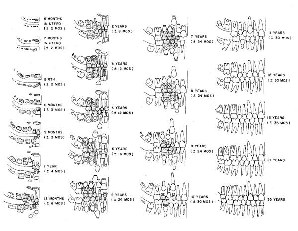
22 Tooth Development When the embryo is 5-6 weeks old, the first sign of tooth development are found in the mandibular anterior region 7th week - all primary teeth are developed and development of permanent teeth begin At birth there are normally 44 teeth in various stages of development. Enamel formation is underway in the primary dentition and just beginning on the permanent first molars 22
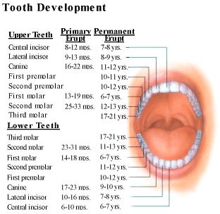
23 Tooth Development 23
24 24
25 Life Cycle of the Tooth The growth periods: the bud, cap, and bell stages Crown - covered by enamel, and each root has dentin covered by cementum Anatomic crown - portion of the tooth covered with enamel Clinical crown - portion of the tooth that is visible in the mouth Root - covered with cementum. Bifurcation means two roots and trifurcation means three roots. Enamel - hardest material in the body. Formed by ameloblasts consisting of 96-99% inorganic matter. Hydroxyapatite which is primarily calcium, is the most abundant mineral component. 25
26 26
27 Enamel Composed of millions of calcified enamel prisms, known as enamel rods. These extend from the surface of the tooth to the DEJ Hunter-Schreger bands - microscopic light and dark bands are caused by enamel prisms changing direction Striae of Retzius - growth rings. The shock of birth is known as the neonatal line Enamel tufts - start at the DEJ and may extend to the inner third of the enamel. Hypocalcified or uncalcified ends of groups of enamel prisms 27
28 Enamel Continued Enamel lamellae are thin structures that extend from the enamel surface towards the DEJ. Organic material primarily Enamel spindles are the ends of odontoblasts that extend across the DEJ a short distance into enamel 28
29 Dentin Extends across the entire length of the tooth, covered by enamel on the crown and by cementum on the root Normally a light yellow but can darken with age Mineralized tissue that is harder than bone and cementum but not as hard as enamel 70% inorganic material and 30% organic Dentin is formed by odontoblasts and these line the walls of the pulp cavity to form and repair dentin Dentinal tubules canals in dentin that contaisn the dentinal fiber 29
30 Dentin Continued Primary dentin forms prior to eruption, forms the bulk of the tooth Secondary dentin begins formation after eruption and continues at a very slow rate throughout life of the tooth. This results in the pulp chamber becoming narrow with age Tertiary dentin reparative dentin, formed in response to trauma on the wall of the pulp chamber. This can also occur in response to attrition. 30
31 Cementum Covers the root of the tooth, it overlies dentin and joins the enamel at the CEJ Primary function is to anchor the tooth to the bony socket with attachment fibers within the periodontium Yellow, and lighter in color than dentin Formed by cementoblasts and DOES NOT resorb and form again Capable of some repair through new layers 31
32 Cementum Primary cementum acellular cementum is formed outward from the cementodentinal junction for the full length of the root Secondary cementum cellular cementum, after the tooth has reached functional occlusion, continues to form on the apical half of the root 32

33 Pulp The pulp and within it the pulp chamber, which follows the contours of the exterior surface of the tooth The pulp is large in primary teeth but becomes smaller in permanent teeth and with age 33
34 Pulp Continued Coronal pulp within the crown portion Radicular pulp apically located, also called root pulp Apical foramen the radicular portion of the pulp in each root is continuous with the tissues of the periapical area via an apical foramen. In young teeth this is not fully formed, and has a wide opening. Secondary dentin eventually decreases the pulp chamber and apical foramen Blood vessels and nerves enter the pulp through the apical foramen. 34
35 Pulp Continued The pulp also contains connective tissue, which consists of cells, intercellular substance, and tissue fluid. Fibroblasts are cells responsible for the formation of the intercellular substance of the pulp. The nerve supply transmits and received pain stimuli. When the stimuli is weak, the response by the system is weak. When the stimulus is great, the reaction is stronger, and pain is present 35
36 Periodontium Supports the teeth in the alveolar bone Consists of cementum, alveolar bone, and periodontal ligaments Consists of the attachment apparatus and the gingival unit Attachment apparatus: cementum, alveolar process and periodontal ligaments Gingival unit: oral mucosa and the three main types are lining, masticatory, and specialized 36
37 Periodontium Alveolar process osteoblasts form bone and osteoclasts resorb and remodel the bone Cortical bone dense outer covering of the spongy bone that makes up the central part of the alveolar process. Denser in the mandible Alveolar crest highest point of the alveolar ridge. In a healthy mouth, the distance between the CEJ and the alveolar crest are consistent but with periodontal disease it will start to flatten Alveolar socket cavity within the alveolar process that surrounds the root of the tooth 37
38 Periodontium Alveolar socket continued the bony projection separating one socket from another is called the interdental septum. The bone separating the roots of a multirooted tooth is called the interradicular septum Lamina dura also known as the cribriform plate, is thin, compact bone that lines the alveolar socket. The nerves and blood vessels go through the tiny spaces. On a radiograph it appears as a thin white line around the root of the tooth 38
39 Periodontium Periodontal ligament connects the cementum covering the root of the tooth with the alveolar bone of the socket wall At one end the fibers are embedded in the cementum and at the other end embedded in the bone The fiber groups support the tooth in its socket and allows the tooth to withstand the pressures and forces of mastication The fibroblasts of the PL helps with remodeling, as do the cementoblasts and osteoblasts (and clasts) 39
40 Periodontium The PL has three different types of fiber groups: The periodontal fiber groups that support the tooth in its socket (horizontal, oblique, apical and interradicular fibers) The transseptal fiber groups support the tooth in relation to the adjacent teeth Gingival fiber groups support the gingiva surrounding the tooth. Also has the dentogingival, alveologingival, circular and dentoperiosteal fibers. 40

41 Periodontal Fiber Groups 41
42 Gingival Unit Oral mucosa is composed of stratified squamous epithelium. The three main types of oral mucosa in the oral cavity are lining, masticatory, and specialized mucosa Lining covers the inside of cheeks, vestibule, lips, soft palate, and ventral of the tongue. Beneath the lining is the submucosa containing blood vessels and nerves. NOT attached to the bone. Is a bright red color. 42
43 Gingival Unit Masticatory mucosa attached gingiva, hard palate, and dorsum of the tongue Light pink in color and keratinized. Lining mucosa IS not (previously mentioned in the last slide) No submucosa in the masticatory mucosa Withstands chewing and swallowing, tough. Specialized mucosa the dorsal of the tongue has both masticatory and specialized mucosa in the form of the lingual papilla. 43
Oral Embryology and Histology
 Oral Embryology and Histology Chapter 8 Copyright 2018, Elsevier Inc. All Rights Reserved. 1 Learning Objectives Lesson 8.1: Oral Embryology 1. Pronounce, define, and spell the key terms. 2. Define embryology
ANATOMY OF THE PERIODONTIUM. Dr. Fatin Awartani
 ANATOMY OF THE PERIODONTIUM Part II Cementum and Alveolar bone Associate Professor Periodontal division King Saud university Cementum Calcified mesenchymal tissue that forms the outer covering of the anatomic
Dental Anatomy and Physiology for Clinical Dental Technicians. with Marnie Hayward
 Dental Anatomy and Physiology for Clinical Dental Technicians with Marnie Hayward Salivary glands Parotid Submandibular Sublingual Salivary glands position Parotid glands Lie below ear and behind angle
Periodontal ligament
 Periodontal ligament The periodontium The periodontium includes: The gingiva Cementum Periodontal ligament Alveolar bone Def: The periodontal ligament is the dense fibrous connective tissue that occupies
Fundamental & Preventive Curvatures of Teeth and Tooth Development. Lecture Three Chapter 15 Continued; Chapter 6 (parts) Dr. Margaret L.
 Dr. Margaret L.") Fundamental & Preventive Curvatures of Teeth and Tooth Development Lecture Three Chapter 15 Continued; Chapter 6 (parts) Dr. Margaret L. Dennis Proximal contact areas Contact areas are on the mesial and
Fundamental & Preventive Curvatures of Teeth and Tooth Development Lecture Three Chapter 15 Continued; Chapter 6 (parts) Dr. Margaret L. Dennis Proximal contact areas Contact areas are on the mesial and
Development of teeth. 5.DM - Pedo
 Development of teeth 5.DM - Pedo Tooth development process of continuous changes in predetermined order starts from dental lamina A band of ectodermal cells growing from the epithelium of the embryonic
Lec. 11 & 12 Dr. Ali H. Murad Dental pulp 1- Coronal pulp
 Lec. 11 & 12 Dr. Ali H. Murad Dental pulp Is the soft connective tissue located in the central portion of each tooth. All pulps have similar morphologic characteristic, such as a soft, gelatinous consistency
Dental Morphology and Vocabulary
 Dental Morphology and Vocabulary Palate Palate Palate 1 2 Hard Palate Rugae Hard Palate Palate Palate Soft Palate Palate Palate Soft Palate 4 Palate Hard Palate Soft Palate Maxillary Arch (Maxilla) (Uppers)
ORAL ANATOMY AND PHYSIOLOGY
 CHAPTER 7 ORAL ANATOMY AND PHYSIOLOGY INTRODUCTION This chapter covers the oral anatomy and physiology of the teeth, the histology of the tissues and supporting structures, and concentrates on the external
SPACE MAINTAINER. Multimedia Health Education. Disclaimer
 Disclaimer This movie is an educational resource only and should not be used to manage your health. All decisions about the management of premature loss of primary teeth and use of space maintainers must
Medical NBDE-II. Dental Board Exams Part I.
 Medical NBDE-II Dental Board Exams Part I http://killexams.com/exam-detail/nbde-ii Question: 149 Anatomically, the term "clinical root" can be defined as which of the following: A. The space in the tooth
6610 NE 181st Street, Suite #1, Kenmore, WA
 660 NE 8st Street, Suite #, Kenmore, WA 9808 www.northshoredentalacademy.com.08.900 READ CHAPTER The Professional Dental Assistant (p.-9) No Key Terms Recall Questions:,,,, and 6 CLASS SYLLABUS DAY READ
06 Tooth Development and Eruption
 + 06 Tooth Development and Eruption Tooth development Root development PDL and alveolar bone development Primary tooth eruption and shedding Permanent tooth eruption Q. Where and how tooth starts to form?
PREMATURE PRIMARY TOOTH LOSS
 Disclaimer This movie is an educational resource only and should not be used to manage your dental health. All decisions about the management of premature primary tooth loss must be made in conjunction
CHAPTER 4 ORAL ANATOMY
 CHAPTER 4 ORAL ANATOMY This chapter covers the oral anatomy and physiology of the teeth, the histology of their tissues and supporting structures, and concentrates on the external features of the teeth.
AP I f2014 E3 c_5 & 6
 AP I f2014 E3 c_5 & 6 Student: Multiple choice questions choose the best answer. True/false answer A for true and B for false 1. The layer within the epidermis that acts as the foundation providing new
Anatomy Sheet: Oral cavity Done by: rasha Rakan edited by: khansaa Mahmoud
 Anatomy Sheet: Oral cavity Done by: rasha Rakan edited by: khansaa Mahmoud The oral cavity has 2 parts: 1. Oral vestibule: outer part that consists of outside the teeth, between the teeth, the cheeks and
CAP STAGE. Ans 1 The following are the stages of tooth development :
 Ans 1 The following are the stages of tooth development : 1. Bud stage 2. Cap stage 3. Bell stage 4. Advanced bell stage 5. Formation of Hertwig s epithelial root sheath BUD STAGE 1. Around the eighth
PERIODONTAL REVIEW DENTALELLE TUTORING ANDREA TWAROWSKI
 PERIODONTAL REVIEW DENTALELLE TUTORING ANDREA TWAROWSKI Periodontics Questions 1. What is the periodontium composed of? 2. What type of mucosa is the gingiva composed of? 3. Is it normal to have melanin
Dental Anatomy and Occlusion
 CHAPTER 53 Dental Anatomy and Occlusion Ma Lou C. Sabino DDS, and Emily G. Smythe, DDS What numerical system is used most commonly in the United States for designating the adult dentition? Pediatric dentition?
Oral Histology. Alveolar bone or process: Functions of alveolar bone: Chemical composition: Development of the alveolar process: Dr.
 Oral Histology Lec.12 Alveolar bone or process: Dr. Nada Al-Ghaban Alveolar bone is a specialized part of the mandibular and maxillary bones that forms the primary support structure for teeth. Although
Tooth eruption and movement
 Tooth eruption and movement Dr. Krisztián Nagy Diphydont dentition Deciduous dentition primary dentition Diphydont dentition Permanent dentition secondary dentition Mixed Dentition: Presence of both dentitions
An Overview of Dental Anatomy
 Continuing Education Brought to you by An Overview of Dental Anatomy Course Author(s): Vickie Parrish Foster, RDH, MEd CE Credits: 1 hour Intended Audience: Dental Hygienists, Dental Assistants, Dental
DENTAL TRAUMA IN DECIDUOUS TEETH
 Disclaimer This movie is an educational resource only and should not be used to manage your health. All decisions about the management of Dental Trauma in Deciduous Teeth must be made in conjunction with
Periodontal Disease. Radiology of Periodontal Disease. Periodontal Disease. The Role of Radiology in Assessment of Periodontal Disease
 Radiology of Periodontal Disease Steven R. Singer, DDS srs2@columbia.edu 212.305.5674 Periodontal Disease! Includes several disorders of the periodontium! Gingivitis! Marginal Periodontitis! Localized
TOOTH DISCOLORATION. Multimedia Health Education. Disclaimer
 Disclaimer This movie is an educational resource only and should not be used to manage dental health. All decisions about the management of tooth discoloration must be made in conjunction with your dentist
Advanced Probing Techniques
 Module 21 Advanced Probing Techniques MODULE OVERVIEW The clinical periodontal assessment is one of the most important functions performed by dental hygienists. This module begins with a review of the
A Single Neuron from the Brain
 Nervous Tissue A Single Neuron from the Brain Dendrites Cell Body Axon Nerve cells, called neurons, transmit signals throughout our bodies. These signals tell our bodies what to do. Dendrites transmit
An Overview of Dental Anatomy
 An Overview of Dental Anatomy Vickie P. Overman, RDH, MEd Continuing Education Units: 1 hour Online Course: www.dentalcare.com/en-us/professional-education/ce-courses/ce500 Disclaimer: Participants must
DENTIN It a hard vital tissue, surrounds the pulp & underlies the enamel on the crown & the cementum on the roots of the teeth.
 Lec. 7 Dr. Ali H.Murad DENTIN It a hard vital tissue, surrounds the pulp & underlies the enamel on the crown & the cementum on the roots of the teeth. Physical properties: 1-Dentin is pale yellow in color,
Skin: The Body s Protection
 Ch 34: Protection, Support and Locomotion 34.1 - Skin: The Body s Protection Inside This Section... The Structure of Skin The Function of Skin Response to Injury Structure and Function of the skin 4 tissue
BIOH111. o Cell Module o Tissue Module o Integumentary system o Skeletal system o Muscle system o Nervous system o Endocrine system
 BIOH111 o Cell Module o Tissue Module o Integumentary system o Skeletal system o Muscle system o Nervous system o Endocrine system Endeavour College of Natural Health endeavour.edu.au 1 TEXTBOOK AND REQUIRED/RECOMMENDED
December 3, Name five bones in your body. Are bones living or dead? Explain. What is the function of bone marrow?
 December 3, 2013 Name five bones in your body. Are bones living or dead? Explain. What is the function of bone marrow? Skeletal, Muscular, and Integumentary Systems Chapter 36: Biology II The Skeleton
NOTES: CH 40 Introduction to Human Anatomy & Physiology
 NOTES: CH 40 Introduction to Human Anatomy & Physiology THE HUMAN BODY Anatomy Physiology (= structures) (= functions or processes) Characteristics of LIFE: 1) Made up of 1 or more CELLS. 2) Obtain and
Upper arch. 1Prosthodontics. Dr.Bassam Ali Al-Turaihi. Basic anatomy & & landmark of denture & mouth
 1Prosthodontics Lecture 2 Dr.Bassam Ali Al-Turaihi Basic anatomy & & landmark of denture & mouth Upper arch Palatine process of maxilla: it form the anterior three quarter of the hard palate. Horizontal
Lab Animal Tissue. LEARNING OBJECTIVES: To understand the relationship between the structure and function of different animal tissues
 Name: Bio A.P. PURPOSE: HYPOTHESIS: NONE Lab Animal Tissue BACKGROUND: In animals, groups of closely related cells specialized to perform the same function are called tissues. There are four general classes
The Skeletal System:Bone Tissue
 The Skeletal System:Bone Tissue Dynamic and ever-changing throughout life Skeleton composed of many different tissues cartilage, bone tissue, epithelium, nerve, blood forming tissue, adipose, and dense
BONE TISSUE. Dr. Heba Kalbouneh Associate Professor of Anatomy and Histology
 BONE TISSUE Dr. Heba Kalbouneh Associate Professor of Anatomy and Histology BONE FUNCTION Support Protection (protect internal organs) Movement (provide leverage system for skeletal muscles, tendons, ligaments
DHYG 121 Winter, 2009 COURSE OUTLINE
 CAMOSUN COLLEGE School of Health & Human Services Dental Hygiene Department DHYG 121 Winter, 2009 COURSE OUTLINE The Approved Course Description is available on the web @ http://www.camosun.bc.ca/calendar/current/web/dhyg.html#dhyg121
part TWO Communication
 part TWO Communication chapter FOUR Dental Terminology OBJECTIVES After completing this chapter, you should be able to do the following: Spell and define key terms Discuss the purposes of teeth Identify
The Histology of Dentin
 The Histology of Dentin Pauline Hayes Garrett, D.D.S. Department of Endodontics, Prosthodontics, and Operative Dentistry University of Maryland, Baltimore This material was taken from: Essentials of Oral
Chapter 3 General Anatomy and Radiographic Positioning Terminology General Anatomy
 Chapter 3 General Anatomy and Radiographic Positioning Terminology General Anatomy Definition of Terms Anatomy- term applied to the science of the structure of the body Physiology- study of the function
Essentials of Anatomy and Physiology, 9e (Marieb) Chapter 1 The Human Body: An Orientation. Short Answer. Figure 1.1
 Chapter 1 The Human Body: An Orientation. Short Answer. Figure 1.1") Essentials of Anatomy and Physiology, 9e (Marieb) Chapter 1 The Human Body: An Orientation Short Answer Figure 1.1 Using Figure 1.1, identify the following: 1) Label A points to the cavity. 2) Label B
Essentials of Anatomy and Physiology, 9e (Marieb) Chapter 1 The Human Body: An Orientation Short Answer Figure 1.1 Using Figure 1.1, identify the following: 1) Label A points to the cavity. 2) Label B
The Human Body. Mrs. Green
 The Human Body Mrs. Green Bell Work Which of the following helps the body to cool down? a) Shivering b) Sweating c) Running a fever d) Taking a deep breath Which of the following is a function of the digestive
B. Incorrect! The ectoderm does not produce the dermis. C. Incorrect! The dermis is derived from the mesoderm.
 Human Anatomy - Problem Drill 04: The Integumentary System Question No. 1 of 10 Instructions: (1) Read the problem and answer choices carefully, (2) Work the problems on paper as 1. From the inner cell
Anatomy Review-INTRODUCTION. The study of the function of the body parts is called. Examples include:
 Anatomy Review-INTRODUCTION The study of the organs and parts of the body is called Examples include: The study of the function of the body parts is called. Examples include: Use the numbers from the diagram
Anatomy & Physiology. An Introduction
 Anatomy & Physiology An Introduction An Overview of Anatomy Anatomy - The study of the structure of the human body Physiology - The study of body function Branches of Anatomy Surface anatomy Gross anatomy
FRACTURES AND LUXATIONS OF PERMANENT TEETH
 FRACTURES AND LUXATIONS OF PERMANENT TEETH 1. Treatment guidelines and alveolar bone Followup Procedures INFRACTION Clinical findings Radiographic findings Treatment Follow-Up Favorable Outcome Unfavorable
Skin and Body Membranes Body Membranes Function of body membranes Cover body surfaces Line body cavities Form protective sheets around organs
 Skin and Body Membranes Body Membranes Function of body membranes Cover body surfaces Line body cavities Form protective sheets around organs Classification of Body Membranes Epithelial membranes Cutaneous
Semester Credits: 3 Lecture Hours: 3. Prerequisites:
 Revised: Fall 2015 Semester Credits: 3 Lecture Hours: 3 21THistology DNH 115 Admission into dental hygiene program. Prerequisites: Course Description: Presents a study of the microscopic and macroscopic
SKELETAL TISSUES CHAPTER 7 INTRODUCTION TO THE SKELETAL SYSTEM TYPES OF BONES
 SKELETAL TISSUES CHAPTER 7 By John McGill Supplement Outlines: Beth Wyatt Original PowerPoint: Jack Bagwell INTRODUCTION TO THE SKELETAL SYSTEM STRUCTURE Organs: Bones Related Tissues: Cartilage and Ligaments
in compact bone, large vertical canals carrying blood vessels and nerves. in compact bone, large horizontal canals carrying blood vessels and nerves.
 Carl Christensen, PhD Skeletal System (Bones`) Bio. 2304 Human Anatomy 1. Identify a term for each of the following: shaft of a long bone ends of a long bone ossified remnant of the "growth plate" connective
ALABAMA DENTAL HYGIENE PROGRAM 50 QUESTIONS PRE ENTRANCE EXAM
 ALABAMA DENTAL HYGIENE PROGRAM 50 QUESTIONS PRE ENTRANCE EXAM NAME DATE RETURN COMPLETED EXAM WITH APPLICATION You may copy for future reference 1. One cause of decay is the Streptococcus mutans bacteria
OpenStax-CNX module: m Bone Structure * Ildar Yakhin. Based on Bone Structure by OpenStax. Abstract
 OpenStax-CNX module: m63474 1 Bone Structure * Ildar Yakhin Based on Bone Structure by OpenStax This work is produced by OpenStax-CNX and licensed under the Creative Commons Attribution License 4.0 By
Skeletal System. Chapter 6.1 Human Anatomy & Physiology
 Skeletal System Chapter 6.1 Human Anatomy & Physiology Overview of Skeletal System Bones Joints Skeletal System Cartilage Tendons (bone to muscle) Ligaments (bone to bone) Function of the Skeletal System
POSTGRADUATE INSTITUTE OF MEDICINE UNIVERSITY OF COLOMBO SELECTION EXAMINATION IN MD (ORAL SURGERY) OCTOBER 2009 PAPER 1.1. Part A (General Anatomy)
 OCTOBER 2009 PAPER 1.1. Part A (General Anatomy)") POSTGRADUATE INSTITUTE OF MEDICINE UNIVERSITY OF COLOMBO SELECTION EXAMINATION IN MD (ORAL SURGERY) OCTOBER 2009 Date : 5 th October 2009 Time : 2.00 p.m. 5.00p.m. PAPER 1.1 Answer three (03) questions
POSTGRADUATE INSTITUTE OF MEDICINE UNIVERSITY OF COLOMBO SELECTION EXAMINATION IN MD (ORAL SURGERY) OCTOBER 2009 Date : 5 th October 2009 Time : 2.00 p.m. 5.00p.m. PAPER 1.1 Answer three (03) questions
Eruption and Shedding of Teeth
 Eruption and Shedding of Teeth Mixed Dentition: Presence of both dentitions Figure from Ten Cate s Oral Histology, Ed., Antonio Nanci, 6 th edition Tooth eruption is the process by which developing teeth
Unit 4 - The Skin and Body Membranes 1
 Unit 4 - The Skin and Body Membranes 1 I. Unit 4: Skin and Body Membranes A. Body Membranes 1. Function of body membranes a) Cover body surfaces b) Line body cavities c) Form protective sheets around organs
Basic Anatomy and Physiology of the Lips and Oral Cavity. Dr. Faghih
 Basic Anatomy and Physiology of the Lips and Oral Cavity Dr. Faghih It is divided into seven specific subsites : 1. Lips 2. dentoalveolar ridges 3. oral tongue 4. retromolar trigone 5. floor of mouth 6.
Dentin Formation(Dentinogenesis)
") Lecture four Dr. Wajnaa Oral Histology Dentin Formation(Dentinogenesis) Dentinogenesis begins at the cusp tips after the odontoblasts have differentiated and begin collagen production. Dentinogenesis growth
Lecture four Dr. Wajnaa Oral Histology Dentin Formation(Dentinogenesis) Dentinogenesis begins at the cusp tips after the odontoblasts have differentiated and begin collagen production. Dentinogenesis growth
PowerPoint Lecture Slide Presentation by Patty Bostwick-Taylor, Florence-Darlington Technical College Skin and Body Membranes
 PowerPoint Lecture Slide Presentation by Patty Bostwick-Taylor, Florence-Darlington Technical College Skin and Body Membranes 4 Body Membranes Function of body membranes Cover body surfaces Line body cavities
How to use this material
 !!!CAUTION!!! This power point presentation is intended to be used as an add on exercise to your standard lab experience. It is not intended to be used in lieu of the hands on lab time. In lab you will
What is histology? HISTOLOGY
 Introduction to Histology What is histology? HISTOLOGY histo = tissue ogy = study So HISTOLOGY = the study of tissues! What is a TISSUE? Tissues are groups of cells with specialized structural and functional
Chapter 6: Skeletal System: Bones and Bone Tissue
 Chapter 6: Skeletal System: Bones and Bone Tissue I. Functions A. List and describe the five major functions of the skeletal system: 1. 2. 3.. 4. 5.. II. Cartilage A. What do chondroblasts do? B. When
DENTIN-PULP COMPLEX. Erlina Sih Mahanani. School of Dental sciences Universiti Sains Malaysia. Erlina Sih Mahanani
 DENTIN-PULP COMPLEX School of Dental sciences Universiti Sains Malaysia Introduction Overview anatomy & histology of dentin and pulp. Development of dentin and pulp Structure of dentin and pulp Dentin
Blood. Hematopoietic Tissue
 Blood Hematopoietic Tissue Is a type of connective tissue in which its cells are suspended in a circulating fluid. Erythrocytes+ leukocytes + platelets (thrombocytes) =formed elements of blood. These formed
Learning Objectives (1&2)
") Learning Objectives (1&2) By the end of the session, students should be able to: 1) Identify anatomical position seated, standing, prone, supine. 2) Pronounce, define and be able to use directional and
Learning Objectives (1&2) By the end of the session, students should be able to: 1) Identify anatomical position seated, standing, prone, supine. 2) Pronounce, define and be able to use directional and
Introduction. Study detail of structure - - Gross Anatomy. Study all structures in one part of body Study of internal structures as relate to skin
 Introduction What is Anatomy and Physiology? Anatomy study of the shape and structure of body parts and their relationships to one another Physiology study of how the body functions individually and cooperatively
Lecture 2 Maxillary central incisor
 Lecture 2 Maxillary central incisor Generally The deciduous tooth appears in the mouth at 3 18 months of age, with 6 months being the average and is replaced by the permanent tooth around 7 8 years of
Diagnosis. overt Examination. Definitive Examination. History. atient interview. Personal History. Clinical Examination.
 Diagnosis overt Examination History Definitive Examination atient interview Personal History Mental Attitude Medical History Dental History Clinical Examination Extra Oral Oral Radiographic Evaluation
Oral Surgery. Basic Techniques of Dental Local Anesthesia. A variety of techniques used in administration and deposition of local anesthesia:
 Oral Surgery Lecture: 9 Dr. Saif Saadedeen Basic Techniques of Dental Local Anesthesia A variety of techniques used in administration and deposition of local anesthesia: 1. Topical anesthesia 2. Infiltration
HISTOLOGY. Simple squamal lungs
 HISTOLOGY Lab Objectives: Students should be able to... 1. Visually identify each class of tissue and examples within each class 2. Indicate the location (in the human body and/or organ) and function of
Digestive Anatomy Lab
 Digestive Anatomy Lab In-Lab Exercises I have included the word list in this document. Any descrepencies between this document and the wordlist, you should default to this document. There is a lot of repetition
Anatomy & Physiology Skeletal System
 I. Functions of the Skeletal System A. the body Anatomy & Physiology Skeletal System B. of vital organs C. Provide for movement D. storage (calcium & phosphate) E. cell formation II. Bone Structure A.
-Ibrahim Al-Naser. -Dr Al- Muhtaseb. 1 P a g e
 -1 -Ibrahim Al-Naser - -Dr Al- Muhtaseb 1 P a g e The Digestive System The doctor started the lecture by talking about the class rules. The GI system is an organ system, it is divided into: The Alimentary
Ch. 4: Skin and Body Membranes
 Ch. 4: Skin and Body Membranes I. Body Membranes A. Function of body membranes 1. Cover body surfaces 2. Line body cavities 3. Form protective sheets around organs II. Classification of Body Membranes
DEVELOPING ANALOGUE/SUBTITUTE FOR THE MANDIBULAR DENTURE BEARING AREA. Dr Muhammad Rizwan Memon FCPS Assistant Professor
 DEVELOPING ANALOGUE/SUBTITUTE FOR THE MANDIBULAR DENTURE BEARING AREA Dr Muhammad Rizwan Memon FCPS Assistant Professor Crest of Residual Ridge Buccal Shelf Shape of supporting structure Mylohyoid Ridge
Introduction in human anatomy
 Introduction in human anatomy Overview of Anatomy Anatomy is the study of the body structure and the relationships of the various parts of the body Gross or macroscopic (visible structures) Microscopic
Applications in Dermatology, Dentistry and LASIK Eye Surgery using LASERs
 Applications in Dermatology, Dentistry and LASIK Eye Surgery using LASERs http://www.medispainstitute.com/menu_laser_tattoo.html http://www.life123.com/bm.pix/bigstockphoto_close_up_of_eye_surgery_catar_2264267.s600x600.jpg
11/8/2012. Chapter 6 Part 1 Objectives: Skin = Integument = Cutaneous Membrane. The Structure of Skin. Epidermis
 Chapter 6 Part 1 Objectives: Define organ, and associate the skin as an organ of the integumentary system. List the general functions of the skin. Describe the structure of the layers of the skin. Summarize
Ex. 7: Integumentary
 Collin County Community College BIOL. 2401 Ex. 7: Integumentary. Skin or Integument Consists of three major regions Epidermis outermost superficial region Dermis middle region Hypodermis (superficial fascia)
The Skeletal System PART A. PowerPoint Lecture Slide Presentation by Patty Bostwick-Taylor, Florence-Darlington Technical College
 PowerPoint Lecture Slide Presentation by Patty Bostwick-Taylor, Florence-Darlington Technical College The Skeletal System 5 PART A The Skeletal System Parts of the skeletal system Bones (skeleton) Joints
The Skeletal System:Bone Tissue
 The Skeletal System:Bone Tissue Dynamic and ever-changing throughout life Skeleton composed of many different tissues cartilage, bone tissue, epithelium, nerve, blood forming tissue, adipose, and dense
The Integumentary System. Mosby items and derived items 2010, 2006, 2002, 1997, 1992 by Mosby, Inc., an affiliate of Elsevier Inc.
 The Integumentary System The Skin Structure two primary layers called epidermis and dermis Epidermis Outermost and thinnest primary layer of skin Composed of several layers of stratified squamous epithelium
Anatomy Ch 6: Integumentary System
 Anatomy Ch 6: Integumentary System Introduction: A. Organs are body structures composed of two or more different tissues. B. The skin and its accessory organs make up the integumentary system. Types of
36 1 The Skeletal System Slide 1 of 40
 1 of 40 The Skeleton All organisms need structural support. Unicellular organisms have a cytoskeleton. Multicellular animals have either an exoskeleton (arthropods) or an endoskeleton (vertebrates). 2
Educational Training Document
 Educational Training Document Table of Contents Part 1: Resource Document Disclaimer Page: 2 Part 2: Line Item Grade Sheets Page: 3 Release: 11/2016 Page 1 of 6 Part 1: Resource Document Disclaimer The
36.3 The Integumentary System The Skin. KEY CONCEPT The integumentary system has many tissues that protect the body.
 36.3 The Integumentary System The Skin KEY CONCEPT The integumentary system has many tissues that protect the body. 36.3 The Integumentary System The Skin The integument is the body system that surrounds
The Integumentary System
 The Integumentary System The Integumentary System Integument is skin Skin and its appendages make up the integumentary system (See if you can name some appendages) A fatty layer (hypodermis) lies deep
14/09/15. Assessment of Periodontal Disease. Outline. Why is Periodontal assessment needed? The Basics of Periodontal assessment
 Assessment of Periodontal Disease Dr Wendy Turner Outline Why is Periodontal assessment needed? The Basics of Periodontal assessment Probing: Basic Periodontal Examination for adults and children. Detailed
TIGHTEN YOUR DENTISTRY KNOWLEDGE Jeanne Perrone, CVT VTS (Dentistry)
") TIGHTEN YOUR DENTISTRY KNOWLEDGE Jeanne Perrone, CVT VTS (Dentistry) DENTISTRY Skeletal Anatomy Skull Types There are three common head shapes in the dog and cat. Mesocephalic or mesaticephalic: the most
TIGHTEN YOUR DENTISTRY KNOWLEDGE Jeanne Perrone, CVT VTS (Dentistry) DENTISTRY Skeletal Anatomy Skull Types There are three common head shapes in the dog and cat. Mesocephalic or mesaticephalic: the most
Functions of the Skeletal System. Chapter 6: Osseous Tissue and Bone Structure. Classification of Bones. Bone Shapes
 Chapter 6: Osseous Tissue and Bone Structure Functions of the Skeletal System 1. Support 2. Storage of minerals (calcium) 3. Storage of lipids (yellow marrow) 4. Blood cell production (red marrow) 5. Protection
Blood and Heart. Student Learning Objectives:
 Blood and Heart Student Learning Objectives: Identify the major components of the blood. Identify the primary structures associated with the heart Follow the blood through the path of the circulation.
Derived copy of Bone *
 OpenStax-CNX module: m57739 1 Derived copy of Bone * Shannon McDermott Based on Bone by OpenStax This work is produced by OpenStax-CNX and licensed under the Creative Commons Attribution License 4.0 By
Chapter 4 :Organization & Regulation of Body Systems
 Chapter 4 :Organization & Regulation of Body Systems 4.1 Types of tissues What is a tissue? A collection of cells of the same type that perform a common function There are 4 major tissue types in the body:
INTEGUMENTARY SYSTEM CHAPTER 4
 INTEGUMENTARY SYSTEM CHAPTER 4 FUNCTIONS Waterproofs Protein called keratin Protection 1 st line of defense against pathogens, chemicals & abrasions Insulation Regulates heat loss by controlling blood
Clinical UM Guideline
 Clinical UM Guideline Subject: Clinical Crown Lengthening Guideline #: 04-206 Current Effective Date: 03/24/2017 Status: New Last Review Date: 02/08/2017 Description This document addresses the procedure
Ch 4. Skin and Body Membranes
 Ch 4 Skin and Body Membranes TITLE HISTOLOGY SLIDES & NOTES ESSENTIAL QUESTION What tissues compose the integumentary system? Stratified Squamous Epithelium Stratified = several layers; Squamous = shape
The Integumentary System
 The Integumentary System The Integumentary System Integument is skin Skin and its appendages make up the integumentary system A fatty layer (hypodermis) lies deep to it Two distinct regions Epidermis Dermis