FA vs. OCTA? The status of OCTA, today. Fukuoka, JSOS 2016 Gerd Klose. Korobelnik J Fr Ophthalmol (2015)
|
|
|
- Cecilia Shaw
- 5 years ago
- Views:
Transcription

1 FA vs. OCTA? The status of OCTA, today Korobelnik J Fr Ophthalmol (2015) Fukuoka, JSOS 2016 Gerd Klose 1

2 2

3 FA / ICGA a well-founded Gold standard! Benefits Useful for many pathologies High contrast, detailed images Supports widefield imaging Choroidal imaging possible (w/ ICG) Well-established methodology 3


4 FA / ICGA with a flip-side Invasive & dye-based! Patient Discomfort Higher Clinical Effort 5% Adverse Effects: (nausea shock death) with many risk factors (age, diabetes, allergies, organ insuffiencies, pregnancy ) 4

5 FA / ICGA with a flip-side There is a constant balance between the clinical need and the risk of the diagnosis! What if you could reduce the number of FA exams 5
6 6


7 OCT Angiography sounds promising Presentations and Publications show so many nice images from OCT Angiography... providing fine structural details and 3D information non-invasive, quick and easy for patients & staff noncritical for sensitive patients, and allows frequent follow-ups 7

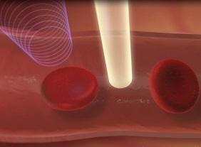

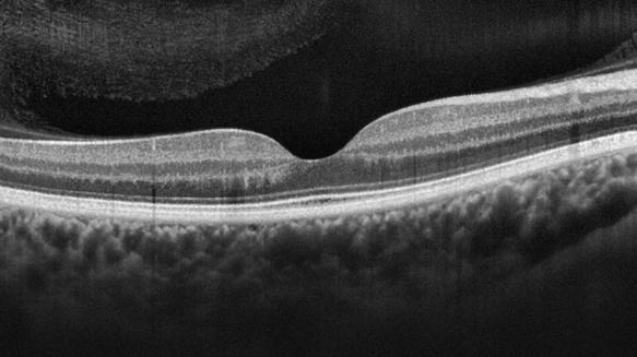
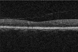
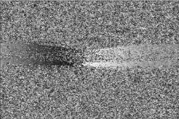
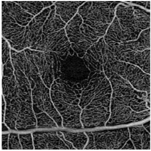
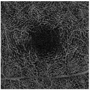
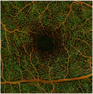
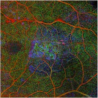
8 How does it work? Difference Bloodflow Average Structure 8

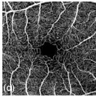
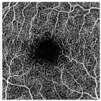
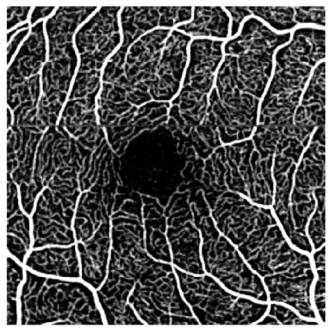
 3x3 mm, 245 Ascans (12")

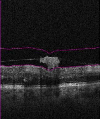
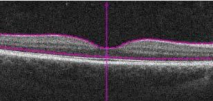
9 How does it work? Horizontal Vasculature Angio Cube Scans: 6x6 mm, 350 Ascans (17 um) 3x3 mm, 245 Ascans (12 um) Angio and Structure are complementary, you want to look at them side-by-side! 9


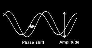


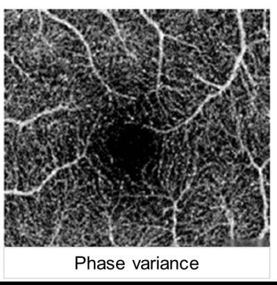
10 Quality Images require care at all levels Fast & Artifact-free Acquisition with Active tracking / Re-scan Reference Tracking (filtered) Amplitude & Phase Image Reconstruction Amplitude Phase Difference Zhang Q, et al., J. Biomed. Opt. 20(6), (2015) 10


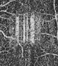

11 Quality Images require care at all levels Post-Acquisition Multi-Step Motion Correction Correction of Projection Artifacts Corrected Deeper Retina 11

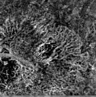
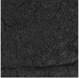
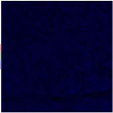
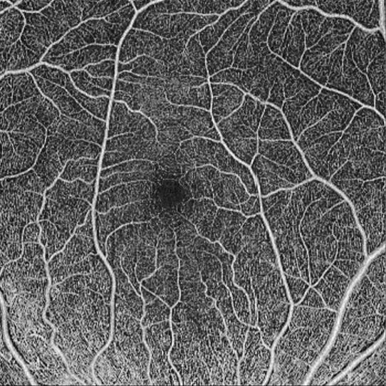
12 OCT-Angiography provides 3D information Choriocapillaris Slab Retina-Slab ILM to VRI- or Custom-Slab 70 um above RPE-fit Choroidor Custom-Slab Superficial Deeper Avascular 12


, (2015) /")

13 the Development will continue Quantification Larger Areas with Stitching ILM to Faster Swept-Source OCT-Angiography 70 um above RPE-fit 3x3 mm 6x6 mm 12 x12 mm Zhang Q, et al., J. Biomed. Opt. 20(6), (2015) / Dr. Philip J. Rosenfeld, BPEI / Dr. Y. Ito, Nagoya U 13


14 AngioPlex OCT Angiography simply another scan that works 14
15 15
16 FA / OCTA how about clinical usability FA detects the presence of dye/blood OCTA detects the flow of blood It is not identical, but similar and both can be utilized to visualize vasculature, (but not leakage or pooling) Pathologies resulting in neo-vascularization, low- or non-perfusion, and occlusions can be visualized! Korobelnik J Fr Ophthalmol (2015) 16
17 FA / OCTA how about clinical aspects many Clinical Cases have already been presented and many more to come: Diabetic Retinopathy (prolific / non-prolific) (DR) Central/Branch Retinal Vessel Occlusion (BRVO/CRVO) Age-related Macular Degeneration (AMD) Polypoidal Choroidal Vasculopathy (PCV) Macular Telangiectasia (Mactel) Coats like Exudative Maculopathy Sickle Cell Maculopathy Ocular tumors Glaucoma 17


18 FA / OCTA DR + Micro-aneurysms + timely follow-up on photo-coagulation + novel combination treatment with anti-vegf + non-perfusion, ischemic areas + Visualization of Neo-vascularization - but often in periphery - limited sensitivity to aneurysms - limited cataract penetration no 100% FA replacement (yet) 18


19 FA / OCTA BRVO, CRVO, etc. + non-perfusion + detailed images of neo-vascularization + remodeling of vasculature + remodeling of vasculature + new treatment possibilities - limited in covered area & periphery - hemorrhages in acute phase - visibility of aneurysms, edema no 100% FA replacement (yet) 19


20 FA / OCTA Wet AMD & PCV + detailed visualization of CNV lesion + visualization of choroidal feeder trunk + GA acts as window to choroid + Treat and Extend follow-up with OCTA + Choroidal NV & polyps - limited in covered area - Treatment decision still triggered by Edema - Limited choroidal and small polyp visualization no 100% FA replacement (yet) 20

21 FA / OCTA Clinical benefits, today FA / ICGA is still needed! but in specific cases OCTA has already helped with differential diagnosis and improved treatment decisions and there are immediate, general benefits: non-invasive patient comfort & simplified clinical work 3D details potential improved treatment no dye risk reduction every FA replaced, helps enabling fast follow-up one last argument no doctor using OCTA wants to give it up afterwards! 21
22 22

23 OCTA the future is bright the benefits are now, and only keep growing 23
Incorporating OCT Angiography Into Patient Care
 Incorporating OCT Angiography Into Patient Care Beth A. Steele, OD, FAAO OCT A: Introduction Isolates microvascular circulation from OCT image data Axial resolution = 5 microns (i.e. fine capillaries visible)
ZEISS AngioPlex OCT Angiography Making the revolutionary, routine.
 ZEISS AngioPlex OCT Angiography Making the revolutionary, routine. The moment that revolutionary insight becomes routine. // OCT ANGIOGRAPHY MADE BY ZEISS CIRRUS with AngioPlex creates a new era in both
ZEISS AngioPlex OCT Angiography Overview ZEISS OCT Angiography
 ZEISS AngioPlex OCT Angiography Overview ZEISS OCT Angiography California, ZEISS AngioPlex Ultra-clear visualization of microvascular blood flow using non-invasive OCT angiography 2 AngioPlex OCT Angiography
OCT Angiography The Next Frontier
 Choroid Retina avascular 5/13/2017 OCT Angiography The Next Frontier Pierce Kenworthy OD, FAAO June 9, 2017 OCT Angiography (OCTA) 2016 Non-invasive, motion contrast imaging Represents erythrocyte movement
10/17/2017. FDA Approved. Zeiss AngioPlex TM Optovue AngioVue TM
 Images retinal microvasculature without dye injection Displays structure and function from a single imaging system Standard of Care-2011 DFE, Fundus Photos, VF 10-2, SD-OCT, FAF, or mferg 2016-AAO Baseline
Visualize. Analyze. Personalize. OCT + OCTA
 Visualize. Analyze. Personalize. OCT + OCTA A New Approach to Protecting Vision AngioVue OCT Angiography brings valuable new information to clinical practice. Non-invasive visualization of retinal vasculature.
Will OCT-Angiography replace FA?
 ASL Roma A PRESIDIO TERRITORIALE NUOVO REGINA MARGHERITA AMBULATORIO PATOLOGIE RETINICHE Resp. Dott.ssa SUSANNA CATALANO CENTRO ITALIANO MACULA Will OCT-Angiography replace FA? Marco Rispoli, Luca di Antonio,
Swept-Source OCT Angiography: SS OCT Angio TM
 Swept-Source OCT Angiography: SS OCT Angio TM Not available in all countries, please check with your distributor. 2015.09 Swept-Source OCT Angiography: SS OCT Angio TM Introduction Optical coherence tomography
Deeper visualizations for intervening with confidence.
 CIRRUS OCT with AngioPlex from ZEISS Making the revolutionary routine New vascular quantification Deeper visualizations for intervening with confidence. CIRRUS OCT with AngioPlex from ZEISS can be a much
OPTICAL COHERENCE TOMOGRAPHY ANGIOGRAPHY OF THE RETINA AND OPTIC NERVE. Lindsay B. Howse, OD
 OPTICAL COHERENCE TOMOGRAPHY ANGIOGRAPHY OF THE RETINA AND OPTIC NERVE Lindsay B. Howse, OD drlindsayhowse@gmail.com None. FINANCIAL DISCLOSURES OUTLINE Introduction/How OCTA works OCTA Analysis Advantages
Introducing ANGIOVUE ESSENTIAL. Built on the Avanti Widefield OCT Platform. OCT Angiography for Primary Eye Care
 Introducing ANGIOVUE ESSENTIAL Built on the Avanti Widefield OCT Platform OCT Angiography for Primary Eye Care Transform Your View of the Retina OCT Angiography (OCTA) is a quick non-invasive test that
Is OCT-A Needed As An Investigative Tool During The Management Of Diabetic Macular Edema
 Is OCT-A Needed As An Investigative Tool During The Management Of Diabetic Macular Edema Ayman M Khattab MD, FRCS Professor of Ophthalmology Cairo University Diabetic Macular Edema (DME) Diabetic macular
OCT Interpretation in Retinal Disease
 OCT Interpretation in Retinal Disease Jay M. Haynie, OD, FAAO Financial Disclosure I have received honoraria or am on the advisory board for the following companies: Carl Zeiss Meditec Advanced Ocular
OCT Angiography in Primary Eye Care
 OCT Angiography in Primary Eye Care An Image Interpretation Primer Julie Rodman, OD, MS, FAAO and Nadia Waheed, MD, MPH Table of Contents Diabetic Retinopathy 3-6 Choroidal Neovascularization 7-9 Central
OCT Fundal Angiography Initial Experience The new era in Medical Retina Imaging Based on Cirrus 5000 AngioPlex 2016 Model Sheena George & Nicholas
 OCT Fundal Angiography Initial Experience The new era in Medical Retina Imaging Based on Cirrus 5000 AngioPlex 2016 Model Sheena George & Nicholas Lee Consultants Ophthalmologist at The Hillingdon Hospital
OCT Angiography. Financial Disclosures: Pre-Test: Which one is Correct?
 OCT Angiography Brandon Lujan, MD Medical Director, Casey Reading Center Assistant Professor of Ophthalmology Financial Disclosures: Genentech (Consultant, Grant support, Educational training) UC Berkeley
Leo Semes, OD, FAAO UAB Optometry
 Leo Semes, OD, FAAO UAB Optometry Safe; inert Has long track record - over 45 years Mixes with plasma and highlights blood vessel compromise Using specific exciting (490 nm)and absorption (510 nm) filters
PLEX Elite 9000 from ZEISS Swept-Source OCT
 PLEX Elite 9000 from ZEISS Swept-Source OCT Uncovering the undiscovered. ZEISS PLEX Elite 9000 // INNOVATION MADE BY ZEISS 2 Ultra-wide angiography En face montage Image courtesy of Prof. G. Querques,
OCT Angiography. SriniVas Sadda, MD
 OCT Angiography SriniVas Sadda, MD Professor of Ophthalmology Director, Medical Retina Unit Ophthalmic Imaging Unit University of Southern California Los Angeles, California, USA Disclosure Consulting
OCT Angiography: The Next Step in Retinal Imaging Jonathan Zelenak D.O.
 OCT Angiography: The Next Step in Retinal Imaging Jonathan Zelenak D.O. Hillsdale Hospital Michigan State University Overview Evolution of OCT How does OCT angiography work? Clinical examples Potential
ANSWERING THE WHY? Clinicians discuss the latest imaging technologies for retina practice BY PETER K. KAISER, MD
 Insert to March 2018 Sponsored by MULTI-MODALITY IMAGING: LATEST EVOLUTIONS IN OCTA AND UWF As the array of safe and efficacious medical and surgical options for retinal diseases expands, so does the need
World Sight Day Case Studies. Mark Frost Screening Manager South East London DESP
 World Sight Day 2015 Case Studies Mark Frost Screening Manager South East London DESP Introduction All of the following cases have been identified in our screening programme over the last 3 years. The
The World s fastest OCT. As simple as pressing. the start button
 The World s fastest OCT As simple as pressing the start button lution continues Optopol engineering team, designers of the first commercially available Spectral Domain OCT in the world, are proud to present
Go With the Flow: An OCT Angiography Primer Lorne Yudcovitch, OD, MS, FAAO
 Go With the Flow: An OCT Angiography Primer Lorne Yudcovitch, OD, MS, FAAO yudcovil@pacificu.edu OCT Angiography (OCTA) History 2000 - First Doppler flowimetry OCT on human retina 2005 Speckle analysis
Widefield Retinal Imaging with Auto Fluorescence Technology in the Optometric Practice
 Widefield Retinal Imaging with Auto Fluorescence Technology in the Optometric Practice This course will define ultra-widefield retinal imaging and autofluorescence for the attendee. Will show how it is
OCT-Angiography Clinical Cases. OCT-Angiography Clinical Cases
 OCT-Angiography Clinical Cases OCT-Angiography Clinical Cases NIDEK RS-3000 Advance AngioScan Daniela Bacherini Andrea Sodi Stanislao Rizzo CONTENTS Page Authors 3 Introduction 4 Case 1 Case 2 Case 3 Case
Visualize. Analyze. Personalize. OCT + OCTA. with
 Visualize. Analyze. Personalize. OCT + OCTA with Avanti Widefield OCT with AngioVue OCTA Imaging Comprehensive Structural and Functional Imaging in a Single Imaging Platform Comprehensive OCT Imaging The
Experience Spectacular Retinal Imaging with the new NIDEK F-10 Digital Ophthalmoscope
 Experience Spectacular Retinal Imaging with the new NIDEK F-10 Digital Ophthalmoscope The F-10 was developed to give Ophthalmologists a high definition (HD) diagnostic imaging system. Designed to provide
Disclosures. Definitions. Goals. Imaging and glaucoma 3/22/2016
 Pinakin Davey OD, PhD, FAAO Professor and Director of Research Disclosures Principal investigator for ivue OCT trial Principal investigator Topcon FDA trials for Maestro and OCT 2000 Consultant for Topcon
The Measure of Confidence
 Heidelberg_936357.qxd:Layout 1 5/9/08 12:01 PM 12:02 Page 1 (Cyan (Magenta (Yellow (Black (UV Five Powerful Solutions to Fit Your Practice PowerCheck Glaucoma FastCheck+ GPS Software and Retina Edema Index
The role of OCT-A in retinal disease management
 Graefe's Archive for Clinical and Experimental Ophthalmology (2018) 256:2019 2026 https://doi.org/10.1007/s00417-018-4109-3 REVIEW ARTICLE The role of OCT-A in retinal disease management Francisco J. Rodríguez
Technologies and Methods for Visualizing the Retina
 Transcript Details This is a transcript of an educational program accessible on the ReachMD network. Details about the program and additional media formats for the program are accessible by visiting: https://reachmd.com/programs/revealing-retina/technologies-and-methods-for-visualizing-theretina/3663/
Optical Coherence Tomography: Pearls for the Anterior Segment Surgeon Basic Science Michael Stewart, M.D.
 Optical Coherence Tomography: Pearls for the Anterior Segment Surgeon Basic Science Michael Stewart, M.D. Disclosure OCT Optical Coherence Tomography No relevant financial relationships I will refer to
Optical Coherence Tomography Angiography In Diagnosis Of Retinal Angiomatous Proliferation
 Optical Coherence Tomography Angiography In Diagnosis Of Retinal Angiomatous Proliferation Stepanov A, (1,4) Jiraskova N, 1,4) Lestak J, 2, 3, 4* 1. Department of Ophthalmology, University Hospital and
THE ROLE OF anti-vegf IN DIABETIC RETINOPATHY AND AGE RELATED MACULAR DEGENERATION
 THE ROLE OF anti-vegf IN DIABETIC RETINOPATHY AND AGE RELATED MACULAR DEGENERATION MOESTIDJAB DEPARTMENT OF OPHTHALMOLOGY SCHOOL OF MEDICINE AIRLANGGA UNIVERSITY DR SOETOMO HOSPITAL SURABAYA INTRODUCTION
Clinical Study Optical Coherence Tomography Angiography in Retinal Vascular Diseases and Choroidal Neovascularization
 Hindawi Publishing Corporation Journal of Ophthalmology Volume 2015, Article ID 343515, 8 pages http://dx.doi.org/10.1155/2015/343515 Clinical Study Optical Coherence Tomography Angiography in Retinal
f.a.q s Q. Why should I have an iwellness exam?
 f.a.q s The iwellness exam is a quick and non-invasive scan of your eye that lets your doctor see the layers of your retina to aid in the diagnosis of sight-threatening eye disease. Q. Why should I have
Building The Retina Company
 Building The Retina Company Optos devices produce ultra-widefield (UWF ), high resolution images (optomap ) of approximately 82% (200 ) of the retina. A single optomap can document the retina from the
11/29/2016 MACULAR MALADIES: TYPICAL & ATYPICAL CASES
 MACULAR MALADIES: TYPICAL & ATYPICAL CASES Dawn Pewitt, OD, FAAO Triad Eye Institute, Grove, OK Dpewitt@triadeye.com Disclosure Statement: No financial disclosures COPE 51218-PS Please silence all mobile
Mark Dunbar: Disclosure
 Important Things to Understand About OCT Mark T. Dunbar, O.D., F.A.A.O. Bascom Palmer Eye Institute University of Miami, School of Medicine Mark Dunbar: Disclosure Optometry Advisory Board for: Allergan
Photodynamic Therapy (PDT) for agerelated
 for agerelated") Page 1 of 5 Photodynamic Therapy (PDT) for agerelated eye conditions Introduction This leaflet gives you information about Photodynamic Therapy (PDT) for the age--related eye conditions macular degeneration
Page 1 of 5 Photodynamic Therapy (PDT) for agerelated eye conditions Introduction This leaflet gives you information about Photodynamic Therapy (PDT) for the age--related eye conditions macular degeneration
Fluorescein Angiography
 Last revision: October 2011 by Luis Arias Fluorescein Angiography Authors: Luis Arias, MD Hospital Universitari de Bellvitge - University of Barcelona. Spain Jordi Monés, MD Institut de la Màcula i de
ZEISS AngioPlex OCT Angiography. Clinical Case Reports
 Clinical Case Reports Proliferative Diabetic Retinopathy (PDR) Case Report 969 PROLIFERATIVE DIABETIC RETINOPATHY 1 1-year-old diabetic female presents for follow-up of proliferative diabetic retinopathy
From Outdated to Updated: A Review of Important Clinical Trials in Ocular Disease from 2014
 From Outdated to Updated: A Review of Important Clinical Trials in Ocular Disease from 2014 1. This course is designed to review the important ophthalmic literature that was released between October 2013
Learn Connect Succeed. JCAHPO Regional Meetings 2017
 Learn Connect Succeed JCAHPO Regional Meetings 2017 How Retinal Imaging Guides Treatment Odette Margit Houghton MD Question 1 Which OCT has the highest resolution? A: Swept source OCT B: Spectral domain
The College of Optometrists - Learning outcomes for the Professional Certificate in Medical Retina
 Learning outcomes for the Professional Certificate in Medical Retina, incorporating diabetic retinopathy screening and age related macular degeneration The professional certificate is a prerequisite to
Optical Coherence Tomography in Diabetic Retinopathy. Mrs Samantha Mann Consultant Ophthalmologist Clinical Lead of SEL-DESP
 Optical Coherence Tomography in Diabetic Retinopathy Mrs Samantha Mann Consultant Ophthalmologist Clinical Lead of SEL-DESP Content OCT imaging Retinal layers OCT features in Diabetes Some NON DR features
The Human Eye. Cornea Iris. Pupil. Lens. Retina
 The Retina Thin layer of light-sensitive tissue at the back of the eye (the film of the camera). Light rays are focused on the retina then transmitted to the brain. The macula is the very small area in
APRIL 8th 2016 Therapy
 APRIL 8th 2016 Therapy 09.00-10.00 SESSION 1: Age-related Macular Degeneration Moderators: A. Brucker, E. Souied, M. Stirpe, F. Boscia 09.00-09.15 TBD A. Brucker 09.15-09.30 Anti-PDGF and CNV fibrosis
Why Is Imaging Critical in My Uveitis Practice?
 Why Is Imaging Critical in My Uveitis Practice? Dilraj S. Grewal, MD Developed in collaboration Imaging Is the Backbone of Uveitis Workup and Monitoring Treatment Response FP FAF B- scan Multimodal Imaging
doi: /s
 doi: 10.1186/s12886-015-0077-0 Miura et al. BMC Ophthalmology (2015) 15:79 DOI 10.1186/s12886-015-0077-0 RESEARCH ARTICLE Open Access Noninvasive vascular imaging of ruptured retinal arterial macroaneurysms
Amber Priority. Image Library
 Amber Priority Image Library Amber flag Diabetic Maculopathy (M1) Pre-proliferative Diabetic Retinopathy (R2) Old, treated and now inactive DR (R1/M0/P1or R0/M0/P1) Where only partial or incomplete images
CENTENE PHARMACY AND THERAPEUTICS NEW DRUG REVIEW 2Q17 April May
 BRAND NAME Lucentis GENERIC NAME ranibizumab MANUFACTURER Genentech, Inc. DATE OF APPROVAL June 30, 2006 PRODUCT LAUNCH DATE July 13, 2006 REVIEW TYPE Review type 1 (RT1): New Drug Review Full review of
Ultrahigh Speed Imaging of the Rat Retina Using Ultrahigh Resolution Spectral/Fourier Domain OCT
 Ultrahigh Speed Imaging of the Rat Retina Using Ultrahigh Resolution Spectral/Fourier Domain OCT The MIT Faculty has made this article openly available. Please share how this access benefits you. Your
Rising to the Challenge of Satisfying Unmet Medical Needs. Back-of-the-eye. Main Back-of-the-Eye Diseases. Uveitis. Behcet s disease.
 Feature Rising to the Challenge of Satisfying Unmet Medical Needs Eyeing Further Specialization Back-of-the-eye Diseases Main Back-of-the-Eye Diseases Age-related macular degeneration Uveitis Behcet s
November Volume 35 - Issue 11
 November 2015 - Volume 35 - Issue 11 pp: 2161-2431,e67-e72 Editorial Optical Coherence Tomography Angiography Spaide, Richard F.; Fujimoto, James G.; Waheed, Nadia K. Original Study IMAGE ARTIFACTS IN
The retinal function imager and clinical applications
 Su and Garg Eye and Vision (2018) 5:20 https://doi.org/10.1186/s40662-018-0114-1 REVIEW Open Access The retinal function imager and clinical applications Daniel Su and Sunir Garg * Abstract Background:
4/27/2010 INTRODUCTION TO RETINAL VASCULAR DISEASE VENOUS/VENULAR CENTRAL RETINAL VEIN OBSTRUCTION / CRVO ADDITIONAL FEATURES /COMPLICATIONS
 INTRODUCTION TO RETINAL VASCULAR DISEASE VENOUS/VENULAR Leo Semes, OD Professor, UAB Optometry 2 CENTRAL RETINAL VEIN OBSTRUCTION CENTRAL RETINAL VEIN OBSTRUCTION / OCCLUSION (CRVO) obstruction of the
LEE EYE CENTRE. YOUR VISION, OUR PASSION LEC EyeNews
 LEE EYE CENTRE YOUR VISION, OUR PASSION LEC EyeNews FOR INTERNAL CIRCULATION ONLY www.lec.com.my ISSUE 51/003 SEPT OCT 2017 The American Society of Cataract and Refractive Surgery is one of the leading
A Patient s Guide to Diabetic Retinopathy
 Diabetic Retinopathy A Patient s Guide to Diabetic Retinopathy 840 Walnut Street, Philadelphia PA 19107 www.willseye.org Diabetic Retinopathy 1. Definition Diabetic retinopathy is a complication of diabetes
The Common Clinical Competency Framework for Non-medical Ophthalmic Healthcare Professionals in Secondary Care
 The Common Clinical Competency Framework for Non-medical Ophthalmic Healthcare Professionals in Secondary Care Medical Retina November 2016 Association of Health Professions in Ophthalmology General basic
 measure of your overall performance. An isolated glucose test is helpful to let you know what your sugar level is at one moment, but it doesn t tell you whether or not your diabetes is under adequate control
Diabetic Management beyond traditional risk factors and LDL-C control: Can we improve macro and microvascular risks?
 Retinopathy Diabetes has a negative effect on eyes in many ways, increasing the risk of cataracts for example, but the most common and serious ocular complication of diabetes is retinopathy. Diabetic retinopathy
The Era of anti- - - VEGF Kirk L. Halvorson, OD
 The Era of anti- - - VEGF Kirk L. Halvorson, OD Introduction: Anti- - - Vascular Endothelial Growth Factor (Anti- - - VEGF) medication is a relatively a new line of medications used in treating a variety
*Pleasesee amendment forpennsylvaniamedicaid at the endofthis CPB.
 1 of 134 Number: 0701 Policy *Pleasesee amendment forpennsylvaniamedicaid at the endofthis CPB. Aetna considers pegaptanib sodium injection (Macugen) medically necessary for the treatment of individuals
OCT Angiography: An Upcoming Tool for Diagnosis and Treatment of Retinal Vascular Diseases
 E-ISSN 2454-2784 Recent Advances OCT Angiography: An Upcoming Tool for Diagnosis and Treatment of Retinal Vascular Diseases Purnima Sood 1, Nalini Saxena 2, Dinesh Talwar 3 1 Vitreo-Retina Consultant,
Case Report Optical Coherence Tomography Angiography of Macular Telangiectasia Type 2 with Associated Subretinal Neovascular Membrane
 Hindawi Case Reports in Ophthalmological Medicine Volume 2017, Article ID 8186134, 4 pages https://doi.org/10.1155/2017/8186134 Case Report Optical Coherence Tomography Angiography of Macular Telangiectasia
DRI OCT Triton Series A Multimodal Swept Source OCT
 DRI OCT Triton Series A Multimodal Swept Source OCT Color Red-Free FA FAF Posterior Anterior See what others can t see. A Multimodal Swept Source OCT DEEP RANGE IMAGING Swept Source OCT imaging massively
Authors. Introduction. Introduction. Materials and Methods. Objective 10/27/2015
 Idiopathic Polypoidal Choroidal Vasculopathy (IPCV) in Thai Population Presenting with Choroidal Neovascularization (CNV) A multicenter study Authors Yonrawee Piyacomn 1, Chavakij Bhoomibunchoo 1, Yosanan
Angio-OCT. Degenerazione Maculare Legata all Eta. Giuseppe Querques
 Angio-OCT Degenerazione Maculare Legata all Eta Giuseppe Querques Department of Ophthalmology, IRCCS Ospedale San Raffaele, University Vita Salute San Raffaele, Milan, Italy Financial Disclosure ADVISORY
Diabetic Retinopathy
 Diabetic Retinopathy Diabetes can be classified into type 1 diabetes mellitus and type 2 diabetes mellitus, formerly known as insulin-dependent diabetes mellitus, and non-insulin diabetes mellitus, respectively.
Ophthalmic VEGF Inhibitors. Eylea (aflibercept), Macugen (pegaptanib) Description
, Macugen (pegaptanib) Description") Federal Employee Program 1310 G Street, N.W. Washington, D.C. 20005 202.942.1000 Fax 202.942.1125 Subject: Ophthalmic VEGF Inhibitors Page: 1 of 5 Last Review Date: September 20, 2018 Ophthalmic VEGF Inhibitors
Federal Employee Program 1310 G Street, N.W. Washington, D.C. 20005 202.942.1000 Fax 202.942.1125 Subject: Ophthalmic VEGF Inhibitors Page: 1 of 5 Last Review Date: September 20, 2018 Ophthalmic VEGF Inhibitors
Chris Brown, M.D. Eye Specialty Group, PLC Continuing Education Series
 Chris Brown, M.D. Eye Specialty Group, PLC 2018 Continuing Education Series Disclaimer I have no financial interests in this lecture or any information discussed therein Objectives Fluorescein Angiogram
Interesting, unusual and eclectic cases from 2017 Robert A. Mittra, MD VitreoRetinal Surgery, P.A. Minneapolis, MN
 Fundus, SG Interesting, unusual and eclectic cases from 2017 Robert A. Mittra, MD VitreoRetinal Surgery, P.A. Minneapolis, MN Which is most likely? A) Age > 65, history of HTN B) Age 40 65, history of
Interesting, unusual, eclectic cases from 2017 Robert A. Mittra, MD VitreoRetinal Surgery, P.A. Minneapolis, MN
 56 yo female, EW Presented to outside Ophthalmologist Diagnosed with viral conjunctivitis, but viral testing was negative. Also had pain around the eye and on the right side of her face Interesting, unusual,
! Honoraria. " Kemin " Nicox " Review of Optometry " Optometric Management " VSP. ! Scientific Advisory Boards
 Financial Disclosure: JP Choroid Chronicles Joseph J. Pizzimenti, OD, FAAO pizzimen@nova.edu! Honoraria " Kemin " Nicox " Review of Optometry " Optometric Management " VSP! Scientific Advisory Boards "
On Different Wavelengths: The Spectrum of Retinal Imaging. On Different Wavelengths: The Spectrum of Retinal Imaging. Wavelength Specific Imaging
 On Different Wavelengths: The Spectrum of Retinal Imaging Timothy J. Bennett, CRA, FOPS, OCT-C Penn State Hershey Eye Center Hershey, PA On Different Wavelengths: The Spectrum of Retinal Imaging Wavelengths
Clinically Significant Macular Edema (CSME)
") Clinically Significant Macular Edema (CSME) 1 Clinically Significant Macular Edema (CSME) Sadrina T. Shaw OMT I Student July 26, 2014 Advisor: Dr. Uwaydat Clinically Significant Macular Edema (CSME) 2
Clinically Significant Macular Edema (CSME) 1 Clinically Significant Macular Edema (CSME) Sadrina T. Shaw OMT I Student July 26, 2014 Advisor: Dr. Uwaydat Clinically Significant Macular Edema (CSME) 2
Common Causes of Vision Loss
 Common Causes of Vision Loss Learning Objectives To identify the most common causes of vision loss in the United States To differentiate the most common forms of agerelated macular degeneration and diabetic
Diabetic Retinopathy A Presentation for the Public
 Diabetic Retinopathy A Presentation for the Public Ray M. Balyeat, MD The Eye Institute Tulsa, Oklahoma The Healthy Eye Light rays enter the eye through the cornea, pupil and lens. These light rays are
Quantitative optical coherence tomography angiography of vascular abnormalities in the living human eye
 Quantitative optical coherence tomography angiography of vascular abnormalities in the living human eye Yali Jia a, Steven T. Bailey a, Thomas S. Hwang a, Scott M. McClintic a, Simon S. Gao a, Mark E.
TOPCON EURETINA Clinical Advances and Applications With Swept Source OCT and Angiography. JANUARY/FEBRUARY 2019 VOL. 17, NO.
 SUPPLEMENT TO SPONSORED BY JANUARY/FEBRUARY 2019 VOL. 17, NO. 1 TOPCON EURETINA 2018 Clinical Advances and Applications With Swept Source OCT and Angiography. This supplement summarizes highlights from
Clinical Study Optical Coherence Tomography and Optical Coherence Tomography Angiography in Monitoring Coats Disease
 Hindawi Ophthalmology Volume 2017, Article ID 7849243, 8 pages https://doi.org/10.1155/2017/7849243 Clinical Study Optical Coherence Tomography and Optical Coherence Tomography Angiography in Monitoring
Adaptive Optics and OCTA: Update on Retinal Imaging. Judy E. Kim, MD Professor of Ophthalmology Medical College of Wisconsin
 Adaptive Optics and OCTA: Update on Retinal Imaging Judy E. Kim, MD Professor of Ophthalmology Medical College of Wisconsin Financial Disclosure Advisory Board Alimera Science, Allergan, Bayer, Novartis
RVO RETINAL VEIN OCCLUSION
 RVO RETINAL VEIN OCCLUSION A guide to understanding RVO Take some time to learn about RVO - it may help you hold on to your vision Retinal vein occlusion is a common disorder of the retina and a leading
You can see clearly now. Heidelberg Retina Angiograph 2
 You can see clearly now Heidelberg Retina Angiograph 2 Wishes come true The way ahead is clear Highest image contrast and detail Lowest light exposure Simultaneous FA and ICGA Infra-red and Blue Reflectance
Diagnosis and treatment of diabetic retinopathy. Blake Cooper MD Ophthalmologist Vitreoretinal Surgeon Retina Associates Kansas City
 Diagnosis and treatment of diabetic retinopathy Blake Cooper MD Ophthalmologist Vitreoretinal Surgeon Retina Associates Kansas City Disclosures Consulted for Novo Nordisk 2017,2018. Will be discussing
Case Report: Indocyanine Green Dye Leakage from Retinal Artery in Branch Retinal Vein Occlusion
 Case Report: Indocyanine Green Dye Leakage from Retinal Artery in Branch Retinal Vein Occlusion Hiroki Fujita, Kyoko Ohno-Matsui, Soh Futagami and Takashi Tokoro Department of Visual Science, Tokyo Medical
Polypoidal choroidal vasculopathy (PCV) is a variation of agerelated
 is a variation of agerelated") Multidisciplinary Ophthalmic Imaging Noninvasive Vascular Imaging of Polypoidal Choroidal Vasculopathy by Doppler Optical Coherence Tomography Masahiro Miura, 1,2 Daisuke Muramatsu, 1,2 Young-Joo Hong,
Multidisciplinary Ophthalmic Imaging Noninvasive Vascular Imaging of Polypoidal Choroidal Vasculopathy by Doppler Optical Coherence Tomography Masahiro Miura, 1,2 Daisuke Muramatsu, 1,2 Young-Joo Hong,
RETINAL CONDITIONS RETINAL CONDITIONS
 GENERAL INFORMATION RETINAL CONDITIONS RETINAL CONDITIONS WHAT ARE RETINAL CONDITIONS? Retinal conditions affect the light-sensitive tissue at the back of eye known as the retina. They include diseases
GENERAL INFORMATION DIABETIC EYE DISEASE
 GENERAL INFORMATION DIABETIC EYE DISEASE WHAT IS DIABETIC EYE DISEASE? Diabetic eye disease is a term used to describe the common eye complications seen in people with diabetes. It includes: Diabetic retinopathy
Long-term Management of AMD. Motasem Al-latayfeh, MD Assistant Prof. Ophthalmology Hashemite University Jordan
 Long-term Management of AMD Motasem Al-latayfeh, MD Assistant Prof. Ophthalmology Hashemite University Jordan DEFINITION 1 Age-related macular degeneration (AMD) is a disorder of the macula characterized
Fundus autofluorescence in exudative age-related macular degeneration
 Fundus autofluorescence in exudative age-related macular degeneration Q. Peng*, Y. Dong* and P.Q. Zhao Department of Ophthalmology, Xinhua Hospital Affiliated to Shanghai JiaoTong University School of
NIH Public Access Author Manuscript Ophthalmology. Author manuscript; available in PMC 2015 January 01.
 NIH Public Access Author Manuscript Published in final edited form as: Ophthalmology. 2014 January ; 121(1): 180 187. doi:10.1016/j.ophtha.2013.09.002. Phase-Contrast Optical Coherence Tomography: A New
Comparison of Neovascular Lesion Area Measurements From Different Swept-Source OCT Angiographic Scan Patterns in Age-Related Macular Degeneration
 Retina Comparison of Neovascular Lesion Area Measurements From Different Swept-Source OCT Angiographic Scan Patterns in Age-Related Macular Degeneration Fang Zheng, 1,2 Qinqin Zhang, 3 Elie H. Motulsky,
Staphylomas in Pathologic Myopia With SS-OCT: A New Classification
 INSERT TO The 1st International Swept Source OCT & Angiography Conference took place in Madrid on Feb. 10-11, 2017. A cadre of renowned international retinal specialists presented new clinical findings
FRANZCO, MD, MBBS. Royal Darwin Hospital
 Diabetes and Eye By Dr. Nishantha Wijesinghe FRANZCO, MD, MBBS Consultant Ophthalmologist Royal Darwin Hospital 98% of Diabetics do not need to suffer from severe visual loss Yet Diabetic eye disease is
PRIMUS 200 from ZEISS The essential OCT
 PRIMUS 200 from ZEISS The essential OCT Seeing beyond the surface. ZEISS PRIMUS 200 // INNOVATION MADE BY ZEISS Clear Visualization. Advanced Technology. Reliability. Essential elements of your first OCT.
Diabetic Retinopatathy
 Diabetic Retinopatathy Jay M. Haynie, OD, FAAO Financial Disclosure I have received honoraria or am on the advisory board for the following companies: Carl Zeiss Meditec Arctic DX Macula Risk Advanced
Choroidal Mapping; a Novel Approach for Evaluating Choroidal Thickness and Volume
 Imaging Technique Choroidal Mapping; a Novel Approach for Evaluating Choroidal Thickness and Volume Jila Noori 1, MD; Mohammad Riazi Esfahani 1,2, MD Fedra Hajizadeh 2, MD; Mohammad-Mehdi Zaferani 1, MD