Retinal Manifestations of Systemic Disease Part 2. Pigmentary retinopathies with systemic associations. The Retina and Systemic diseases
|
|
|
- Chad Simpson
- 5 years ago
- Views:
Transcription
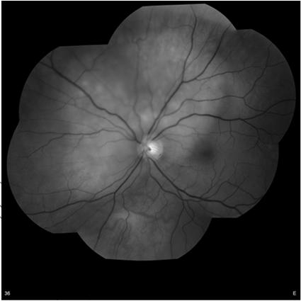
1 Retinal Manifestations of Systemic Disease Part 2 Sundeep Dev, MD VRSF Retinal Update 2019 VitreoRetinal Surgery, PA The Retina and Systemic diseases Retinitis/Vasculitis Vitreous cells Serous detachments Choroidal lesions Pigmentary retinopathies Choroidal folds/choroidal masses Retinal Vascular abnormalities yo with unilateral VA loss. Va = 20/400 Pigmentary retinopathies with systemic associations 3 Other eye normal 4 Psudoretinitis pigmentosa Syphillis Other inflammatory disease Lyme, AZOOR, DUSN Phenothiazine toxicity Congenital rubella Autoimmune / Cancer associated retinopathy Vitamin A deficiency Resolved exudative detachment VKH, toxemia of pregnancy 5 Retained foreign body after penetrating trauma 6 1
 worm Unilateral progressive pigmentary retinopathy, vascular")
 Fecal-oral route 7 8 Rubella Measles Can be unilateral")


2 Diffuse Unilateral Subacute Neuroretinitis (DUSN) Diffuse Unilateral Subacute Neuroretinitis (DUSN) worm Unilateral progressive pigmentary retinopathy, vascular attenuation, optic atrophy, vitritis Baylisascaris procyonis (raccoon parasite), Ancylostoma caninum (dog hookworm) Fecal-oral route 7 8 Rubella Measles Can be unilateral or bilateral after congenital or acquired infection. Salt and Pepper appearance. Vision and ERG usually normal. Vessels usually normal. Cataract may be present. 9 Typically bilateral. More common after acquired infection. Salt and pepper appearance. ERG/VF diminished, but may recover. 10 Vitamin A Deficiency Acute Zonal Occult Outer Retinopathy (AZOOR) Night blindness, severe dry eye. Common in children in developing world. Can occur after bariatric surgery, with malabsorption, or liver disease. ERG/VF changes. Pigmentary retinopathy. Treatment: Vitamin A. 11 Late stage 12 2


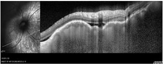
3 Acute Zonal Occult Outer Retinopathy (AZOOR) AZOOR Typically young females in mid 30 s. Present with photopsias, scotomas, VF loss with ERG changes, minimal fundus changes with delayed development of zones of atrophic RPE. Start unilateral, but often become bilateral. Poor prognosis. 13 Early stage 14 Autoimmune Retinopathy high degree of suspicion HVF / multifocal ERG Gardner s syndrome Familial polyposis coli autosomal dominant 17 Systemic Diseases associated with Reitinitis Pigmentosa Consider in young patients with RP Refsum s disease recessive, associated with increased phytanic acid levels. Hereditary abetalipoprotenemia recessive, RP with fat intolerance, deficiency of vitamins A, D, E, and K Kearns-Sayre syndrome - recessive, salt and pepper appearance, normal arterioles, CPEO, ptosis, heart block by age


4 Kearns Sayre syndrome Angioid Streaks Pseudoxanthoma Elasticum % Ehlers Danlos syndrome Paget s disease Sickle cell disease Idiopathic Angioid Streaks Hypercalcemia Marfan syndrome Hemochromatosis Acromegaly Sturge-Weber Myopia Pseudoxanthoma Elasticum Choroidal Folds Peau d orange Main concerns: Cardiovascular disease from abnormal elastic tissue in blood 23 vessel walls and GI bleeds Hypotony Choroidal Inflammation Posterior Scleritis Hyperopia Hyperthyroidism Hyperopia Postoperative Retinal detachment repair CNV/Disciform scar Choroidal mass Orbital mass Idiopathic 24 4


5 Posterior Scleritis Posterior Scleritis 50 % have associated systemic disease collagen vascular disease, infectious disease (including Lyme, TB, parasitic) 25 Present with pain, tenderness, vision loss. Choroidal folds/serous RD. Papilledema may be present. Posterior scleral thickening on B scan. Anlge closure can occur from choroidal thickening. 26 Posterior Scleritis Posterior Scleritis Hypotony Maculopathy associated with Carotid insufficiency Other Choroidal lesions and Choroidal masses 70 yo. Vsiion CF, IOP =2. No eye surgery. 95% carotid obstruction. May develop rebound glaucoma after reperfusion surgery


 33 34 Choroidal Mass")


6 Choroidal mass lesion 55 yo male with photopsias and distortion for 2 months Choroidal Mass- OCT Choroidal Mass (EDI) Choroidal Mass (Ultrasound) Ocular Melanoma worrisome findings on exam/testing Thickness - > 2 mm Fluid/ Fluorescein leakage Symptoms - Photopsias Orange Pigment Margins near the optic disc (within 3 mm) Acoustic hollowness (Low internal reflectivity) Absence of Halo/ Absence of Drusen


7 Uveal Melanoma Ocular Melanosis 37 Congenital, may be associated with glaucoma and increased risk of melanoma 38 Astrocytic Hamartoma in Tuberous Sclerosis Astrocytic Hamartoma Classic calcific Mulberry lesion. CNS astrocytoma, seizures, mental retardation, adenoma sebaceum, ash leaf spot, subungual fibroma, café au lait spots. Uusually bilateral in TS. 39 Non calcified apperance 40 Astrocytic Hamartoma Astrocytic Hamarotoma Can be isolated and idiopathic. Neurofibromatosis. Less common, but can be associated with 41 Isolated lesions possible, but need to rule out systemic TS and NF 42 7


8 Retinal capillary hemangioblastoma in Von Hippel-Lindau Syndrome Von Hippel Lindau disease Often earliest manifestation of VHL. Mean age is % unilateral. Most that have multifocal or bilateral, have VHL. Can also be isolated. Genetic testing is best way to screen for the condition. 43 Feeder arteriole and draining venule. FA shows rapid filling and leakage. Va loss from exudation, traction, VH, NVG. Small ones can be observed. Laser/Cryo/Radiation/ surgical excision. +/- seteroids/ anti-vegf. Prognosis depends on tumor size. 44 Wyburn-Mason Syndrome Wyburn-Mason Syndrome Non-hereditary, Congenital Unilateral, can progress in childhood.- involves retina and midbrain seizures, hemiparesis. Vision can be normal to severe loss due to intraretinal heme and NVG. No clear Rx Cavernous Hemangioma Systemic Histiocystosis 20/20 OD 20/400 OS Benign vascular hamartoma. Most are sporadic, they can be autosomal dominant and associated with brain and skin hemangiomas. Grape like clusters with stagnant blood flow cause layering on FA. No leakage. Usually no Rx and asymptomatic unless traction or VH develops in which case cryo/laser 47 may help. May involve optic nerve or macula. 48 8


. Often associated with rapidly progressive cataracts.")


9 Sclerochoroidal Calcification Sclerochoroidal Calcification B-Scan OD Most cases are idiopathic should test patients for abnormal calciumphosphorous metabolism. Check parathyroid hormone, vitamin D and calcium levels. Chronic Kidney disease can be associated Choroidal Osteoma BDUMP A benign ossifying tumor where bone replaces the choroid. More commonly unilateral females in late teens, or early twenties. Oval/round, peripapillary with 51 well defined borders. Slow growing with CNVM in 30-40%. A rare paraneoplastic condition with diffuse uveal melanocytic infiltration associated with systemic malignancy (ovarian, uterine, lung most common). Often associated with rapidly progressive cataracts. Cutaneous and mucosal involvement has been observed. 52 Cirumscribed Choroidal Hemangioma Choroidal Hemangioma Red-orange, round or oval in posterior ½ of fundus. Usually 1-3 mm thick and 3-7mm in diameter. Can get fibrous changes on surface. Often get can a serous detachment and cystic degeneration. PDT is quite effective. Radiation in severe 53 cases. Can also combine anti-vegf with PDT. FA very early hyperfluorescence of large choroidal vessels with late staining of entire lesion 54 9


10 Choroidal Hemangioma (EDI) Circumscribed Choroidal Hemangioma Most are within 2DD of the nerve. Cystoid degeneration and SRF on OCT. B scan shows high internal reflectivity. 55 Exudation and serous RD cause vision loss. Diffuse choroidal hemangioma occurs in Stuge-Weber syndrome in which case glaucoma can result. 56 Choroidal Hemangioma Retinoblastoma Would treat with PDT reduced fluence B cell lymphoma Metastasis (Breast) Mass Infiltration Yellow spots 59 Amelanotic, shallow, round-oval, usually posterior to the equator, may be multifocal 60 10


11 Metastasis Metastatic Breast Cancer Usually bright, hyperfluorecent late, hyperechoic with high internal reflectivity Metastatic Breast Cancer Metastatic Breast Cancer Metastatic Breast Cancer Lung Metastasis


12 Metastatic Renal Carcinoma Metastatic Renal Carcinoma Metastatic Lung Cancer Metastatic Lung Cancer (OCT) Metastatic Lung Cancer (EDI) GI Metastasis


13 Macroaneurysm with old subretinal hemorrhage Macroaneurysm Varix Retinal Vascular Disorders Will fluctuate with valsalva maneuver Cotton wool spot Cotton Wool spots Hypertension usually DBP >110 Diabetes seen in 44% of cases of DR Retinal vein occlusion Inflammatory GCA, Wegener s granulomatosis, Polyarteritis nodosa, Systemic Lupus, Scleroderma Infectious CMV, HIV retinopathy, Lyme, Toxoplasmosis, Mucormycosis, Leptospirosis Coagulopathies Sickle cell disease, omocysteinemia, Lupus anticoagulant syndrome, Proteins C, S, antithrombin III deficiencies Embolic Carotid and Cardiac disease Miscellaneous Migraine, severe anemia, Leukemia/lymphoproliferative disorders, Interferon therapy, Radiation, Purtscher s retinopathy, Papilledema 50 yo with new blurry spot and no known medical problems


14 Hypertensive Crisis Acute Leukocytic Leukemia Multiple Myeloma Purtscher s Retinopathy ischemia Classic is after crush injury, long bone fractures. Associated with pancreatitis, amniotic fluid embolism, collagen vascular disease, TTP Radiation Retinopathy Other Systemic Retinal Vascular Disease


15 Ocular ischemic syndrome Retinal Arterial Macroaneurysm Symptoms of vision loss, transient vision loss, pain. May be asymptomatic. Delayed choroidal filling and A/V transit. Unilateral cataract, iritis, low IOP initially, but may go into NVG. Asymmetric Diabetic Retinopathy. 85 Elderly females with HTN. Treat those with leakage/exudation. Laser, but anti-vegf seems to help regress as well. Those that bleed often fibrose. 86 Emboli Susac s Syndrome Recommend Carotid imaging and Transesophogeal echo. Anticoagulation. 87 Multiple mid arteriolar occlusions, tinnitus, mental status changes. MRI. Immunosuppression. 88 Susac s syndrome Lipemia Retinalis mid-arteriolar obstruction 89 Hypertriglyceridemia with levels > 1000 mg/dl. Genetic hyperlipoproteinemia. Starts peripherally, usually affects posterior vessels at levels of
 Sickle")


16 Peripheral Retinal Neovascularization Peripheral Retinal Neovascularization Diabetes Vein occlusion (branch) Sickle cell retinopathy (SC) Sarcoidosis Drug abuse embolization/ talc retinopathy Chronic uveitis/pars planitis Vasculitis Leukemia/anemia Eales disease PDR PDR after West Nile Virus 93 Diabetic Retinopathy rapidly progresses after ocular inflammatory disease 94 Talc Retinopathy SS Disease: Macular infarct year old HM vision after Sickle crisis lead to NVG within 1 month 96 16

")


17 SC disease Retinal Vasculitis Black sunburst lesions, salmon patches, peripheral ischemia, neovascularization. Hgb electrophoresis Familial Exudative Vitreoretinopathy (FEVR) Familial Exudative Vitreoretinopathy (FEVR) 99 OCT may show temporal thinning 100 Conclusions Many retinal - systemic associations Keep in mind patterns of retinal abnormalities that should raise clinical suspicion May be bilateral or unilateral Often no overt systemic symptoms at the time of ocular presentation. However, many times the symptoms are there if specifically looked for. The ophthalmologist can have a life-saving role Thanks very much for attending!
CLINICAL PEARLS IN OCULAR ONCOLOGY
 CLINICAL PEARLS IN OCULAR ONCOLOGY IRIS NEVUS - Two kinds circumscribed and diffuse - Photodocumentation important to monitor growth - Risk Factors for iris nevus growth to melanoma (ABCDEF) A Age (young),
Dispelling Rumors about Tumors. Case
 Dispelling Rumors about Tumors Jesse L. Berry, MD Arizona Ophthalmology Society 2017 Associate Director, Ocular Oncology Service Associate Program Director USC/CHLA, Keck School of Medicine Case 65 year
ZEISS AngioPlex OCT Angiography. Clinical Case Reports
 Clinical Case Reports Proliferative Diabetic Retinopathy (PDR) Case Report 969 PROLIFERATIVE DIABETIC RETINOPATHY 1 1-year-old diabetic female presents for follow-up of proliferative diabetic retinopathy
Ocular Pathology. I. Congenital and/or developmental. A. Trisomy 21. Hypertelorism (widely spaced eyes) Keratoconus (cone shaped cornea)
 Keratoconus (cone shaped cornea)") I. Congenital and/or developmental Robbins Pathologic Basis of Disease, 6 th Ed. A. Trisomy 21 Hypertelorism (widely spaced eyes) Keratoconus (cone shaped cornea) Focal hypoplasia of iris Cataracts frequently
I. Congenital and/or developmental Robbins Pathologic Basis of Disease, 6 th Ed. A. Trisomy 21 Hypertelorism (widely spaced eyes) Keratoconus (cone shaped cornea) Focal hypoplasia of iris Cataracts frequently
Posterior Segment Update
 Posterior Segment Update Featured Speaker: Dr. Kyle Cheatham, FAAO, DIP ABO DISCLOSURE STATEMENT We have no direct financial or proprietary interest in any companies, products or services mentioned in
2009 REIMBURSEMENT GUIDE, VISUCAM and VISUCAM NM/FA
 2009 REIMBURSEMENT GUIDE FF 450 PLUS PRO NM, VISUCAM and VISUCAM NM/FA Zeiss Fundus Cameras INTRODUCTION The following guide provides an overview of billing and reimbursement for procedures performed with
Differential diagnosis of posterior uveitis
 Differential diagnosis of posterior uveitis Diagnostic approach 45-year old male. Floaters and decreased vision since 1 week Fever, lymphadenopathy, myalgias, night sweats, two months ago Oral ulcer sporadically
Pediatric Ocular Sonography
 Pediatric Ocular Sonography Cicero J Torres A Silva, MD Associate Professor of Radiology 2016 SPR Pediatric Ultrasound Course Yale University School of Medicine None Disclosures Objectives of Presentation
Central venous occlusion
 Central venous occlusion Central venous occlusion (right eye) There are dark haemorrhages at the macula and all over the retina. Choroidal haemangioma A choroidal haemangioma has salmon pink colour. There
Dr/ Marwa Abdellah EOS /16/2018. Dr/ Marwa Abdellah EOS When do you ask Fluorescein angiography for optic disc diseases???
 When do you ask Fluorescein angiography for optic disc diseases??? 1 NORMAL OPTIC DISC The normal optic disc on fluorescein angiography is fluorescent due to filling of vessels arising from the posterior
Top Pediatric Retinal Diseases you don t want to miss! Retinopathy of Prematurity (ROP) Aggressive, Posterior ROP (AP ROP)
 Aggressive, Posterior ROP (AP ROP)") Top 10 10 Pediatric Retinal Diseases you don t want to miss! Polly Quiram MD, PhD Vitreoretinal Surgery, PA Retinal Update Jan 26th, 2018 ROP Retinoblastoma Coats disease Persistent fetal vasculature Familial
Top 10 10 Pediatric Retinal Diseases you don t want to miss! Polly Quiram MD, PhD Vitreoretinal Surgery, PA Retinal Update Jan 26th, 2018 ROP Retinoblastoma Coats disease Persistent fetal vasculature Familial
Retinal Manifestations of Systemic Disease Part 1
 The Retina and Systemic diseases Retinal Manifestations of Systemic Disease Part 1 Sundeep Dev, MD VRSF Retinal Update 2019 VitreoRetinal Surgery, PA 1 Retinitis/Vasculitis Vitreous cells Serous detachments
Moncef Khairallah, MD
 Moncef Khairallah, MD Department of Ophthalmology, Fattouma Bourguiba University Hospital Faculty of Medicine, University of Monastir Monastir, Tunisia INTRODUCTION IU: anatomic form of uveitis involving
What Is O.C.T. and Why Should I Give A Rip? OCT & Me How Optical Coherence Tomography Changed the Life of a Small Town Optometrist 5/19/2014
 OCT & Me How Optical Coherence Tomography Changed the Life of a Small Town Optometrist Email: myoder@wcoil.com Mark A. Yoder, O.D. 107 N. Main Street PO Box 123 Bluffton, OH 45817 @yoderod 115.02 Histoplasma
Mild NPDR. Moderate NPDR. Severe NPDR
 Diabetic retinopathy Diabetic retinopathy is the most common cause of blindness in adults aged 35-65 years-old. Hyperglycaemia is thought to cause increased retinal blood flow and abnormal metabolism in
Ophthalmology. Juliette Stenz, MD
 Ophthalmology Juliette Stenz, MD Required Slide Disclosures NO SIGNIFICANT FINANCIAL, GENERAL, OR OBLIGATION INTERESTS TO REPORT Required Slide At the end of this session, students will be able to: 1.
The Human Eye. Cornea Iris. Pupil. Lens. Retina
 The Retina Thin layer of light-sensitive tissue at the back of the eye (the film of the camera). Light rays are focused on the retina then transmitted to the brain. The macula is the very small area in
OCT Angiography in Primary Eye Care
 OCT Angiography in Primary Eye Care An Image Interpretation Primer Julie Rodman, OD, MS, FAAO and Nadia Waheed, MD, MPH Table of Contents Diabetic Retinopathy 3-6 Choroidal Neovascularization 7-9 Central
Patient AB. Born in 1961 PED
 Clinical Atlas Patient AB Born in 1961 PED Autofluorescence Dilated 45 EasyScan Zero-dilation IR 45 Fundus Dilated 45 In the fundus photos (Canon CX1) the PED is not able to be seen. However, the extent
Outline. Brief history and principles of ophthalmic ultrasound. Types of ocular ultrasound. Examination techniques. Types of Ultrasound
 Ultrasound and Intraocular Tumors 2015 Ophthalmic Photographers' Society Mid-Year Program Cagri G. Besirli MD, PhD Kellogg Eye Center University of Michigan Outline Brief history and principles of ophthalmic
What You Should Know About Angioid Streaks By David J. Browning, MD, PhD
 What You Should Know About Angioid Streaks By David J. Browning, MD, PhD The eye wall has several layers, as shown in figure 1. Proceeding from the inside of the eye to the outside, the layers are as follows:
Interesting, unusual and eclectic cases from 2017 Robert A. Mittra, MD VitreoRetinal Surgery, P.A. Minneapolis, MN
 Fundus, SG Interesting, unusual and eclectic cases from 2017 Robert A. Mittra, MD VitreoRetinal Surgery, P.A. Minneapolis, MN Which is most likely? A) Age > 65, history of HTN B) Age 40 65, history of
Interesting, unusual, eclectic cases from 2017 Robert A. Mittra, MD VitreoRetinal Surgery, P.A. Minneapolis, MN
 56 yo female, EW Presented to outside Ophthalmologist Diagnosed with viral conjunctivitis, but viral testing was negative. Also had pain around the eye and on the right side of her face Interesting, unusual,
World Sight Day Case Studies. Mark Frost Screening Manager South East London DESP
 World Sight Day 2015 Case Studies Mark Frost Screening Manager South East London DESP Introduction All of the following cases have been identified in our screening programme over the last 3 years. The
ACTIVATED OR NOT? RETINAL CASE PRESENTATION Shorye Payne, MD Medical Retinal Specialist Robley Rex VA Eye Clinic
 ACTIVATED OR NOT? RETINAL CASE PRESENTATION Shorye Payne, MD Medical Retinal Specialist Robley Rex VA Eye Clinic C We anticipate that the future management of posterior uveal melanoma (PUM) will focus
OPTIC DISC PIT Pathogenesis and Management OPTIC DISC PIT
 OPTIC DISC PIT Pathogenesis and Management Abdel-Latif Siam Ain Shams University Cairo Egypt OPTIC DISC PIT Congenital pit is an atypical coloboma usually located on the temporal edge of the disc, associated
FRANZCO, MD, MBBS. Royal Darwin Hospital
 Diabetes and Eye By Dr. Nishantha Wijesinghe FRANZCO, MD, MBBS Consultant Ophthalmologist Royal Darwin Hospital 98% of Diabetics do not need to suffer from severe visual loss Yet Diabetic eye disease is
Course # Flashes and Floaters and Curtains, Oh My!
 Course # 132 Flashes and Floaters and Curtains, Oh My! FLASHES and FLOATERS and CURTAINS, OH MY!!! FLASHES OF LIGHT Vitreous is the villain Retinal traction Retinal hole Retinal tear Migraine Classic migraine
Course # Flashes and Floaters and Curtains, Oh My!
 Course # 132 Flashes and Floaters and Curtains, Oh My! FLASHES and FLOATERS and CURTAINS, OH MY!!! FLASHES OF LIGHT Vitreous is the villain Retinal traction Retinal hole Retinal tear Migraine Classic migraine
Retro-bulbar visual anatomy Optic nerves carry. Normal left ocular fundus. Retinal nerve fiber layer anatomy
 The Patient with Visual Loss: Localization of Neuropathologic Disease and Select Diseases of Neuropathologic Interest Steven A. Kane, M.D., Ph.D. The Edward S. Harkness Eye Institute Shared embryology
Retina Center of Oklahoma Sam S. Dahr, M.D. Adult Intraocular Tumors
 Adult Intraocular Tumors Sam S. Dahr, M.D. Retina Center of Oklahoma www.retinacenteroklahoma.com www.rcoklahoma.com Table of Contents Posterior uveal malignant melanoma Uveal metastasis Uveal melanoma
Optical Coherence Tomography in Diabetic Retinopathy. Mrs Samantha Mann Consultant Ophthalmologist Clinical Lead of SEL-DESP
 Optical Coherence Tomography in Diabetic Retinopathy Mrs Samantha Mann Consultant Ophthalmologist Clinical Lead of SEL-DESP Content OCT imaging Retinal layers OCT features in Diabetes Some NON DR features
Retina Conference. Janelle Fassbender, MD, PhD University of Louisville Department of Ophthalmology and Visual Sciences 09/04/2014
 Retina Conference Janelle Fassbender, MD, PhD University of Louisville Department of Ophthalmology and Visual Sciences 09/04/2014 Subjective CC/HPI: 64 year old Caucasian female referred by outside ophthalmologist
Shared embryology Eye and brain develop from neuro-ectoderm
 The Patient with Visual Loss: Localization of Neuropathologic Disease and Select Diseases of Neuropathologic Interest Steven A. Kane, M.D., Ph.D. The Edward S. Harkness Eye Institute Shared embryology
OCCLUSIVE VASCULAR DISORDERS OF THE RETINA
 OCCLUSIVE VASCULAR DISORDERS OF THE RETINA Learning outcomes By the end of this lecture the students would be able to Classify occlusive vascular disorders (OVD) of the retina. Correlate the clinical features
Financial Disclosures. The Eye in Neoplastic Disease. Course Goal. We wish to acknowledge and thank: Tumor Definition
 The Eye in Neoplastic Disease Carlo J. Pelino, OD, FAAO Joseph J. Pizzimenti, OD, FAAO cpelino@salus.edu pizzimen@uiwtx.edu Financial Disclosures! Speakers have no relevant financial relationships to declare.
Neurocutaneous Syndromes. Phakomatoses
 Neurocutaneous Syndromes Phakomatoses Financial Disclosures I have NO SIGNIFICANT FINANCIAL, GENERAL, OR OBLIGATION INTERESTS TO REPORT Neurocutaneous Syndomes Definition Entities Diagnosis/ Presentation
Diabetic Retinopathy
 Diabetic Retinopathy Diabetes can be classified into type 1 diabetes mellitus and type 2 diabetes mellitus, formerly known as insulin-dependent diabetes mellitus, and non-insulin diabetes mellitus, respectively.
Tuberous sclerosis presenting as atypical aggressive retinal astrocytoma with proliferative retinopathy and vitreous haemorrhage
 Case Report Brunei Int Med J. 2015; 11 (1): 49-53 Tuberous sclerosis presenting as atypical aggressive retinal astrocytoma with proliferative retinopathy and vitreous haemorrhage Pui Ling TANG and Mae-Lynn
Diagnosis and treatment of diabetic retinopathy. Blake Cooper MD Ophthalmologist Vitreoretinal Surgeon Retina Associates Kansas City
 Diagnosis and treatment of diabetic retinopathy Blake Cooper MD Ophthalmologist Vitreoretinal Surgeon Retina Associates Kansas City Disclosures Consulted for Novo Nordisk 2017,2018. Will be discussing
Contractor Information. LCD Information. Local Coverage Determination (LCD): Ophthalmic Angiography (Fluorescein and Indocyanine Green) (L34426)
: Ophthalmic Angiography (Fluorescein and Indocyanine Green) (L34426)") Local Coverage Determination (LCD): Ophthalmic Angiography (Fluorescein and Indocyanine Green) (L34426) Links in PDF documents are not guaranteed to work. To follow a web link, please use the MCD Website.
Local Coverage Determination (LCD): Ophthalmic Angiography (Fluorescein and Indocyanine Green) (L34426) Links in PDF documents are not guaranteed to work. To follow a web link, please use the MCD Website.
PHAKOMATOSES SYNDROMES Michelle Willis, COMT, OSA
 PHAKOMATOSES SYNDROMES Michelle Willis, COMT, OSA This article and its accompanying quiz are worth.5 JCAHPO Group A continuing education credit. CONTINUING EDUCATION CREDITS ARE SUBJECT TO CHANGE. Internet
Local Coverage Determination (LCD): Scanning Computerized Ophthalmic Diagnostic Imaging (SCODI) (L34431)
: Scanning Computerized Ophthalmic Diagnostic Imaging (SCODI) (L34431)") Local Coverage Determination (LCD): Scanning Computerized Ophthalmic Diagnostic Imaging (SCODI) (L34431) Links in PDF documents are not guaranteed to work. To follow a web link, please use the MCD Website.
Local Coverage Determination (LCD): Scanning Computerized Ophthalmic Diagnostic Imaging (SCODI) (L34431) Links in PDF documents are not guaranteed to work. To follow a web link, please use the MCD Website.
COEXISTENCE OF OPTIC NERVE HEAD DRUSEN
 COEXISTENCE OF OPTIC NERVE HEAD DRUSEN AND COMBINED HAMARTOMA OF THE RETINA AND RETINAL PIGMENT EPITHELIUM IN A TAIWANESE MALE Yo-Chen Chang 1 and Rong-Kung Tsai 2,3 1 Department of Ophthalmology, Kaohsiung
Objectives. Unexplained Vision Loss: Where Do I Go From Here. History. History. Drug Induced Vision Loss
 Objectives Unexplained Vision Loss: Where Do I Go From Here Denise Goodwin, OD, FAAO Coordinator, Neuro-ophthalmic Disease Clinic Pacific University College of Optometry goodwin@pacificu.edu Know the importance
Neuro-Ocular Grand Rounds Anthony B. Litwak,OD, FAAO VA Medical Center Baltimore, Maryland
 Neuro-Ocular Grand Rounds Anthony B. Litwak,OD, FAAO VA Medical Center Baltimore, Maryland Dr. Litwak is on the speaker and advisory boards for Alcon and Zeiss Meditek COMMON OPTIC NEUROPATHIES THAT CAN
Neuro-Ocular Grand Rounds
 Neuro-Ocular Grand Rounds Anthony B. Litwak,OD, FAAO VA Medical Center Baltimore, Maryland Dr. Litwak is on the speaker and advisory boards for Alcon and Zeiss Meditek COMMON OPTIC NEUROPATHIES THAT CAN
Misdiagnosed Vogt-Koyanagi-Harada (VKH) disease and atypical central serous chorioretinopathy (CSC)
 disease and atypical central serous chorioretinopathy (CSC)") HPTER 12 Misdiagnosed Vogt-Koyanagi-Harada (VKH) disease and atypical central serous chorioretinopathy (S) linical Features VKH disease is a bilateral granulomatous panuveitis often associated with exudative
HPTER 12 Misdiagnosed Vogt-Koyanagi-Harada (VKH) disease and atypical central serous chorioretinopathy (S) linical Features VKH disease is a bilateral granulomatous panuveitis often associated with exudative
OCULAR HEMORRHAGES. ROSCOE J. KENNEDY, M.D. Department of Ophthalmology
 OCULAR HEMORRHAGES ROSCOE J. KENNEDY, M.D. Department of Ophthalmology Ocular hemorrhages are important not only because they produce visual loss but also because they usually indicate a disorder elsewhere
PART 1: GENERAL RETINAL ANATOMY
 PART 1: GENERAL RETINAL ANATOMY General Anatomy At Ora Serrata At Optic Nerve Head Fundoscopic View Of Normal Retina What Is So Special About Diabetic Retinopathy? The WHO definition of blindness is
Contractor Information. LCD Information. Local Coverage Determination (LCD): Ophthalmic Angiography (Fluorescein and Indocyanine Green) (L34426)
: Ophthalmic Angiography (Fluorescein and Indocyanine Green) (L34426)") Local Coverage Determination (LCD): Ophthalmic Angiography (Fluorescein and Indocyanine Green) (L34426) Links in PDF documents are not guaranteed to work. To follow a web link, please use the MCD Website.
Local Coverage Determination (LCD): Ophthalmic Angiography (Fluorescein and Indocyanine Green) (L34426) Links in PDF documents are not guaranteed to work. To follow a web link, please use the MCD Website.
Michael P. Blair, MD Retina Consultants, Ltd Libertyville/Des Plaines, Illinois Clinical Associate University of Chicago 17 October 2015
 Michael P. Blair, MD Retina Consultants, Ltd Libertyville/Des Plaines, Illinois Clinical Associate University of Chicago 17 October 2015 So What Parts of the Eye Retina are Affected by VHL Neural tissue
Chris Brown, M.D. Eye Specialty Group, PLC Continuing Education Series
 Chris Brown, M.D. Eye Specialty Group, PLC 2018 Continuing Education Series Disclaimer I have no financial interests in this lecture or any information discussed therein Objectives Fluorescein Angiogram
OCT Interpretation in Retinal Disease
 OCT Interpretation in Retinal Disease Jay M. Haynie, OD, FAAO Financial Disclosure I have received honoraria or am on the advisory board for the following companies: Carl Zeiss Meditec Advanced Ocular
Funduscopic Interpretation Understanding the Fundus: is that normal?
 Funduscopic Interpretation Understanding the Fundus: is that normal? Gillian McLellan BVMS PhD DVOphthal DECVO DACVO MRCVS With thanks to Christine Heinrich and all who contributed images Fundus Retina
4/27/2010 INTRODUCTION TO RETINAL VASCULAR DISEASE VENOUS/VENULAR CENTRAL RETINAL VEIN OBSTRUCTION / CRVO ADDITIONAL FEATURES /COMPLICATIONS
 INTRODUCTION TO RETINAL VASCULAR DISEASE VENOUS/VENULAR Leo Semes, OD Professor, UAB Optometry 2 CENTRAL RETINAL VEIN OBSTRUCTION CENTRAL RETINAL VEIN OBSTRUCTION / OCCLUSION (CRVO) obstruction of the
Amber Priority. Image Library
 Amber Priority Image Library Amber flag Diabetic Maculopathy (M1) Pre-proliferative Diabetic Retinopathy (R2) Old, treated and now inactive DR (R1/M0/P1or R0/M0/P1) Where only partial or incomplete images
Clinical Case Presentation. Branch Retinal Vein Occlusion. Sarita M. Registered Nurse Whangarei Base Hospital
 Clinical Case Presentation on Branch Retinal Vein Occlusion Sarita M. Registered Nurse Whangarei Base Hospital Introduction Case Study Pathogenesis Clinical Features Investigations Treatment Follow-up
DOME SHAPED MACULOPATHY. Ιωάννης Ν. Βαγγελόπουλος Χειρ. Οφθαλμίατρος - Βόλος
 DOME SHAPED MACULOPATHY Ιωάννης Ν. Βαγγελόπουλος Χειρ. Οφθαλμίατρος - Βόλος DOME SHAPED MACULOPATHY-DEFINITIONS The entity Dome Shaped Macula ( DSM ) was first described by Gaucher and associates in 2008
OCULAR MANIFESTATIONS OF SYSTEMIC DISEASES THUCANH MULTERER, MD
 OCULAR MANIFESTATIONS OF SYSTEMIC DISEASES THUCANH MULTERER, MD UNDERGRADUATE: Philadelphia College of Pharmacy and Science 1996 MEDICAL SCHOOL: MCP Hahnemann School of Medicine, Philadelphia PA 2000 RESIDENCY:
 measure of your overall performance. An isolated glucose test is helpful to let you know what your sugar level is at one moment, but it doesn t tell you whether or not your diabetes is under adequate control
Factors that lead to progression. Evaluation of Diabetics. Ocular Circulation. Ischemic Optic Neuropathy. Proliferative Diabetic Retinopathy
 Non proliferative Diabetic Retinopathy Proliferative Diabetic Retinopathy Factors that lead to progression Puberty and pregnancy Systolic and diastolic blood pressure Hyperlipidemia : hard exudates in
White-Spot Syndromes of the Retina Lee Jampol, M.D. Chicago, IL
 Objectives At the conclusion of the program, the attendees will be able to: 1. recognize the various white-spot syndromes of the retina 2. initiate appropriate diagnostic tests of patients with the white-spot
White Dot Syndromes Noninfectious Chorioretinopathies Update 2019
 White Dot Syndromes Noninfectious Chorioretinopathies Update 2019 Kelly T. Mitchell, MD Retina Service TTUHSC Definition Noninfectious disease Inflammation of choroid, choriocapillaris, RPE, and Retina
Year 2 MBChB Clinical Skills Session Ophthalmoscopy. Reviewed & ratified by: Mr M Batterbury Consultant Ophthalmologist
 Year 2 MBChB Clinical Skills Session Ophthalmoscopy Reviewed & ratified by: o Mr M Batterbury Consultant Ophthalmologist Learning objectives o To understand the anatomy and physiology of the external and
Imaging in uveitis. Anthony Hall
 Imaging in uveitis Anthony Hall Causes of Vision Loss in Uveitis 1. Cystoid macular oedema 26% 2. Cataract 19% 3. Glaucoma 11% 4. Permanent macular damage 5% Rothova et al BJO 1996; 80: 332-336 Macular
Diabetic Retinopathy A Presentation for the Public
 Diabetic Retinopathy A Presentation for the Public Ray M. Balyeat, MD The Eye Institute Tulsa, Oklahoma The Healthy Eye Light rays enter the eye through the cornea, pupil and lens. These light rays are
Leo Semes, OD, FAAO UAB Optometry
 Leo Semes, OD, FAAO UAB Optometry Safe; inert Has long track record - over 45 years Mixes with plasma and highlights blood vessel compromise Using specific exciting (490 nm)and absorption (510 nm) filters
EyePACS Grading System (Part 2): Detecting Presence and Severity of Background (Non-Proliferative) Diabetic Retinopathy Lesion
: Detecting Presence and Severity of Background (Non-Proliferative) Diabetic Retinopathy Lesion") EyePACS Grading System (Part 2): Detecting Presence and Severity of Background (Non-Proliferative) Diabetic Retinopathy Lesion George Bresnick MD MPA Jorge Cuadros OD PhD Anatomy of the eye: 3 Normal Retina
EyePACS Grading System (Part 2): Detecting Presence and Severity of Background (Non-Proliferative) Diabetic Retinopathy Lesion George Bresnick MD MPA Jorge Cuadros OD PhD Anatomy of the eye: 3 Normal Retina
Rare Presentation of Ocular Toxoplasmosis
 Case Report Rare Presentation of Ocular Toxoplasmosis Rakhshandeh Alipanahi MD From Department of Ophthalmology, Nikookari Eye Hospital, Tabriz University of Medical Sciences, Tabriz, Iran. Correspondence:
Incorporating OCT Angiography Into Patient Care
 Incorporating OCT Angiography Into Patient Care Beth A. Steele, OD, FAAO OCT A: Introduction Isolates microvascular circulation from OCT image data Axial resolution = 5 microns (i.e. fine capillaries visible)
Alan G. Kabat, OD, FAAO (901)
") THE SWOLLEN OPTIC DISC: EMERGENCY OR ANOMALY? Alan G. Kabat, OD, FAAO (901) 252-3691 Memphis, Tennessee alan.kabat@alankabat.com Course description: The swollen disc presents a diagnostic dilemma. While
THE SWOLLEN OPTIC DISC: EMERGENCY OR ANOMALY? Alan G. Kabat, OD, FAAO (901) 252-3691 Memphis, Tennessee alan.kabat@alankabat.com Course description: The swollen disc presents a diagnostic dilemma. While
Deep Trouble. Thomas Stone, MD Retina Associates of Kentucky River City Retina Conference May 15, 2014
 Deep Trouble Thomas Stone, MD Retina Associates of Kentucky River City Retina Conference May 15, 2014 History 20 yo WM Decreased vision OU, OD>OS Sudden onset blurred central vision 12 days prior 4 days
Diabetic Retinopathy. Barry Emara MD FRCS(C) Giovanni Caboto Club October 3, 2012
 Giovanni Caboto Club October 3, 2012") Diabetic Retinopathy Barry Emara MD FRCS(C) Giovanni Caboto Club October 3, 2012 Outline Statistics Anatomy Categories Assessment Management Risk factors What do you need to do? Objectives Summarize the
Diabetic Retinopathy Barry Emara MD FRCS(C) Giovanni Caboto Club October 3, 2012 Outline Statistics Anatomy Categories Assessment Management Risk factors What do you need to do? Objectives Summarize the
Early detection of Retinoblastoma in children. Max Mantik
 Early detection of Retinoblastoma in children Max Mantik Introduction The most common primary intraocular malignancy of childhood 10 to 15 % of cancers that occur within the first year of life Typical
When is eye screening performed
 Why is a regular ophthalmological exam critical in VHL Screening Department of Ophthalmology, University of South Florida VHLA Family Meeting in Tampa 2017 Screening is the testing of individuals at risk
Clinico-Pathological Atlas of Congenital Fundus Disorders
 Clinico-Pathological Atlas of Congenital Fundus Disorders Juan Orellana Alan H. Friedman Clinico-Pathological Atlas of Congenital Fundus Disorders With 236 Illustrations 196 in Color Springer Science+
Contractor Information. LCD Information. Local Coverage Determination (LCD): Ophthalmoscopy (L34017) Document Information
: Ophthalmoscopy (L34017) Document Information") Local Coverage Determination (LCD): Ophthalmoscopy (L34017) Links in PDF documents are not guaranteed to work. To follow a web link, please use the MCD Website. Contractor Information Contractor Name Contract
Local Coverage Determination (LCD): Ophthalmoscopy (L34017) Links in PDF documents are not guaranteed to work. To follow a web link, please use the MCD Website. Contractor Information Contractor Name Contract
11/29/2016 MACULAR MALADIES: TYPICAL & ATYPICAL CASES
 MACULAR MALADIES: TYPICAL & ATYPICAL CASES Dawn Pewitt, OD, FAAO Triad Eye Institute, Grove, OK Dpewitt@triadeye.com Disclosure Statement: No financial disclosures COPE 51218-PS Please silence all mobile
Principle of OCT. Reading Between the Lines: OCT Interpretation. Initial Concept. Advantage: High Resolution Cross Section Images
 Principle of OCT Reading Between the Lines: OCT Interpretation Mohammad Rafieetary, OD, FAAO mrafieetary@charlesretina.com Introduction Optical Biopsy Morphologic Evaluation of Live Tissue Measurements
EyePACS Grading System (Part 3): Detecting Proliferative (Neovascular) Diabetic Retinopathy. George Bresnick MD MPA Jorge Cuadros OD PhD
: Detecting Proliferative (Neovascular) Diabetic Retinopathy. George Bresnick MD MPA Jorge Cuadros OD PhD") EyePACS Grading System (Part 3): Detecting Proliferative (Neovascular) Diabetic Retinopathy George Bresnick MD MPA Jorge Cuadros OD PhD Anatomy of the eye: 3 Normal Retina Retinal Arcades Macula Optic
EyePACS Grading System (Part 3): Detecting Proliferative (Neovascular) Diabetic Retinopathy George Bresnick MD MPA Jorge Cuadros OD PhD Anatomy of the eye: 3 Normal Retina Retinal Arcades Macula Optic
Acute Retinal Necrosis Secondary to Varicella Zoster Virus in an Immunosuppressed Post-Kidney Transplant Patient
 CM&R Rapid Release. Published online ahead of print September 20, 2012 as Aperture Acute Retinal Necrosis Secondary to Varicella Zoster Virus in an Immunosuppressed Post-Kidney Transplant Patient Elizabeth
Diabetic Management beyond traditional risk factors and LDL-C control: Can we improve macro and microvascular risks?
 Retinopathy Diabetes has a negative effect on eyes in many ways, increasing the risk of cataracts for example, but the most common and serious ocular complication of diabetes is retinopathy. Diabetic retinopathy
Venous Occlusive Diseases
 Venous Occlusive Diseases Bruce R. Saran, MD Adjunct Assistant Clinical Professor of Medicine Scheie Eye Institute University of Pennsylvania School of Medicine Philadelphia, PA -a division of: RVO Demographics
FROM OUTDATED TO UPDATED Eminence-Based Medicine
 FROM OUTDATED TO UPDATED Eminence-Based Medicine Evidence-Based Medicine A REVIEW OF KEY CLINICAL TRIALS Anthony DeWilde, OD FAAO 1 EMINENCE BASED MEDICINE 2 EVIDENCE BASED MEDICINE 3 4 CLINICAL TRIALS
Clinically Significant Macular Edema (CSME)
") Clinically Significant Macular Edema (CSME) 1 Clinically Significant Macular Edema (CSME) Sadrina T. Shaw OMT I Student July 26, 2014 Advisor: Dr. Uwaydat Clinically Significant Macular Edema (CSME) 2
Clinically Significant Macular Edema (CSME) 1 Clinically Significant Macular Edema (CSME) Sadrina T. Shaw OMT I Student July 26, 2014 Advisor: Dr. Uwaydat Clinically Significant Macular Edema (CSME) 2
Eyes on Diabetics: How to Avoid Blindness in Diabetic Patient
 Eyes on Diabetics: How to Avoid Blindness in Diabetic Patient Rova Virgana FK Unpad Pusat Mata Nasional RS Mata Cicendo Bandung Eye Center (Hospital and Clinic) PIT IDI Jabar 2018 Keys Facts from WHO
CHAPTER 13 CLINICAL CASES INTRODUCTION
 2 CHAPTER 3 CLINICAL CASES INTRODUCTION The previous chapters of this book have systematically presented various aspects of visual field testing and is now put into a clinical context. In this chapter,
Laboratory Testing for the Primary Care Optometrist
 General lab principles Indications for testing - Confirm a clinical diagnosis - Rule out a diagnosis - Manage a disease - Screen for a disease Sensitivity is the ability to show a positive result in a
Update on Central Serous Chorioretinopathy
 Update on Central Serous Chorioretinopathy Steve Bennett Retina Update 2017 No financial interest Off label use of Spironolactone, Eplerenone, Melatonin, Verteporfin, Rifampin 1 2 Classic Central Serous
Ocular imaging in acquired retinopathy with multiple myeloma
 Ocular imaging in acquired retinopathy with multiple myeloma ABDELRAHMAN GABER SALMAN MD- FRCS (GLASG)- MRCS (ED) ASSOCIATE PROFESSOR AIN SHAMS UNIVERSITY EVRS 2015 Immunogammopathies Immunogammopathies
Applying structure-function to solve clinical cases
 Applying structure-function to solve clinical cases Professor Michael Kalloniatis Centre for Eye Health, and, School of Optometry and Vision Science Acknowledgements Some material prepared by Nayuta Yoshioka
Vascular Disease Ocular Manifestations of Systemic Hypertension
 Vascular Disease Ocular Manifestations of Systemic Hypertension Maynard L. Pohl, OD, FAAO Pacific Cataract & Laser Institute 10500 NE 8 th Street, Suite 1650 Bellevue, WA 98004 USA 425-462-7664 Cerebrovascular
Objectives. Myth 1: Weiss ring = PVD. Myths 1/23/2018. To review misconceptions and clinical pearls regarding common vitreoretinal presentations.
 Objectives David RP Almeida MD MBA PhD VitreoRetinal Surgery, PA To review misconceptions and clinical pearls regarding common vitreoretinal presentations. Retina Update Minneapolis MN January 2018 Myths
Learn Connect Succeed. JCAHPO Regional Meetings 2017
 Learn Connect Succeed JCAHPO Regional Meetings 2017 How Retinal Imaging Guides Treatment Odette Margit Houghton MD Question 1 Which OCT has the highest resolution? A: Swept source OCT B: Spectral domain
Diabesity A Public Health Crisis: AOA Evidence Based Translation to Care Series
 Diabesity A Public Health Crisis: AOA Evidence Based Translation to Care Series Joseph J. Pizzimenti, OD, FAAO Associate Professor Nova Southeastern University The Eye Care Institute pizzimen@nova.edu
Acknowledgements. Outline. Who were von Hippel and Lindau? Eugen von Hippel German Ophthalmologist
 Ophthalmic Therapies & Standard of Care Acknowledgements Eric Jonasch, MD & Surena Matin, MD Collaborators Franco DeMonte, MD Marcy Johnson Ian McCutcheon, MD Chaan Ng, MD Nancy Perrier, MD Dawid Schellingerhout,
Optic Nerve Anomalies
 Optic Nerve Anomalies Raman Bhakhri, OD, FAAO Southern California College of Optometry Marshall B. Ketchum University Goals for today Review some of the optic nerve anomalies that can be seen in practice
Retinal Vein Occlusion
 Retinal Update 2018 Retinal Vein Occlusion Case Presentations to Myself Branch Vein Occlusion What medical evaluation do you recommend for this 72 year old patient? Is there anything you ask of your medical
Diabetes & Your Eyes
 Diabetes & Your Eyes Diabetes is a disease that occurs when the pancreas does not secrete enough insulin or the body is unable to process it properly. Insulin is the hormone that regulates the level of