OCT Evaluation of the Retina Alison Bozung, OD, FAAO
|
|
|
- Alban Ford
- 6 years ago
- Views:
Transcription
 Internal Limiting Membrane 9. (NFL) Nerve Fiber Layer 8.")
1 Alison Bozung, OD, FAAO Rob Wooldridge, OD, FAAO Bozung: No relevant financial relationships with commercial interests Wooldridge: Speakers Bureau/honoraria from Aerie, Alcon, Allergan, Bausch & Lomb, Biotissue, Centervue, Oculus, Optovue, Reichert, Synemed Retina Refresher OCT Evaluation of the Retina Alison Bozung, OD, FAAO 10. (ILM) Internal Limiting Membrane 9. (NFL) Nerve Fiber Layer 8. (GCL) Ganglion Cell Layer 7. (IPL) Inner Plexiform Layer 6. (INL) Inner Nuclear Layer 5. (OPL) Outer Plexiform Layer 4. (ONL) Outer Nuclear Layer 3. (ELM) External Limiting Membrane 2. (IS/OS or PIL) Inner/Outer Segment 1. (RPE) Retina Pigmented Epithelium Topics 1. Where is the fluid 2. Common entities 3. Masqueraders 4. Going below and beyond 5. Take a second look Topics 1. Where is the fluid 2. Common entities 3. Masqueraders 4. Going below and beyond 5. Take a second look 1

 RPE")
 Key Features: Hyporeflective space Directly above RPE C.")
 SRF PED (likely")

2 1. Where is the Fluid? A. Sub-RPE Fluid Describing fluid location A. Sub-RPE B. Subretinal C. Intraretinal Key Features: Hyporeflective space Directly below RPE Often bubble-like, but can be irregular Aka Pigment epithelial detachment (PED) RPE Sub-RPE space B. Subretinal Fluid (SRF) Key Features: Hyporeflective space Directly above RPE C. Intraretinal Fluid Key Features: Rounded, hyporeflective spaces Within central retinal layers SRF RPE Aka Cystoid Macular Edema (CME) SRF PED (likely fibrovascular) Topics 1. Where is the fluid 2. Common entities 3. Masqueraders 4. Going below and beyond 5. Take a second look Retina Choroidal nevus Retinal detachment 2. Common entities Macula Age related macular degeneration Macular edema Vitreoretinal interface Epiretinal membrane Macular hole 2


3 Choroidal Nevus Common features on OCT Increased choroidal hyperreflectivity Posterior blocking defect Overlying drusen Minimal thickness Choroidal Nevus Choroidal Nevus Choroidal Nevus Hyper-reflectivity due to increased pigment Blocking defect Pinpoint druse Choroidal Nevus Juxtapapillary choroidal nevus Choroidal Melanoma OCT features of small choroidal melanoma include SRF, increased thickness, subretinal lipofuscin, and structural retinal alterations. RPE alteration without SRF Increased thickness Shields, et al. Lipofuscin Shaggy photoreceptors Shields, et al. SRF Shields, et al. Shields CL, Kaliki S, Rojanaporn D, Ferenczy SR, Shields JA. Enhanced Depth Imaging Optical Coherence Tomography of Small Choroidal Melanoma Comparison With Choroidal Nevus. Arch Ophthalmol. 2012;130(7): doi: /archophthalmol Shields CL, Kaliki S, Rojanaporn D, Ferenczy SR, Shields JA. Enhanced Depth Imaging Optical Coherence Tomography of Small Choroidal Melanoma Comparison With Choroidal Nevus. Arch Ophthalmol. 2012;130(7): doi: /archophthalmol


4 Retinal Detachment Case 1: Retinal Detachment Common features on OCT Extensive subretinal fluid Flat or corrugated retina Intact RPE/Bruchs complex 19 yo male 1 day of flashes, floaters, and vision loss OD Ophthalmic history: Congenital cataract OU PCIOL OD, Aphakia OS Nystagmus Glaucoma HSVK OD BCVA OD 20/70 20/200 Macula on or off? Case 1: Retinal Detachment Case 2: Retinal Detachment Inferior Superior Fovea attached SRF 76 yo male 1 week vision loss OD Ophthalmic history: PCIOL OD, NSC OS Medical history: CAD, COPD, HTN Clopidogrel, ASA 325mg BCVA OD 20/40 Macula on or off? Case 2: Retinal Detachment Case 3: Retinal Detachment 82 yo female 2 days vision loss OD Ophthalmic history: PCIOL OU Retinal Detachment Choroidal hemorrhage Fovea detached Corrugation suggests rhegmatogenous component Medical history: Clopidogrel Coronary stent CHF, HTN, DM BCVA OD 20/50 4
 Bilateral peripheral")


5 Case 3: Retinal Detachment Age Related Macular Degeneration Subretinal fluid with increased hyperreflectivity = mixed hemorrhage Exudates Peripheral exudative hemorrhagic chorioretinopathy (PEHCR) Bilateral peripheral degenerative retinal condition Caucasians >70yoa Often referred to Pseudomelanoma due to dome-shaped exudative lesion Ultrasound necessary Common features on OCT Drusen Drusenoid PEDs Fibrovascular PEDs Geographic atrophy Subretinal fluid Sub-RPE, normal BM Classic Drusen Sub-RPE, thickened BM Basal Laminar Drusen Sub-retinal Reticular Pseudodrusen Types of Drusen Sub-RPE, normal BM Classic Drusen Sub-RPE, thickened BM Basal Laminar Drusen Sub-retinal Reticular Pseudodrusen Types of Drusen Mixed qualities Geographic Atrophy Case 1: Non-exudative ARMD Increased choroidal penetrance Outer retinal atrophy: loss of RPE and photoreceptors Bilateral RPE mottling and macular drusen 5


 SRF BM")


6 Case 1: Non-exudative ARMD Case 1: Non-exudative ARMD Sub RPE drusen Sub retinal drusen Color fundus photo Infrared reflectance Combined classic and reticular pseudodrusen Case 2: Non-exudative ARMD Case 2: Non-exudative ARMD Case 2: Non-exudative ARMD Case 3: Exudative ARMD BM thickening Right eye RPE PED (likely fibrovascular) SRF BM thickening Left eye Basal laminar drusen with vitelliform macular detachment PED (likely fibrovascular) SRF 6


 Hemicentral retinal vein occlusion with CME")


 Macular edema")
 Posterior hyaloid attachment to macula NO")
 Posterior hyaloid attachment to macula")

7 Cystoid Macular Edema Common features on OCT Rounded hyporeflective spaces Typically located in outer plexiform layer Case 1: CME Common etiologies Cataract surgery Diabetes Retinal vein occlusion Posterior uveitis Before and after single anti- VEGF injection of Avastin (bevacizumab) Hemicentral retinal vein occlusion with CME Case 2: CME Case 3: CME Day 1: 20/50 Day 1: 20/70 Day 9: 20/250 Day 3: 20/100 1 mo s/p IVA 20/20 Central retinal vein occlusion with CME Central retinal vein occlusion with CME CME: Diabetic Retinopathy Vitreomacular Interface OCT findings: Exudates Diffuse thickening Disorganized retinal structure Aka Diabetic macular edema (DME) Macular edema Exudates Thickening with disorganization Vitreomacular Adhesion (VMA) Posterior hyaloid attachment to macula NO distortion of macular contour Asymptomatic Vitreomacular Traction (VMT) Posterior hyaloid attachment to macula Change in foveal contour or retinal morphology i.e. surface distortion, pseudocysts, elevation of retina from RPE May benefit from Jetrea (ocriplasmin, Thrombogenics) 7
 Epiretinal Membrane (ERM) Aaron")

 Complications of Vitreomacular Traction Topics 1.")


 Macular telangiectasia (Mac Tel)")
8 Vitreomacular Interface Complications of Vitreomacular Traction Vitreomacular Adhesion (VMA) Epiretinal Membrane (ERM) Aaron Gold, OD, FAAO Vitreomacular Traction (VMT) Stage 2 Macular Hole Aaron Gold, OD, FAAO Aaron Gold, OD, FAAO Vitreomacular Traction (VMT) with pseudocyst Aaron Gold, OD, FAAO Stage 4 Macular Hole (Full Thickness) Complications of Vitreomacular Traction Topics 1. Where is the fluid 2. Common entities 3. Masqueraders 4. Going below and beyond 5. Take a second look Before and after surgical repair 20/400 20/30 3. Masqueraders of leakage Similar appearances, but underlying process differs True leakage is a VEGF-mediated process Masqueraders are typically degenerative processes 2. Masqueraders of leakage Common degenerative mimickers Outer retinal tubulations (ORTs) Macular telangiectasia (Mac Tel) Retinoschisis Why does it matter?? Masqueraders typically do not respond to anti-vegf injections 8
 versus CME (below) Comprised of photoreceptor")
 versus CME (below) Intraretinal spaces, no encircling band Intraretinal spaces, no encircling band Case 3 Case")
 versus CME (below) OS Rounded spaces, overall")
 versus CME (below) OS Rounded spaces, overall retinal")
9 Outer Retinal Tubulations Outer Retinal Tubulations Key Features: Bright-banded encircling ring Located in outer retina Atrophic area Intact outer retina Geographic atrophy (above) versus CME (below) Comprised of photoreceptor portions and ELM Causes: Geographic atrophy Outer retinal disease Atrophic area Intact outer retina Geographic atrophy (above) versus CME (below) Intraretinal spaces, no encircling band Intraretinal spaces, no encircling band Case 3 Case 3: Mac Tel 54 yo female Blur OU 20/30 OD, 20/25 OS OD Key features: Cavitary foveal spaces ILM drape Mac Tel causes THINNING CME causes THICKENING ILM drape Cavitary spaces Mac Tel (above) versus CME (below) OS Rounded spaces, overall retinal thickening Case 3: Mac Tel Case 4 Female > male Vision > 20/50 Treatment: Monitor ILM drape Cavitary spaces 12 yo male Reduced vision OU 20/70 OD, 20/50 OS OD Mac Tel (above) versus CME (below) OS Rounded spaces, overall retinal thickening 9

 Treatment: Varied (+/-) Carbonic anhydrase inhibitors Rounded spaces Rounded spaces Topics 1.")

:325]. Am J Ophthalmol.")
10 Case 4: Retinoschisis Case 4: Retinoschisis Key Features: Elongated spaces Inner/middle retina Strand-like separations Muller cell strands Causes: Inherited retinal conditions Pathologic myopia Surgical Muller cell strands Juvenile X-linked Retinoschisis (above) vs CME (below) Juvenile X-linked Retinoschisis (above) vs CME (below) Treatment: Varied (+/-) Carbonic anhydrase inhibitors Rounded spaces Rounded spaces Topics 1. Where is the fluid 2. Common entities 3. Masqueraders 4. Going below and beyond 5. Take a second look Thin Normal ~320 to 330um 1 Going below and beyond Thick Spaide RF, Koizumi H, Pozzoni MC. Enhanced depth imaging spectral-domain optical coherence tomography [published correction appears in Am J Ophthalmol. 2009; 148(2):325]. Am J Ophthalmol.2008;146(4): Choroidal Thinning Thinning with age Pathologic myopia AMD Choroidal Thinning Pathologic Myopia Increased axial length 10


11 Choroidal Thinning Choroidal Thickening AMD Rate of thinning surpasses age-related controls Central Serous (CSCR) Polypoidal choroidal vasculopathy (PCV) Extreme choroidal thinning Case 5 35 yo female Blurred vision OS x 10 days 20/15 OD, 20/20 OS Key Features: PEDs SRF Thickened choroid Case 5: Central Serous Chorioretinopathy PED Serous macular detachment OS Choroidal thickening Guttering Chronic signs Pigmentary changes Guttering Treatment Discontinue steroids Monitor Case 5: Central Serous Chorioretinopathy PED Serous macular detachment Guttering Case 6 43 yo female Blurred vision for >1 year OU 20/80 OD, 20/25 OS Diagnosed with AMD last year AMD in a 43 year old? 11


12 Case 6 Case 6 OD OS Choroidal thickening Case 6: Polypoidal Choroidal Vasculopathy Key Features: Multifocal, hemorrhagic PEDs (+/-) SRF Intraretinal Hemorrhage Sub RPE Hemorrhage Case 6: Polypoidal Choroidal Vasculopathy ICG remains classic imaging technique Choroidal polyps Treatment: Anti-VEGF PDT Combined PDT + Anti- VEGF* Intraretinal Hemorrhage Sub RPE Hemorrhage Multifocal PEDs Multifocal PEDs Topics 1. Where is the fluid 2. Common entities 3. Masqueraders 4. Going below and beyond 5. Take a second look 5. Take a Second Look How does what we look for guide what we see? 12


13 Case 7 27 yo African female Blurred vision x 2 weeks OD Mild eye pain and photophobia OD Case 7 Case 7 Vitreous cell OD Diagnosis: Toxoplasmosis OD Treatment: Variable, but oral antibiotics Case 7 OS Case 8: Presumed Ocular Histoplasmosis Syndrome 32 yo white male Longstanding visual decrease OS Case 8: Presumed Ocular Histoplasmosis Syndrome PPA and punched out lesions OU Macular scar OS Visual Acuity? 20/40 13


 GCC is: Nerve")
14 Usually, it doesn t take us long to figure out what s wrong....but don t let your initial glance fool you. Source: Source: Questions/Comments? alison-bozung@uiowa.edu Patient Information RNFL Thickness Map RNFL Sector Analysis Optic Disc Analysis Parameter Tables Thank you for your time and attention. TSNIT graph Asymmetry Analysis 50% of ganglion cells Disc located in central 4.5mm Peak ganglion cell density is 15,000 cells/mm 2 in macula(white region) GCC map covers central 6mm area RNFL Ganglion cell layer Inner plexiform layer Inner nuclear layer Outer plexiform layer Outer nuclear layer IS / OS Junction Ganglion cell axons Ganglion cell bodies Ganglion cell dendrites Ganglion cell complex (GCC) GCC is: Nerve Fiber Layer Ganglion cell axons Ganglion cell layer Cell bodies Inner-Plexiform Layer - Dendrites RPE Layer 14


15 Patient Information GCC Full Retina Thickness Blood vessel ILM NFL GCL IPL INL OPL ONL PR IS/OS RPE Choriocapillaris and choroid GCC Thickness Map Deviation Map GCC is inner retinal layers Nerve Fiber Layer Ganglion cell axons Ganglion cell layer Cell bodies Inner-Plexiform Layer - Dendrites Parameter Table Significance Map Rao et al. found GCC had similar accuracy levels as FD RNFL (AROC = 0.81 for GCC vs 0.88 for RNFL) Seong et al. found similar results (AROC = 0.95 for GCC and 0.97 for RNFL) Kim et al. found AROC values were higher for RNFL vs GCC in a group of advanced glaucoma patients (AROC = 0.92 for GC vs 0.96 for RNFL), but GCC values were higher than RNFL in a group of early glaucoma patients (AROC = 0.83 for GCC vs 0.78 for RNFL) Rao HL, Zangwill LM, Weinreb RN et al. Ophthalmology 2010; in press. Seong M, Sung KR, Choi EH, et al. Invest Ophthalmol Vis Sci 2010; 51: Kim NR, Lee ES, Sung GJ, et al. Invest Ophthalmol Vis Sci 2010; in press Huang et al. compared the diagnostic accuracy for GCC, optic disc, and RNFL from the RTVue AROC for RNFL was highest (AROC = 0.92), with GCC second (AROC = 0.86), and vertical C/D ratio a close third (AROC = 0.854) They found the accuracy improved when they combined all three structures in an LDF (AROC = 0.97) Huang JY, Pekmezci M, Mesiwala N, Kao A, Lin S. J of Glaucoma patients with different stages of glaucoma; 30 normals Imaged NFL and GCC with Optovue RTVue-100 Conclusions: GCC and NFL thickness measurements performed by FD-OCT showed high diagnostic ability in detecting glaucoma. Mean thickness values can be determined for each glaucoma stage. Sevim MS; Buttanri B Journal of Glaucoma. 22(7): , September Thickness Maps Deviation Maps Significance Maps Glaucoma Progression Analysis (GCC of stable glaucomatous eye) GCC parameter change analysis 15
. GCC thickness decreased 0.25 +/- 0.05 um per year (P < 0.001) GCC thickness was 0.17 +/- 0.05 um thinner per year of baseline age (P < 0.001) Equivalent to 0.")


16 Studied longitudinal (4 years) and cross sectional age and IOP effects on 192 normals (40-75yo) NFL thickness decreased / um per year (P = 0.04) NFL was / um thinner (P < 0.001). GCC thickness decreased / um per year (P < 0.001) GCC thickness was / um thinner per year of baseline age (P < 0.001) Equivalent to 0.2% per year IOP had no effect on rate of thinning 1.Glaucoma Diagnosis Glaucoma Surgery 3. Angle Closure 4. IOP 5. Glaucoma Screening 6. Ocular Blood Flow Medical Treatment 8 Progression 9. Childhood Glaucoma 10. Diagnosis of POAG 2013 Zhang X, Francis BA, et al. Trans Vis Sci Tech. 2016;5(2):1, doi: /5.2.1 Clinical evaluation and documentation of the optic nerve head is essential for the diagnosis and the monitoring of glaucoma. Clinical diagnosis of glaucoma is predicated on the detection of a thinned RNFL and narrowed neuroretinal rim. These features often appear first in the supero- or inferotemporal quadrants. Detecting progressive glaucomatous RNFL thinning and neuroretinal rim narrowing are the best currently available gold standards for glaucoma diagnosis. Disease-related damaged should be differentiated from age-related change Diagnosis of POAG pp 1-19 Weinreb RN, Garway-Heath D et al Klugler Publications Diagnosis of POAG pp 1-19 Weinreb RN, Garway-Heath D et al Klugler Publications RNFL is the most clinically useful parameter of the ones currently available with OCT. Macular RGC loss in glaucoma also can be detected by OCT. RNFL thickness and RGC loss are complementary Diagnosis of POAG pp 1-19 Weinreb RN, Garway-Heath D et al Klugler Publications Pre-LPI Post-LPI 16


17 CORNEA Full 6x6mm Pachymetry Mapping Minimum Thickness Marker Change & Symmetry Analysis CORNEA Mild Moderate Severe 62yoF referred as glaucoma suspect S/P LASIK OD only VAcc 20/15 OU GAT R 18 L 19 CCT R 628 L 635 ORA R 14, L 17 CH R12.6 L 13.1 Cindy 17


18 VA sc OD 20/25 OS 20/30 SLE Penetrating scar with retained metallic debris From galvanized nail PXE noted OU IOP R 20 L 38 DFE C/D R 0.2 L 0.3 x 0.2 healthy OU IOP repeated one week later R 22 L 32 Rx Travatan-Z QHS OU 18


19 5/16/17 19


20 Diane OD yo WF with PDS IOP R 20 L 22 Diane OD Diane OS


21 Diane OS /18/08 Is there any value in watching for progression analysis in severe glaucoma? 50yo M Pigmentary Glaucoma Pre-Tx IOP R 30 L 30 ORAcc R 32 L 28 CCT R 600 L 602 S/P SLT OU Travatan-Z, Simbrinza, timolol Current IOP R 14 L 14 ORAcc R 17 L 17 21


22 22


23 60yo AAM treated many years for OAG Stable or progressing? 23


24 Don t just watch for the slope of the NFL and GCC Rate of Change line! Need to look at the NFL curve and GCC images! Taking Travatan OS IOP: R 14 L 13 IMP: +response to Travatan Plan: Continue Travatan OS for now 24


25 8/25/05 25

26 Definitely can detect damage on OCT prior to VF Check OCT, VF, ONP/NFL photos for correlation May not correlate in early stage glaucoma Watch both NFL and GCC Can see damage better with NFL in some cases Can see damage better with GCC in others Watch for asymmetry! OD v OS Superior rim v Inferior rim 26
The Quick Guide to OCT Mastery 50 Real Cases with Expert Analysis
 OPTICAL COHERENCE TOMOGRAPHY The Quick Guide to OCT Mastery 50 Real Cases with Expert Analysis VOL 1 Sanjay Sharma, MD, FRCS, MSc (Epid), MBA Ophthalmologist, Epidemiologist Queen s University, Canada
11/29/2016 MACULAR MALADIES: TYPICAL & ATYPICAL CASES
 MACULAR MALADIES: TYPICAL & ATYPICAL CASES Dawn Pewitt, OD, FAAO Triad Eye Institute, Grove, OK Dpewitt@triadeye.com Disclosure Statement: No financial disclosures COPE 51218-PS Please silence all mobile
OCT Interpretation. Financial Disclosure. Jay M. Haynie, OD, FAAO. OCT Image Layers 7/21/2014
 OCT Interpretation Jay M. Haynie, OD, FAAO Financial Disclosure I have received honoraria or am on the advisory board for the following companies: Olympia Tacoma Renton Kennewick - Washington Carl Zeiss
Optical Coherence Tomography in Diabetic Retinopathy. Mrs Samantha Mann Consultant Ophthalmologist Clinical Lead of SEL-DESP
 Optical Coherence Tomography in Diabetic Retinopathy Mrs Samantha Mann Consultant Ophthalmologist Clinical Lead of SEL-DESP Content OCT imaging Retinal layers OCT features in Diabetes Some NON DR features
Course # Getting to Know Your OCT
 Course # 140 Getting to Know Your OCT Course Title: Lecturer: Getting to Know Your OCT Brad Sutton, OD, FAAO IU School of Optometry Financial Disclosures No financial disclosures Optical Coherence Tomography-OCT
OCT Interpretation in Retinal Disease
 OCT Interpretation in Retinal Disease Jay M. Haynie, OD, FAAO Financial Disclosure I have received honoraria or am on the advisory board for the following companies: Carl Zeiss Meditec Advanced Ocular
Mark Dunbar: Disclosure
 Important Things to Understand About OCT Mark T. Dunbar, O.D., F.A.A.O. Bascom Palmer Eye Institute University of Miami, School of Medicine Mark Dunbar: Disclosure Optometry Advisory Board for: Allergan
Optical Coherence Tomography: Pearls for the Anterior Segment Surgeon Basic Science Michael Stewart, M.D.
 Optical Coherence Tomography: Pearls for the Anterior Segment Surgeon Basic Science Michael Stewart, M.D. Disclosure OCT Optical Coherence Tomography No relevant financial relationships I will refer to
Advances in OCT Murray Fingeret, OD
 Disclosures Advances in OCT Murray Fingeret, OD Consultant Alcon, Allergan, Bausch & Lomb, Carl Zeiss Meditec, Diopsys, Heidelberg Engineering, Reichert, Topcon Currently Approved OCT Devices OCT Devices
Posterior Segment Update
 Posterior Segment Update Featured Speaker: Dr. Kyle Cheatham, FAAO, DIP ABO DISCLOSURE STATEMENT We have no direct financial or proprietary interest in any companies, products or services mentioned in
ATLAS OF OCT. Retinal Anatomy in Health & Pathology by Neal A. Adams, MD. Provided to you by:
 ATLAS OF OCT Retinal Anatomy in Health & Pathology by Neal A. Adams, MD Provided to you by: Atlas of OCT The OCT Atlas is written by Neal A. Adams, MD, and produced by Heidelberg Engineering, Inc. to help
OCT Angiography in Primary Eye Care
 OCT Angiography in Primary Eye Care An Image Interpretation Primer Julie Rodman, OD, MS, FAAO and Nadia Waheed, MD, MPH Table of Contents Diabetic Retinopathy 3-6 Choroidal Neovascularization 7-9 Central
Ganglion cell analysis by optical coherence tomography (OCT) Jonathan A. Micieli, MD Valérie Biousse, MD
 Jonathan A. Micieli, MD Valérie Biousse, MD") Ganglion cell analysis by optical coherence tomography (OCT) Jonathan A. Micieli, MD Valérie Biousse, MD Figure 1. Normal OCT of the macula (cross section through the line indicated on the fundus photo)
Ganglion cell analysis by optical coherence tomography (OCT) Jonathan A. Micieli, MD Valérie Biousse, MD Figure 1. Normal OCT of the macula (cross section through the line indicated on the fundus photo)
Principle of OCT. Reading Between the Lines: OCT Interpretation. Initial Concept. Advantage: High Resolution Cross Section Images
 Principle of OCT Reading Between the Lines: OCT Interpretation Mohammad Rafieetary, OD, FAAO mrafieetary@charlesretina.com Introduction Optical Biopsy Morphologic Evaluation of Live Tissue Measurements
Diagnosis in AMD. Managing your AMD Patients
 Managing your AMD Patients Robert W. Dunphy, O.D., F.A.A.O. Diagnosis in AMD Have suspicion Identify relative risk Conduct surveillance Biometry Utilize technology to facilitate detection of change / stability
Cirrus TM HD-OCT. Details defi ne your decisions
 Cirrus TM HD-OCT Details defi ne your decisions 2 With high-defi nition OCT Carl Zeiss Meditec takes you beyond standard spectral domain Built on 10 years experience at the vanguard of innovation, Carl
8/6/17. Disclosures Aerie Pharmaceuticals Alcon BioTissue Diopsys Optovue Shire
 Nathan Lighthizer, O.D., F.A.A.O. Associate Professor Assistant Dean for Clinical Care Director of Continuing Education Chief of Specialty Care Clinics Oklahoma College of Optometry Tahlequah, OK lighthiz@nsuok.edu
Cirrus TM HD-OCT. Details define your decisions
 Cirrus TM HD-OCT Details define your decisions 2 With high-definition OCT Carl Zeiss Meditec takes you beyond standard spectral domain Built on 10 years experience at the vanguard of innovation, Carl Zeiss
History/principles of the OCT What does the normal retinal OCT look like Vitreal disorders Retinal/RPE disorders Choroidal disorders
 Nathan Lighthizer, O.D., F.A.A.O. Assistant Professor Assistant Dean for Clinical Care Director of Continuing Education Chief of Specialty Care Clinics Chief of Electrodiagnostics Clinic Oklahoma College
Vitreomacular interface disorders. Ghanbari MD 1393:10:25
 Vitreomacular interface disorders Ghanbari MD 1393:10:25 Human vitreous after dissection of the sclera, choroid, and retina. Lamellar structure of the posterior vitreous cortex (PVC) in the monkey. V =
OCT Image Analysis System for Grading and Diagnosis of Retinal Diseases and its Integration in i-hospital
 Progress Report for1 st Quarter, May-July 2017 OCT Image Analysis System for Grading and Diagnosis of Retinal Diseases and its Integration in i-hospital Milestone 1: Designing Annotation tool extraction
ACTIVATED OR NOT? RETINAL CASE PRESENTATION Shorye Payne, MD Medical Retinal Specialist Robley Rex VA Eye Clinic
 ACTIVATED OR NOT? RETINAL CASE PRESENTATION Shorye Payne, MD Medical Retinal Specialist Robley Rex VA Eye Clinic C We anticipate that the future management of posterior uveal melanoma (PUM) will focus
What Is O.C.T. and Why Should I Give A Rip? OCT & Me How Optical Coherence Tomography Changed the Life of a Small Town Optometrist 5/19/2014
 OCT & Me How Optical Coherence Tomography Changed the Life of a Small Town Optometrist Email: myoder@wcoil.com Mark A. Yoder, O.D. 107 N. Main Street PO Box 123 Bluffton, OH 45817 @yoderod 115.02 Histoplasma
ZEISS AngioPlex OCT Angiography. Clinical Case Reports
 Clinical Case Reports Proliferative Diabetic Retinopathy (PDR) Case Report 969 PROLIFERATIVE DIABETIC RETINOPATHY 1 1-year-old diabetic female presents for follow-up of proliferative diabetic retinopathy
Learn Connect Succeed. JCAHPO Regional Meetings 2017
 Learn Connect Succeed JCAHPO Regional Meetings 2017 How Retinal Imaging Guides Treatment Odette Margit Houghton MD Question 1 Which OCT has the highest resolution? A: Swept source OCT B: Spectral domain
Glaucoma Evaluation. OCT Pearls for Glaucoma. OCT: Retinal Nerve Fiber Layer. Financial Disclosures. OCT: Macula. Case Example
 OCT Pearls for Glaucoma using OCT of the macula for glaucoma Glaucoma Evaluation Right eye Visual Acuity 20/25 20/25 IOP 13 13 Central corneal 530 530 thickness Anterior exam Normal with PCIOL Normal with
OCT Angiography The Next Frontier
 Choroid Retina avascular 5/13/2017 OCT Angiography The Next Frontier Pierce Kenworthy OD, FAAO June 9, 2017 OCT Angiography (OCTA) 2016 Non-invasive, motion contrast imaging Represents erythrocyte movement
Managing the Vitreomacular Interface
 Managing the Vitreomacular Interface A Guide to VMA, VMT, Holes and ERM Anna K. Bedwell, OD, FAAO Indiana University School of Optometry Please silence all mobile devices and remove items from chairs so
Optical Coherence Tomography (OCT)
") Understanding and Interpreting OCT Mark Dunbar: Disclosure The Swiss Army Pocket Knife of Eye Care Mark T. Dunbar, O.D., F.A.A.O. Bascom Palmer Eye Institute University of Miami, School of Medicine Consultant
Understanding and Interpreting OCT Mark Dunbar: Disclosure The Swiss Army Pocket Knife of Eye Care Mark T. Dunbar, O.D., F.A.A.O. Bascom Palmer Eye Institute University of Miami, School of Medicine Consultant
VITREOMACULAR UPDATE FOR THE PRIMARY CARE OD
 VITREOMACULAR UPDATE FOR THE PRIMARY CARE OD VITREOMACULAR UPDATE FOR THE PRIMARY CARE OD 1 2 DISCLOSURE STATEMENT I have received lecture honoraria from TearScience. I have no direct financial or proprietary
OPTIC DISC PIT Pathogenesis and Management OPTIC DISC PIT
 OPTIC DISC PIT Pathogenesis and Management Abdel-Latif Siam Ain Shams University Cairo Egypt OPTIC DISC PIT Congenital pit is an atypical coloboma usually located on the temporal edge of the disc, associated
Retina Conference. Janelle Fassbender, MD, PhD University of Louisville Department of Ophthalmology and Visual Sciences 09/04/2014
 Retina Conference Janelle Fassbender, MD, PhD University of Louisville Department of Ophthalmology and Visual Sciences 09/04/2014 Subjective CC/HPI: 64 year old Caucasian female referred by outside ophthalmologist
VMA at the macula resulting in VMT
 Ocriplasmina for pharmacologic treatment in VMT Teresio Avitabile 1 Introduction PVD is a normal, physiologic process that occurs with aging; however, in some cases, PVD is incomplete Incomplete PVD localized
CHAPTER 13 CLINICAL CASES INTRODUCTION
 2 CHAPTER 3 CLINICAL CASES INTRODUCTION The previous chapters of this book have systematically presented various aspects of visual field testing and is now put into a clinical context. In this chapter,
Why Is Imaging Critical in My Uveitis Practice?
 Why Is Imaging Critical in My Uveitis Practice? Dilraj S. Grewal, MD Developed in collaboration Imaging Is the Backbone of Uveitis Workup and Monitoring Treatment Response FP FAF B- scan Multimodal Imaging
IN NICU OCT UTILIZES A CONCEPT KNOWN AS INTERFEROMETRY APPLICATIONS FOR OCT THE PRIMARY USE IN THE EYE - RETINA
 2016 25 YEARS OF OPTICAL COHERENCE TOMOGRAPHY OPTICAL COHERENCE TOMOGRAPHY IN NICU Marcin Stopa, MD, PhD, FEBO Department of Ophthalmology, Chair of Ophthalmology and Optometry. Poznan University of Medical
How to Be Efficient and Effective. Disclosure. Topics CASE CM. Case JF 2007 OHTN / POAG? How to Be Efficient and Effective with. with New Technology
 How to Be Efficient and Effective with Disclosure COPE Course ID: 40750 GL Michael Chaglasian has the following disclosures: 1. Advisory Board: Allergan, Inc., Alcon Labs, B+L Carl Zeiss Meditec 2. Research:
How Strongly Do You Feel That This Patient Has Glaucoma? % % % % %
 My Favorite Cases Anthony B. Litwak, OD, FAAO VA Medical Center Baltimore, Maryland Dr. Litwak is a speaker and on advisory boards for Alcon and Zeiss Meditek CASE CR 35 yohf Neg PMH +FOH mother and grandmother
Title: OCT Analysis Workshop: Interpretation of OCT printouts
 Title: OCT Analysis Workshop: Interpretation of OCT printouts Authors: David Yang, OD, FAAO Staff Optometrist, VA Palo Alto Health Care System Associate Clinical Professor, UC Berkeley School of Optometry
My Favourite Cases Anthony B. Litwak, OD, FAAO VA Medical Center Baltimore, MD
 My Favourite Cases Anthony B. Litwak, OD, FAAO VA Medical Center Baltimore, MD Dr. Litwak is a speaker and on advisory boards for Alcon and Zeiss Meditek CASE CR! 35 YOHF! Neg PMH! +FOH mother and grandmother
Moving forward with a different perspective
 Moving forward with a different perspective The Leader In Vision Diagnostics Offers A New Perspective Marco has served the eyecare community by offering exceptional lane products and automated high tech
Macular Ganglion Cell Complex Measurement Using Spectral Domain Optical Coherence Tomography in Glaucoma
 Med. J. Cairo Univ., Vol. 83, No. 2, September: 67-72, 2015 www.medicaljournalofcairouniversity.net Macular Ganglion Cell Complex Measurement Using Spectral Domain Optical Coherence Tomography in Glaucoma
EPIRETINAL MEMBRANE & VITREOMACULAR TRACTION
 EPIRETINAL MEMBRANE & VITREOMACULAR TRACTION Management of ERM and VMT K.V.Chalam,MD,PhD,MBA,FACS Professor and Director of Retina Loma Linda Eye Institute Los Angeles, USA REVIEW ANATOMY The vitreous
Ganglion cell complex scan in the early prediction of glaucoma
 Original article in the early prediction of glaucoma Ganekal S Nayana Super Specialty Eye Hospital and Research Center, Davangere, Karnataka, India Abstract Objective: To compare the macular ganglion cell
Method for comparing visual field defects to local RNFL and RGC damage seen on frequency domain OCT in patients with glaucoma.
 Method for comparing visual field defects to local RNFL and RGC damage seen on frequency domain OCT in patients with glaucoma. Donald C. Hood 1,2,* and Ali S. Raza 1 1 Department of Psychology, Columbia
The Future of Retinal Imaging Has Arrived!
 Financial Disclosure The Future of Retinal Imaging Has Arrived! Joseph J. Pizzimenti, OD, FAAO Carlo Pelino, OD, FAAO! Pizzimenti:! Honoraria! Kemin! Nicox! Review of Optometry! Optometric Management!
Optical Coherence Tomograpic Features in Idiopathic Retinitis, Vasculitis, Aneurysms and Neuroretinitis (IRVAN)
") Columbia International Publishing Journal of Ophthalmic Research (2014) Research Article Optical Coherence Tomograpic Features in Idiopathic Retinitis, Vasculitis, Aneurysms and Neuroretinitis (IRVAN)
Columbia International Publishing Journal of Ophthalmic Research (2014) Research Article Optical Coherence Tomograpic Features in Idiopathic Retinitis, Vasculitis, Aneurysms and Neuroretinitis (IRVAN)
When optical coherence tomography (OCT)
") Macular Imaging: SD-OCT in nterior Segment Surgical Practice Many pathologic processes of the macula can be visualized or quantified only with this modality. y Steven G. Safran, MD When optical coherence
Macular Imaging: SD-OCT in nterior Segment Surgical Practice Many pathologic processes of the macula can be visualized or quantified only with this modality. y Steven G. Safran, MD When optical coherence
DOME SHAPED MACULOPATHY. Ιωάννης Ν. Βαγγελόπουλος Χειρ. Οφθαλμίατρος - Βόλος
 DOME SHAPED MACULOPATHY Ιωάννης Ν. Βαγγελόπουλος Χειρ. Οφθαλμίατρος - Βόλος DOME SHAPED MACULOPATHY-DEFINITIONS The entity Dome Shaped Macula ( DSM ) was first described by Gaucher and associates in 2008
Often asymptomatic but can cause a reduction in BCVA and distortion of vision.
 Christopher Wolfe, OD, FAAO, Dipl. ABO Epiretinal Membrane (ERM) and Vitreomacular Traction (VMT) Epiretinal membrane (macular pucker, cellophane maculopathy, premacular fibrosis) consists of a layer of
The Optic Nerve Head In Glaucoma. Clinical Pearl #1. Characteristics of Normal Disk 9/26/2017. Initial detectable damage Structure vs function
 The Optic Nerve Head In Glaucoma Clinical Pearl #1 Eric E. Schmidt, O.D., F.A.A.O. Omni Eye Specialists Wilmington,NC schmidtyvision@msn.com Glaucoma is an optic neuropathy Initial detectable damage Structure
Applying structure-function to solve clinical cases
 Applying structure-function to solve clinical cases Professor Michael Kalloniatis Centre for Eye Health, and, School of Optometry and Vision Science Acknowledgements Some material prepared by Nayuta Yoshioka
evaluation of vitreoretinal adhesions in exudative AMD using optical coherence tomography
 evaluation of vitreoretinal adhesions in exudative AMD using optical coherence tomography Dr. Mahmoud Alaa Abouhusssein, FRCO Lecturer of ophthalmology, Alexandria university Dr. Amir Ramadan Gomaa, MD
3/6/2014. Hoda MH Mostafa MD Associate Professor of Ophthalmology Cairo University. The author has no proprietary interest. Today s Objectives
 Hoda MH Mostafa MD Associate Professor of Ophthalmology Cairo University The author has no proprietary interest Today s Objectives Identify the CLINICAL SCENARIOS IN MACULAR EDEMA where OCT plays a MAJOR
Disclosures. Definitions. Goals. Imaging and glaucoma 3/22/2016
 Pinakin Davey OD, PhD, FAAO Professor and Director of Research Disclosures Principal investigator for ivue OCT trial Principal investigator Topcon FDA trials for Maestro and OCT 2000 Consultant for Topcon
University Hospital Basel. Optical Coherence Tomography Emerging Role in the Assessment of MS PD Dr. Konstantin Gugleta
 University Hospital Basel Optical Coherence Tomography Emerging Role in the Assessment of MS PD Dr. Konstantin Gugleta 15th State of the Art SMSS, Lucerne January 2013 Retinal Nerve Fiber Layer 1.200.000
Optical Coherence Tomography (OCT) in Uveitis Piergiorgio Neri, BMedSc, MD, PhD Head Ocular Immunology Unit
 in Uveitis Piergiorgio Neri, BMedSc, MD, PhD Head Ocular Immunology Unit") The Eye Clinic Polytechnic University of Marche Head: Prof Alfonso Giovannini November, 1991 Optical Coherence Tomography (OCT) in Uveitis Piergiorgio Neri, BMedSc, MD, PhD Head Ocular Immunology Unit
The Eye Clinic Polytechnic University of Marche Head: Prof Alfonso Giovannini November, 1991 Optical Coherence Tomography (OCT) in Uveitis Piergiorgio Neri, BMedSc, MD, PhD Head Ocular Immunology Unit
Splitting of Differentials: a New Type of Foveal Retinoschisis
 Splitting of Differentials: a New Type of Foveal Retinoschisis Tobin Ansel, OD, MS Belinda Weinberg, OD Nancy Shenouda-Awad, OD, FAAO Abstract West Haven VA Medical Center Stellate Nonhereditary Idiopathic
Analysis of Peripapillary Atrophy Using Spectral Domain Optical Coherence Tomography
 Analysis of Peripapillary Atrophy Using Spectral Domain Optical Coherence Tomography The MIT Faculty has made this article openly available. Please share how this access benefits you. Your story matters.
Objectives. Myth 1: Weiss ring = PVD. Myths 1/23/2018. To review misconceptions and clinical pearls regarding common vitreoretinal presentations.
 Objectives David RP Almeida MD MBA PhD VitreoRetinal Surgery, PA To review misconceptions and clinical pearls regarding common vitreoretinal presentations. Retina Update Minneapolis MN January 2018 Myths
Pearls, Pitfalls and Advances in Neuro-Ophthalmology
 Pearls, Pitfalls and Advances in Neuro-Ophthalmology Nancy J. Newman, MD Emory University Atlanta, GA Consultant for Gensight Biologics, Santhera Data Safety Monitoring Board for Quark AION Study Medical-legal
We are IntechOpen, the world s leading publisher of Open Access books Built by scientists, for scientists. International authors and editors
 We are IntechOpen, the world s leading publisher of Open Access books Built by scientists, for scientists 3,900 116,000 120M Open access books available International authors and editors Downloads Our
OCT in the Diagnosis and Follow-up of Glaucoma
 OCT in the Diagnosis and Follow-up of Glaucoma Karim A Raafat MD. Professor Of Ophthalmology Cairo University Hmmmm! Do I have Glaucoma or not?! 1 Visual Function 100% - N Gl Structure : - 5000 axon /
Non-arteritic anterior ischemic optic neuropathy (NAION) with segmental optic disc edema. Jonathan A. Micieli, MD Valérie Biousse, MD
 with segmental optic disc edema. Jonathan A. Micieli, MD Valérie Biousse, MD") Non-arteritic anterior ischemic optic neuropathy (NAION) with segmental optic disc edema Jonathan A. Micieli, MD Valérie Biousse, MD A 75 year old white woman lost vision in the inferior part of her visual
Non-arteritic anterior ischemic optic neuropathy (NAION) with segmental optic disc edema Jonathan A. Micieli, MD Valérie Biousse, MD A 75 year old white woman lost vision in the inferior part of her visual
Technicians & Nurses Program
 ASCRS ASOA Symposium & Congress Technicians & Nurses Program April 17-21, 2015 San Diego, California Optical Coherence Tomography: Essentials in Anterior and Posterior Segment Imaging Michael Stewart,
Patient AB. Born in 1961 PED
 Clinical Atlas Patient AB Born in 1961 PED Autofluorescence Dilated 45 EasyScan Zero-dilation IR 45 Fundus Dilated 45 In the fundus photos (Canon CX1) the PED is not able to be seen. However, the extent
Glaucoma Pearls and Grand Rounds Vision Expo East 2016
 Glaucoma Pearls and Grand Rounds Vision Expo East 2016 Murray Fingeret, Ben Gaddie, Richard Madonna Disclosures Murray Fingeret - Consultant Alcon, Allergan, Bausch & Lomb, Carl Zeiss Meditec, Dyopsys,
Authors. Introduction. Introduction. Materials and Methods. Objective 10/27/2015
 Idiopathic Polypoidal Choroidal Vasculopathy (IPCV) in Thai Population Presenting with Choroidal Neovascularization (CNV) A multicenter study Authors Yonrawee Piyacomn 1, Chavakij Bhoomibunchoo 1, Yosanan
Introducing ANGIOVUE ESSENTIAL. Built on the Avanti Widefield OCT Platform. OCT Angiography for Primary Eye Care
 Introducing ANGIOVUE ESSENTIAL Built on the Avanti Widefield OCT Platform OCT Angiography for Primary Eye Care Transform Your View of the Retina OCT Angiography (OCTA) is a quick non-invasive test that
Intro to Glaucoma/2006
 Intro to Glaucoma/2006 Managing Patients with Glaucoma is Exciting Interesting Challenging But can often be frustrating! Clinical Challenges To identify patients with risk factors for possible glaucoma.
Fluorescein Angiography
 Last revision: October 2011 by Luis Arias Fluorescein Angiography Authors: Luis Arias, MD Hospital Universitari de Bellvitge - University of Barcelona. Spain Jordi Monés, MD Institut de la Màcula i de
Collaboration in the care of glaucoma patients and glaucoma suspects. Barry Emara MD FRCS(C) Nico Ristorante November 29, 2012
 Nico Ristorante November 29, 2012") Collaboration in the care of glaucoma patients and glaucoma suspects Barry Emara MD FRCS(C) Nico Ristorante November 29, 2012 Goals of Collaboration Patient-centred and evidence based approach Timely access
Collaboration in the care of glaucoma patients and glaucoma suspects Barry Emara MD FRCS(C) Nico Ristorante November 29, 2012 Goals of Collaboration Patient-centred and evidence based approach Timely access
Vanderbilt Eye Institute Clinical Trials
 April, 2010 Vanderbilt Eye Institute Clinical Trials Ophthalmology Actively Recruiting Studies For information on our clinical trials and other studies, please contact: Sandy Owings, COA, CCRP Clinic Director
Available online at Pelagia Research Library. Advances in Applied Science Research, 2013, 4(6):
:") Available online at www.pelagiaresearchlibrary.com Advances in Applied Science Research, 2013, 4(6):201-206 ISSN: 0976-8610 CODEN (USA): AASRFC Comparison of glaucoma diagnostic ability of retinal nerve
Available online at www.pelagiaresearchlibrary.com Advances in Applied Science Research, 2013, 4(6):201-206 ISSN: 0976-8610 CODEN (USA): AASRFC Comparison of glaucoma diagnostic ability of retinal nerve
The College of Optometrists - Learning outcomes for the Professional Certificate in Medical Retina
 Learning outcomes for the Professional Certificate in Medical Retina, incorporating diabetic retinopathy screening and age related macular degeneration The professional certificate is a prerequisite to
Angio-OCT. Degenerazione Maculare Legata all Eta. Giuseppe Querques
 Angio-OCT Degenerazione Maculare Legata all Eta Giuseppe Querques Department of Ophthalmology, IRCCS Ospedale San Raffaele, University Vita Salute San Raffaele, Milan, Italy Financial Disclosure ADVISORY
Glaucoma Diagnosis. Definition of Glaucoma. Diagnosing Glaucoma. Vision Institute Annual Fall Conference
 Glaucoma Diagnosis Vision Institute Annual Fall Conference Mitchell W. Dul, OD, MS, FAAO mdul@sunyopt.edu Richard J. Madonna, MA, OD, FAAO rmadonna@sunyopt.edu Definition of Glaucoma Glaucoma can be regarded
Outline. Outline. Vitreous Development & Anatomy OPT - 243
 2010 OPT - 243 Vitreous Disorders & Vitreoretinal Disorders of the Posterior Pole I Leo Semes, OD, FAAO 100% 0% 0% 0% 0% Which of these gives the best resolution for studying vitreoretinal disorders of
In 1990 our group first described reticular pseudodrusen as a. Choroidal Changes Associated with Reticular Pseudodrusen. Retina
 Retina Choroidal Changes Associated with Reticular Pseudodrusen Giuseppe Querques, 1,2 Lea Querques, 1,2 Raimondo Forte, 1 Nathalie Massamba, 1 Florence Coscas, 1 and Eric H. Souied 1 PURPOSE. To analyze
Local Coverage Determination (LCD): Scanning Computerized Ophthalmic Diagnostic Imaging (SCODI) (L34431)
: Scanning Computerized Ophthalmic Diagnostic Imaging (SCODI) (L34431)") Local Coverage Determination (LCD): Scanning Computerized Ophthalmic Diagnostic Imaging (SCODI) (L34431) Links in PDF documents are not guaranteed to work. To follow a web link, please use the MCD Website.
Local Coverage Determination (LCD): Scanning Computerized Ophthalmic Diagnostic Imaging (SCODI) (L34431) Links in PDF documents are not guaranteed to work. To follow a web link, please use the MCD Website.
Vitreo-retinal interface pathologies and fibrinolytic treatment approaches
 Vitreo-retinal interface pathologies and fibrinolytic treatment approaches Constantin J. Pournaras Memorial A. de Rothschild Clinical Research Group La Colline Ophthalmology Center Vitreoretinal Interface
NIH Public Access Author Manuscript Retin Cases Brief Rep. Author manuscript; available in PMC 2012 January 1.
 NIH Public Access Author Manuscript Published in final edited form as: Retin Cases Brief Rep. 2011 ; 5(1): 46 48. doi:10.1097/icb.0b013e3181cafc49. Intact Retinal Tissue and Retinal Pigment Epithelium
OCT Assessment of the Vitreoretinal Relationship in CSME
 December 2007 Sonia Rani John et al. - IFIS 375 ORIGINAL ARTICLE OCT Assessment of the Vitreoretinal Relationship in CSME Dr. Manoj S. DNB FRCS, Dr. Unnikrishnan Nair MS DO FRCS, Dr. Gargi Sathish MS Introduction
Acquired vitelliform detachment in patients with subretinal drusenoid deposits (reticular pseudodrusen)
") Zurich Open Repository and Archive University of Zurich Main Library Strickhofstrasse 39 CH-8057 Zurich www.zora.uzh.ch Year: 2011 Acquired vitelliform detachment in patients with subretinal drusenoid
Zurich Open Repository and Archive University of Zurich Main Library Strickhofstrasse 39 CH-8057 Zurich www.zora.uzh.ch Year: 2011 Acquired vitelliform detachment in patients with subretinal drusenoid
Glaucoma: a disease of the macula?
 Glaucoma: a disease of the macula? Derek MacDonald, OD, FAAO Waterloo, Ontario, Canada Please silence all mobile devices and remove items from chairs so others can sit. Unauthorized recording of this session
Case report 12/10/2014. Delphine Lam ; Dr Mayer Srour Service d ophtalmologie Professeur E.Souied Université Paris Est
 Case report 12/10/2014 Delphine Lam ; Dr Mayer Srour Service d ophtalmologie Professeur E.Souied Medical history Man, 75 years old Complaint: Vision loss in left eye in June 2014 Past ophthalmologic history:
Early diagnosis and treatment of VMT with single Intravitreal Injection of Pharmacologic Vitreolysis. Stratos Gotzaridis MD Athens
 Early diagnosis and treatment of VMT with single Intravitreal Injection of Pharmacologic Vitreolysis Stratos Gotzaridis MD Athens The Vitreous Body Gel composed of 98-99% water 1% macromolecules Glycoproteins
Financial Disclosures
 Financial Disclosures Consultant Genentech, Regeneron, Allergan, Thrombogenics, Optos, and ArcticDx Grant Support Regeneron, Allergan Mathew W. MacCumber, MD, PhD Professor & Assoc. Chair for Research
Translating data and measurements from stratus to cirrus OCT in glaucoma patients and healthy subjects
 Romanian Journal of Ophthalmology, Volume 60, Issue 3, July-September 2016. pp:158-164 GENERAL ARTICLE Translating data and measurements from stratus to cirrus OCT in glaucoma patients and healthy subjects
Age-related Macular Degeneration Update
 Age-related Macular Degeneration Update AMD: The Burden of Disease Carlo J. Pelino, OD, FAAO cpelino@salus.edu Joseph J. Pizzimenti, OD, FAAO pizzimen@nova.edu Course Goals Statement of the problem Epidemiology
To assess the glaucoma diagnostic ability of Fourier Domain Optical Coherence Tomography
 American Journal of Engineering Research (AJER) e-issn : 2320-0847 p-issn : 2320-0936 Volume-02, Issue-11, pp-104-110 www.ajer.org Research Paper Open Access To assess the glaucoma diagnostic ability of
af Diagnostic Atlas A Retinal Reference Guide Building The Retina Company
 af Diagnostic Atlas A Retinal Reference Guide Building The Retina Company af Diagnostic Atlas A Retinal Reference Guide Optos core devices produce ultra-widefield (UWF ), high resolution digital images
Longitudinal Changes of Retinal Thicknesses in Branch Retinal Artery Occlusion: Spectral-Domain Optical Coherence Tomography Study METHODS.
 Retina Longitudinal Changes of Retinal Thicknesses in Branch Retinal Artery Occlusion: Spectral-Domain Optical Coherence Tomography Study Min-Su Kim, 1 Kyeung-Min Kim, 1 Hyung-Bin Lim, 1,2 Young-Joon Jo,
You can C-ME after Uveitis
 You can C-ME after Uveitis Abstract: Approximately 50% of uveitis patients will present with vision loss secondary to cystoid macular edema[1]. Two patients with uveitis present with a constant decrease
Mariam Raouf Fadel M.B., B.Ch. M.Sc., Cairo University. A thesis. Submitted by. For partial fulfillment of. MD Degree in Ophthalmology
 Correlation of fundus autofluorescence and spectral domain OCT findings of the macula with visual outcome after successful repair of rhegmatogenous retinal detachment A thesis Submitted by Mariam Raouf
Case : The glaucoma consult Case: The Glaucoma Consult Case: The Glaucoma Consult Case : The Glaucoma Consult Case : The weekend call you don t want
 1 2 3 4 5 6 Case : The glaucoma consult CC: second opinion on glaucoma HPI: OU/ 1 mos/optometrist thinks I have glaucoma/ no Rx yet / no POAG family hx 62 BM, VA OD = 20/40, VA OS = 20/30 NS +1 IOP: 20
Ophthalmic Multimedia Systems. Technology and Digital Retinal Imaging: A Changing Dynamic In Eye Care. Digital Imaging Technologies
 1 Technology and Digital Retinal Imaging: A Changing Dynamic In Eye Care University of Milan Spring 2007 Anthony Cavallerano, O.D. VA Boston Health Care System The New England College of Optometry Boston
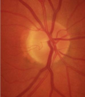
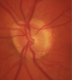
Two Pits in a Pod: Using EDI-OCT to evaluate the lamina cribrosa in a patient with openangle glaucoma and multiple optic pits
 Two Pits in a Pod: Using EDI-OCT to evaluate the lamina cribrosa in a patient with openangle glaucoma and multiple optic pits Abstract: EDI-OCT imaging is used to evaluate glaucoma by examining the thinning
Do You See What I See!!! Shane R. Kannarr, OD
 Do You See What I See!!! Shane R. Kannarr, OD skannarr@kannarreyecare.com Define Specialty Testing Additional Test to: Prove/Disprove Diagnosis To monitor progression of a condition To document a condition
The Foundation WHAT IS THE RETINA? continued next page. RETINA HEALTH SERIES Facts from the ASRS
 The Foundation American Society of Retina Specialists Committed to improving the quality of life of all people with retinal disease. Vitreomacular Traction Syndrome The vitreous humor is a transparent,