Introduction. Pardon the Objection: Posterior Segment Update. Disclosures. Introduction. Steven Ferrucci, OD, FAAO 5/23/2014. Fast paced! Interactive!
|
|
|
- Alexandra Webster
- 6 years ago
- Views:
Transcription
1 Introduction Pardon the Objection: Posterior Segment Update Steven Ferrucci, OD, FAAO Mark Dunbar, OD, FAAO Maynard Pohl, OD, FAAO Diana Shechtman, OD, FAAO Fast paced! Interactive! Panel participation! Audience participation! Apologies in advance! The opinions and comments expressed in this lecture do not reflect those of the AOA or any sensible person Introduction Disclosures FERRUCCI: Chief, Optometry, Sepulveda VA; Professor, MBKU/SCCO DUNBAR: Optometric Director, Bascom Palmer Eye Institute and University of Miami School of Medicine POHL: Director of Optometry, Pacific Cataract & Laser Institute SHECTHMAN: Professor, Nova Southeastern University FERRUCCI: Alcon, Arctic DX, Maculogix, Kemin, RetnaGene, ThromboGenics DUNBAR: Allergan, Arctic Dx, CZM, Sucampo; Co Chair Education, Reed Exhibits POHL: SHECHTMAN: Allergan, Alcon, Arctic DX, B&L, CZM, Zeavision 1
 OCT/IMAGING MARK DUNBAR, OD, FAAO")


2 What s New in Imaging: AMD u Advances in SDOCT u Maculogics (dark adaption for AMD) OCT/IMAGING MARK DUNBAR, OD, FAAO Advances in SD-OCT OCT I u Progression analysis software u Wider and deeper scans v Traditional scan area is 6 mm X 6 mm, now >12 mm scans v Software evolving to include whole posterior pole u Greater density in the scans u Higher resolution Stratus SD OCT (Spectralis) 2


3 The multi dimensional platform for Widefield Enface OCT High speed Multi layered assessment of peripheral retina pathology Glaucoma Anterior segment analysis Meeting the patient eye care needs of today and the future. XR Avanti MCT with OCTA 1 Detailed vasculature of the fovea without any dye or contrast enhancement injection RPE/Drusen Analysis Software OCT En face image SSADA / OCTA Imaging SSADA / OCTA Imaging Flow rate directly relates to appearance (brightness) of vessel in the image 2 *Images courtesy of David Huang, M.D. OHSU. 1 OCTA(SSADA) is not yet commercially available. 3


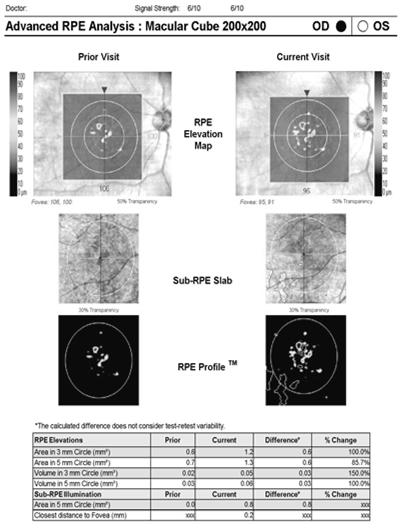
4 RPE Elevations: If the RPE is raised, a new proprietary algorithm for Cirrus maps and measures the area and volume of the elevations. Advanced RPE Analysis Gain new insights on your AMD patients RPE Elevations Sub-RPE Illumination 2009 Esther: Geographic Atrophy Sub-RPE Illumination. If the RPE is absent or has lost integrity a new proprietary algorithm for Cirrus can map and measure the affected area. Cirrus /28/2010 vs. 1/18/ /28/2010 1/18/2013 4


5 3/20/10 6/5/2012 3/20/10 6/5/2012 5


6 Drusen Analysis Dark Adaptation Impairment in Patients with Age related Macular Degeneration 6

 is severely impaired in AMD Biology of")

7 Maculogics FDA Statement Cleared 510(k) number K The AdaptDx is an AC powered, automated adaptometer (biophotometer) intended to measure the time for retinal adaptation after exposure to an adapting light. Next-generation dark adaptometer for rapid, routine clinical use Simple, objective tool to measure dark adaptation as earliest functional correlate of AMD Two clinical protocols - 5 minute Rapid test - 20 minute Staging test AdaptDx Dark Adaptation is an Established Marker of AMD Dark adaptation (DA) is severely impaired in AMD Biology of DA impairment is well understood and linked to breakdown of RPE/Bruch s membrane complex - Impairment tracks with severity - Impairment is rod-mediated DA represents earliest functional correlate with AMD Traditional 90 minute test too slow and impractical for clinical use Late AMD Early AMD Normal Dark Adaptation Contrast Sensitivity Photopic Visual Field Scotopic Visual Field Visual Acuity Diagnostic Sensitivity 0% 20% 40% 60% 80% 100% Owsley et al. (2001) Ophthalmology 108:1196 7

8 Rapid Dark Adaptation Test Results Patients classified as having AMD if dark adaptation >6.5 minutes High Sensitivity: correctly identified 90.6% of confirmed AMD cases High Specificity: correctly identified 90.5% of confirmed normal cases AMD cases exhibit no rod recovery of dark adaptation AdaptDx Validation Study Results Screening Test 5 Staging Test 20 minutes minutes Jackson et al. (2014) IOVS 55:1427 Jackson et al. (2014) IOVS 55:1427 CHOROIDAL LESIONS DIANA SHECHTMAN, OD, FAAO 8


9 When the NEVOMA PRESENTS Choroidal nevus vs choroidal melanoma ~30% of choroidal melanomas are small with <3mm thickness Tumor development Benign Growth restricted to that area & no organ functional damage Malignant Cell proliferation results in organ function damage. Cell proliferation may be due to: lack of responses to normal apoptosis genetic alteration cause by mutations Metastasis Rapid growth with infiltration into other tissues So which is which & how do I know? What are features of a choroidal nevus? Choroidal Nevi: BENIGN proliferation (neoplams) of choroidal melanocytes 6 10% (blue mnt eye study) prevalence among whites No correlation w (+)FHx of any melanoma < 3 mm elevation Typically FLAT < 3 DD in size 90% are <2 DD; so if note 3 5DD it is questionable (NEVOMA) 9


10 20 yo asymptomatic Disappears in RF Ultrasound revealed no thickness OCT shows no fluid Nevus or melanoma? Management 6:3:6 Nevus or melanoma? What are the associated findings? What do you think? Is that LP or drusen? There s drusen Is it benign? In general, nevi rarely enlarge Blue mountain Study (n= 160 nevi s/p with 5 yr follow up) If no camera document BORDERS 10


11 Choroidal Melanoma What are features of a choroidal melanoma? Observe in white pts with a peak incidence in 50 60yo >3 mm elevation Usually >5mm in size Increase risk in light eyes, fair skin, propensity to burn, pts with numerous freckles Growth over 7 ½ months Melanomas grow RAPIDLY In height &/or diameter How do I know the choroidal lesion today is a melanoma tomorrow? 11


 Thickness >2mm Increase")
 38% chance of growth FAF")

 pathognomonic Why is a referral")
 or <5 8mm diameter =")
 or 10 15mm diameter = 10yr mortality")


12 Nevus or melanoma? To Find Small Ocular Melanoma Shields CL, et al "Choroidal nevus transformation into melanoma. Analysis of 2,514 consecutive cases" Arch Ophthalmol 2009; 127(8): Symptoms VFD, metamorphopsia Photopsia & VL (dep on location) No signs: annual exam <3% chance of growth Fluid (subretinal) Thickness >2mm Increase risk of metastasis 1 2 signs: 4 6M f/u (sooner after initial Dx) 38% chance of growth FAF margins near ON >3 signs: consultation (W/I 3mm) 50% chance of growth over 5 yrs Management depends on # of features noted Orange pigment (lipofuscin) pathognomonic Why is a referral important with regards to a small melanoma? S: <3 mm thickness (height) or <5 8mm diameter = 10yr mortality rate ~10% M: 3 5 mm thickness (height) or 10 15mm diameter = 10yr mortality rate ~20% L: > 5 mm thickness (height) or >16mm diameter = 10yr mortality rate 40% Diameter = largest linear tumor dimension (basal diameter) SAVE LIVES You save vision, you save eyes & you save Highest incidence of metastatic detection is 1 3 yr s s/p Dx of the choroidal melanoma COMS
 3.")

13 Why is identifying a small melanoma critical? SIZE does mater 1. THE 5 yr mortality rate after enucleation: 16% small choroidal melanoma, 32% medium & 53% for large 2. 1mm increase thickness = 5% increase risk of metastatic dz ( 10 yrs) 3. Despite direct tx of the choroidal melanoma, 30 50% of pts develop metastatic disease COMS 1998 What is the most common location of primary uveal melanoma to metastasize? Metastatic choroidal melanoma Liver Brain Lungs Bone Breast Skin median survival for a hepatic metastasis: 20% at 1 year 10% at 2 years <1% after 5yrs, REGARDLESS OF TX Arch of Ophthalmol May (5):670 >25% of patients with ocular melanoma will develop metastases within 5 yrs s/p Dx & 90% of those will metastasize to the liver Incidence of metastasis increase to 34% at 10yr BRVO MAYNARD POHL, OD, FAAO 13


14 Branch Retinal Vein Occlusion (BRVO) BRVO: Acute Findings Thrombus formation at arteriovenous crossing Systemic hypertension commonly associated Age most common Sectoral superficial hemorrhages Sectoral retinal edema Sectoral cotton wool spots BRVO: Chronic Findings Chronic BRVO Microvascular abnormalities Macular edema Intraretinal collaterals Sclerosis and sheathing of retinal vessels 14


15 BVOS BRVO: Laser Treatment Techniques Branch Retinal Vein Occlusion Study Group, American Journal of Ophthalmology 1984; 98: Branch Retinal Vein Occlusion Study Group, Archives Ophthalmology 1986; 104:34 41 Macular Grid Laser Photocoagulation: BRVO present for more than 3 months absence of foveal hemorrhage vision worse than 20/40 vision loss due to macular edema 15


16 Macular grid laser photocoagulation remains the criterion standard treatment of eyes with perfused macular edema secondary to BRVO. 16
17 BRVO: Laser Treatment Techniques BRVO: Other Treatment Techniques Scatter Photocoagulation: presence of neovascularization presence of vitreous hemorrhage Laser induced chorioretinal anastomosis Arteriovenous decompression (sheathotomy) Vitrectomy Intravitreal Kenalog (triamcinolone acetonide) SCORE Study Ozurdex (0.7 mg dexamethasone intravitreal implant) Avastin, Lucentis, Eylea Vascular Endothelial Growth Factor (VEGF) VEGF is a potent inductor of vascular permeability and intraocular neovascularization. Human aqueous levels of VEGF and interleukin 6 (IL 6) are correlated with the degree of retinal ischemia and the severity of macular edema in BRVO. Therefore, VEGF inhibition is a promising treatment modality for macular edema. Clinical Evidence Based Conclusions Timing of diagnosis and management of BRVO is important. Eyes with macular edema secondary to BRVO should be offered VEGF inhibition upon diagnosis to achieve the best possible visual outcome (BRAVO Study, HORIZON Trial, RETAIN Study). Eyes are eligible for laser after 3 months if hemorrhages have sufficiently cleared to allow safe laser treatment and if vision acuity remains worse than 20/40. Retinal nonperfusion is related to intravitreal VEGF levels and may result in loss of visual gains. The prevention of worsening retinal nonperfusion should be a treatment objective as important as the resolution of macular edema. Periodic fluorescein angiograms should be performed to monitor perfusion status. 17


18 Central Retinal Vein Occlusion (CRVO) CRVO MAYNARD POHL,OD, FAAO Thrombus formation in retinal vein at lamina cribosa Etiology of thrombus formation unclear: arteriosclerosis, vasculitis Primary open angle glaucoma: 20% have POAG, 20% develop POAG Central Retinal Vein Occlusion (CRVO) Non Ischemic (Partial) CRVO Systemic associations: CVD 74% HTN 57% DM 34% Risk factors include oral contraceptives and diuretics 90% of patients are over 50 18
 Ischemic CRVO Marked optic disc, retinal, and macular edema Marked venous dilatation and")

19 Non Ischemic CRVO Ischemic (Complete) CRVO 30+% convert to ischemic type (CVOS) < 10 dd of retinal non perfusion (CVOS) Ischemic CRVO Marked optic disc, retinal, and macular edema Marked venous dilatation and tortuosity Many retinal hemorrhages, cotton wool spots VA worse than 20/200 Afferent pupillary defect CRVO: Incidence of Neovascularization Non Ischemic: Any NV in < 5% NV glaucoma in < 2% Ischemic: Any NV in > 60% NV glaucoma in 33% 19
20 CVOS Central Retinal Vein Occlusion Study Group, Ophthalmology 1995; 102: CVOS Conclusions Grid photocoagulation for macular edema: effective in decreasing retinal thickening but ineffective in improving VA Pan retinal photocoagulation to prevent neovascular complications: indicated only in eyes with apparent iris NV, angle NV, or ischemic eyes that cannot be followed monthly CRVO: Other Advocated Treatments Aspirin Anti inflammatory agents Isovolemic hemodilution Plasmapheresis Systemic anticoagulation with warfarin, heparin, and alteplase Fibrinolytic agents Systemic corticosteroids Intravitreal treatments the standard of care CRVO: Intravitreal Treatments Local anticoagulation with intravitreal injection of alteplase (Activase) Intravitreal injection of triamcinolone (Kenalog) Ozurdex intravitreal implant Intravitreal injection of Lucentis Intravitreal injection of Avastin Intravitreal injection of Eylea 20

 CRVO: Pre and Post OCT SCORE Study: Conclusion Pre Injection: VA 20/400,")


21 Intravitreal Injection of Triamcinolone Intravitreal Injection (IVI) SCORE Study CRVO Trial demonstrated effectivity in resolving perfused macular edema and improving vision 1 mg dose and retreatment prn may be considered up to 12 months (preferred over 4 mg dose due to fewer adverse effects) CRVO: Pre and Post OCT SCORE Study: Conclusion Pre Injection: VA 20/400, thickness > 600 microns Post Injection (6 weeks): VA 20/80, thickness 350 microns No difference in longterm outcome between triamcinolone injections and grid photocoagulation with BRVO. Ozurdex biogradable implant (Allergan, June 2009) is considered superior to triamcinolone as a delivery method, with fewer injections. Triamcinolone remains a viable option for patients with financial troubles. 21
. 12 month data: vision gained at 6 months continued after 6 months of subsequent prn dosage.")
 Anti VEGF therapy ranks as the preferred first line therapy for RVO.")
22 Pre and Post Avastin Injection Anti VEGF Trials For RVO After 6 months of Lucentis therapy, between 55% and 61% of BRVO patients and 47% of CRVO patients gained at least 3 lines of BCVA (BRAVO and CRUISE studies). 12 month data: vision gained at 6 months continued after 6 months of subsequent prn dosage. From a strictly evidenced based perspective, slightly better visual outcomes and huge safety profile, relative to steroids. Lucentis approved for treatment of macular edema following RVO in June Eylea approved for macular edema following CRVO in September 2012 (COPERNICUS and GALILEO trials) Anti VEGF therapy ranks as the preferred first line therapy for RVO. Head to Head Studies in RVO COMO and COMRADE B comparing Lucentis with dexamethosone IVT in BRVO patients COMRADE C in CRVO patients RABAMES comparing Lucentis, argon laser monotherapy, and Lucentis plus adjunctive argon laser therapy in BRVO patients (completed) BRIGHTER (EUDRACT 2011) European studies with similar treatment arms AMD TREATMENT MARK DUNBAR, OD, FAAO 22

 A monoclonal antibody that inhibits Complement Factor D A rate-limiting enzyme involved in the alternative complement pathway A key-driver for the development of geographic")
23 AMD: Management Options for Today and the Future u Dry AMD treatment v Nutritional options v Targeting genetic factors u Wet AMD treatment v Laser, PDT, Anti-angiogenic therapy v Prognosis for successful outcome is dependent on early diagnosis and treatment of CNV COL8A1 TIMP3 Key Genes Involved in the Development of AMD LIPC ARMS2 APOE CETP ABCA1 CFH CFI CFB C2 CFH C3 C3 C2 CFB CFI Macula Risk NXG 12 genes ARMS2 TIMP3 COL8A1 LIPC APOE CETP ABCA1 Complement Oxygen Metabolism Extracellular Matrix Cholesterol Metabolism Ongoing Dry AMD Trials Lampalizumab (LAMP) Anti-factor D (Genentech) A monoclonal antibody that inhibits Complement Factor D A rate-limiting enzyme involved in the alternative complement pathway A key-driver for the development of geographic atrophy 23

24 MAHALO Trial 129 pts with bilateral geographic atrophy randomized to assess the safety, tolerability, and activity of lampalizumab (LAMP) Biomarkers for complement factor H (CFH), C3, C2/CFB and CFI were also determined Participants, y/o randomized to lampalizumab 10 mg or sham injections MAHALO Trial A positive treatment effect was seen in the monthly lampalizumab group beginning at month 6, and was maintained through month 18 with primary and secondary imaging end points Subgroup analysis of monthly injections showed overall reduction in GA area in pts with a specific biomarkers was more than double that of the study cohort as a whole (44.0% vs. 20.4%) Geographic Atrophy and Complement Factor I (CFI) 57% of genotype samples collected from 93 patients were positive for the CFI biomarker CFI biomarker is both prognostic for GA area progression and predictive for lampalizumab treatment response ACQUIRED MACULOPATHIES DIANA SHECHTMAN, OD, FAAO 24


25 Stages of HCQ/ CQ Maculopathy Early Moderate Advance MEDICATION toxicity What was the previous tests used for plaquenil pts? What are today s tests? Protocol change in 2011 Why were screening recommendations change Typically retinopathy not observed until 5yrs s/p use of med Analysis of 4,000 study 1.8% develop retinopathy s/p LONG term use of med Risk of toxicity increases 5x s/p 7yrs of usage or at 1,000 GRAMS (1,000,000 mg) of exposure 25


26 Baseline So when do YOU see the pt back? within 1 st yr medication that the has been started to get baseline detects any pre existing maculopathy & other HR signs Monitoring: determine if si/s maculopathy have occurred Recommendations 2011 new protocols Not plaquenil module VFD should have a low threshold LOW RISK Year 0 Year 1 Year 2 Year 3 Year 4 Year 5 Year 6 Year 7 HIGH RISK FAF shows decreased autofl perifoveally: photoreceptor damaging denoted by increase FAF What does the SDOCT damage Associated with toxicity look like? 26


27 Reading the OCT associated with plaquenil toxicity Normal Patient OCT: Saucerazation & Sinkhole appearance Where s the damage? Plaquenil Patient perifoveal outer retinal abnormalities displacement of the inner retinal structures toward the RPE with increase retinal atrophy Chen et al. Clinical Ophthalmology BF DFE: What s the Dx? CC: Decreased VA X 2 yrs PMHx: Lupus Meds: 8 9yrs Plaquenil 200mg/QD Pred 15mg / QD BCVA: 20/20 OD, OS OU D/N 27
 Duration of use Cumulative Dose Daily Dose Age Systemic Disease/High BMI Ocular Disease > 5 years > 1000 g (total) = 400x365X7 yr = 1mill mg > 400 mg/day")

28 OCT: Does she have early toxicity? WHAT IS HIGH RISK? WHEN DO YOU SEE HER BACK? What do you communicate to the treating doctor? What is HR pt? Factors Increasing Risk of Retinopathy (HR) Duration of use Cumulative Dose Daily Dose Age Systemic Disease/High BMI Ocular Disease > 5 years > 1000 g (total) = 400x365X7 yr = 1mill mg > 400 mg/day Older Kidney or liver dysfunction Retinal disease or maculopathy Daily Dosage Daily Dosage Standard dose: 200mg/day or 400 mg/day Typically Rx as 6.5mg/kg 1 kg = 2.2 pounds 6.5mg/2.2 pounds = 2.95mg/pound 400 mg HCQ/2.95 = 135 pounds 400mg/Day is given to a 135lb pt (IDEAL weight) IDEAL weight At 5ft a women weighs 100 lb women & a male weighs 110 men 5lb added thereafter for every ft OBESITY (BMI) & toxicity Our pts are NOT at the ideal weight Pt with higher BMI is at risk because they are Rx based on 6.5mg/kg (& not based on LEAN muscle weight) Hence, they are OVERDOSE DRUG is not retained in Adipose (fat) tissue & thus, there are higher plasma levels of the drug. Height recently has also been an issue Hence, if you re short, the standard Rx of 200 mg BID is TOO MUCH! 28
 How")


29 How would you handle this case? (HCQ, 48 yrs, 400mg/day for 25 years) How do you deal with initial VF? Do you repeat them initially q6m? What does a plaquenil toxicity VFD look like? What do you communicate to the treating doctor? Sup inferior Sup inferior 29
.")


30 THE DILEMMA of when to D/c meds Making the right decision First presentation Safe & effective True maculopathy is rare Sign of early ocular toxicity May cause VL even after d/c 3 years AFTER D/C drug Although there s a speculation that toxicity can be reversed if detected earlier, there s no conclusive evidence (no longitudinally studies). Yet, there s conclusive evidence that progression of toxicity occurs despite cessation of drug in many cases Marmor stated, Goal is to catch changes at a very early stage when there s just a minimal amount of damage. Then, when you stop the drug, the damage won t progress enough to cross the fovea and affect reading vision. The likelihood of toxicity is LOW Dr. Marmor has seen only seen 10 cases in the past year. Impact of the guidelines on today's practice! AJO 8/2013 n=183 pts came for f/u & 36 were evaluated for baseline Evaluated by 26 ophthalmologist & 3 ODs Results 40% increase on health care cost 50% of doctors perform 6M rather than yrly f/u No additional pts discover with toxicity in accordance to new guideline. Incidence of toxicity remains at 1%, as noted in f/u pts No pts was followed at recommended guidelines of 5-year period after baseline (even if low-risk patient) Impact of the guidelines on today's practice! AJO 8/2013 Small sample size No one perform FAF Pt s height, weight, and daily dose were not determined in 1/3rd o the pts 30


31 DIABETIC RETINOPATHY MAYNARD POHL, OD, FAAO Evidence Based WHO Guidelines (2012) South Asian and Pacific Island populations now considered high risk populations All Type 2 DM patients examined at diagnosis Type 1 DM patients examined at puberty Women with DM examined before pregnancy and during first trimester All DM patients regardless of degree of DR examined at least every 2 years Prevalence of blindness from DR is escalating in developing countries with a younger age of onset of DM Detection of referrable retinopathy with single 45 degree non mydriatic camera using trained operator with off site grading by ophthalmologist, or trained ophthalmic medical officer or optometrist performing dilated fundus exam Advanced Diabetic Retinopathy Laser Photocoagulation & Vitrectomy in Diabetic Retinopathy Laser in Type 1 and Type 2 DM patients with NVE with vitreous heme, or with NVD with/without vitreous heme PRP considered in severe or very severe NPDR Modified ETDRS macular laser in CSME when macular ischemia absent Vitrectomy in advanced DR: severe PDR with nonresolving vitreous heme or fibrosis, RD, or areas of retinal traction threatening macula Vitrectomy in persistent diffuse macular edema 31
 NHMRC consider anti VEGF as an adjunct to laser and prior to vitrectomy Eylea Triamcinolone (IVTA) in refractory")

32 Intraocular Steroids & Anti VEGF Agents in PDR and CSME Macugen Avastin BOLT study (monotherapy) Lucentis RESOLVE study (monotherapy), READ 2 study (Lucentis vs laser vs combo) DRCR.net greatest improvement in VA is Lucentis and deferred laser (>6 mos after injection) NHMRC consider anti VEGF as an adjunct to laser and prior to vitrectomy Eylea Triamcinolone (IVTA) in refractory DME, weighing risk vs benefit; as an adjunct to PRP in PDR Ophthalmology: August 2013 AMD GENETICS MARK DUNBAR, OD, FAAO More than double the benefit of AREDS: We estimate that genotype directed therapy of the study population would have more than doubled the reduction in AMD progression rate compared with treatment with the AREDS formulation. A clear role for genetic testing Ivana Kim (co author) This data demonstrates that the composition of supplements recommended to AMD patients should be guided by an individual s genetic risk profile, indicating a clear role for genetic testing in clinical management. 32


33 Genetic analysis of 995 patients with intermediate (moderate) AMD who were in the original AREDS 1 study Followed for 12 years Evaluated the interaction of genetics and type of nutritional supplement on progression from moderate to advanced AMD Patients with genetically determined CFH high risk alleles, zinc was associated with increased progression to advanced AMD Patients with ARMS2 high risk alleles, zinc was associated with DECREASED progression to advanced AMD antioxidants worsened the outcome Patients with CFH risk alleles benefit from antioxidants without zinc but patients with ARMS2 risk alleles benefit from zinc without 49% derive more benefit from treatment other than AREDS Estimate Probability of Progression 33


34 Estimate Probability of Progression Estimate Probability of Progression Genetic Testing and Recommending Nutritional Supplements Of the estimated 15 million Americans taking the AREDS formula, more than ten million should not be on either zinc or antioxidants Only about 23% of patients taking AREDS formula should be while the vast majority should not Genotype-directed therapy of the study population would have more than doubled the reduction in AMD progression rate compared to treatment with the AREDS formulation VMT DIANA SHECHTMAN, OD, FAAO 34

 But PPV is associated with a #")


35 20/30 20/40 THE SLEEPING CONDITION m o m o n i What are the common management options? Option 1 is to REFER: Given it may progress (associated with various complications) But PPV is associated with a # of complications WHAT ARE THE COMPLICATIONS ASSOCAITED WITH PPV? Option 2 is to MONITOR Given it may resolve on its own WHEN DO YOU REFER? Tx Options for Symptomatic VMT MILD THE SLEEPING CONDITION SEVERE LET US NOT FORGET THE BURDEN ON THE PT. Most surgeons RISK vs BENEFITS advise pts to keep a head down ~7 days Outcome depends on: initial VA, complications, onset PPV risks: cataracts, RD, infection, no resolution Clinical Monitoring But may miss therapeutic window Vitrectomy Associated complications


36 VR interface contains lamin, collagen, fibronectin, which facilitate adhesion b/t ILM & vitreous cortex Jetrea has been commercially available Jan 2013 to tx symptomatic VMA Excluded PDR, high M, wet AMD & aphakia Included symptomatic VMA, ERM, smaller MH Truncated form of active PLASMIN recombinant protease with activity against fibronectin/lamin (components of VR interference) functions as a thrombolytic agent causing an enzyme pharmacological induced vitreolysis Nonsurgical PVD The enzymatic agents alter the biochemistry of vitreous Liquefaction of the vitreous occurs LYSIS between vitreous cortex and ILM is the final outcome Stalman 2012 N > 650 pts Inclusion: VMT w VA < 20/25 & OCT showing thickness PPV advised if: MD deemed it to be necessary s/p 1M VA worsen by 2 lines or No improvement s/p tx 36


37 Primary outcome Case Sample: MIVI Study pt 20/32 20/32 20/20 baseline 7 days 14 days 20/25 20/16 20/20 Among those pts that resolved s/p injection, 73% were w/i 1 st week ½ OF THE DRUG IS SHORT Treatment related Adverse Events Postinjection Day 0 to 7 Proportion of Patients with Retinal Tear or Retinal Detachment Patients, % Placebo N= Ocriplasmin N=465 ~10% adverse affects. Most affects noted within the 1st 7 days due to localize injection. They were transient and after 8d the adverse effects were minimal & equal b/t tx & placebo Patients, % n= Placebo N= Ocriplasmin N=465 Retinal Detachment Retinal Tear Retinal Detachment Retinal Tear Abbreviations: Ant., anterior; VA, visual acuity. Data on file. ThromboGenics, Inc Pre vitrectomy Occurrences All Occurrences


38 Case Sample: MIVI Study pt 20/32 20/32 20/20 Terminology: VMA/VMT Sub classified by size of adhesion Focal vs broad baseline 7 days 14 days 20/25 20/16 20/20 Note primary outcome was PVD w VA improvement as SECONDARY outcome Resolution of VMA at Day 28 By Predictors of Response Ray 2012 Patients, % Placebo Ocriplasmin 60 p=0.008 p=0.006 p< p<0.001 p<0.001 Increasing the # of (+) features 37.4 was associated with increase odds VMA resolution % respond rate with >3 predictive factors Singh Ophthal SO DO YOU REFER FOR JETREA? IF YES, WHEN? n=10 N=38 N=12 N=53 N= N=18 N=109 N=17 N=100 0 <65 years FTMH No ERM 1500 μm Phakic um N= * 400 μm with VMA Data on file. ThromboGenics, Inc

39 Thank you! 39
Vascular Disease Ocular Manifestations of Systemic Hypertension
 Vascular Disease Ocular Manifestations of Systemic Hypertension Maynard L. Pohl, OD, FAAO Pacific Cataract & Laser Institute 10500 NE 8 th Street, Suite 1650 Bellevue, WA 98004 USA 425-462-7664 Cerebrovascular
Diabetic Retinopatathy
 Diabetic Retinopatathy Jay M. Haynie, OD, FAAO Financial Disclosure I have received honoraria or am on the advisory board for the following companies: Carl Zeiss Meditec Arctic DX Macula Risk Advanced
OCCLUSIVE VASCULAR DISORDERS OF THE RETINA
 OCCLUSIVE VASCULAR DISORDERS OF THE RETINA Learning outcomes By the end of this lecture the students would be able to Classify occlusive vascular disorders (OVD) of the retina. Correlate the clinical features
Diabetic Retinopathy: Managing the Extremes. J. Michael Jumper, MD West Coast Retina
 Diabetic Retinopathy: Managing the Extremes J. Michael Jumper, MD West Coast Retina Case 1: EC 65 y.o. HM No vision complaints Meds: Glyburide Metformin Pioglitazone Va: 20/20 OU 20/20 Case 2: HS 68 y.o.
The Era of anti- - - VEGF Kirk L. Halvorson, OD
 The Era of anti- - - VEGF Kirk L. Halvorson, OD Introduction: Anti- - - Vascular Endothelial Growth Factor (Anti- - - VEGF) medication is a relatively a new line of medications used in treating a variety
THE ROLE OF anti-vegf IN DIABETIC RETINOPATHY AND AGE RELATED MACULAR DEGENERATION
 THE ROLE OF anti-vegf IN DIABETIC RETINOPATHY AND AGE RELATED MACULAR DEGENERATION MOESTIDJAB DEPARTMENT OF OPHTHALMOLOGY SCHOOL OF MEDICINE AIRLANGGA UNIVERSITY DR SOETOMO HOSPITAL SURABAYA INTRODUCTION
Jay M. Haynie, O.D.; F.A.A.O. Olympia Tacoma Renton Kennewick Washington
 Jay M. Haynie, O.D.; F.A.A.O. Olympia Tacoma Renton Kennewick Washington I Jay M. Haynie, OD, FAAO have received honoraria from the following companies: Reichert Technologies Notal Vision Carl Zeiss Meditec
VMA at the macula resulting in VMT
 Ocriplasmina for pharmacologic treatment in VMT Teresio Avitabile 1 Introduction PVD is a normal, physiologic process that occurs with aging; however, in some cases, PVD is incomplete Incomplete PVD localized
An Update on Branch Retinal Vein Occlusion Treatment Studies. Amiee Ho, O.D. Pacific University College of Optometry
 An Update on Branch Retinal Vein Occlusion Treatment Studies Amiee Ho, O.D. Pacific University College of Optometry Course Description This course focuses on current treatment options available for macular
Early diagnosis and treatment of VMT with single Intravitreal Injection of Pharmacologic Vitreolysis. Stratos Gotzaridis MD Athens
 Early diagnosis and treatment of VMT with single Intravitreal Injection of Pharmacologic Vitreolysis Stratos Gotzaridis MD Athens The Vitreous Body Gel composed of 98-99% water 1% macromolecules Glycoproteins
Diagnosis and treatment of diabetic retinopathy. Blake Cooper MD Ophthalmologist Vitreoretinal Surgeon Retina Associates Kansas City
 Diagnosis and treatment of diabetic retinopathy Blake Cooper MD Ophthalmologist Vitreoretinal Surgeon Retina Associates Kansas City Disclosures Consulted for Novo Nordisk 2017,2018. Will be discussing
Macular edema (ME) is the most common
 is the most common") MANAGEMENT OF RETINAL VEIN OCCLUSIONS * Peter A. Campochiaro, MD ABSTRACT Macular edema (ME) is the most common cause of reduced vision in patients with retinal vein occlusions (RVOs). The primary cause
MANAGEMENT OF RETINAL VEIN OCCLUSIONS * Peter A. Campochiaro, MD ABSTRACT Macular edema (ME) is the most common cause of reduced vision in patients with retinal vein occlusions (RVOs). The primary cause
Diabetic Retinopathy A Presentation for the Public
 Diabetic Retinopathy A Presentation for the Public Ray M. Balyeat, MD The Eye Institute Tulsa, Oklahoma The Healthy Eye Light rays enter the eye through the cornea, pupil and lens. These light rays are
Venous Occlusive Diseases
 Venous Occlusive Diseases Bruce R. Saran, MD Adjunct Assistant Clinical Professor of Medicine Scheie Eye Institute University of Pennsylvania School of Medicine Philadelphia, PA -a division of: RVO Demographics
From Outdated to Updated: A Review of Important Clinical Trials in Ocular Disease from 2014
 From Outdated to Updated: A Review of Important Clinical Trials in Ocular Disease from 2014 1. This course is designed to review the important ophthalmic literature that was released between October 2013
Mark Dunbar: Disclosure
 Important Things to Understand About OCT Mark T. Dunbar, O.D., F.A.A.O. Bascom Palmer Eye Institute University of Miami, School of Medicine Mark Dunbar: Disclosure Optometry Advisory Board for: Allergan
FROM OUTDATED TO UPDATED Eminence-Based Medicine
 FROM OUTDATED TO UPDATED Eminence-Based Medicine Evidence-Based Medicine A REVIEW OF KEY CLINICAL TRIALS Anthony DeWilde, OD FAAO 1 EMINENCE BASED MEDICINE 2 EVIDENCE BASED MEDICINE 3 4 CLINICAL TRIALS
Clinically Significant Macular Edema (CSME)
") Clinically Significant Macular Edema (CSME) 1 Clinically Significant Macular Edema (CSME) Sadrina T. Shaw OMT I Student July 26, 2014 Advisor: Dr. Uwaydat Clinically Significant Macular Edema (CSME) 2
Clinically Significant Macular Edema (CSME) 1 Clinically Significant Macular Edema (CSME) Sadrina T. Shaw OMT I Student July 26, 2014 Advisor: Dr. Uwaydat Clinically Significant Macular Edema (CSME) 2
Optical Coherence Tomography in Diabetic Retinopathy. Mrs Samantha Mann Consultant Ophthalmologist Clinical Lead of SEL-DESP
 Optical Coherence Tomography in Diabetic Retinopathy Mrs Samantha Mann Consultant Ophthalmologist Clinical Lead of SEL-DESP Content OCT imaging Retinal layers OCT features in Diabetes Some NON DR features
OCT Interpretation in Retinal Disease
 OCT Interpretation in Retinal Disease Jay M. Haynie, OD, FAAO Financial Disclosure I have received honoraria or am on the advisory board for the following companies: Carl Zeiss Meditec Advanced Ocular
OCT Interpretation. Financial Disclosure. Jay M. Haynie, OD, FAAO. OCT Image Layers 7/21/2014
 OCT Interpretation Jay M. Haynie, OD, FAAO Financial Disclosure I have received honoraria or am on the advisory board for the following companies: Olympia Tacoma Renton Kennewick - Washington Carl Zeiss
FA Conference. Lara Rosenwasser Newman, M.D. 10/2/14 University of Louisville Department of Ophthalmology and Visual Sciences
 FA Conference Lara Rosenwasser Newman, M.D. 10/2/14 University of Louisville Department of Ophthalmology and Visual Sciences Patient Presentation CC: (sent by optometrist) Blurry/foggy vision HPI: 62 yo
Diabesity A Public Health Crisis: AOA Evidence Based Translation to Care Series
 Diabesity A Public Health Crisis: AOA Evidence Based Translation to Care Series Joseph J. Pizzimenti, OD, FAAO Associate Professor Nova Southeastern University The Eye Care Institute pizzimen@nova.edu
Retinal Vein Occlusion (RVO) Treatment pathway- Northeast England. Retinal Vein Occlusion (RVO) with Macular oedema (MO)
 Treatment pathway- Northeast England. Retinal Vein Occlusion (RVO) with Macular oedema (MO)") Retinal Vein Occlusion (RVO) Treatment pathway- Northeast England (Royal Victoria Infirmary, Sunderland Eye Infirmary, James Cook University Hospital, Darlington Memorial Hospital, University Hospital
Retinal Vein Occlusion (RVO) Treatment pathway- Northeast England (Royal Victoria Infirmary, Sunderland Eye Infirmary, James Cook University Hospital, Darlington Memorial Hospital, University Hospital
Mark T. Dunbar Disclosures. Hot Topics in Retina. The Challenge. Where is the Fluid? 4/12/2016
 A PANEL DISCUSSION: RAPID-FIRE POSTERIOR SEGMENT UPDATE! Steve Ferrucci, OD Chief, Optometry Sepulveda VA 16111 Plummer St #112e Sepulveda CA, 91343 Mark T. Dunbar, OD Bascom Palmer Eye Institute Miami,
Marcus Gonzales, OD, FAAO Cedar Springs Eye Clinic
 Marcus Gonzales, OD, FAAO Cedar Springs Eye Clinic 25.6 million adults 11.3% of the adult population 10.9 million adults 65 years and older 26.9% of this age population 79 million people are Pre-diabetic!!
OCT Angiography in Primary Eye Care
 OCT Angiography in Primary Eye Care An Image Interpretation Primer Julie Rodman, OD, MS, FAAO and Nadia Waheed, MD, MPH Table of Contents Diabetic Retinopathy 3-6 Choroidal Neovascularization 7-9 Central
OCT Angiography The Next Frontier
 Choroid Retina avascular 5/13/2017 OCT Angiography The Next Frontier Pierce Kenworthy OD, FAAO June 9, 2017 OCT Angiography (OCTA) 2016 Non-invasive, motion contrast imaging Represents erythrocyte movement
Retinal Complications of Obstructive Sleep Apnea A Growing Concern!
 Retinal Complications of Obstructive Sleep Apnea A Growing Concern! Jay M. Haynie, OD, FAAO Financial Disclosure I have received honoraria or am on the advisory board for the following companies: Carl
RETINA. OCT in the Optometric Prac4ce OCT. New stuff 11/7/17 AMD. Disclosure Statement
 Disclosure Statement OCT in the Optometric Prac4ce Steven Ferrucci, OD, FAAO Chief, Optometry Sepulveda VA Professor, SCCO/MBKU Speakers bureau/advisory Board Alcon B&L Centervue Genentech MacuLogix Optovue
New Developments in the treatment of Diabetic Retinopathy
 New Developments in the treatment of Diabetic Retinopathy B. Jeroen Klevering University Medical Centre Nijmegen - The Netherlands Topics Management of diabetic retinopathy Interventions a. primary (prevention)
PART 1: GENERAL RETINAL ANATOMY
 PART 1: GENERAL RETINAL ANATOMY General Anatomy At Ora Serrata At Optic Nerve Head Fundoscopic View Of Normal Retina What Is So Special About Diabetic Retinopathy? The WHO definition of blindness is
Managing the Vitreomacular Interface
 Managing the Vitreomacular Interface A Guide to VMA, VMT, Holes and ERM Anna K. Bedwell, OD, FAAO Indiana University School of Optometry Please silence all mobile devices and remove items from chairs so
CENTENE PHARMACY AND THERAPEUTICS NEW DRUG REVIEW 2Q17 April May
 BRAND NAME Lucentis GENERIC NAME ranibizumab MANUFACTURER Genentech, Inc. DATE OF APPROVAL June 30, 2006 PRODUCT LAUNCH DATE July 13, 2006 REVIEW TYPE Review type 1 (RT1): New Drug Review Full review of
Clinical Case Presentation. Branch Retinal Vein Occlusion. Sarita M. Registered Nurse Whangarei Base Hospital
 Clinical Case Presentation on Branch Retinal Vein Occlusion Sarita M. Registered Nurse Whangarei Base Hospital Introduction Case Study Pathogenesis Clinical Features Investigations Treatment Follow-up
Retinal Vein Occlusion
 Retinal Update 2018 Retinal Vein Occlusion Case Presentations to Myself Branch Vein Occlusion What medical evaluation do you recommend for this 72 year old patient? Is there anything you ask of your medical
EU Regulatory workshop Ophthalmology clinical development and scientific advice. Industry view on DME and macular edema secondary to RVO
 EU Regulatory workshop Ophthalmology clinical development and scientific advice. Industry view on DME and macular edema secondary to RVO Yehia Hashad, M.D. Vice President and Global Therapeutic Area Head
FRANZCO, MD, MBBS. Royal Darwin Hospital
 Diabetes and Eye By Dr. Nishantha Wijesinghe FRANZCO, MD, MBBS Consultant Ophthalmologist Royal Darwin Hospital 98% of Diabetics do not need to suffer from severe visual loss Yet Diabetic eye disease is
ZEISS AngioPlex OCT Angiography. Clinical Case Reports
 Clinical Case Reports Proliferative Diabetic Retinopathy (PDR) Case Report 969 PROLIFERATIVE DIABETIC RETINOPATHY 1 1-year-old diabetic female presents for follow-up of proliferative diabetic retinopathy
1/25/2018. Case Management Strategies in Diabetic Retinopathy. Case Study #1: Severe DME. DDOS: 3/31/2016 Va 20/400. Disclosures
 Case Management Strategies in Diabetic Retinopathy Disclosures No financial conflict of interest Will discuss off label use of intraocular Bevacizumab (Avastin) for Diabetic Retinopathy Sundeep Dev, MD
Course # Getting to Know Your OCT
 Course # 140 Getting to Know Your OCT Course Title: Lecturer: Getting to Know Your OCT Brad Sutton, OD, FAAO IU School of Optometry Financial Disclosures No financial disclosures Optical Coherence Tomography-OCT
Treatment Options for VMT and Macular Holes Observation, Surgery, and Pharmacotherapy
 Treatment Options for VMT and Macular Holes Observation, Surgery, and Pharmacotherapy Andrew Moshfeghi, MD, MBA Bascom Palmer Eye Institute Palm Beach Gardens, FL Financial Disclosures Salary/Honoraria:
Diabetic Retinopathy
 Diabetic Retinopathy Diabetes can be classified into type 1 diabetes mellitus and type 2 diabetes mellitus, formerly known as insulin-dependent diabetes mellitus, and non-insulin diabetes mellitus, respectively.
Steven Ferrucci, OD. FAAO; Jeffry Gerson, OD, FAAO; Robert Prouty, OD, FAAO; Leo semes OD, FAAO
 PARDON THE OBJECTION: RETINA Steven Ferrucci, OD. FAAO; Jeffry Gerson, OD, FAAO; Robert Prouty, OD, FAAO; Leo semes OD, FAAO 1. Introductions/Disclosures (Ferrucci) 2. The genetics of AMD (Gerson) a. Background
4/27/2010 INTRODUCTION TO RETINAL VASCULAR DISEASE VENOUS/VENULAR CENTRAL RETINAL VEIN OBSTRUCTION / CRVO ADDITIONAL FEATURES /COMPLICATIONS
 INTRODUCTION TO RETINAL VASCULAR DISEASE VENOUS/VENULAR Leo Semes, OD Professor, UAB Optometry 2 CENTRAL RETINAL VEIN OBSTRUCTION CENTRAL RETINAL VEIN OBSTRUCTION / OCCLUSION (CRVO) obstruction of the
The Human Eye. Cornea Iris. Pupil. Lens. Retina
 The Retina Thin layer of light-sensitive tissue at the back of the eye (the film of the camera). Light rays are focused on the retina then transmitted to the brain. The macula is the very small area in
 measure of your overall performance. An isolated glucose test is helpful to let you know what your sugar level is at one moment, but it doesn t tell you whether or not your diabetes is under adequate control
Financial Disclosures
 Financial Disclosures Consultant Genentech, Regeneron, Allergan, Thrombogenics, Optos, and ArcticDx Grant Support Regeneron, Allergan Mathew W. MacCumber, MD, PhD Professor & Assoc. Chair for Research
Goals/Objectives. Disclosures. Risk Factors RAO and RVO. Risk Factors. Retinal Artery Occlusions Branch and Central
 Jeffrey D. Perotti, OD, MS Indiana University School of Optometry Goals/Objectives RETINAL VASCULAR OCCLUSIONS FOR THE PRIMARY CARE CLINICIAN Using cases as a framework, review current evaluation and management
ABCs. ABCs of retinal disease !"#$"%!& Disclosures. ABCs three major threats to vision where 1 o care intervention may be helpful!a = AMD !
 Disclosures Stockholder: HPO ABCs of retinal disease Honoraria, Consultant or Advisory Board: Alcon, Allergan, B&L, Arctic Dx, Sucampo, Zeiss. Idaho Optometric Physicians 2016 Leo Semes, OD, FAAO ABCs
Often asymptomatic but can cause a reduction in BCVA and distortion of vision.
 Christopher Wolfe, OD, FAAO, Dipl. ABO Epiretinal Membrane (ERM) and Vitreomacular Traction (VMT) Epiretinal membrane (macular pucker, cellophane maculopathy, premacular fibrosis) consists of a layer of
Past ocular history. DME Case 1. Patient presents blurred VA. Hemoglobin A1c 11.5% -- patient states sugars have not been in good control
 Past ocular history Patient presents blurred VA DME Case 1 Hemoglobin A1c 11.5% -- patient states sugars have not been in good control PDR with macular edema OU Rishi Singh MD Cleveland Clinic OD OS 1
Eyes on Diabetics: How to Avoid Blindness in Diabetic Patient
 Eyes on Diabetics: How to Avoid Blindness in Diabetic Patient Rova Virgana FK Unpad Pusat Mata Nasional RS Mata Cicendo Bandung Eye Center (Hospital and Clinic) PIT IDI Jabar 2018 Keys Facts from WHO
Recalcitrant Diabetic Macular Oedema: Therapeutic Options
 December 2007 A. Giridhar et al. - Recalcitrant DME 451 CONSULTATION S E C T I O N Recalcitrant Diabetic Macular Oedema: Therapeutic Options Dr. Cyrus M Shroff 1, Dr. N S Muralidhar 2, Dr. R Narayanan
Is OCT-A Needed As An Investigative Tool During The Management Of Diabetic Macular Edema
 Is OCT-A Needed As An Investigative Tool During The Management Of Diabetic Macular Edema Ayman M Khattab MD, FRCS Professor of Ophthalmology Cairo University Diabetic Macular Edema (DME) Diabetic macular
Facts About Diabetic Eye Disease
 Facts About Diabetic Eye Disease Points to Remember 1. Diabetic eye disease comprises a group of eye conditions that affect people with diabetes. These conditions include diabetic retinopathy, diabetic
EyePACS Grading System (Part 3): Detecting Proliferative (Neovascular) Diabetic Retinopathy. George Bresnick MD MPA Jorge Cuadros OD PhD
: Detecting Proliferative (Neovascular) Diabetic Retinopathy. George Bresnick MD MPA Jorge Cuadros OD PhD") EyePACS Grading System (Part 3): Detecting Proliferative (Neovascular) Diabetic Retinopathy George Bresnick MD MPA Jorge Cuadros OD PhD Anatomy of the eye: 3 Normal Retina Retinal Arcades Macula Optic
EyePACS Grading System (Part 3): Detecting Proliferative (Neovascular) Diabetic Retinopathy George Bresnick MD MPA Jorge Cuadros OD PhD Anatomy of the eye: 3 Normal Retina Retinal Arcades Macula Optic
EPIRETINAL MEMBRANE & VITREOMACULAR TRACTION
 EPIRETINAL MEMBRANE & VITREOMACULAR TRACTION Management of ERM and VMT K.V.Chalam,MD,PhD,MBA,FACS Professor and Director of Retina Loma Linda Eye Institute Los Angeles, USA REVIEW ANATOMY The vitreous
Optical Coherence Tomography: Pearls for the Anterior Segment Surgeon Basic Science Michael Stewart, M.D.
 Optical Coherence Tomography: Pearls for the Anterior Segment Surgeon Basic Science Michael Stewart, M.D. Disclosure OCT Optical Coherence Tomography No relevant financial relationships I will refer to
ACTIVATED OR NOT? RETINAL CASE PRESENTATION Shorye Payne, MD Medical Retinal Specialist Robley Rex VA Eye Clinic
 ACTIVATED OR NOT? RETINAL CASE PRESENTATION Shorye Payne, MD Medical Retinal Specialist Robley Rex VA Eye Clinic C We anticipate that the future management of posterior uveal melanoma (PUM) will focus
The Diabetic Retinopathy Clinical Research Network. Management of DME in Eyes with PDR
 The Diabetic Retinopathy Clinical Research Network Management of DME in Eyes with PDR 1 What Has Been Learned? Diabetic Retinopathy Treatment Protocol F: Results suggest that clinically meaningful differences
Retinal Diseases. Age-Related Macular Degeneration. What Is AMD? Risk Factors for AMD
 Santosh C. Patel, M.D. Retina Specialists The Latest And Greatest in the Management of Retinal Diseases February 13, 2007 Retinal Diseases Majority of Blindness in Civilized World AMD Diabetic Retinopathy
8/6/17. Disclosures Aerie Pharmaceuticals Alcon BioTissue Diopsys Optovue Shire
 Nathan Lighthizer, O.D., F.A.A.O. Associate Professor Assistant Dean for Clinical Care Director of Continuing Education Chief of Specialty Care Clinics Oklahoma College of Optometry Tahlequah, OK lighthiz@nsuok.edu
Outline. Preventing & Treating Diabetes Related Blindness. Eye Care Center Doctors. Justin Kanoff, MD. Eye Care Center of Northern Colorado
 Outline Preventing & Treating Diabetes Related Blindness Justin Kanoff, MD Eye Care Center of Northern Colorado 303 974 4302 Introduction to Eye Care Center of Northern Colorado How the eye works Eye problems
Posterior Segment Update
 Posterior Segment Update Featured Speaker: Dr. Kyle Cheatham, FAAO, DIP ABO DISCLOSURE STATEMENT We have no direct financial or proprietary interest in any companies, products or services mentioned in
Scott M. Pfahler D.O. Dayton Vitreo-Retinal Associates AOCOO-HNS Palm Springs, CA 2012
 Scott M. Pfahler D.O. Dayton Vitreo-Retinal Associates AOCOO-HNS Palm Springs, CA 2012 Proliferative Diabetic Retinopathy Laser Treatments Medical Treatment Surgical Treatment Diabetic Macular Edema Laser
Serious Eye diseases, New treatments. Mr. M. Usman Saeed MBBS, FRCS, FRCOphth Consultant Ophthalmologist
 Serious Eye diseases, New treatments Mr. M. Usman Saeed MBBS, FRCS, FRCOphth Consultant Ophthalmologist 5 major causes of loss of vision Cataracts Glaucoma Macular degeneration Retinal Vein occlusions
Diabetic Retinopathy. Barry Emara MD FRCS(C) Giovanni Caboto Club October 3, 2012
 Giovanni Caboto Club October 3, 2012") Diabetic Retinopathy Barry Emara MD FRCS(C) Giovanni Caboto Club October 3, 2012 Outline Statistics Anatomy Categories Assessment Management Risk factors What do you need to do? Objectives Summarize the
Diabetic Retinopathy Barry Emara MD FRCS(C) Giovanni Caboto Club October 3, 2012 Outline Statistics Anatomy Categories Assessment Management Risk factors What do you need to do? Objectives Summarize the
Diabetic eye disease. Diabetic retinopathy. Sam S. Dahr, M.D. Retina Center of Oklahoma.
 Diabetic eye disease Sam S. Dahr, M.D. Retina Center of Oklahoma www.rcoklahoma.com Downloaded from: The Retina (on 28 May 2007 12:48 AM) 2007 Elsevier Diabetic retinopathy Downloaded from: The Retina
Management of Neovascular AMD
 Kapusta AMD Part 1 Management of Neovascular AMD Dr. Michael A. Kapusta, MD, FRCSC Ophthalmologist in Chief Jewish General Hospital Vitreoretinal Surgeon 1 FINANCIAL DISCLOSURES Consulting honoraria Bayer,
Comparison of BRVO and CRVO management
 Comparison of BRVO and CRVO management Francesco Bandello, MD, FEBO Department of Ophthalmology University Vita-Salute Scientific Institute San Raffaele Milan, Italy 1 Financial Disclosure Advisory Board
Diabetic Macular Edema Treatment in the 21st Century
 Transcript Details This is a transcript of a continuing medical education (CME) activity accessible on the ReachMD network. Additional media formats for the activity and full activity details (including
Medical Retina 2011 Nicholas Lee
 Medical Retina 2011 Nicholas Lee 1 Diabetic Retinopathy Epidemiology 1000 registered blind each year 2% diabetics registered as blind (8% of all Blind Registrations) 42% with Mild Background DR will progress
Charles C. Wykoff MD PhD Rahul N. Khurana MD
 HDWallpapers Suprachoroidal Triamcinolone Acetonide with & without Intravitreal Aflibercept for DME: Results of the 6 Month Prospective Phase 1/2 Hulk trial Blanton Eye Institute Charles C. Wykoff MD PhD
Incorporating OCT Angiography Into Patient Care
 Incorporating OCT Angiography Into Patient Care Beth A. Steele, OD, FAAO OCT A: Introduction Isolates microvascular circulation from OCT image data Axial resolution = 5 microns (i.e. fine capillaries visible)
International Journal of Health Sciences and Research ISSN:
 International Journal of Health Sciences and Research www.ijhsr.org ISSN: 2249-9571 Original Research Article A Multivariate Analysis of Intravitreal Injection of Anti-VEGF Bevacizumab in the Treatment
11/29/2016 MACULAR MALADIES: TYPICAL & ATYPICAL CASES
 MACULAR MALADIES: TYPICAL & ATYPICAL CASES Dawn Pewitt, OD, FAAO Triad Eye Institute, Grove, OK Dpewitt@triadeye.com Disclosure Statement: No financial disclosures COPE 51218-PS Please silence all mobile
Applications of Sustained Release Delivery Systems in Ocular Disease
 Applications of Sustained Release Delivery Systems in Ocular Disease Formulation and Delivery Systems For Peptide and Protein 2012 December 5, 2012 Christopher A. Rhodes, Ph.D. Principal, Christopher A
Darren J. Bell, M.D. Texas A&M University College of Medicine College Station, Texas Doctor of Medicine
 Darren J. Bell, M.D. Education Texas A&M University College of Medicine College Station, Texas Doctor of Medicine 1997-2001 Texas A&M University College Station, Texas Bachelor of Science, Biomedical Engineering
OCT Assessment of the Vitreoretinal Relationship in CSME
 December 2007 Sonia Rani John et al. - IFIS 375 ORIGINAL ARTICLE OCT Assessment of the Vitreoretinal Relationship in CSME Dr. Manoj S. DNB FRCS, Dr. Unnikrishnan Nair MS DO FRCS, Dr. Gargi Sathish MS Introduction
10/17/2017. FDA Approved. Zeiss AngioPlex TM Optovue AngioVue TM
 Images retinal microvasculature without dye injection Displays structure and function from a single imaging system Standard of Care-2011 DFE, Fundus Photos, VF 10-2, SD-OCT, FAF, or mferg 2016-AAO Baseline
Retina Grandrounds. Retinal Grand Rounds Exhibition of Common Entities
 Retina Grandrounds Retinal Grand Rounds Exhibition of Common Entities Mohammad R Rafieetary, O.D. mrafieetary@charlesrteina.com Exhibitions 1) Choroidal Neovascular Membrane 2) Vitreomacular Interface
25% of normal 2/20/2018. Practical Guidelines for the Treatment of AMD 78% 22% Overview. AMD Information Overload
 I am a consultant / or have financial interest in: Practical Guidelines for the Treatment of Maculogix J&J Acuvue Vision Source Ocusoft Pogo Tech Jeffrey W. Jones, OD Longview, TX 75605 jjoneseye@gmail.com
Vitreo-retinal interface pathologies and fibrinolytic treatment approaches
 Vitreo-retinal interface pathologies and fibrinolytic treatment approaches Constantin J. Pournaras Memorial A. de Rothschild Clinical Research Group La Colline Ophthalmology Center Vitreoretinal Interface
ROLE OF LASER PHOTOCOAGULATION VERSUS INTRAVITREAL TRIAMCINOLONE ACETONIDE IN ANGIOGRAPHIC MACULAR EDEMA IN DIABETES MELLITUS
 ORIGINAL ARTICLE ROLE OF LASER PHOTOCOAGULATION VERSUS INTRAVITREAL TRIAMCINOLONE ACETONIDE IN ANGIOGRAPHIC MACULAR EDEMA IN DIABETES MELLITUS Aggarwal Somesh VP 1, Shah Sonali N 2, Bharwada Rekha M 3,
EFFICACY OF ANTI-VASCULAR ENDOTHELIAL GROWTH FACTOR AGENTS IN RETINAL DISORDER FOR BETTER VISUAL ACUITY
 EFFICACY OF ANTI-VASCULAR ENDOTHELIAL GROWTH FACTOR AGENTS IN RETINAL DISORDER FOR BETTER VISUAL ACUITY Diwakar chaudhary *1, 2, Hu shuqiong, Long Yuan and Xiong kun 1 Yangtze University, 1 Nanhuan Road
VITREOMACULAR UPDATE FOR THE PRIMARY CARE OD
 VITREOMACULAR UPDATE FOR THE PRIMARY CARE OD VITREOMACULAR UPDATE FOR THE PRIMARY CARE OD 1 2 DISCLOSURE STATEMENT I have received lecture honoraria from TearScience. I have no direct financial or proprietary
Diabetic maculopathy 11/ An update on. Miss Vasuki Sivagnanavel
 Miss Vasuki Sivagnanavel Consultant Ophthalmologist An update on Diabetic maculopathy Despite advances in the management of diabetes, diabetic retinopathy is already the commonest cause of blindness among
There are no published randomized, double-blind trials comparing aflibercept to other therapies in neovascular AMD.
 Subject: Eylea (aflibercept) Original Effective Date: 7/11/2014 Policy Number: MCP-191 Revision Date(s): 10/11/2016 Review Date(s): 12/16/2015; 10/11/2016, 6/22/2017, 7/10/2018 DISCLAIMER This Medical
Study of clinical significance of optical coherence tomography in diagnosis & management of diabetic macular edema
 Original Research Article Study of clinical significance of optical coherence tomography in diagnosis & management of diabetic macular edema Neha Kantilal Desai 1,*, Somesh Vedprakash Aggarwal 2, Sonali
Ophthalmology Macular Pathways
 Ophthalmology Macular Pathways Age related Macular Degeneration Diabetic Macular Oedema Macular Oedema secondary to Central Retinal Macular Oedema secondary to Branch Retinal CNV associated with pathological
Ophthalmology: Questions and Answers. Current Topics in Ophthalmology. Disclosures: Common Questions:
 Current Topics in Ophthalmology Ophthalmology: Questions and Answers Jacque Duncan, MD Professor, Clinical Ophthalmology UCSF Primary Care Medicine: Principles and Practice October 30, 2013 With help from:
Paradigm Shift in the treatment of Diabetic Retinopathy. Haytham I. S. Salti, MD Associate Professor
 Paradigm Shift in the treatment of Diabetic Retinopathy Haytham I. S. Salti, MD Associate Professor Disclosure No financial interests related to the subject matter of this talk This presentation includes
NATIONAL INSTITUTE FOR HEALTH AND CARE EXCELLENCE. Health Technology Appraisal. Aflibercept for treating diabetic macular oedema.
 NATIONAL INSTITUTE FOR HEALTH AND CARE EXCELLENCE Health Technology Appraisal Aflibercept for treating diabetic macular oedema Final scope Final remit/appraisal objective To appraise the clinical and cost
ILUVIEN IN DIABETIC MACULAR ODEMA
 1 ILUVIEN IN DIABETIC MACULAR ODEMA Marie Tsaloumas Consultant Ophthalmic Surgeon Queen Elizabeth Hospital, Birmingham bars conference 2104 1 2 Declaration of interest I have sat on Advisory boards for
Diabetic Retinopathy: Recent Advances in Treatment and Treatment Approaches
 Diabetic Retinopathy: Recent Advances in Treatment and Treatment Approaches Dr. David Wong Associate Professor Retina Specialist, Department of Ophthalmology & Vision Sciences, University of Toronto, Canada
A Patient s Guide to Diabetic Retinopathy
 Diabetic Retinopathy A Patient s Guide to Diabetic Retinopathy 840 Walnut Street, Philadelphia PA 19107 www.willseye.org Diabetic Retinopathy 1. Definition Diabetic retinopathy is a complication of diabetes
Fundus Autofluorescence. Jonathan A. Micieli, MD Valérie Biousse, MD
 Fundus Autofluorescence Jonathan A. Micieli, MD Valérie Biousse, MD The retinal pigment epithelium (RPE) has many important functions including phagocytosis of the photoreceptor outer segments Cone Rod
RETINA 2018 OBJECTIVES OCT VERY USEFUL INFORMATION SAFE AND FRIENDLY 1/11/2018 KELLY MITCHELL
 RETINA 2018 KELLY MITCHELL OBJECTIVES HIGHLIGHT NEW DIAGNOSTIC & TREATMENT OPTIONS REVIEW DIAGNOSTIC KEYS OF SELECT RETINAL DISEASES DISCUSS USE OF IMAGING AND REFERRAL RECOURSES FOR PATIENT BENEFIT OCT
Vitreomacular interface disorders. Ghanbari MD 1393:10:25
 Vitreomacular interface disorders Ghanbari MD 1393:10:25 Human vitreous after dissection of the sclera, choroid, and retina. Lamellar structure of the posterior vitreous cortex (PVC) in the monkey. V =