실험동물을이용한분자영상 (In Vivo Molecular Imaging)
|
|
|
- Eugene Martin
- 5 years ago
- Views:
Transcription

")

1 실험동물을이용한분자영상 (In Vivo Molecular Imaging) Laboratory Animal Medicine May

2 Contents 1. Tissue sampling for in vivo experiment 2. What s the molecular imaging 3. Molecular imaging modalities 4. In vivo molecular imaging: MRI/fMRI/MRS


3 What is the molecular Imaging?


4 What is the molecular imaging Molecular imaging is a new biomedical research discipline enabling the visualization, characterization, and quantification of biologic processes taking place at the cellular and molecular levels within intact living subjects including patients. The society for molecular imaging
5 Marriage of; Imaging technology In vivo molecular imaging Molecular biology Non-invasive and repetitive imaging of target macromolecules (cells) and biological processes (cellular processes) in living organism The in vivo characterization and measurement of biological process at the molecular & cellular level.
6 In Medicine In Classical Method for Assessing Disease Anatomic changes and Physiologic changes that are a late manifestation of the molecular changes that truly underlie disease Using Molecular Imaging Method Focus on to probe the molecular abnormalities that are the basis of disease than to image the end effects of these molecular abnormalities
7 Translational Research: bench-to-bedside (i) Basic Scientists, Who discover new genes & their functions Who discover new materials Imaging Scientists, Who could transform these discoveries into non-invasive imaging method Clinical Scientist, Who transfer the above into patients care
8 Translational Research: bench-to-bedside (ii) Biophysics Molecular biology Radiology Biomathematics Cell biology Medicine Molecular Imaging Lab Chemistry Veterinary medicine Pharmacology Bioinformatics Computer science

9 The GOALS of the field are; Early diagnosis Life science medicine Gene therapy Targeted cancer drug Molecular Imaging Stem cell research The ultimate outcome of Drug this technique Clinical should be for early diagnosis, development and pre-disease state therapeutic response pathology at the molecular level


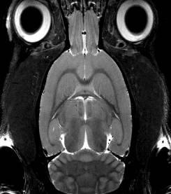

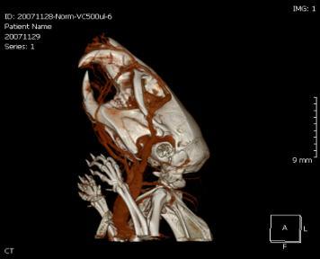
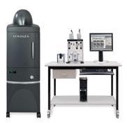
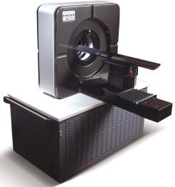
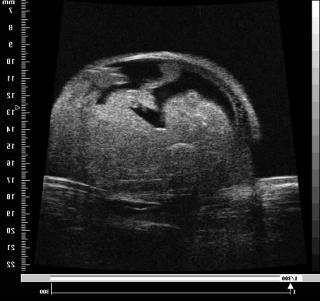
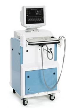
10 Micro US Micro MR In Vivo Animal Imaging Modalities Micro PET-CT Micro CT Optical Imaging Micro PET
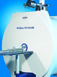
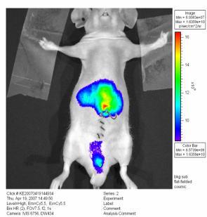
11 Commonly used 1. MR Imaging / Spectroscopy small animal imaging - at high field ( > 4.7T) human scanner field (1.5T ~ 3.0T) 2. PET Imaging 3. Optical Imaging bioluminescence imaging fluorescence imaging tomographic imaging.

12 Detection range of imaging modalities Things to be considered: 1. Spatial & temporal resolution 2. Depth 3. Sensitivity 4. Type of molecular probe 5. Perturbation of biological system

 fmri (functional")
13 Magnetic Resonance in Molecular Imaging MRI (magnetic resonance image) MRS (magnetic resonance spectroscopy) fmri (functional MRI)
14 Anatomic MR Gross morphology Specific target Metabolic MR Tissue functionality MRI/MRS/fMRI Functional MR Function Activity Molecular MR Target imaging Probe development

15 Gyromagnetic Ratio Nucleus Spin Quantum Number Gyromagnetic Ratio (MHz/1T) Relative Sensitivity at Constant Field Natural Abunda nce (%) 1 H 1/ C 1/ P 1/ Na 3/ F 1/ Li 3/ K 3/ ex: 1 H, 3T MHz, 1H, 11.7T 500 MHz
 is a relatively new procedure that uses MR imaging to measure the tiny")
.")
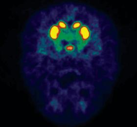
16 WHAT IS fmri? Functional magnetic resonance imaging (fmri) is a relatively new procedure that uses MR imaging to measure the tiny metabolic changes that take place in an active part of the brain (Ogawa, et al, 1990 a and b, 1992, 1993; Belliveau, et al, 1990, 1991). Functional MRI is based on the increase in blood flow to the local vasculature that accompanies neural activity in the brain. Since deoxyhemoglobin is paramagnetic, it alters the T2* weighted magnetic resonance image signal
17 In vivo magnetic resonance spectroscopy
18 In vivo MRS provide a noninvasive window into brain In vivo magnetic resonance spectroscopy (MRS) directly measures chemically specific information. It is the only noninvasive technique for measuring concentration of metabolites from the living brain. Our MRS development at 11.7 Tesla, the highest field strength at which in vivo MRS has ever been attempted, has allowed, for the first time, detection of GABA turnover in vivo. In addition to the static levels of metabolites and metabolic fluxes measured by proton or 13 C MRS, our recent discovery of 13 C magnetization (saturation) transfer effect of specific enzyme reactions has made it possible to probe the action of several enzymes in vivo, pointing to new directions in 13 C MRS technology development and applications.
19 In vivo evidence for reduced cortical glutamate-glutamine cycling in rats treated with the antidepressant/antipanic drug phenelzine Yang J and Shen J. Neuroscience 135: (2005) Metabolite a Group A Group B alanine ** 0.05 ± ± 0.38 aspartate 2.85 ± ± 0.30 creatine 3.39 ± ± 0.25 GABA c** 1.02 ± ± 0.26 glutamate d** ± ± 0.28 glutamine d** 5.17 ± ± 0.34 lactate 0.59 ± ± 0.24 myo-inositol 4.52 ± ± 0.33 N-acetylaspartate ± ± 0.40 phosphocreatine 5.11 ± ± 0.25 phosphorylethanola mine 2.31 ± ± 0.33 taurine 4.94 ± ± 0.17 Among 12 metabolites, Glu, Gln, GABA and Ala were the only metabolite which showed statistically significant changes. As a result, we became the first group in the world to detect turnover of the major inhibitory neurotransmitter GABA in the brain in vivo.
 13 C/ 1 H: 11.1 x 2.8, 24.2 x 3.")

20 Home-built surface radiofrequency transceiver coils 1 H: circular, diameter 15 mm 13 C: square, 25 x 25 mm 2 ) 13 C/ 1 H: 11.1 x 2.8, 24.2 x 3.2 mm (diameter x conductor width, respectively) RF coil integrated in-house-built animal handling system capable of rat head fixation, body support, physiology maintenance, coil tuning, and RF shieding.

21 Animal preparation femoral vein and artery cannulation Two femoral veins (left and right) were also cannulated for intravenous infusion of α- chloralose (initial dose: 80 mg/kg supplemented with a constant infusion of 26.7 mg/kg/hr throughout the experiment) and [1,6-13C2]glucose, respectively.


22 Animal preparation monitoring of physiological condition Arterial blood po2, pco2, mean blood pressure, and ph were maintained at approximately mm Hg, mm Hg, 150±30 mm Hg, and , respectively. Heart rate, end-tidal CO2, and tidal pressure of ventilation were monitored continuously.
23 Blood glucsoe (mm/l) Mean blood pressure (mmhg) ph pco2 (mmhg) The change of arterial ph, pco 2, glucose and mean blood pressure in each group Control PHA ACZ Time (min) Control PHA ACZ Time (min) Control PHA ACZ Control PHA ACZ Time (min) Time (min)
24 Elevated Endogenous GABA Concentration Attenuates Glutamate-Glutamine Cycling between Neurons and Astroglia J Neural Tansm 116: (2009) Jehoon Yang and Jun Shen Fig. 1. Comparison of in vivo 1 H short-te spectra from a control rat of Group I (bottom trace) and a Group III rat 24 hours after vigabatrin injection (top trace; 500 mg/kg, i.p., 24 hours prior to data acquisition). Voxel size = mm 3. Total number of scans = 128. lb = 3, gb = 0.1. In the 1 H short-te spectrum of vigabatrin-treated rat, the elevation of GABA α-methylene proton signal at 2.28 ppm, the GABA β-methylene proton signal at 1.91 ppm due to vigabatrin treatment were clearly observed.
25 Detection of reduced GABA synthesis following inhibition of GABA transaminase using in vivo magnetic resonance signal of [13C]GABA C1. J Neurosci Methods 182(2) 15: (2009) Jehoon Yang, Christopher J and Jun Shen
26
27 Fast isotopic exchange between mitochondria and cytosol in brain revealed by relayed 13 C magnetization transfer spectroscopy J Cereb Blood Flow Metab 29(4):661-9 (2009) Jehoon Yang, Su Xu and Jun Shen Fig. The relationship between the TCA cycle and V x used in brain metabolic models to describe the kinetics of 13 C label incorporation from mitochondrial TCA cycle intermediates into predominantly cytosolic glutamate and aspartate pools (adapted from Figure 43 in Siesjo (1978)). V x represents the lumped exchange between mitochondrial -ketoglutarate/oxaloacetate and cytosolic glutamate/aspartate.
28 Increased oxygen consumption in the somatosensory cortex of α-chloralose anesthetized rats during forepaw stimulation determined using MRS at 11.7 Tesla Neuroimage 32 (3) (2006) Jehoon Yang and Jun Shen Fig. Typical in vivo time course of proton-detected, 13C-edited spectra following intravenous infusion of [1, 6-13C2]glucose (TR/TE = 2000/22 ms, NS = 128 2, AQ = 196 ms, lb = 10, gb = 0.175). The [4-13C]glutamine signal at 2.46 ppm and the [2-13C]GABA signal at 2.30 ppm are spectrally resolved in vivo from the target [4-13C]glutamate signal at 2.35 ppm


29 All procedures were approved by the National Institute of Mental Health Animal Care and Use Committee and the physiological data should be recorded by certificated person.
![In vivo 13C magnetic resonance spectroscopy of human brain on a clinical 3 T scanner using [2-13C]glucose infusion and low-power stochastic decoupling.](/docs-images/94/119580780/images/30-2.jpg "Magnetic Resonance in Medicine 62(3): 565-73 (2009) Li S, Zhang Y, Wang S, Yang J, Ferraris Araneta M, Farris A, Johnson C, Fox S, Innis R, Shen J.")
30 In vivo 13C magnetic resonance spectroscopy of human brain on a clinical 3 T scanner using [2-13C]glucose infusion and low-power stochastic decoupling. Magnetic Resonance in Medicine 62(3): (2009) Li S, Zhang Y, Wang S, Yang J, Ferraris Araneta M, Farris A, Johnson C, Fox S, Innis R, Shen J. Time course spectra of glutamate, glutamine, and aspartate turnover detected in the occipital lobe during intravenous infusion of [2-3C]glucose.
31 Weaknesses of MRS Limited sensitivity = large voxels (nucleus dependent) = high metabolite concentration (millimolar) Limited specificity = multiple species in many resources = spectral overlap may be difficult to overcome Larger data acquisition windows =spectra are the sums of large numbers at averages =scans are expensive (several hundred dollars/hour) Special equipment may be required =software ( 1 H MRS) =hardware (RF coils, amplifiers, decouplers) Local expertise critical = automated methods variably reliable = multiple issues in data acquisition, analysis
32 Molecular imaging probe


33 Future drug Therapeutic drug Multimodality Imaging Imaging Drug

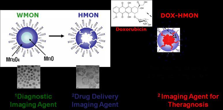
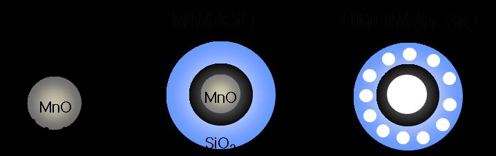
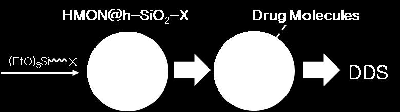
34 HMON as an MRI contrast agent HMON (Hollow Manganese Oxide Nanoparticles) Newly developed T1 contrast agent Gd 3+ or Mn 2+ containing colloidal nanoparticles : recently reported as potent T1 MRI contrast agents A nanometer-sized colloid particle with small size, large surface area & internal void spaces Large water accessible surface areas Able to carry high payloads of MR-active magnetic centers with an ability to take up a large amount of drug molecules within the internal void space Cetuximab-conjugated HMON

 Phospholipid ph4.")
 Mn3O4 WMON (Water")
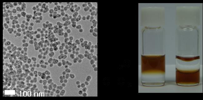
 HMON (dia : ~20nm,")
35 Synthesis of HMON Scheme MnCl 2 + Na-Oleate Manganese oleate Thermal decomposition (used schlenk technique) Phospholipid ph4.6 MON (Manganese Oxide Nanoparticle) Mn3O4 WMON (Water soluble MON) MnO HMON (Hollow MON) MON (~20nm) HMON (dia : ~20nm, core : ~10nm) HMON in water

36 In vivo molecular MR image after treatment with Cetuximab-conjugated HMON T1-Pre T1-30min T1-1hrs T1-3hrs T1-6hrs T1-24hrs T2-Pre T2-6hrs T2-24hrs
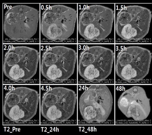
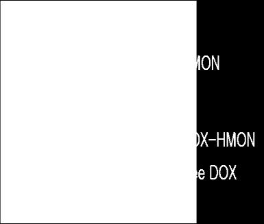

37 표적항암제전달경로및분포의영상화기술개발 약물담체용분자영상프로브개발 HMON-(Doxorubicin) 표적항암제용분자영상프로브개발 HMON-(small molecule drug) - sorafenib: 간암치료경구용표적항암제 1. Lee JH and Lee IS et al., Angew Chem Int Ed. (2007) 2. Lee JH and Lee IS et al., Angew Chem Int Ed. (2009) Work-in-progress 2010 년하반기보건의료연구개발사업 - 구두평가 4. 연구개발내용


38 Molecular imaging probe (ii) J Control Release Oct 10;155(1):11-7. Fig. 6. T 2 *-weighted MR imaging of the rat brain with MCAO treatment and enhanced by Fe 3 O 4 -PEG-PAEA10. The polymeric micelles were dissolved in the acidic area of the ischemic brain and the Fe 3 O 4 nanoparticles were accumulated over time (arrows).
 First described by Dr.")


39 Ongoing sudy 100 Years of Research on Alzheimer s Disease Alzheimer s Disease (AD) First described by Dr. Alois Alzheimer on November 3rd, 1906 in Tübingen, Germany The most common cause of dementia among people age 65 and older Progressive, irreversible decline in memory, loss of orientation and changes in personality and behavior No disease-modifying therapeutics available to date Page 39
40 Detection of reduced GABA synthesis following inhibition of GABA transaminase using in vivo magnetic resonance signal of [ 13 C]GABA C1 J Neurosci Methods (2009) 182(2): , Yang J, Johnson C and Shen J Lin AP et al, 2007
41 Prospective results Metabolite Wild type AD Drug evaluation Alanine 0.05± Aspartate 2.85± Creatine 3.39± GABA 1.02± Glutamate 10.22± Glutamine 5.17± Lactate 0.59± myo-inositol 4.52± N-acetylaspartate 10.50± Phosphocreatine 5.11± Phosphorylethanolamine 2.31± Taurine 4.94± Detection of potential biomarkers for early diagnosis of AD Evaluation of the effect of new developed drugs for AD

42 Thank you for your attention!
Report CAEN Category 1A: Visit by the Applicant to Another Laboratory
 Report CAEN Category 1A: Visit by the Applicant to Another Laboratory Visitor: Gustavo Ferreira, Federal University of Rio de Janeiro, Brazil Host: Prof. Mary C. McKenna, University of Maryland, Baltimore,
Emerging contrasts at ultrahigh fields" A. Dean Sherry
 Emerging contrasts at ultrahigh fields" A. Dean Sherry Advanced Imaging Research Center Department of Radiology UT Southwestern Medical Center Department of Chemistry & Biochemistry, UT Dallas ADVANCED
Magnetic Resonance Imaging. Alex MacKay University of British Columbia
 Magnetic Resonance Imaging Alex MacKay University of British Columbia Magnetic Resonance Imaging A) What is MRI? B) Why do MRI? C) What can we do with an MRI scanner? What is MRI? Magnetic Resonance Imaging
P2 Visual - Perception
 P2 Visual - Perception 2014 SOSE Neuroimaging of high-level visual functions gyula.kovacs@uni-jena.de 11/09/06 Functional magnetic resonance imaging (fmri) The very basics What is fmri? What is MRI? The
Introduction to the Course and the Techniques. Jeffry R. Alger, PhD Ahmanson-Lovelace Brain Mapping Center Department of Neurology
 Introduction to the Course and the Techniques Jeffry R. Alger, PhD Ahmanson-Lovelace Brain Mapping Center Department of Neurology (jralger@ucla.edu) CTSI Neuroimaging April 2014 Rationale for the Course
Biennial SPM course The BOLD signal. Cyril Pernet. Centre for Clinical Brain Sciences (CCBS) Neuroimaging Sciences
 Neuroimaging Sciences") Biennial SPM course 2017 The BOLD signal Cyril Pernet Centre for Clinical Brain Sciences (CCBS) Neuroimaging Sciences Overview 1. MRI physics 2. Neurovascular coupling 3. Neural activity and BOLD 4. Experimental
Biennial SPM course 2017 The BOLD signal Cyril Pernet Centre for Clinical Brain Sciences (CCBS) Neuroimaging Sciences Overview 1. MRI physics 2. Neurovascular coupling 3. Neural activity and BOLD 4. Experimental
Neurovascular Physiology and Pathophysiology
 Neurovascular Physiology and Pathophysiology The physiological questions aim at understanding the molecular and biochemical mechanisms, by which the brain adapts local blood flow to neuronal activity and
Metabonomics and MRS BCMB/CHEM 8190
 Metabonomics and MRS BCMB/CHEM 8190 Metabolomics, Metabonomics, Metabolic Profiling! Definition: The quantitative measurement of the dynamic multi parametric metabolic response of living systems to physiological
BOLD signal dependence on blood flow and metabolism. Outline
 BOLD signal dependence on blood flow and metabolism R. Hoge, MGH NMR Center Outline physiological events accompanying neuronal activation factors affecting BOLD signal sensitivity BOLD response dynamics
Concurrent near-infrared spectroscopy (NIRS) and functional magnetic resonance imaging (fmri) of the brain
 and functional magnetic resonance imaging (fmri) of the brain") Motor cortex activation fmri Near-infrared imaging Concurrent near-infrared spectroscopy (NIRS) and functional magnetic resonance imaging (fmri) of the brain Sergio Fantini s group, Department of Biomedical
Motor cortex activation fmri Near-infrared imaging Concurrent near-infrared spectroscopy (NIRS) and functional magnetic resonance imaging (fmri) of the brain Sergio Fantini s group, Department of Biomedical
Syllabus References. Resources. Video: MRI Introduction
 MRI Lesson Outline Syllabus References 9.6.4.2.5 Define precessing and relate the frequency of the precessing to the composition of the nuclei and the strength of the applied external magnetic field 9.6.4.2.6
neurotransmitter cycling in humans
 Special Issue Review Article Received: 10 December 2010, Revised: 9 June 2011, Accepted: 14 June 2011, Published online in Wiley Online Library: 2011 (wileyonlinelibrary.com) DOI: 10.1002/nbm.1772 13 C
Molecular Imaging and the Brain
 Molecular imaging technologies are playing an important role in neuroimaging, a branch of medical imaging, by providing a window into the living brain. Where CT and conventional MR imaging provide important
Outline. Why Image Animals?
 Small Animal Magnetic Resonance Imaging: Current Trends, Challenges and Perspectives for Pathological Imaging C. Chad Quarles Vanderbilt University Institute of Imaging Science Outline Why image animals?
Technology Summary: Dansyl Molecular Probe
 Opportunity Statement Cell apoptosis refers to the active death process of cells controlled by genes for multi-cellular organisms to regulate development and maintain homeostasis. It is one of the main
Increased tricarboxylic acid cycle flux in rat brain during forepaw stimulation detected with 1 H[ 13 C] NMR
![Increased tricarboxylic acid cycle flux in rat brain during forepaw stimulation detected with 1 H[ 13 C] NMR](/thumbs/73/69489900.jpg "Increased tricarboxylic acid cycle flux in rat brain during forepaw stimulation detected with 1 H[ 13 C] NMR") Proc. Natl. Acad. Sci. USA Vol. 93, pp. 7612 7617, July 1996 Biophysics Increased tricarboxylic acid cycle flux in rat brain during forepaw stimulation detected with 1 H[ 13 C] NMR FAHMEED HYDER*, JENNIFER
Proc. Natl. Acad. Sci. USA Vol. 93, pp. 7612 7617, July 1996 Biophysics Increased tricarboxylic acid cycle flux in rat brain during forepaw stimulation detected with 1 H[ 13 C] NMR FAHMEED HYDER*, JENNIFER
fmri: Interpretation, Limits and Potential Pitfalls
 fmri: Interpretation, Limits and Potential Pitfalls Seong-Gi Kim kimsg@pitt.edu www.kimlab.pitt.edu Mapping Brain Functions Stimulation/Task Functional Map (MRI) Pre-synaptic activity Post-synaptic activity
Neuroimaging. BIE601 Advanced Biological Engineering Dr. Boonserm Kaewkamnerdpong Biological Engineering Program, KMUTT. Human Brain Mapping
 11/8/2013 Neuroimaging N i i BIE601 Advanced Biological Engineering Dr. Boonserm Kaewkamnerdpong Biological Engineering Program, KMUTT 2 Human Brain Mapping H Human m n brain br in m mapping ppin can nb
Intrinsic Signal Optical Imaging
 Intrinsic Signal Optical Imaging Introduction Intrinsic signal optical imaging (ISOI) is a technique used to map dynamics in single cells, brain slices and even and most importantly entire mammalian brains.
MRI - functional MRI, spectroscopy, etc. Lecture 24
 MRI - functional MRI, spectroscopy, etc Lecture 24 Functional MRI: Principles Magnetic susceptibility χ: M = χ H M is the magnetization of the material, H is the strength of the external magnetic field
Importance of X-Nuclei for Broadening Uses of Ultrahigh Field MR Imaging in Humans
 UHF Funding Conference 2015, NIH, Washington DC Importance of X-Nuclei for Broadening Uses of Ultrahigh Field MR Imaging in Humans Keith R. Thulborn, MD, PhD Professor of Radiology, Physiology & Biophysics
University of Groningen. Neuro-imaging of visual field defects Boucard, Christine
 University of Groningen Neuro-imaging of visual field defects Boucard, Christine IMPORTANT NOTE: You are advised to consult the publisher's version (publisher's PDF) if you wish to cite from it. Please
Biophysical and physiological bases of fmri signals: challenges of interpretation and methodological concerns
 Biophysical and physiological bases of fmri signals: challenges of interpretation and methodological concerns Antonio Ferretti aferretti@itab.unich.it Institute for Advanced Biomedical Technologies, University
Non-Invasive Techniques
 Non-Invasive Techniques Key: Does not hurt the organism Psychology 372 Physiological Psychology Steven E. Meier, Ph.D. Listen to the audio lecture while viewing these slides or view the video presentation
Non-Invasive Techniques
 Many Procedures Non-Invasive Techniques Key: Does not hurt the organism Psychology 372 Physiological Psychology Steven E. Meier, Ph.D. Listen to the audio lecture while viewing these slides or view the
Cerebral Glucose Is Detectable by Localized Proton NMR Spectroscopy in Normal Rat Brain in Vivo
 MAGNETIC RESONANCE IN MEDICINE 19,489-495 ( 1991 ) Cerebral Is Detectable by Localized Proton NMR Spectroscopy in Normal Rat Brain in Vivo M. L. GYNGELL, T. MICHAELIS, D. H~RSTERMANN, H. BRUHN, W. HANICKE,
1) Diffusion weighted imaging DWI is a term used to describe moving molecules due to random thermal motion. This motion is restricted by boundaries
 Diffusion weighted imaging DWI is a term used to describe moving molecules due to random thermal motion. This motion is restricted by boundaries") 1) Diffusion weighted imaging DWI is a term used to describe moving molecules due to random thermal motion. This motion is restricted by boundaries such as ligaments, membranes and macro molecules. Diffusion
1) Diffusion weighted imaging DWI is a term used to describe moving molecules due to random thermal motion. This motion is restricted by boundaries such as ligaments, membranes and macro molecules. Diffusion
Physiological and Physical Basis of Functional Brain Imaging 6. EEG/MEG. Kâmil Uludağ, 20. November 2007
 Physiological and Physical Basis of Functional Brain Imaging 6. EEG/MEG Kâmil Uludağ, 20. November 2007 Course schedule 1. Overview 2. fmri (Spin dynamics, Image formation) 3. fmri (physiology) 4. fmri
RECENT ADVANCES IN CLINICAL MR OF ARTICULAR CARTILAGE
 In Practice RECENT ADVANCES IN CLINICAL MR OF ARTICULAR CARTILAGE By Atsuya Watanabe, MD, PhD, Director, Advanced Diagnostic Imaging Center and Associate Professor, Department of Orthopedic Surgery, Teikyo
Daniel Bulte. Centre for Functional Magnetic Resonance Imaging of the Brain. University of Oxford
 Daniel Bulte Centre for Functional Magnetic Resonance Imaging of the Brain University of Oxford Overview Signal Sources BOLD Contrast Mechanism of MR signal change FMRI Modelling Scan design details Factors
Titelmaster The physics of functional magnetic resonance imaging (fmri)
") Titelmaster The physics of functional magnetic resonance imaging (fmri) Outline 1.Introduction 2.The fmri experiment 2 3.The physics basis of fmri 4.Application Outline 3 1.Introduction Introduction Phrenology
Titelmaster The physics of functional magnetic resonance imaging (fmri) Outline 1.Introduction 2.The fmri experiment 2 3.The physics basis of fmri 4.Application Outline 3 1.Introduction Introduction Phrenology
Molecular Imaging and Cancer
 Molecular Imaging and Cancer Cancer causes one in every four deaths in the United States, second only to heart disease. According to the U.S. Department of Health and Human Services, more than 512,000
LESSON 1.3 WORKBOOK. How can we study the behaving brain?
 LESSON 1.3 WORKBOOK How can we study the behaving brain? We are in the middle of a technological revolution when it comes to how closely we can look at the behaving brain. Scientists and doctors now have
Introduction to Brain Imaging
 Introduction to Brain Imaging Human Brain Imaging NEUR 570 & BIC lecture series September 9, 2013 Petra Schweinhardt, MD PhD Montreal Neurological Institute McGill University Montreal, Canada Various techniques
Metabolites in Proton Spectroscopy of the Brain: Neurochemistry and Physiology
 Metabolites in Proton Spectroscopy of the Brain: Neurochemistry and Physiology Josef Pfeuffer Max-Planck Institute for Biological Cybernetics, Department of Neurophysiology, Tübingen, Germany josef.pfeuffer@tuebingen.mpg.de
Laurent Itti: CS564 Brain Theory and Artificial Intelligence. Lecture 4: Experimental techniques in visual neuroscience. Reading Assignments: None!
 CS 564 Brain Theory and Artificial Intelligence Lecture 4: Experimental techniques in visual neuroscience Reading Assignments: None! 1 Today we will briefly review - electrophysiological recording and
Multimodality Imaging in Cardiac Stem Cell Research
 Multimodality Imaging in Cardiac Stem Cell Research IL SUK SOHN, MD, PhD Department of Cardiology Kyung Hee University Hospital at Gangdong Kyung Hee University School of Medicine, Seoul, Korea Stem Cell
Table 1. Summary of PET and fmri Methods. What is imaged PET fmri BOLD (T2*) Regional brain activation. Blood flow ( 15 O) Arterial spin tagging (AST)
 Regional brain activation. Blood flow ( 15 O) Arterial spin tagging (AST)") Table 1 Summary of PET and fmri Methods What is imaged PET fmri Brain structure Regional brain activation Anatomical connectivity Receptor binding and regional chemical distribution Blood flow ( 15 O)
Table 1 Summary of PET and fmri Methods What is imaged PET fmri Brain structure Regional brain activation Anatomical connectivity Receptor binding and regional chemical distribution Blood flow ( 15 O)
Electronic Supplementary Information
 Electronic Supplementary Material (ESI) for Nanoscale. This journal is The Royal Society of Chemistry 2016 Electronic Supplementary Information MnO 2 -induced synthesis of fluorescent polydopamine nanparticles
COGNITIVE SCIENCE 17. Peeking Inside The Head. Part 1. Jaime A. Pineda, Ph.D.
 COGNITIVE SCIENCE 17 Peeking Inside The Head Part 1 Jaime A. Pineda, Ph.D. Imaging The Living Brain! Computed Tomography (CT)! Magnetic Resonance Imaging (MRI)! Positron Emission Tomography (PET)! Functional
NIH Public Access Author Manuscript NMR Biomed. Author manuscript; available in PMC 2011 August 23.
 NIH Public Access Author Manuscript Published in final edited form as: NMR Biomed. 2010 October ; 23(8): 977 985. doi:10.1002/nbm.1524. 13C MRS of occipital and frontal lobes at 3 T using a volume coil
Cover Page. The handle holds various files of this Leiden University dissertation.
 Cover Page The handle http://hdl.handle.net/1887/35124 holds various files of this Leiden University dissertation. Author: Wokke, Beatrijs Henriette Aleid Title: Muscle MRI in Duchenne and Becker muscular
Functional aspects of anatomical imaging techniques
 Functional aspects of anatomical imaging techniques Nilendu Purandare Associate Professor & Consultant Radiologist Tata Memorial Centre Functional/metabolic/molecular imaging (radioisotope scanning) PET
Newborn Hypoxic Ischemic Brain Injury. Hisham Dahmoush, MBBCh FRCR Lucile Packard Children s Hospital at Stanford
 Newborn Hypoxic Ischemic Brain Injury Hisham Dahmoush, MBBCh FRCR Lucile Packard Children s Hospital at Stanford NO DISCLOSURES INTRODUCTION Neonatal hypoxic-ischemic encephalopathy (HIE) is a major cause
PHYSICS OF MRI ACQUISITION. Alternatives to BOLD for fmri
 PHYSICS OF MRI ACQUISITION Quick Review for fmri HST-583, Fall 2002 HST.583: Functional Magnetic Resonance Imaging: Data Acquisition and Analysis Harvard-MIT Division of Health Sciences and Technology
Intra-renal Oxygenation. in Human Subjects
 MRI-based Mapping of Intra-renal Oxygenation BOLD in Human Subjects OEF Xiang He, PhD Department of Radiology Background Cortex Brain CBF ~ 1.0 ml/min/g Brain PO 2 ~ 25-35 mm Hg Medullary hypoxia is an
Deakin Research Online
 Deakin Research Online This is the published version: Silberstein, Morry, Lane, Dianne, Dodd, Seetal and Opeskin, Kenneth 2002, Identification of a by-product of nitric oxide synthase activity in human
Lactate Chemical Exchange Saturation Transfer (LATEST) Imaging in vivo: A. Biomarker for LDH Activity
 Imaging in vivo: A. Biomarker for LDH Activity") Lactate Chemical Exchange Saturation Transfer (LATEST) Imaging in vivo: A Biomarker for LDH Activity Catherine DeBrosse 1, Ravi Prakash Reddy Nanga 1, Puneet Bagga 1, Kavindra Nath 2, Mohammad Haris 3,
Lactate Chemical Exchange Saturation Transfer (LATEST) Imaging in vivo: A Biomarker for LDH Activity Catherine DeBrosse 1, Ravi Prakash Reddy Nanga 1, Puneet Bagga 1, Kavindra Nath 2, Mohammad Haris 3,
Problem Set #8 Rad 226
 Problem Set #8 1. J-editing. In class, we discussed J-editing for the doublet resonance of lactate. Other in vivo peaks (e.g. GABA) are more complicated (triplets, quartets, etc.). In this problem, we
Control vs. HFD-lipid
 Animals Control vs. T1D vs. T1D-leptin and Hyperinsulinemic-diabetic vs. hyperinsulinemic-diabetic-leptin STZ ± nicotinamide injection Leptin or saline 6 8 1 12 2 4 6 8 1 12 2 4 6 8 1 12 Control vs. HFD-lipid
APPLICATIONS OF ASL IN NEUROSCIENCE
 APPLICATIONS OF ASL IN NEUROSCIENCE Luis Hernandez-Garcia, Ph.D. Functional MRI laboratory University of Michigan 1 OVERVIEW Quick review of ASL The niche for ASL Examples of practical applications in
Brain Energy State and Lactate Metabolism during Status Epilepticus in the Neonatal Dog: Resonance Study1
 303 1-399819 112902-0 19 1 $03.00/0 PEDIATRIC RESEARCH Copyright 0 199 1 International Pediatric Research Foundation, Inc Vol. 29, No. 2, 199 1 Printed in U. S. A. Brain Energy State and Lactate Metabolism
High-field MR imaging systems such as 3T strength aid in
 TECHNICAL NOTE H.-S. Liu H.-W. Chung C.-J. Juan S.-Y. Tsai C.-Y. Wang C.-C. Chan G.-S. Huang M.-C. Chou C.-S. Lee C.-W. Ko N.-Y. Cho C.-Y. Chen Anomalous J-Modulation Effects on Amino Acids in Clinical
controls. <Conclusions> These data support the hypothesis that JME and FLE involve neuronal dysfunction within the temporal lobe as well as the
 A single-voxel spectroscopy study of hippocampal metabolic dysfunction in patients with juvenile myoclonic epilepsy, frontal lobe epilepsy, and psychogenic nonepileptic seizures Epilepsy Center, National
Outline. Biological Psychology: Research Methods. Dr. Katherine Mickley Steinmetz
 Biological Psychology: Research Methods Dr. Katherine Mickley Steinmetz Outline Neuroscience Methods Histology Electrophysiological Recordings Lesion Neuroimaging Neuroanatomy Histology: Brain structure
The neurolinguistic toolbox Jonathan R. Brennan. Introduction to Neurolinguistics, LSA2017 1
 The neurolinguistic toolbox Jonathan R. Brennan Introduction to Neurolinguistics, LSA2017 1 Psycholinguistics / Neurolinguistics Happy Hour!!! Tuesdays 7/11, 7/18, 7/25 5:30-6:30 PM @ the Boone Center
ASL Perfusion Imaging: Concepts and Applications
 ASL Perfusion Imaging: Concepts and Applications David C. Alsop, Ph.D. Beth Israel Deaconess Medical Center and Harvard Medical School, Boston USA INTRODUCTION Arterial Spin Labeling (ASL) perfusion imaging
Introduction to Functional MRI
 Introduction to Functional MRI Douglas C. Noll Department of Biomedical Engineering Functional MRI Laboratory University of Michigan Outline Brief overview of physiology and physics of BOLD fmri Background
In vivo optical imaging : revealing endogeneous optical contrast at depth
 In vivo optical imaging : revealing endogeneous optical contrast at depth Anne PLANAT-CHRÉTIEN, Jean-Marc DINTEN CEA-LETI, MINATEC, Grenoble Jérôme GATEAU Université Paris Descartes, Paris 1 Why using
Advances in Clinical Neuroimaging
 Advances in Clinical Neuroimaging Joseph I. Tracy 1, PhD, ABPP/CN; Gaelle Doucet 2, PhD; Xaiosong He 2, PhD; Dorian Pustina 2, PhD; Karol Osipowicz 2, PhD 1 Department of Radiology, Thomas Jefferson University,
BioMedical quantitative X-Ray Imaging. Emmanuel Brun Researcher Inserm Université Grenoble Alpes
 BioMedical quantitative X-Ray Imaging Emmanuel Brun Researcher Inserm Université Grenoble Alpes 1 Outline Introduction K-Edge Imaging Patient imaging at the European synchrotron Medical Phase Contrast
Supplementary Online Content
 Supplementary Online Content Gregg NM, Kim AE, Gurol ME, et al. Incidental cerebral microbleeds and cerebral blood flow in elderly individuals. JAMA Neurol. Published online July 13, 2015. doi:10.1001/jamaneurol.2015.1359.
The Effect of Zn2+ Binding on the Chemistry of Tm3+ and Eu3+ Chelates
 Portland State University PDXScholar University Honors Theses University Honors College 3-1-2018 The Effect of Zn2+ Binding on the Chemistry of Tm3+ and Eu3+ Chelates Diana King Portland State University
MRI and CT of the CNS
 MRI and CT of the CNS Dr.Maha ELBeltagy Assistant Professor of Anatomy Faculty of Medicine The University of Jordan 2018 Computed Tomography CT is used for the detection of intracranial lesions. CT relies
Contact: Course outline: Contact for other times.
 Contact: kdelaney@uvic.ca Course outline: http://web.uvic.ca/~kdelaney/b367 Scheduled office hours: 1:00-3:00, M&Th Cunn. 259A Contact kdelaney@uvic.ca for other times. Quiz (0.5 hrs) midterm (1.4 hrs)
Brain and Cognition. Cognitive Neuroscience. If the brain were simple enough to understand, we would be too stupid to understand it
 Brain and Cognition Cognitive Neuroscience If the brain were simple enough to understand, we would be too stupid to understand it 1 The Chemical Synapse 2 Chemical Neurotransmission At rest, the synapse
Hybrid Nanomaterials for Biomedical Imaging and Cancer Therapy Wenbin Lin
 Hybrid anomaterials for Biomedical Imaging and Cancer Therapy Wenbin Lin Department of Chemistry University of orth Carolina Chapel Hill, C 27599 wlin@unc.edu Magnetic Resonance Imaging Precontrast Post-contrast
Final Report. Title of Project: Quantifying and measuring cortical reorganisation and excitability with post-stroke Wii-based Movement Therapy
 Final Report Author: Dr Penelope McNulty Qualification: PhD Institution: Neuroscience Research Australia Date: 26 th August, 2015 Title of Project: Quantifying and measuring cortical reorganisation and
Positron Emission Tomography: Tool to Facilitate Drug Development and to Study Pharmacokinetics
 Positron Emission Tomography: Tool to Facilitate Drug Development and to Study Pharmacokinetics Robert B. Innis, MD, PhD Molecular Imaging Branch National Institute Mental Health 1 Outline of Talk 1. PET
PHY3111 Mid-Semester Test Study. Lecture 2: The hierarchical organisation of vision
 PHY3111 Mid-Semester Test Study Lecture 2: The hierarchical organisation of vision 1. Explain what a hierarchically organised neural system is, in terms of physiological response properties of its neurones.
Molecular Imaging and Breast Cancer
 Molecular Imaging and Breast Cancer Breast cancer forms in tissues of the breast usually in the ducts, tubes that carry milk to the nipple, and lobules, the glands that make milk. It occurs in both men
Effects of Brain Region and Gender on Proton Magnetic Resonance Spectroscopy in Normal Subjects
 Yale University EliScholar A Digital Platform for Scholarly Publishing at Yale Yale Medicine Thesis Digital Library School of Medicine 12-13-2002 Effects of Brain Region and Gender on Proton Magnetic Resonance
Clinical application of 3.0 T proton MR spectroscopy in evaluation of pancreatic diseases
 Clinical application of 3.0 T proton MR spectroscopy in evaluation of pancreatic diseases Award: Cum Laude Poster No.: C-1762 Congress: ECR 2012 Type: Scientific Paper Authors: T. Su, E. Jin; Beijing/CN
Methods of Visualizing the Living Human Brain
 Methods of Visualizing the Living Human Brain! Contrast X-rays! Computerized Tomography (CT)! Magnetic Resonance Imaging (MRI)! Positron Emission Tomography (PET)! Functional MRI! Magnetoencephalography
Comparison of 1.5T and 3T 1 H MR Spectroscopy for Human Brain Tumors
 Comparison of 1.5T and 3T 1 H MR Spectroscopy for Human Brain Tumors Ji-hoon Kim, MD 1 Kee-Hyun Chang, MD 2-4 Dong Gyu Na, MD 2 In Chan Song, PhD 2,3 Seung Ja Kim, MD 2 Bae Ju Kwon, MD 2 Moon Hee Han,
Boron-gadolinium binary system as a magnetic resonance imaging boron carrier*
 Pure Appl. Chem., Vol. 75, No. 9, pp. 1343 1348, 2003. 2003 IUPAC Boron-gadolinium binary system as a magnetic resonance imaging boron carrier* Yoshinori Yamamoto Department of Chemistry, Graduate School
Clinically Available Optical Topography System
 Clinically Available Optical Topography System Clinically Available Optical Topography System 18 Fumio Kawaguchi Noriyoshi Ichikawa Noriyuki Fujiwara Yûichi Yamashita Shingo Kawasaki OVERVIEW: Progress
Laura Tormoehlen, M.D. Neurology and EM-Toxicology Indiana University
 Laura Tormoehlen, M.D. Neurology and EM-Toxicology Indiana University Disclosures! No conflicts of interest to disclose Neuroimaging 101! Plain films! Computed tomography " Angiography " Perfusion! Magnetic
MRI qbold Based Evaluation. Renal Oxidative Metabolism. Department of Radiology and Hernando Gomez, MD Critical Care Medicine
 MRI qbold Based Evaluation of Renal Oxidative Metabolism Xiang He, PhD Department of Radiology and Hernando Gomez, MD Critical Care Medicine Background High oxygen-demand and lower medullary blood flow
Multimodal Spectroscopic Tissue Scanner for diagnosis of ex vivo surgical specimens
 Multimodal Spectroscopic Tissue Scanner for diagnosis of ex vivo surgical specimens Collaborating researcher: Dr. Maryann Fitzmaurice (Case Western Reserve University, Cleveland, OH), Dr. Tulio Valdez
Magnetic Resonance Imaging on Soft Tissue. Jiten K. Mistry Calvin Gan
 Magnetic Resonance Imaging on Soft Tissue 1 Jiten K. Mistry Calvin Gan Outline Background of Medical Imaging Introduction to MRI How MRI works MRI of Soft Tissue Benefits & Risks Recent Advances 2 The
Essentials of Clinical MR, 2 nd edition. 99. MRA Principles and Carotid MRA
 99. MRA Principles and Carotid MRA As described in Chapter 12, time of flight (TOF) magnetic resonance angiography (MRA) is commonly utilized in the evaluation of the circle of Willis. TOF MRA allows depiction
Fig. 1. Localized single voxel proton MR spectroscopy was performed along the long axis of right hippocampus after extension of patient s head to
 125 A B C Fig. 1. Localized single voxel proton MR spectroscopy was performed along the long axis of right hippocampus after extension of patient s head to obtain entire dimension of the hippocampal body.
Turbo ASL: Arterial Spin Labeling With Higher SNR and Temporal Resolution
 COMMUNICATIONS Magnetic Resonance in Medicine 44:511 515 (2000) Turbo ASL: Arterial Spin Labeling With Higher SNR and Temporal Resolution Eric C. Wong,* Wen-Ming Luh, and Thomas T. Liu A modified pulsed
Advanced multimodal imaging in malformations of cortical development
 Advanced multimodal imaging in malformations of cortical development Seok Jun Hong (sjhong@bic.mni.mcgill.ca) NOEL Neuroimaging of Epilepsy Lab MICA Multimodal Imaging and Connectome Analysis Lab w4 w5
Positron Emission Tomography: Tool to Facilitate Drug Development and to Study Pharmacokinetics
 Positron Emission Tomography: Tool to Facilitate Drug Development and to Study Pharmacokinetics Robert B. Innis, MD, PhD Molecular Imaging Branch National Institute Mental Health 1 Outline of Talk 1. PET
Non-Invasive MR-based Evaluation of Kidney Function without Exogenous Contrast Agent. Xiang He, PhD Department of Radiology University of Pittsburgh
 Non-Invasive MR-based Evaluation of Kidney Function without Exogenous Contrast Agent Xiang He, PhD Department of Radiology University of Pittsburgh Contents MR-based non-invasive estimation of single kidney
The Central Nervous System
 The Central Nervous System Cellular Basis. Neural Communication. Major Structures. Principles & Methods. Principles of Neural Organization Big Question #1: Representation. How is the external world coded
Name: Period: Chapter 2 Reading Guide The Biology of Mind
 Name: Period: Chapter 2 Reading Guide The Biology of Mind The Nervous System (pp. 55-58) 1. What are nerves? 2. Complete the diagram below with definitions of each part of the nervous system. Nervous System
METABOLITE CHARACTERIZATION IN SERUM SAMPLES FROM NORMAL HEALTHY HUMAN SUBJECTS BY 1 H AND 13 C NMR SPECTROSCOPY. Divya Misra * and Usha Bajpai
 , 211-221. ISSN 1011-3924 Printed in Ethiopia 2009 Chemical Society of Ethiopia METABOLITE CHARACTERIZATION IN SERUM SAMPLES FROM NORMAL HEALTHY HUMAN SUBJECTS BY 1 H AND 13 C NMR SPECTROSCOPY Divya Misra
Experimental Assessment of Infarct Lesion Growth in Mice using Time-Resolved T2* MR Image Sequences
 Experimental Assessment of Infarct Lesion Growth in Mice using Time-Resolved T2* MR Image Sequences Nils Daniel Forkert 1, Dennis Säring 1, Andrea Eisenbeis 2, Frank Leypoldt 3, Jens Fiehler 2, Heinz Handels
The effect of diets on neurodegenerative diseases
 The effect of diets on neurodegenerative diseases Results from the FP7 project LIPIDIDIET A Kiliaan Donders Institute for Brain, Cognition and Behaviour, Centre for Neuroscience Dept Anatomy and dept Cognitive
Organization of the nervous system. [See Fig. 48.1]
![Organization of the nervous system. [See Fig. 48.1]](/thumbs/90/103926552.jpg "Organization of the nervous system. [See Fig. 48.1]") Nervous System [Note: This is the text version of this lecture file. To make the lecture notes downloadable over a slow connection (e.g. modem) the figures have been replaced with figure numbers as found
Nervous System [Note: This is the text version of this lecture file. To make the lecture notes downloadable over a slow connection (e.g. modem) the figures have been replaced with figure numbers as found
Jana Novotná, Bruno Sopko. Department of the Medical Chemistry and Clinical Biochemistry The 2nd Faculty of Medicine, Charles Univ.
 Amino acid metabolism II. Urea cycle Jana Novotná, Bruno Sopko Department of the Medical Chemistry and Clinical Biochemistry The 2nd Faculty of Medicine, Charles Univ. Nitrogen balance Tissue proteins
Perfusion MRI. Youngkyoo Jung, PhD Associate Professor Radiology, Biomedical Engineering, and Clinical & Translational Science Institute
 Perfusion MRI Youngkyoo Jung, PhD Associate Professor Radiology, Biomedical Engineering, and Clinical & Translational Science Institute Perfusion The delivery of blood to a capillary bed in tissue Perfusion
INTRO TO BOLD FMRI FRANZ JOSEPH GALL ( ) OUTLINE. MRI & Fast MRI Observations Models Statistical Detection
 OUTLINE. MRI & Fast MRI Observations Models Statistical Detection") INTRO TO BOLD FMRI 2014 M.S. Cohen all rights reserved mscohen@g.ucla.edu OUTLINE FRANZ JOSEPH GALL (1758-1828) MRI & Fast MRI Observations Models Statistical Detection PAUL BROCA (1824-1880) WILLIAM JAMES
INTRO TO BOLD FMRI 2014 M.S. Cohen all rights reserved mscohen@g.ucla.edu OUTLINE FRANZ JOSEPH GALL (1758-1828) MRI & Fast MRI Observations Models Statistical Detection PAUL BROCA (1824-1880) WILLIAM JAMES
University of Groningen. Biomarkers in premanifest Huntington's disease van Oostrom, Joost Cornelis Hendricus
 University of Groningen Biomarkers in premanifest Huntington's disease van Oostrom, Joost Cornelis Hendricus IMPORTANT NOTE: You are advised to consult the publisher's version (publisher's PDF) if you
Case reports functional imaging in epilepsy
 Seizure 2001; 10: 157 161 doi:10.1053/seiz.2001.0552, available online at http://www.idealibrary.com on Case reports functional imaging in epilepsy MARK P. RICHARDSON Medical Research Council Fellow, Institute
Principles of Haemodynamic Coupling for fmri
 Principles of Haemodynamic Coupling for fmri Paul M. Matthews Head, Global Imaging Unit, GlaxoSmithKline and Professor of Clinical Neurosciences, Imperial College paul.m.matthews@gsk.com Regulation of