Anatomy Skull and Spinal Cord
|
|
|
- David Whitehead
- 6 years ago
- Views:
Transcription
1 1 Anatomy Skull and Spinal Cord 1. Skull The skull is anterior to the spinal column and is the bony structure that encases the brain. Its purpose is to protect the brain and allow attachments for the facial muscles. The two regions of the skull are the cranial and facial region. The cranial portion is the part of the skull that directly houses the brain and the facial portion includes the rest of the bones of the skull. The skull is supported on the summit of the vertebral column, and is of an oval shape, wider behind than in front. It is composed of a series of flattened or irregular bones which, with one exception (the mandible), are immovably jointed together. It is divisible into two parts: (1) the cranium, which lodges and protects the brain, consists of eight bones, and (2) the skeleton of the face, of fourteen, as follows: Skull, 22 bones Cranium, 8 bones - Occipital. - Two Parietals. - Frontal. - Two Temporals - Sphenoidal - Ethmoida Face, 14 bones - Two Nasals - Two Maxillæ - Two Lacrimals - Two Zygomatics - Two Palatines. - Two Inferior Nasal Conchæ - Vomer - Mandible

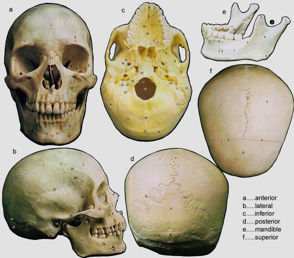
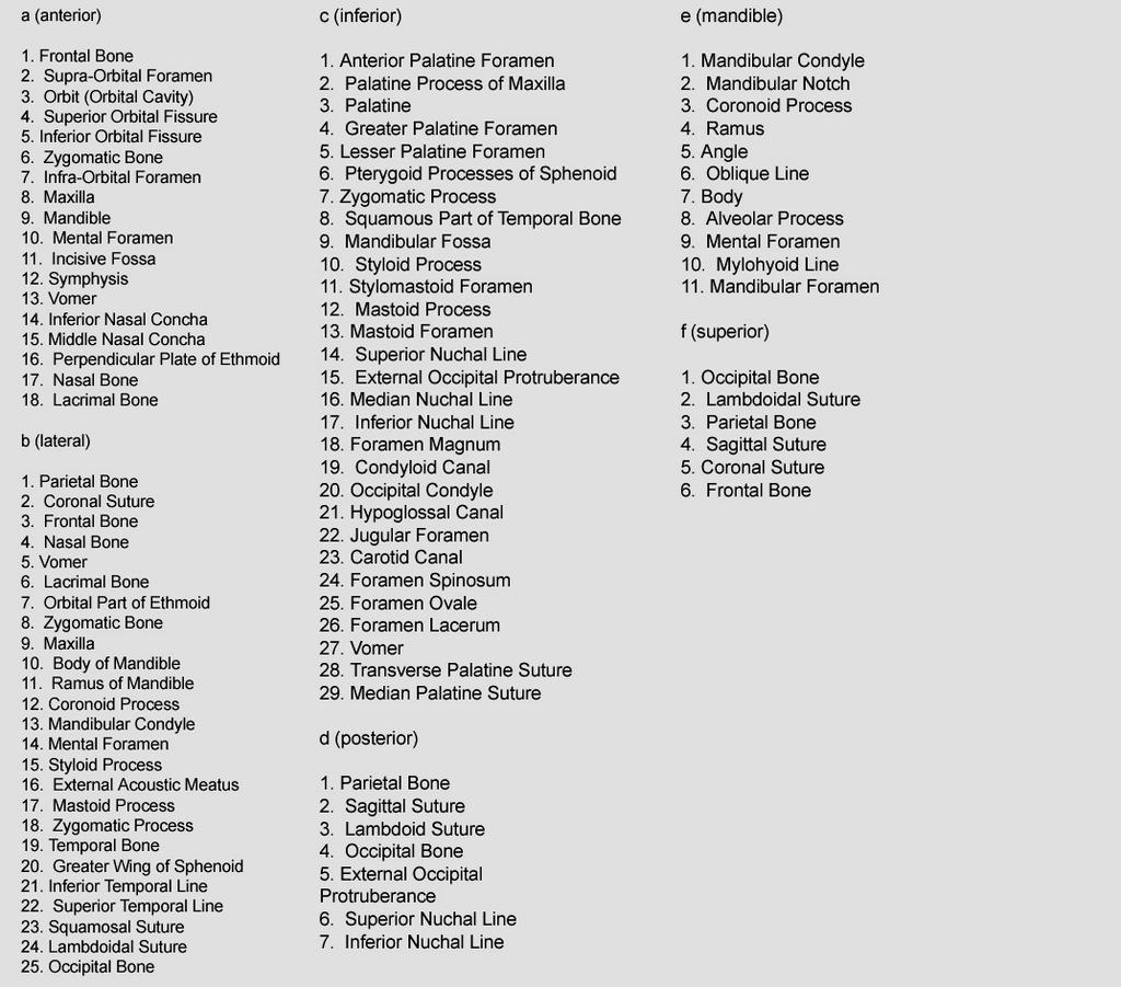
2 2 SKULL 1. Frontal Bone 2. Supra-Orbital Foramen 3. Orbit (Orbital Cavity) 4. Superior Orbital Fissure 5. Inferior Orbital Fissure 6. Zygomatic Bone 7. Infra-Orbital Foramen 8. Maxilla 9. Mandible 10. Mental Foramen 11. Incisive Fossa 12. Symphysis 13. Vomer 14. Inferior Nasal Concha 15. Middle Nasal Concha 16. Perpendicular Plate of Ethmoid 17. Nasal Bone 18. Lacrimal Bone
3 3
4 4
5 5

6 6 1. Parietal Bone 2. Temporal Bone 3. Occipital Bone 4. Mandible Maxilla 5. Frontal Bone Sphenoid 6. Bone zygomatic bone A. Frontal bone During development there are two frontal bones which fuse after birth. This bone forms the ridges above the orbits and the roof of the orbit. Branches of the ophthalmic division of the trigeminal nerve emerge from the orbit to innervate the skin of the forehead (supra-orbital and supra-trochlear nerves). The supra-orbital nerve lies in the supra-orbital notch of the frontal bone. B. Parietal bone
.")
7 7 The paired parietal bones form the top and sides of the skull. The suture between them is the sagittal suture. The suture between the pair of parietal bones and the frontal bone is the coronal suture. On the outside the bones carry large muscles which elevate the mandible during chewing (temporalis muscles). On the inside, running along the sagittal suture, lies the superior sagittal sinus, a major route of venous drainage from the brain. At birth there is a soft spot where the sagittal suture meets the coronal suture, the anterior fontanelle. In the adult this fills in and the point is then termed the bregma.

8 8 C. Temporal bone The temporal bone has a vertical squamous part forming part of the side of the skull, and a horizontal base, the petrous temporal bone. On the exterior the temporal bone has an opening which leads into the middle ear, the external auditory meatus. The ear is attached at this point. A process runs forward forming part of the cheek bone, the zygomatic process of the temporal bone. Projecting inferiorly from behind the external auditory meatus is the mastoid process. The mastoid process is a spongy bone containing air cells. The drawing out of the mastoid process is related to the insertion of the sternocleidomastoid muscle, responsible for rotation of the head to the opposite side. Within the substance of the petrous temporal bone lie the components of the inner ear, the cochlear and vestibular systems. On the inside of the squamous part run branches of the middle meningeal artery which may be ruptured in bone fractures. The sigmoid sinus, a continuation of the transverse sinus lies in a groove on the inside of the bone. The head of the mandible articulates with the temporal bone forming the temporomandibular joint (TMJ). Anterior to the joint is the articular tubercle. At the internal auditory meatus the vestibulo-cochlear (VIII) and facial nerves (VII) enter the bone.

9 9 On the inferior surface of the bone between the mastoid and styloid processes is the stylomastoid foramen through which the facial nerve leaves the temporal bone. The temporal bone is commonly fractured, leading to complications. The facial nerve enters the temporal bone at the internal auditory meatus. A common site of injury is close to the geniculate ganglion. The integrity of the greater superficial petrosal nerve and chorda tympani should be tested. The integrity of the motor component of the facial nerve can be tested by testing the muscles of facial expression. Hearing loss is common in fractures of the temporal bone since the auditory mechanism lies entirely within the temporal bone. Vertigo may follow damage to the vestibular apparatus in the temporal. D. Occipital bone This bone forms part of the base and the back of the skull. A fall on the back of the head might cause a fracture of this bone. The basal part of the bone forms a ring (the foramen magnum) through which passes the spinal cord. On the inside surface of the occipital bone, venous drainage from the brain runs in the transverse sinuses. The hypoglossal nerve leaves the skull through the hypoglossal canal.

10 10 E. The sphenoid The sphenoid lies in the base of the skull forming part of the floor of the middle cranial fossa. On its upper surface the bone carries a chamber for the pituitary gland. Anterior to the pituitary fossa the bone is hollowed out as the sphenoidal air sinus. Foramina in the bone transmit nerves and vessels. The superior orbital fissure transmits the ophthalmic division of the trigeminal nerve, the abducent, trochlear and oculomotor nerves. The foramen rotundum transmits the maxillary division of the trigeminal nerve. The pterygoid canal transmits the nerve of the pterygoid canal (sympathetic fibres plus the greater superficial petrosal branch of the facial nerve). The foramen ovale transmits the mandibular division of the trigeminal nerve. The optic foramen transmits the optic nerve and ophthalmic artery. The ophthalmic veins drain back to the cavernous sinus which lies on either side of the sella turcica. The cavernous sinus drains through the superior petrosal sinus to the transverse sinus and through the inferior petrosal sinus to the jugular vein. The internal carotid artery lies for part of its course in the cavernous sinus. Running along the dura of the cavernous sinus are the ophthalmic, oculomotor and trochlear nerves. The abducent nerve runs through the sinus. Although the sphenoid lays centrally it is still prone to fracture. The many neurovascular structures that traverse the sphenoid bone are vulnerable to injury.
11 11 Sphenoidal fractures are frequently associated with vascular injuries such as traumatic pseudo-aneurysm of the internal carotid artery, carotid-cavernous sinus fistula, bleeding from the middle meningeal artery, and nerve palsies due to damage to the optic, trigeminal, oculomotor, abducent and trochlear nerves. F. The maxilla The maxilla forms the upper jaw and part of the cheek bone. It carries the upper set of teeth. The bone forms the medial floor of the orbit and part of the lateral wall and floor of the nasal cavity. The maxillary sinus is a chamber within the bone lined by a mucus producing epithelium. The drain of the maxillary sinus in the adult is above the level of the floor. Cilia in the epithelium move the mucus up and out of the ostium into the hiatus semilunaris below the middle concha. The opening can become blocked resulting in painful sinusitis. The opening can be cleared or another opening created closer to the floor of the maxillary sinus through the lateral wall of the nose below the inferior concha. Several nerves pass through the maxilla, including the infraorbital and superior alveolar nerves. Facial trauma may result in maxillary fractures. The maxillae are hollowed out as the maxillary sinuses. The bone also transmits the maxillary artery and branches of the maxillary nerve.

12 12 G. The mandible The mandible articulates through its head with the articular process of the temporal bone. The bone is flattened as the ramus and changes direction at the angle. On its inside surface the mandible has a foramen for the inferior alveolar nerve and vessels. The mandibular foramen is partly covered over by a spike of bone, the lingula. As the inferior alveolar nerve enters the mandibular foramen it gives off a motor nerve to mylohyoid and the anterior belly of digastric. This nerve lies in a shallow groove in the bone. Anteriorly on the external surface lies the mental foramen. The terminal branches of the inferior alveolar nerve emerge to supply the skin as the mental nerve. The mandible can be dislocated or fractured. The mandible is most commonly dislocated in an anterior direction, with the head of the mandible passing over the articular tubercle. The mandible may be fractured anterior to the masseter muscle. Fracture will result in injury to the inferior alveolar nerve and vessels. Normal action of the mandible in chewing is produced by the muscles of mastication. The temporalis muscle fills the temporal fossa on the side of the skull and inserts onto the coronoid process and anterior edge of the ramus of the mandible. It elevates and retracts the mandible. The masseter muscle arises from the inside of the zygomatic arch and inserts into the lower edge of the body of the mandible at the angle. It elevates the jaw. The medial pterygoid muscle arises from the medial side of the lateral pterygoid plate to insert on the medial side of the angle of the mandible, forming a sling together with masseter.

13 13 The lateral pterygoid muscle arises from the lateral side of the pterygoid plate and neighbouring maxilla to insert into the capsule and disc of the TMJ and the neck of the mandible. It protracts the jaw, and unilaterally moves the jaw from side to side. The mandibular division of the trigeminal nerve supplies all these muscles. H. The zygomatic bone The zygomatic bone forms the lateral part of the orbit and part of the zygomatic arch.

14 14 2. The Vertebrae The vertebral column or spine is made up of a series of bones called vertebrae. The spine is a strong and flexible column of bones made up of the cervical, thoracic, lumbar, sacral, and coccygeal regions. The cervical or neck region is normally made up of 7 bones, the thoracic or chest region has 12 bones, and the lumbar or low back region is made up of 5 bones. The sacral region consists of 5 fused bones, and the coccygeal region 3 to 5 tiny bones. A. Purpose of the Vertebrae Although vertebrae range in size; cervical the smallest, lumbar the largest, vertebral bodies are the weight bearing structures of the spinal column. Upper body weight is distributed through the spine to the sacrum and pelvis. The natural curves in the spine, kyphotic and lordotic, provide resistance and elasticity in distributing body weight and axial loads sustained during movement. The vertebrae are composed of many elements that are critical to the overall function of the spine, which include the intervertebral discs and facet joints.
, also known as the Occipital Bone, is a flat bone that forms the back of the head. 1.")
15 15 B. The Cervical Column The cervical spine is further divided into two parts; the upper cervical region (C1 and C2), and the lower cervical region (C3 through C7). C1 is termed the Atlas and C2 the Axis. The Occiput (CO), also known as the Occipital Bone, is a flat bone that forms the back of the head. 1. Atlas (C1) The Atlas is the first cervical vertebra and therefore abbreviated C1. This vertebra supports the skull. Its appearance is different from the other spinal vertebrae. The atlas is a ring of bone made up of two lateral masses joined at the front and back by the anterior arch and the posterior arch.

16 16 2. Axis (C2) The Axis is the second cervical vertebra or C2. It is a blunt tooth like process that projects upward. It is also referred to as the dens (Latin for tooth ) or odontoid process. The dens provides a type of pivot and collar allowing the head and atlas to rotate around the dens. There are seven vertebrae forming the cervical vertebral column. The first of these, the atlas, forms a ring since the body is absent. It articulates above by a pair of articular facets, with the occipital bone. Nodding movements are possible at these joints. The transverse processes transmit the vertebral arteries which run around the posterior arch to enter the skull through the foramen magnum. The atlas articulates inferiorly with the axis. The dense of the axis projects upwards to articulate with the anterior arch of the atlas. Movements of rotation are possible at the atlanto-axial joint.

17 17 The other cervical vertebrae are more alike in that they all show a small body and perforated transverse processes. They articulate with each other through the intervertebral discs and through facets on the articular processes. The facets lie in the coronal plane, permitting lateral bending and flexion/extension. The spinous processes of the cervical vertebrae are bifid, C7 being easily palpable.
 The thoracic vertebrae increase in size from T1 through T12.")
18 18 C. The Spinal column or the Vertebral column The spinal column (or vertebral column) extends from the skull to the pelvis and is made up of 33 individual bones termed vertebrae. The vertebrae are stacked on top of each other group into four regions: 1. Thoracic Vertebrae (T1 T12) The thoracic vertebrae increase in size from T1 through T12. They are characterized by small pedicles, long spinous processes, and relatively large intervertebral foramen (neural passageways), which result in less incidence of nerve compression. The rib cage is joined to the thoracic vertebrae. At T11 and T12, the ribs do not attach and are so are called "floating ribs." The thoracic spine's range of motion is limited due to the many rib/vertebrae connections and the long spinous processes.

19 19 1 Vertebral Body 2 Spinous Process 3 Transverse Process 4 Pedicle 5 Foramin 6 Articular Process 2. Lumbar Vertebrae (L1 L5) The lumbar vertebrae graduate in size from L1 through L5. These vertebrae bear much of the body's weight and related biomechanical stress. The pedicles are longer and wider than those in the thoracic spine. The spinous processes are horizontal and more squared in shape. The intervertebral foramen (neural passageways) are relatively large but nerve root compression is more common than in the thoracic spine.
 Extension (backward bending) Side bending (left and right) Rotation")
20 20 Functions of the Vertebral or Spinal Column Include Protection Base for Attachment Structural Support Flexibility and Mobility Other Spinal Cord and Nerve Roots Many internal organs Ligaments Tendons Muscles Head, shoulders, chest Connects upper and lower body Balance and weight distribution Flexion (forward bending) Extension (backward bending) Side bending (left and right) Rotation (left and right) Combination of above Bones produce red blood cells Mineral storage The spine has several special roles in the human body. It protects the spinal cord (which connects nerves to the brain); provides the support needed to walk upright; enables the torso to bend; and supports the head. Viewed from the side, the spine has a natural "S" curve.


21 21 3. There are three main parts to a vertebra: Vertebral Body (weight bearing structure) Vertebral Arch (protective in function) Vertebral Processes A. Vertebral Body The vertebral body is a thick, disc shaped cylindrical block of bone flattened at the back and with roughened top and bottom surfaces. It is made up of spongy bone on the inside, which enables it to resist compression, and a thin outer covering of compact bone. It is the weight bearing part of a vertebrae. VERTEBRA Side View View From Above B. Vertebral arch The vertebral arch, or neural arch as it is sometimes called, extends backwards from the vertebral body. It is made up of two short, thick processes called pedicles that stick out backwards from the vertebral body and join the laminae. The laminae are flat and join together to form the back part of the vertebral arch. Together the vertebral arch and vertebral body surround the spinal column. The space occupied by the spinal column is called the vertebral foramen. The vertebral foramen, when stacked on top of each other, forms the vertebral or spinal canal. On each side of the vertebral column there is an opening between each vertebra called the intervertebral foramen which enables the spinal nerves to pass through.


22 22 VERTEBRA View From Above Side View C. Vertebral processes There are seven different processes that come from the vertebral arch. A transverse process extends sideways on each side from the junction of a lamina and pedicle. A single spinous process extends back and downwards from the junction of the laminae. These three processes have spinal muscles attached to them. The other four articular processes form joints with other vertebrae. The two articular processes on top form a joint with the two articular processes on the bottom of the vertebrae above, and the two bottom processes form a joint with the two processes on top of the vertebrae below. Vertebral discs Adjacent vertebral bodies are attached to each other by an intervertebral disc. The disc is made up of a central mass of pulpy tissue called the nucleus pulposus and a tough outer covering of fibrocartilage called the annulous fibrosis. The layers of the annulus are thinner at the back, making this area potentially more prone to damage.


23 23 Intervertebral disc Front View View from Above Side View The discs are shock absorbers, giving resilience to the spinal column as well as flexibility. When a body is erect, the various parts of the disc are under uniform pressure; but when the spine is flexed, extended or bent to the side, one part of the disc is under increased compression whereas another part is under tension.

24 24 Vertebral Discs under Various Loads Spinal cord & spinal nerves The spinal cord is cylindrical in shape and starts as an extension of the lower part of the brain stem and finishes at around the second lumbar vertebrae. The cord is made up of some 31 segments that each give rise to a pair of spinal nerves. The spinal cord is protected by the spinal column, the spinal meninges (dense fibrous covering), the cerebrospinal fluid (special fluid surrounding the brain and spine), and the vertebral ligaments. The main function of the spinal cord is to convey motor impulses from the brain to the muscles, and sensory information from the body to the brain. There are 31 pairs of spinal nerves named and numbered according to the level and region of the spine from which they emerge. There are 8 pairs of cervical nerves, 12 pairs of thoracic nerves, 5 pairs of lumbar and sacral nerves, and 1 pair of coccygeal nerves. With the exception of the first pair of cervical nerves, all spinal nerves leave the vertebral column from the intervertebral foramen.


25 25 SPINAL CORD AND NERVES - BACK VIEW

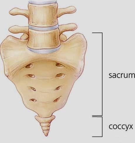
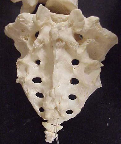
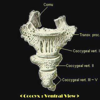
26 26 4. Sacrum and Coccyx Sacral/Sacrum There are five vertebrae that join together to form the sacrum, a wedgeshaped part of the spine that rests at the top of the pelvis. In the female the sacrum is shorter and wider than in the male. Coccyx The coccyx is often referred to as the tailbone. It consists of four vertebrae. Lateral surfaces of sacrum and coccyx.
27 27
Biology 218 Human Anatomy. Adapted from Martini Human Anatomy 7th ed. Chapter 6 The Skeletal System: Axial Division
 Adapted from Martini Human Anatomy 7th ed. Chapter 6 The Skeletal System: Axial Division Introduction The axial skeleton: Composed of bones along the central axis of the body Divided into three regions:
Chapter 7. Skeletal System
 Chapter 7 Skeletal System 1 Skull A. The skull is made up of 22 bones: 8 cranial bones, 13 facial bones, and the mandible. B. The Cranium encloses and protects the brain, provides attachments for muscles,
APPENDICULAR SKELETON 126 AXIAL SKELETON SKELETAL SYSTEM. Cranium. Skull. Face. Skull and associated bones. Auditory ossicles. Associated bones.
 SKELETAL SYSTEM 206 AXIAL SKELETON 80 APPENDICULAR SKELETON 26 Skull Skull and associated s 29 Cranium Face Auditory ossicles 8 4 6 Associated s Hyoid Thoracic cage 25 Sternum Ribs 24 Vertebrae 24 column
Human Anatomy and Physiology - Problem Drill 07: The Skeletal System Axial Skeleton
 Human Anatomy and Physiology - Problem Drill 07: The Skeletal System Axial Skeleton Question No. 1 of 10 Which of the following statements about the axial skeleton is correct? Question #01 A. The axial
Bones of the skull & face
 Bones of the skull & face Cranium= brain case or helmet Copyright The McGraw-Hill Companies, Inc. Permission required for reproduction or display. The cranium is composed of eight bones : frontal Occipital
ANATOMY & PHYSIOLOGY I Laboratory Version B Name Section. REVIEW SHEET Exercise 10 Axial Skeleton
 ANATOMY & PHYSIOLOGY I Laboratory Version B Name Section REVIEW SHEET Exercise 10 Axial Skeleton 1 POINT EACH. THE SKULL MULTIPLE CHOICE 1. The major components of the axial skeleton include the 7. The
Cranium Facial bones. Sternum Rib
 Figure 7.1 The human skeleton. Skull Thoracic cage (ribs and sternum) Cranium Facial bones Sternum Rib Bones of pectoral girdle Vertebral column Sacrum Vertebra Bones of pelvic girdle (a) Anterior view
THE SKELETAL SYSTEM. Focus on the Skull
 THE SKELETAL SYSTEM Focus on the Skull Review Anatomical Terms Anterior/Posterior Dorsal/Ventral Medial/Lateral Superior/Inferior Bone Markings - Review Projections for attachment of muscles, ligaments
Anatomy and Physiology. Bones, Sutures, Teeth, Processes and Foramina of the Human Skull
 Anatomy and Physiology Chapter 6 DRO Bones, Sutures, Teeth, Processes and Foramina of the Human Skull Name: Period: Bones of the Human Skull Bones of the Cranium: Frontal bone: forms the forehead and the
University of Palestine. Midterm Exam 2013/2014 Total Grade:
 Course No: DNTS2208 Course Title: Head and Neck Anatomy Date: 09/11/2013 No. of Questions: (50) Time: 1hour Using Calculator (No) University of Palestine Midterm Exam 2013/2014 Total Grade: Instructor
Chapter 7 Part A The Skeleton
 Chapter 7 Part A The Skeleton Why This Matters Understanding the anatomy of the skeleton enables you to anticipate problems such as pelvic dimensions that may affect labor and delivery The Skeleton The
Skeletal system. Prof. Abdulameer Al-Nuaimi. E. mail:
 Skeletal system Prof. Abdulameer Al-Nuaimi E-mail: a.al-nuaimi@sheffield.ac.uk E. mail: abdulameerh@yahoo.com Functions of Bone and The Skeletal System Support: The skeleton serves as the structural framework
Skull-2. Norma Basalis Interna Norma Basalis Externa. Dr. Heba Kalbouneh Associate Professor of Anatomy and Histology
 Skull-2 Norma Basalis Interna Norma Basalis Externa Dr. Heba Kalbouneh Associate Professor of Anatomy and Histology Norma basalis interna Base of the skull- superior view The interior of the base of the
ACTIVITY 3: AXIAL SKELETON AND LONG BONE DISSECTION COW BONE DISSECTION
 ACTIVITY 3: AXIAL SKELETON AND LONG BONE DISSECTION Objectives: 1) How to get ready: Read Chapter 7, McKinley et al., Human Anatomy, 4e. All text references are for this textbook. Learning the meanings
AXIAL SKELETON SKULL
 AXIAL SKELETON SKULL CRANIAL BONES (8 total flat bones w/ 2 paired) 1. Frontal forms forehead & upper portion of eyesocket (orbital) 2. Parietal paired bones; form superior & lateral walls of cranium 3.
Infratemporal fossa: Tikrit University college of Dentistry Dr.Ban I.S. head & neck Anatomy 2 nd y.
 Infratemporal fossa: This is a space lying beneath the base of the skull between the lateral wall of the pharynx and the ramus of the mandible. It is also referred to as the parapharyngeal or lateral pharyngeal
YOU MUST BRING YOUR OWN GLOVES FOR THIS ACTIVITY.
 ACTIVITY 3: AXIAL SKELETON AND LONG BONE DISSECTION Objectives: 1) How to get ready: Read Chapter 7, McKinley et al., Human Anatomy, 5e. All text references are for this textbook. Learning the meanings
Chapter 8A. The Skeletal System: The Axial Skeleton. The Skeletal System: The Axial Skeleton. Types of Bones. Types of Bones
 Chapter 8A The Skeletal System: The Axial Skeleton The Skeletal System: The Axial Skeleton 206 named bones Axial Skeleton 80 bones lie along longitudinal axis skull, hyoid, vertebrae, ribs, sternum, ear
o Diaphysis o Area where red marrow is found o Area where yellow marrow is found o Epiphyseal plate AXIAL SKELETON Skull
 64 Anatomy & Physiology Coloring Workbook 7. Figure 5-2A is a midlevel, cross-sectional view of the diaphysis of the femur. Label the membrane that lines the cavity and the membrane that covers the outside
Dr. Sami Zaqout, IUG Medical School
 The skull The skull is composed of several separate bones united at immobile joints called sutures. Exceptions? Frontal bone Occipital bone Vault Cranium Sphenoid bone Zygomatic bones Base Ethmoid bone
Structure Location Function
 Frontal Bone Cranium forms the forehead and roof of the orbits Occipital Bone Cranium forms posterior and inferior portions of the cranium Temporal Bone Cranium inferior to the parietal bone forms the
Crafton Hills College Human Anatomy & Physiology Axial Skeleton
 A. Major Divisions Crafton Hills College Human Anatomy & Physiology Axial keleton 1. Axial: Part of skeleton lies along long axis of body 2. Appendicular: Bones & features of the appendages B. AXIAL KELETON
Skeletal System -Axial System. Chapter 7 Part A
 Skeletal System -Axial System Chapter 7 Part A Skeleton Learn: Names of the s. Identify specific landmarks that allow: Bones to fit into each other, Organs to fit into the cavities, Muscles to attach,
Parotid Gland, Temporomandibular Joint and Infratemporal Fossa
 M1 - Anatomy Parotid Gland, Temporomandibular Joint and Infratemporal Fossa Jeff Dupree Sanger 9-057 jldupree@vcu.edu Parotid gland: wraps around the mandible positioned between the mandible and the sphenoid
Exercise 10. The Axial Skeleton
 Exercise 10 The Axial Skeleton The Axial Skeleton Consists of the skeletal structures found along the midline of the body. Includes the skull, hyoid, vertebrae, ribs, sternum, and sacrum. The cartilages
Chapter 7 The Skeletal System:The Axial Skeleton
 Chapter 7 The Skeletal System:The Axial Skeleton Axial Skeleton 80 bones lie along longitudinal axis skull, hyoid, vertebrae, ribs, sternum, ear ossicles Appendicular Skeleton 126 bones upper & lower limbs
Chapter 7: Skeletal System: Gross Anatomy
 Chapter 7: Skeletal System: Gross Anatomy I. General Considerations A. How many bones in an average adult skeleton? B. Anatomic features of bones are based on II. Axial Skeleton A. Skull 1. Functionally
Temporal region. temporal & infratemporal fossae. Zhou Hong Ying Dept. of Anatomy
 Temporal region temporal & infratemporal fossae Zhou Hong Ying Dept. of Anatomy Temporal region is divided by zygomatic arch into temporal & infratemporal fossae. Temporal Fossa Infratemporal fossa Temporal
Human Anatomy - Problem Drill 06: The Skeletal System Axial Skeleton & Articualtions
 Human Anatomy - Problem Drill 06: The Skeletal System Axial Skeleton & Articualtions Question No. 1 of 10 Instructions: (1) Read the problem and answer choices carefully, (2) Work the problems on paper
Anatomy and Physiology 1 Chapter 7 self quiz Pro, Dima Darwish,MD.
 Anatomy and Physiology 1 Chapter 7 self quiz Pro, Dima Darwish,MD. 1) How many bones make up the axial skeleton? A) 50 B) 60 C) 70 D) 80 E) 90 2) Which of the following is a function of the axial skeleton?
in compact bone, large vertical canals carrying blood vessels and nerves. in compact bone, large horizontal canals carrying blood vessels and nerves.
 Carl Christensen, PhD Skeletal System (Bones`) Bio. 2304 Human Anatomy 1. Identify a term for each of the following: shaft of a long bone ends of a long bone ossified remnant of the "growth plate" connective
Chapter 7: Head & Neck
 Chapter 7: Head & Neck Osteology I. Overview A. Skull The cranium is composed of irregularly shaped bones that are fused together at unique joints called sutures The skull provides durable protection from
Dr.Noor Hashem Mohammad Lecture (5)
") Dr.Noor Hashem Mohammad Lecture (5) 2016-2017 If the mandible is discarded, the anterior part of this aspect of the skull is seen to be formed by the hard palate. The palatal processes of the maxillae
Dr.Noor Hashem Mohammad Lecture (5) 2016-2017 If the mandible is discarded, the anterior part of this aspect of the skull is seen to be formed by the hard palate. The palatal processes of the maxillae
Copyright 2010 Pearson Education, Inc.
 E. VERTEBRAL COLUMN 1. The vertebral column extends from the skull to the pelvis and forms the vertical axis of the skeleton. 2. The vertebral column is composed of vertebrae that are separated by intervertebral
Human Anatomy & Physiology I Dr. Sullivan Unit VIIIa The Axial Skeleton Chapter 8 (Sections )
") Human Anatomy & Physiology I Dr. Sullivan Unit VIIIa The Axial Skeleton Chapter 8 (Sections 8.1-8.3) I. Divisions of the skeletal system a) An adult human skeleton has 206 named bones b) Most are paired
Human Anatomy & Physiology I Dr. Sullivan Unit VIIIa The Axial Skeleton Chapter 8 (Sections 8.1-8.3) I. Divisions of the skeletal system a) An adult human skeleton has 206 named bones b) Most are paired
The Axial Skeleton. C h a p t e r. PowerPoint Lecture Slides prepared by Jason LaPres Lone Star College - North Harris
 C h a p t e r 7 The Axial Skeleton PowerPoint Lecture Slides prepared by Jason LaPres Lone Star College - North Harris Copyright 2009 Pearson Education, Inc., publishing as Pearson Benjamin Cummings An
Copyright 2010 Pearson Education, Inc. Copyright 2010 Pearson Education, Inc. Figure Sectioned spinous process. Interspinous.
 PowerPoint Lecture Slides prepared by Janice Meeking, Mount Royal College C H A P T E R 7 The Skeleton: Part B Vertebral Column Transmits weight of trunk to lower limbs Surrounds and protects spinal cord
Labs 9 and 10. Classification of Bones. Bone Shapes 1/05/13. Skeletal system overview. Bone are identified by:
 Labs 9 and 10 Skeletal system overview Classification of Bones Bone are identified by: shape internal tissues bone markings 1. Flat bones 2. Long bones 3. Short bones 4. Irregular bones 5. Sutural bones
The Skull and Temporomandibular joint II Prof. Abdulameer Al-Nuaimi. E. mail:
 The Skull and Temporomandibular joint II Prof. Abdulameer Al-Nuaimi E-mail: a.al-nuaimi@sheffield.ac.uk E. mail: abdulameerh@yahoo.com Temporal fossa The temporal fossa is a depression on the temporal
A. Incorrect! The axial skeleton includes bones of the skull, inner ear, chest and spinal column.
 Anatomy and Physiology - Problem Drill 07: The Skeletal System II No. 1 of 10 1. Which of the following statements about the axial skeleton is correct? A. The axial skeleton includes bones of the skull,
Parotid Gland. Parotid Gland. Largest of 3 paired salivary glands (submandibular; sublingual) Ramus of Mandible. Medial pterygoid.
 Ramus of Mandible. Medial pterygoid.") Parotid region Parotid Gland Largest of 3 paired salivary glands (submandibular; sublingual) Ramus of Mandible Medial pterygoid Cross section of mandible Masseter D S SCM Parotid Gland Mastoid Process
Parotid region Parotid Gland Largest of 3 paired salivary glands (submandibular; sublingual) Ramus of Mandible Medial pterygoid Cross section of mandible Masseter D S SCM Parotid Gland Mastoid Process
Skeletal system overview. Classification of Bones
 Skeletal system overview BIOL241 Lab #9 Classification of Bones Bone are identified by: shape internal tissues bone markings 1 1. Flat bones 2. Long bones 3. Short bones 4. Irregular bones 5. Sutural bones
External Acoustic Meatus. Mastoid Process. Zygomatic Process. Temporal Bone
 Bone lab review 1. Frontal Bone 2. Supra-Orbital Foramen 3. Orbit (Orbital Cavity) 4. Superior Orbital Fissure 5. Inferior Orbital Fissure 6. Zygomatic Bone 7. Infra-Orbital Foramen 8. Maxilla 9. Mandible
SKULL AS A WHOLE + ANTERIOR CRANIAL FOSSA
 SKULL AS A WHOLE + ANTERIOR CRANIAL FOSSA LEARNING OBJECTIVES At the end of this lecture, the student should be able to know: Parts of skeleton (axial and appendicular) Parts of skull Sutures of skull
Biology 2401 The Skeletal System
 Biology 2401 The Skeletal System Purpose: The lab will describe the microscopic and gross anatomy of bone, identify bones of the body, and identify important bone markings. I. Overview of the Skeleton
CHAPTER 7, PART II (BONES)
") Anatomy Name: CHAPTER 7, PART II (BONES) Entry #: INSTRUCTIONS: 1) READ Chapter 7, pg. 140-161. 2) Using the outline, make a note card for each underlined bone name or phrase. 3) On each note card, put
Anatomy Name: CHAPTER 7, PART II (BONES) Entry #: INSTRUCTIONS: 1) READ Chapter 7, pg. 140-161. 2) Using the outline, make a note card for each underlined bone name or phrase. 3) On each note card, put
VERTEBRAL COLUMN VERTEBRAL COLUMN
 VERTEBRAL COLUMN FUNCTIONS: 1) Support weight - transmits weight to pelvis and lower limbs 2) Houses and protects spinal cord - spinal nerves leave cord between vertebrae 3) Permits movements - *clinical
PTERYGOPALATINE FOSSA
 PTERYGOPALATINE FOSSA Outline Anatomical Structure and Boundaries Foramina and Communications with other spaces and cavities Contents Pterygopalatine Ganglion Especial emphasis on certain arteries and
Vertebral Column. Backbone consists of 26 vertebrae. Five vertebral regions. Cervical
 Vertebral Column Backbone consists of 26 vertebrae. Five vertebral regions Cervical vertebrae (7) in the neck. Thoracic vertebrae (12) in the thorax. Lumbar vertebrae (5) in the lower back. Sacrum (5,
Introduction to Local Anesthesia and Review of Anatomy
 5-Sep Introduction and Anatomy Review 12-Sep Neurophysiology and Pain 19-Sep Physiology and Pharmacology part 1 26-Sep Physiology and Pharmacology part 2 Introduction to Local Anesthesia and Review of
The Skeletal System: Axial Skeleton
 The Skeletal System: Axial Skeleton The Big Idea The Axial Skeleton & Homeostasis The bones of the axial skeleton contribute to homeostasis by protecting many of the body s organs such as the brain, spinal
Axial Skeleton: Vertebrae and Thorax
 Axial Skeleton: Vertebrae and Thorax Function of the vertebral column (spine or backbone): 1) 2) 3) Composition of Vertebral column The vertebral column is formed by 33 individual vertebrae (some of which
Dr. Sami Zaqout Faculty of Medicine IUG
 Auricle External Ear External auditory meatus The Ear Middle Ear (Tympanic Cavity) Auditory ossicles Internal Ear (Labyrinth) Bony labyrinth Membranous labyrinth External Ear Auricle External auditory
Omran Saeed. Luma Taweel. Mohammad Almohtaseb. 1 P a g e
 2 Omran Saeed Luma Taweel Mohammad Almohtaseb 1 P a g e I didn t include all the photos in this sheet in order to keep it as small as possible so if you need more clarification please refer to slides In
Bone Flashcards for 10a
 Bone Flashcards for 0a CLAVICLE (collar bone). Sternal extremity (end) flat end. Acromial extremity (end) rounded end. SCAPULA (shoulder blade). Right or left scapula?. Superior border (superior margin).
Skull-2. Norma Basalis Interna. Dr. Heba Kalbouneh Assistant Professor of Anatomy and Histology
 Skull-2 Norma Basalis Interna Dr. Heba Kalbouneh Assistant Professor of Anatomy and Histology Norma basalis interna Base of the skull- superior view The interior of the base of the skull is divided into
Anatomy and Physiology II. Review Spine and Neck
 Anatomy and Physiology II Review Spine and Neck Spine regions How many cervical vertibrae are there? 7 The curvature is the cervical region posterior? Concave posterior How many thoracic? And curvature?
Temporal fossa Infratemporal fossa Pterygopalatine fossa Terminal branches of external carotid artery Pterygoid venous plexus
 Outline of content Temporal fossa Infratemporal fossa Pterygopalatine fossa Terminal branches of external carotid artery Pterygoid venous plexus Boundary Content Communication Mandibular division of trigeminal
University of Palestine. Midterm Exam 2013/2014 Total Grade:
 [ Course No: DNTS2208 Course Title: Head and Neck Anatomy Date: 17/11/1024 No. of Questions: (52) Time: 2hours Using Calculator (No) University of Palestine Midterm Exam 2013/2014 Total Grade: Instructor
2. The vertebral arch is composed of pedicles (projecting from the body) and laminae (uniting arch posteriorly).
 and laminae (uniting arch posteriorly).") VERTEBRAL COLUMN 2018zillmusom I. VERTEBRAL COLUMN - functions to support weight of body and protect spinal cord while permitting movements of trunk and providing for muscle attachments. A. Typical vertebra
VERTEBRAL COLUMN 2018zillmusom I. VERTEBRAL COLUMN - functions to support weight of body and protect spinal cord while permitting movements of trunk and providing for muscle attachments. A. Typical vertebra
BONE CHALLENGE DANIL HAMMOUDI.MD
 BONE CHALLENGE DANIL HAMMOUDI.MD Bone Basic functions? A. support B. protection C. movement assistance in D. RBC formation-hemopoiesis E. mineral homeostasis +importance of calcium F. energy supply -yellow
AXIAL SKELETON FORM THE VERTICAL AXIS OF THE BODY CONSISTS OF 80 BONES INCLUDES BONES OF HEAD, VERTEBRAL COLUMN, RIBS,STERNUM
 AXIAL SKELETON FORM THE VERTICAL AXIS OF THE BODY CONSISTS OF 80 BONES INCLUDES BONES OF HEAD, VERTEBRAL COLUMN, RIBS,STERNUM APPENDICULAR SKELETON BONES OF THE FREE APPENDAGES & THEIR POINTS OF ATTACHMENTS
Lec [8]: Mandibular nerve:
![Lec [8]: Mandibular nerve:](/thumbs/94/121295776.jpg "Lec [8]: Mandibular nerve:") Lec [8]: Mandibular nerve: The mandibular branch from the trigeminal ganglion lies in the middle cranial fossa lateral to the cavernous sinus. With the motor root of the trigeminal nerve [motor roots lies
Lec [8]: Mandibular nerve: The mandibular branch from the trigeminal ganglion lies in the middle cranial fossa lateral to the cavernous sinus. With the motor root of the trigeminal nerve [motor roots lies
Major Anatomic Components of the Orbit
 Major Anatomic Components of the Orbit 1. Osseous Framework 2. Globe 3. Optic nerve and sheath 4. Extraocular muscles Bony Orbit Seven Bones Frontal bone Zygomatic bone Maxillary bone Ethmoid bone Sphenoid
11/25/2012. Chapter 7 Part 2: Bones! Skeletal Organization. The Skull. Skull Bones to Know Cranium
 Chapter 7 Part 2: Bones! 5) Distinguish between the axial and appendicular skeletons and name the major parts of each 6) Locate and identify the bones and the major features of the bones that compose the
Chapter 7 Part B The Skeleton
 Chapter 7 Part B The Skeleton 7.2 The Vertebral Column General Characteristics Extends from skull to pelvis Also called spine or spinal column Functions to transmit weight of trunk to lower limbs, surround
TEST YOURSELF- Chapter 7
 TEST YOURSELF- Chapter 7 Cranial Bones 1. Give the name of the bone for each of the following markings. Some of the markings are found on more than one bone. List all that apply. Cranium a. Frontal squama:
The Skeletal System. Parts of the skeletal system. Bones (Skeleton) Joints Cartilages Ligaments
 Joints Cartilages Ligaments") The Skeletal System Parts of the skeletal system Bones (Skeleton) Joints Cartilages Ligaments Functions of the Bones Support Internal framework of the body Protection Skull and vertebrae protect brain
The Skeletal System Parts of the skeletal system Bones (Skeleton) Joints Cartilages Ligaments Functions of the Bones Support Internal framework of the body Protection Skull and vertebrae protect brain
Tikrit University collage of dentistry Dr.Ban I.S. head & neck anatomy 2 nd y. Lec [5] / Temporal fossa :
![Tikrit University collage of dentistry Dr.Ban I.S. head & neck anatomy 2 nd y. Lec [5] / Temporal fossa :](/thumbs/88/115294566.jpg "Tikrit University collage of dentistry Dr.Ban I.S. head & neck anatomy 2 nd y. Lec [5] / Temporal fossa :") Lec [5] / Temporal fossa : Borders of the Temporal Fossa: Superior: Superior temporal line. Inferior: gap between zygomatic arch and infratemporal crest of sphenoid bone. Anterior: Frontal process of the
Lec [5] / Temporal fossa : Borders of the Temporal Fossa: Superior: Superior temporal line. Inferior: gap between zygomatic arch and infratemporal crest of sphenoid bone. Anterior: Frontal process of the
THE THORACIC WALL. Boundaries Posteriorly by the thoracic part of the vertebral column. Anteriorly by the sternum and costal cartilages
 THE THORACIC WALL Boundaries Posteriorly by the thoracic part of the vertebral column Anteriorly by the sternum and costal cartilages Laterally by the ribs and intercostal spaces Superiorly by the suprapleural
Anatomic Relations Summary. Done by: Sohayyla Yasin Dababseh
 Anatomic Relations Summary Done by: Sohayyla Yasin Dababseh Anatomic Relations Lecture 1 Part-1 - The medial wall of the nose is the septum. - The vestibule lies directly inside the nostrils (Nares). -
human anatomy 2015 lecture four Dr meethak ali ahmed neurosurgeon
 The Vertebral Column the vertebral columnis central pillar of the body.it serve to protect the spinal cord and support the weight of the head trunk, which it transmits to the hip bones & the lower limbs.
Musculoskeletal System (Part A-1) Module 7 -Chapter 10 Overview. Functions
 Module 7 -Chapter 10 Overview. Functions") Musculoskeletal System (Part A-1) Module 7 -Chapter 10 Overview Susie Turner, M.D. 1/8/13 Muscles Attachments Bones Bone types Surface features of bones Divisions of the skeletal system Joints or Articulations
Musculoskeletal System (Part A-1) Module 7 -Chapter 10 Overview Susie Turner, M.D. 1/8/13 Muscles Attachments Bones Bone types Surface features of bones Divisions of the skeletal system Joints or Articulations
Any of the vertebra in the cervical (neck) region of the spinal column. The cervical vertebra are the smallest vertebra in the spine, reflective of th
 region of the spinal column. The cervical vertebra are the smallest vertebra in the spine, reflective of th") Any of the vertebra in the cervical (neck) region of the spinal column. The cervical vertebra are the smallest vertebra in the spine, reflective of the fact that they support the least load. In humans,
Any of the vertebra in the cervical (neck) region of the spinal column. The cervical vertebra are the smallest vertebra in the spine, reflective of the fact that they support the least load. In humans,
Bones Ethmoid bone Inferior nasal concha Lacrimal bone Maxilla Nasal bone Palatine bone Vomer Zygomatic bone Mandible
 splanchnocranium - Consists of part of skull that is derived from branchial arches - The facial bones are the bones of the anterior and lower human skull Bones Ethmoid bone Inferior nasal concha Lacrimal
UNIT 4 - SKELETAL SYSTEM LECTURE NOTES
 UNIT 4 - SKELETAL SYSTEM LECTURE NOTES 4.01 FUNCTIONS OF THE SKELETAL SYSTEM A. Support 1. Provides a framework for the body 2. Supports soft tissue 3. Serves as a point of attachment for ligaments, tendons,
BIO 137 AXIAL SKELETON BONE STUDY THE HUMAN SKELETON
 BIO 137 THE AXIAL SKELETON MARY CATHERINE FLATH, Ph.D. THE HUMAN SKELETON AXIAL SKULL HYOID THORACIC CAGE VERTEBRAL COLUMN APPENDICULAR PECTORAL GIRDLE UPPER LIMBS PELVIC GIRDLE LOWER LIMBS AXIAL SKELETON
Overview of the Skeleton: Bone Markings
 Name Overview of the Skeleton: Bone Markings Match the terms in column B with the appropriate description in column A. Column A 1. sharp, slender process* 2. small rounded projection* 3. narrow ridge of
THEME 2. VERTEBRAE (GENERAL DATA). CERVICAL, THORACIC AND LUMBAR VERTEBRAE. SACRUM. COCCYX. THE VERTEBRAL COLUMN AS A WHOLE
. CERVICAL, THORACIC AND LUMBAR VERTEBRAE. SACRUM. COCCYX. THE VERTEBRAL COLUMN AS A WHOLE") THEME 2. VERTEBRAE (GENERAL DATA). CERVICAL, THORACIC AND LUMBAR VERTEBRAE. SACRUM. COCCYX. THE VERTEBRAL COLUMN AS A WHOLE Osteology of the Vertebral Column Bone Description vertebra Notes a vertebra
THEME 2. VERTEBRAE (GENERAL DATA). CERVICAL, THORACIC AND LUMBAR VERTEBRAE. SACRUM. COCCYX. THE VERTEBRAL COLUMN AS A WHOLE Osteology of the Vertebral Column Bone Description vertebra Notes a vertebra
Skull basic structures. Neurocranium
 Assoc. Prof. Květuše Lovásová, M.V.D., PhD. Skull basic structures Skull consists of two groups of bones: neurocranium (bones forming the brain box) splanchnocranium (bones forming the facial skeleton)
Skeletal System: Skull.
 Skeletal System: Skull www.fisiokinesiterapia.biz Bones of the Skull SPLANCHNOCRANIUM Nasal (2) Maxilla (2) Lacrimal (2) Zygomatic (2) Palatine (2) Inferior concha (2) Vomer Mandible NEUROCRANIUM Frontal
Anatomy images for MSS practical exam- 2019
 Anatomy images for MSS practical exam- 2019 Ilium Ischium Pubis Acetabulaum Iliac crest Iliac tubercle ASIS (muscle and ligament attached) AIIS (muscle attached) PSIS PIIS Ischial spine Ischial tuberosity
The sebaceous glands (glands of Zeis) open directly into the eyelash follicles, ciliary glands (glands of Moll) are modified sweat glands that open
 open directly into the eyelash follicles, ciliary glands (glands of Moll) are modified sweat glands that open") The Orbital Region The orbits are a pair of bony cavities that contain the eyeballs; their associated muscles, nerves, vessels, and fat; and most of the lacrimal apparatus upper eyelid is larger and more
The Orbital Region The orbits are a pair of bony cavities that contain the eyeballs; their associated muscles, nerves, vessels, and fat; and most of the lacrimal apparatus upper eyelid is larger and more
Axial skeleton bones and markings
 Axial skeleton bones and markings Skull Cranial bones Frontal x 1 Supraorbital foramen Occipital x 1 Foramen magnum Occipital condyles Superior nuchal line Inferior nuchal line Anterior cranial fossa External
THE VERTEBRAL COLUMN. Average adult length: In male: about 70 cms. In female: about 65 cms.
 THE VERTEBRAL COLUMN Average adult length: In male: about 70 cms. In female: about 65 cms. 1 Vertebral Column (Regions and Curvatures) Curvatures of the vertebral column: A. Primary curvature: C-shaped;
Bio 5/6 5 The Skeletal System Study Guide
 Name: THE SKELETAL SYSTEM: 5 The Skeletal System Study Guide Period: The skeleton is constructed of two of the most supportive tissues found in the human body - cartilage and bone. Besides supporting and
SKULL / CRANIUM BONES OF THE NEUROCRANIUM (7) Occipital bone (1) Sphenoid bone (1) Temporal bone (2) Frontal bone (1) Parietal bone (2)
 Occipital bone (1) Sphenoid bone (1) Temporal bone (2) Frontal bone (1) Parietal bone (2)") Important! 1. Memorizing these pages only does not guarantee the succesfull passing of the midterm test or the semifinal exam. 2. The handout has not been supervised, and I can not guarantee, that these
Important! 1. Memorizing these pages only does not guarantee the succesfull passing of the midterm test or the semifinal exam. 2. The handout has not been supervised, and I can not guarantee, that these
Maxilla, ORBIT and infratemporal fossa. Neophytos C Demetriades MD, DDS, MSc Associate professor European University of Cyprus School of Medicine
 Maxilla, ORBIT and infratemporal fossa Neophytos C Demetriades MD, DDS, MSc Associate professor European University of Cyprus School of Medicine MAXILLA Superior, middle, and inferior meatus Frontal sinus
The SKELETAL System. The framework of bones and cartilage which protect organs, and provides a lever system that allows locomotion.
 The SKELETAL System The framework of bones and cartilage which protect organs, and provides a lever system that allows locomotion. Functions of the Skeletal System Support Protection Movement Facilitation
Bony orbit Roof The orbital plate of the frontal bone Lateral wall: the zygomatic bone and the greater wing of the sphenoid
 Bony orbit Roof: Formed by: The orbital plate of the frontal bone, which separates the orbital cavity from the anterior cranial fossa and the frontal lobe of the cerebral hemisphere Lateral wall: Formed
Dr.Ban I.S. head & neck anatomy 2 nd y جامعة تكريت كلية طب االسنان مادة التشريح املرحلة الثانية أ.م.د. بان امساعيل صديق 6102/6102
 جامعة تكريت كلية طب االسنان مادة التشريح املرحلة الثانية أ.م.د. بان امساعيل صديق 6102/6102 Pterygopalatine fossa: The pterygopalatine fossa is a cone-shaped depression, It is located between the maxilla,
Superior View of the Skull (Norma Verticalis) Anteriorly the frontal bone articulates with the two parietal bones AT THE CORONAL SUTURE
 Anteriorly the frontal bone articulates with the two parietal bones AT THE CORONAL SUTURE") Superior View of the Skull (Norma Verticalis) Anteriorly the frontal bone articulates with the two parietal bones AT THE CORONAL SUTURE 1 The two parietal bones articulate in the midline AT THE SAGITTAL
Superior View of the Skull (Norma Verticalis) Anteriorly the frontal bone articulates with the two parietal bones AT THE CORONAL SUTURE 1 The two parietal bones articulate in the midline AT THE SAGITTAL
locomotice system Plastinated specimensⅠ: Silicone specimens Regional specimens and organs
 locomotice system Plastinated specimensⅠ: Silicone specimens Regional specimens and organs Art-No. Name Description The locomotor system SL001 Two hundred pieces of plastinated bones (without six The bones
The orbit-1. Dr. Heba Kalbouneh Assistant Professor of Anatomy and Histology
 The orbit-1 Dr. Heba Kalbouneh Assistant Professor of Anatomy and Histology Orbital plate of frontal bone Orbital plate of ethmoid bone Lesser wing of sphenoid Greater wing of sphenoid Lacrimal bone Orbital
The Thoracic Cage ANATOMY 2: THORACIC CAGE AND VERTEBRAL COLUMN
 ANATOMY 2: THORACIC CAGE AND VERTEBRAL COLUMN PSK 4U Mr. S. Kelly North Grenville DHS The Thoracic Cage 7 true ribs 3 false ribs 2 floating ribs Clavicle = collarbone Manubrium Sternum Xiphoid Process
Perpendicular Plate Zygomatic Bone. Mental Foramen Mandible
 Glabella Frontal Middle Nasal Concha Nasal Lacrimal Perpendicular Plate Zygomatic Inferior Nasal Concha Maxilla Mental Mandible Skull (anterior view) Squamosal Suture Coronal Suture Frontal Parietal Nasal
Dr.Ban I.S. head & neck anatomy 2 nd y. جامعة تكريت كلية طب االسنان املرحلة الثانية أ.م.د. بان امساعيل صديق 6102/6102
 جامعة تكريت كلية طب االسنان التشريح مادة املرحلة الثانية أ.م.د. بان امساعيل صديق 6102/6102 Parotid region The part of the face in front of the ear and below the zygomatic arch is the parotid region. The
Skeletal System. Axial Division
 Skeletal System Axial Division The Axial Skeleton You will see that each bone has special features (overviewed in section I below) that provide Sites of Attachment (for muscles, ligaments, tendons, etc.)
Mohammad Hisham Al-Mohtaseb. Lina Mansour. Reyad Jabiri. 0 P a g e
 2 Mohammad Hisham Al-Mohtaseb Lina Mansour Reyad Jabiri 0 P a g e This is only correction for the last year sheet according to our record. If you already studied this sheet just read the yellow notes which