Echocardiographic evaluation of mitral stenosis
|
|
|
- Ophelia Hunt
- 5 years ago
- Views:
Transcription
1 Echocardiographic evaluation of mitral stenosis Euroecho 2011 Philippe Unger, MD, FESC Erasme Hospital, ULB, Brussels, Belgium

2 I have nothing to declare
3
4 EuroHeart Survey Etiology of single native left-sided valve disease 100% 80% 60% 40% 20% 0% AS n=1197 AR n=369 MS n=336 MR n=877 Other Ischemic Congenital Inflamatory Endocarditis Rheumatic Degenerative Eur Heart J 2003 Jul;24(13):

5 Typical M-mode and 2D echocardiographic features of rheumatic MS
6 Degenerative MS
7 Echocardiographic differential diagnosis of LV inflow obstruction LV diastolic dysfunction Rheumatic Degenerative/Calcific Congenital Carcinoid heart disease SLE Rheumatoid arthritis Hunter-Hurler Fabry disease Whipple disease Methysergide therapy Myxoma Thrombus Cor triatriatum
8 Echocardiographic evaluation of the patient with MS MS severity Pressure gradient Valve area Exercise Presence and severity of coexisting MR Mitral valve anatomy LA thrombus? (before PBV) LA, LV, RV size and function Pulmonary systolic pressures (Rheumatic) involvement of other valves?
9
10 Pressure gradient: simplified Bernoulli equation ΔP=4v² Mean gradient 18 mm Hg Nishimura RA et al. J Am Coll Cardiol 1994;24:&(é
11 Pitfalls in evaluation of MS severity Pressure gradient Intercept angle between MS jet and ultrasound beam Beat-to-beat variability in AF Dependence on transvalvular volume flow rate and on diastolic filling period exercise, infection, pregnancy, coexisting MR
12 Relation between pressure gradient and mitral valve area: Role of transmitral diastolic flow rate Gradient 1.0 cm² Increasing severity 1.5 cm² 2.0 cm² Flow

13 Planimetry Reference method Faletra F, et al. J Am Coll Cardiol 1996;28:1190

14 Pitfalls: Image orientation and tomographic plane MVA 1.1 cm² MVA 1.5 cm²


15 Pitfalls: 2D gain settings 1.05 cm² 0.72 cm²
16 Pitfalls Poor acoustic access, severe distortion of valve anatomy, severe ca++ on leaflet tips Feasibility in the elderly? r = 0.39 Shaw TR, et al. Heart 2003;89:1430
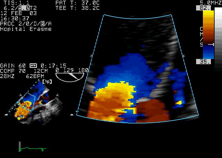
17 Pressure half-time method Moderate MS Severe MS LV LA
18 Pressure half-time method MVA = 220/PHT Peak PG Peak V Peak PG/2 Peak V/1.4 Hatle et al. Circulation 1979
19 Accuracy of Doppler PHT method in assessing severity of MS in patients with and without prior commissurotomy Unoperated patients (N=37) Previous commissurotomy (11.2±5.4 yrs before) (N=37) Smihth MD, et al. Circulation 1986;73:100
20 Pitfalls in PHT-derived valve area calculation LV compliance/relaxation/pressure Coexisting moderate-to-severe AR MS severity Immediately after commissurotomy In the elderly (AHT, associated AS) LA compliance


21 Pitfalls of PHT method Curvilinear slope Atrial Fibrillation


22 Pitfalls of PHT method E/A fusion Carotid Sinus Massage HR 85 bpm Mean gradient 17 mm Hg HR 73 bpm Mean gradient 10 mm Hg


23 Pitfalls of PHT method Lutembacher syndrome MS+ASD
")
/Peak V")

24 Proximal Isovelocity Surface Area (PISA) Method MVA = 2Π(r²)(V aliasing )/Peak V mitral Rodriguez L et al. Circulation 1993;88:1157

25 1. Zoom on the flow convergence

26 2. Aliasing velocity cm/s
27 3. Measure the radius of the flow convergence and the transmitral velocity
28 4. Measure the angle formed by the mitral leaflets MVA = (2 r²) x aliasv / maxv x /180 LV LA = 90 = 180
29 4. Measure the angle formed by the mitral leaflets MVA = (2 r²) x aliasv / maxv x /180 LV LA = 100 = 100
30 5. Apply the formula MVA = R². 2. aliasv/v max. /180 MVA = (1.71)² / /180 = 1.01 cm²
31 MVA assessed by the PISA method using a fixed correction angle of 100 versus MVA measured by the PISA method with the exact angle measured using a protractor r=0.97, P< MVA = (2 r²) x aliasv / maxv x 100/180 Messika-Zeitoun D et al. Eur J Echocardiogr 2007;8:
32 MVA assessed by the PISA method using a fixed angle correction of 100 versus planimetry r=0.91, P< MVA = (2 r²) x aliasv / maxv x 100/180 Messika-Zeitoun D et al. Eur J Echocardiogr 2007;8:
33
34 The continuity equation Mitral stroke volume = Aortic stroke volume X = X Mitral valve area Mitral TVI LVOT diameter LVOT TVI
35 Continuity equation: Caveats Inaccuracy of LVOT diameter determination (Π.r²) Caution if AF, as mitral and aortic stroke volumes are calculated from different beats Invalidated by aortic or mitral regurgitation


36 AR MR MVA = LVOT diameter X LVOT TVI Mitral TVI Understimates severity Overstimates severity

37 Diagnostic caveats in patients with multivalve lesions Unger P et al. Heart 2011;97:

38 70 y-o woman Severe HF LVOT VTI 21.2 cm LVOT diam 1.7 cm SV 48 ml SVI 30 ml/m²

39 Max V 3.60 m/s Mean PG: 32 mm Hg Ao VTI: 96 cm AVA: 0.5 cm² Catheterization: AVA 0.6 cm² Low flow low gradient AS
: 2.4 cm² MVA (continuity): 0.92 cm² R=1.")

40 Mean PG: 8 mm Hg Mi VTI: 52 cm PHT: 90 ms MVA (PHT): 2.4 cm² MVA (continuity): 0.92 cm² R=1.1 cm (alias 31 cm) Max V 2.1 m/s MVA (PISA) 1.12 cm² Catheterization: MVA 1.04 cm² Low flow low gradient MS
41 Low flow low gradient MS Low flow low gradient AS
42 Pressure gradient Planimetry (2D) PHT Level 1 EAE/ASE Recommendation appropriate and recommended for all patients Continuity equation PISA Level 2 EAE/ASE Recommendation reasonnable when additional informations are needed in selected patients


43 What about 3D echocardiography?

44 Real-time 3D color-defined planimetry using TTE in calcific MS N = 34 mean age 72 ± 13 years Chu JW, et al. J Am Soc Echocardiogr 2008;21:1006

45 Real-time 3D transthoracic echocardiography Chu JW, et al. J Am Soc Echocardiogr 2008;21:1006
46 Real-time 3D transesophageal echocardiography

47 Real-time 3D TEE Schlosshan D et al. JACC Cardiovasc Imaging Jun;4(6):580-8.
:580-8.")
48 3D-TEE Planimetry MV leaflet tips using the short-axis view determined after multiplanar reconstruction Schlosshan D, et al. JACC Img 2011 Jun;4(6):580-8.
49 3D TEE versus other techniques Feasibility Mean difference vs other techniques (cm²) % D Continuity 2D PHT Feasibility 0,05 0-0,05-0,1-0,15-0,2-0,25 P< P< P= D PHT cont N=43 Intraclass correlation coefficient interobs 0.93; intraobs 0.96 Schlosshan D, et al. JACC Img 2011 Jun;4(6):580-8.
 and by a")

50 Correlation between MVA measured using 2D-TTE and real-time 3D-TEE measured by an experienced operator (upper panel) and by a non-experienced operator (lower panel) Experienced operator Non-experienced operator Dreyfus J et al. Eur J Echocardiogr 2011;12:
51 Role of TEE? When acoustic windows are poor using TTE Percutaneous mitral commissurotomy To exclude LA or LAA thrombus Occasionally performed during MBV to guide transseptal puncture or position of a balloon.
52 Conclusions Evaluation of MS should determine Its severity Its suitability for PMC Strengths, pitfalls and limitations of each method of severity assessement should be known The decision-making process should also include Associated valve lesions Pulmonary pressure Age Heart rhythm NYHA class Predicted operative mortality
53
54 Thank you for your attention!
55 Recommendations for classification of MS severity Specific findings Mild Moderate Severe Valve area (cm²) > <1.0 Supportive findings < 1 cm²/m² Mean gradient (mm Hg)* < >10 Pulmonary artery pressure (mm Hg) < >50 *At heart rate bpm and in sinus rhythm Baumgartner H et al. Eur J Echocardiogr 2009;10:1

56 The echo examination in MS: ACC/AHA guidelines 2006 Pressure gradient Hemodynamic severity MVA TTE (TEE) Class I, Level B PAP Valve morphology Suitability for PMBV TTE Class I, Level B If discrepency between resting Doppler echo, clinical findings, symptoms and signs Mean gradient MVA Exercise TTE Class I, Level C LA thrombus Suitability for PMBV TEE Class I, Level C Co-existing MR Evaluation of severity TTE (TEE) Class I, Level C Routine evaluation of MV morphology and hemodynamics when TTE satisfactory TEE Class III

57 Images Displaying Different Views of the MV Commissure Using 3DTEE Schlosshan D, et al. JACC Img 2011 Jun;4(6):580-8.

58 (A) RT3D view of a rheumatic mitral valve. Pérez de Isla L et al. Eur J Echocardiogr 2007;8: Copyright 2007, The European Society of Cardiology
Bogdan A. Popescu. University of Medicine and Pharmacy Bucharest, Romania. EAE Course, Bucharest, April 2010
 Bogdan A. Popescu University of Medicine and Pharmacy Bucharest, Romania EAE Course, Bucharest, April 2010 This is how it started Mitral stenosis at a glance 2D echo narrow diastolic opening of MV leaflets
Quantification of Mitral Stenosis: Planimetry, pressure Half time, Continuity Common Errors
 Quantification of Mitral Stenosis: Planimetry, pressure Half time, Continuity Common Errors Christopher J Kramer RDCS Advanced Cardiovascular Services Aurora Health Care Milwaukee, WI No Disclosures Baumgartner,
Balloon mitral valvuloplasty
 Echo for selecting and monitoring procedures in mitral valve diseases Balloon mitral valvuloplasty Eric Brochet,MD Cardiology Department Hôpital Bichat. Paris France Conflict of interest : none Balloon
Prosthesis-Patient Mismatch or Prosthetic Valve Stenosis?
 EuroValves 2015, Nice Prosthesis-Patient Mismatch or Prosthetic Valve Stenosis? Philippe Pibarot, DVM, PhD, FACC, FAHA, FASE FESC Canada Research Chair in Valvular Heart Diseases Université LAVAL Disclosure
Hemodynamic Assessment. Assessment of Systolic Function Doppler Hemodynamics
 Hemodynamic Assessment Matt M. Umland, RDCS, FASE Aurora Medical Group Milwaukee, WI Assessment of Systolic Function Doppler Hemodynamics Stroke Volume Cardiac Output Cardiac Index Tei Index/Index of myocardial
ECHO HAWAII. Role of Stress Echo in Valvular Heart Disease. Not only ischemia! Cardiomyopathy. Prosthetic Valve. Diastolic Dysfunction
 Role of Stress Echo in Valvular Heart Disease ECHO HAWAII January 15 19, 2018 Kenya Kusunose, MD, PhD, FASE Tokushima University Hospital Japan Not only ischemia! Cardiomyopathy Prosthetic Valve Diastolic
MITRAL STENOSIS. Joanne Cusack
 MITRAL STENOSIS Joanne Cusack BSE Breakdown Recognition of rheumatic mitral stenosis Qualitative description of valve and sub-valve calcification and fibrosis Measurement of orifice area by planimetry
Mitral Valve Stenosis: What do I need to know? ACC Latin American Conference 2017
 Mitral Valve Stenosis: What do I need to know? ACC Latin American Conference 2017 Athena Poppas, MD FACC FASE Past ACC Scientific Sessions Chair, ACC Board of Trustee Professor of Medicine, Brown University
Prosthetic valve dysfunction: stenosis or regurgitation
 Prosthetic valve dysfunction: stenosis or regurgitation Jean G. Dumesnil MD, FRCP(C), FACC, FASE(Hon) Quebec Heart and Lung Institute, Québec, Québec No disclosures Possible Causes of High Gradients in
Echo Doppler Assessment of Right and Left Ventricular Hemodynamics.
 Echo Doppler Assessment of Right and Left Ventricular Hemodynamics. Itzhak Kronzon, MD, FASE, FACC, FESC, FAHA, FACP, FCCP Northwell, Lenox Hill Hospital, New York Professor of Cardiology Hofstra University
Comprehensive Hemodynamics By Doppler Echocardiography. The Echocardiographic Swan-Ganz Catheter.
 Comprehensive Hemodynamics By Doppler Echocardiography. The Echocardiographic Swan-Ganz Catheter. Itzhak Kronzon, MD, FASE, FACC, FESC, FAHA, FACP, FCCP North Shore HS, LIJ/Lenox Hill Hospital, New York
PISA Evaluation of Mitral Regurgitation. Raymond Graber, MD Cardiac Anesthesia Group University Hospitals Case Medical Center 4/07/2011
 PISA Evaluation of Mitral Regurgitation Raymond Graber, MD Cardiac Anesthesia Group University Hospitals Case Medical Center 4/07/2011 Introduction Evaluation of MR. What is PISA? Physiologic basis Issues
P = 4V 2. IVC Dimensions 10/20/2014. Comprehensive Hemodynamic Evaluation by Doppler Echocardiography. The Simplified Bernoulli Equation
 Comprehensive Hemodynamic Evaluation by Doppler Echocardiography Itzhak Kronzon, MD North Shore LIJ/ Lenox Hill Hospital New York, NY Disclosure: Philips Healthcare St. Jude Medical The Simplified Bernoulli
DOPPLER HEMODYNAMICS (1) QUANTIFICATION OF PRESSURE GRADIENTS and INTRACARDIAC PRESSURES
 QUANTIFICATION OF PRESSURE GRADIENTS and INTRACARDIAC PRESSURES") THORAXCENTRE DOPPLER HEMODYNAMICS (1) QUANTIFICATION OF PRESSURE GRADIENTS and INTRACARDIAC PRESSURES J. Roelandt DOPPLER HEMODYNAMICS Intracardiac pressures and pressure gradients Volumetric measurement
THORAXCENTRE DOPPLER HEMODYNAMICS (1) QUANTIFICATION OF PRESSURE GRADIENTS and INTRACARDIAC PRESSURES J. Roelandt DOPPLER HEMODYNAMICS Intracardiac pressures and pressure gradients Volumetric measurement
Congenital. Unicuspid Bicuspid Quadricuspid
 David Letterman s Top 10 Aortic Stenosis The victim can be anyone: Echo is the question and the answer!!!! Hilton Head Island Echocardiography Conference 2012 Timothy E. Paterick, MD, JD, MBA Christopher
HEMODYNAMIC ASSESSMENT
 HEMODYNAMIC ASSESSMENT INTRODUCTION Conventionally hemodynamics were obtained by cardiac catheterization. It is possible to determine the same by echocardiography. Methods M-mode & 2D echo alone can provide
Quantification of Aortic Regurgitation
 Quantification of Aortic Regurgitation ASE Review 2018 Boston Susan E Wiegers, MD, FASE, FACC Professor of Medicine And thanks to Dr. Roberto Lang Disclosure None related to this presentation 1 Objectives
The Doppler Examination. Katie Twomley, MD Wake Forest Baptist Health - Lexington
 The Doppler Examination Katie Twomley, MD Wake Forest Baptist Health - Lexington OUTLINE Principles/Physics Use in valvular assessment Aortic stenosis (continuity equation) Aortic regurgitation (pressure
Tricuspid and Pulmonary Valve Disease
 Tricuspid and Pulmonary Valve Disease Lawrence Rudski MD FRCPC FACC FASE Professor of Medicine Director, Division of Cardiology Jewish General Hospital McGill University Question 1 All of the following
When Does 3D Echo Make A Difference?
 When Does 3D Echo Make A Difference? Wendy Tsang, MD, SM Assistant Professor, University of Toronto Toronto General Hospital, University Health Network 1 Practical Applications of 3D Echocardiography Recommended
Disclosures Rebecca T. Hahn, MD, FASE
 The New ASE Guidelines for Native Valvular Regurgitation Mitral Regurgitation The New ASE Guidelines: Role of 2D/3D and CMR (With caveats and comments from R. Hahn) William A. Zoghbi MD, FASE, MACC Professor
Aortic Stenosis: Spectrum of Disease, Low Flow/Low Gradient and Variants
 Aortic Stenosis: Spectrum of Disease, Low Flow/Low Gradient and Variants Martin G. Keane, MD, FASE Professor of Medicine Lewis Katz School of Medicine at Temple University Basic root structure Parasternal
Clinical Outcome of Tricuspid Regurgitation. David Messika-Zeitoun
 Clinical Outcome of Tricuspid Regurgitation David Messika-Zeitoun I have financial relationships to disclose Consultant for: Edwards, Symetis and Valtech Tricuspid Regurgitation is a Common Finding Tricuspid
Aortic Stenosis: LVOT Obstruction
 Aortic Stenosis: LVOT Obstruction Raymond Stainback, MD 7 th annual Houston Echo Review 2016: Boot Camp for the Echo Board Murmur: Additional heart or vascular sound due to normal or abnormal turbulent
TAVR: Echo Measurements Pre, Post And Intra Procedure
 2017 ASE Florida, Orlando, FL October 10, 2017 8:00 8:25 AM 25 min TAVR: Echo Measurements Pre, Post And Intra Procedure Muhamed Sarić MD, PhD, MPA Director of Noninvasive Cardiology Echo Lab Associate
Dobutamine Stress testing In Low Flow, Low EF, Low Gradient Aortic Stenosis Case Studies
 Dobutamine Stress testing In Low Flow, Low EF, Low Gradient Aortic Stenosis Case Studies Mitral Regurgitation The New ASE Guidelines: Role of 2D/3D and CMR William A. Zoghbi MD, FASE, MACC Professor and
Valvular Regurgitation: Can We Do Better Than Colour Doppler?
 Valvular Regurgitation: Can We Do Better Than Colour Doppler? A/Prof David Prior St Vincent s Hospital Melbourne Sports Cardiology Valvular Regurgitation Valve regurgitation volume loads the ventricles
Role of Stress Echo in Valvular Heart Disease. Satoshi Nakatani Osaka University Graduate School of Medicine Osaka, Japan
 Role of Stress Echo in Valvular Heart Disease Satoshi Nakatani Osaka University Graduate School of Medicine Osaka, Japan Exercise echocardiography Dobutamine echocardiography Usefulness of exercise echo
13/06/2018. Rheumatic Mitral Stenosis: What does the ESC Guideline say? Mitral Stenosis: Echo Assessment. Mitral Stenosis ESC Guidance 2017
 Rheumatic Mitral Stenosis: What does the ESC Guideline say? Mitral Stenosis: Echo Assessment Dave Northridge Edinburgh Heart Centre Rheumatic mitral stenosis Prosthetic mitral dysfunction Calcific/degenerative
How to assess ischaemic MR?
 ESC 2012 How to assess ischaemic MR? Luc A. Pierard, MD, PhD, FESC, FACC Professor of Medicine Head, Department of Cardiology University Hospital Sart Tilman, Liège ESC 2012 No conflict of interest Luc
MITRAL REGURGITATION ECHO PARAMETERS TOOL
 Comprehensive assessment of qualitative and quantitative parameters, along with the use of standardized nomenclature when reporting echocardiographic findings, helps to better define a patient s MR and
25 different brand names >44 different models Sizes mm
 Types of Prosthetic Valves BIOLOGIC STENTED Porcine xenograft Pericardial xenograft STENTLESS Porcine xenograft Pericardial xenograft Homograft (allograft) Autograft PERCUTANEOUS MECHANICAL Bileaflet Single
Comprehensive Echo Assessment of Aortic Stenosis
 Comprehensive Echo Assessment of Aortic Stenosis Smonporn Boonyaratavej, MD, MSc King Chulalongkorn Memorial Hospital Bangkok, Thailand Management of Valvular AS Medical and interventional approaches to
Back to Basics: Common Errors In Quantitation In Everyday Practice
 Back to Basics: Common Errors In Quantitation In Everyday Practice Deborah Agler, ACS, RDCS, FASE October 9, 2017 ASE: Echo Florida Rebecca T. Hahn, MD Director of Interventional Echocardiography Professor
Valve Disease Board Review Questions
 Valve Disease Board Review Questions Dennis A. Tighe, MD, FASE University of Massachusetts Medical School Worcester, MA Case 1 History A 61 year-old man Presents to hospital with worsening shortness of
Organic mitral regurgitation
 The best in heart valve disease Organic mitral regurgitation Ewa Szymczyk Department of Cardiology Medical University of Lodz, Poland I have nothing to declare Organic mitral regurgitation leaflet abnormality
What echo measurements are key prior to MitraClip?
 APHP CHU Bichat - Claude Bernard What echo measurements are key prior to MitraClip? Eric Brochet,MD Cardiology Department Hopital Bichat Paris France No disclosure Conflict of interest Case 69 y.o man
ECHOCARDIOGRAPHY DATA REPORT FORM
 Patient ID Patient Study ID AVM - - Date of form completion / / 20 Initials of person completing the form mm dd yyyy Study period Preoperative Postoperative Operative 6-month f/u 1-year f/u 2-year f/u
Aortic stenosis aetiology: morphology of calcific AS,
 How to improve patient selection in aortic stenosis? Fausto J. Pinto, FESC Aortic stenosis aetiology: morphology of calcific AS, bicuspid valve, and rheumatic AS (Adapted from C. Otto, Principles of
Case Reviews: Hemodynamic Calculations in Valvular Regurgitation
 Case Reviews: Hemodynamic Calculations in Valvular Regurgitation Case 5 History: 69-year-old man with orthotopic heart transplant 15 years ago. Inferior MI several years ago. Recurrent CHF. Currently dyspneic
LUST trial. Echocardiography USER S MANUAL
 LUST trial Echocardiography USER S MANUAL Rosa Sicari, Luna Gargani Ins1tute of Clinical Physiology Na1onal Council of Research, Pisa, Italy Parameters required (1) Aortic root Measurement of aortic root
Tricuspid and Pulmonary Valve Disease
 Tricuspid and Pulmonary Valve Disease Lawrence Rudski MD FRCPC FACC FASE Professor of Medicine Director, Division of Cardiology Jewish General Hospital McGill University Right Sided Failure Edema Gut congestion
Stress Testing in Valvular Disease
 2017 ASE Florida Orlando, FL October 10, 2017 2:40 2:50 PM 10 min Grand Harbor Ballroom South Stress Testing in Valvular Disease Muhamed Sarić MD, PhD, MPA Director of Noninvasive Cardiology Echo Lab Associate
Primary Mitral Regurgitation
 EURO VALVE Madrid News from Valves Guidelines 2012: What s new and Why? Primary Mitral Regurgitation Luc A. Pierard, MD, PhD Professor of Medicine Head of the Department of Cardiology Heart Valve Clinic,
Doppler Basic & Hemodynamic Calculations
 Doppler Basic & Hemodynamic Calculations August 19, 2017 Smonporn Boonyaratavej MD Division of Cardiology, Department of Medicine Chulalongkorn University Cardiac Center, King Chulalongkorn Memorial Hospital
Revealing new insights. irotate electronic rotation and xplane adjustable biplane imaging. Ultrasound cardiology. irotate and xplane
 Ultrasound cardiology irotate and xplane Revealing new insights irotate electronic rotation and xplane adjustable biplane imaging Annemien van den Bosch and Jackie McGhie Department of Cardiology, Erasmus
Workshop Facing the challenge of TAVI 2016
 Workshop Facing the challenge of TAVI 2016 Congrès annuel de la SSC Lausanne 15 Juin 2016 Pitfalls in the severity assessment of aortic stenosis by echocardiography Hajo Müller, unité d échocardiographie,
MAKING SENSE OF MODERATE GRADIENTS IN PATIENTS WITH SYMPTOMATIC AORTIC STENOSIS
 MAKING SENSE OF MODERATE GRADIENTS IN PATIENTS WITH SYMPTOMATIC AORTIC STENOSIS David A. Orsinelli, MD, FACC, FASE Professor, Internal Medicine Director, Structural Heart Imaging The Ohio State University
Prof. Patrizio LANCELLOTTI, MD, PhD Heart Valve Clinic, University of Liège, CHU Sart Tilman, Liège, BELGIUM
 The Patient with Aortic Stenosis and Mitral Regurgitation Prof. Patrizio LANCELLOTTI, MD, PhD Heart Valve Clinic, University of Liège, CHU Sart Tilman, Liège, BELGIUM Aortic Stenosis + Mitral Regurgitation?
Quantification of MR
 Valvular Regurgitation: Putting the New Guidelines into Practice James D. Thomas, MD, FACC, FASE, FESC Director, Center for Heart Valve Disease Bluhm Cardiovascular Institute Professor of Medicine, Feinberg
Swan Song: Echocardiography as a Pulmonary Artery Catheter? Interdepartmental Division of Critical Care Medicine
 Swan Song: Echocardiography as a Pulmonary Artery Catheter? The swan is without spot, and it sings sweetly as it dies, that song ending its life Leonardo Da Vinci Curr Opin Anesthesiol 2016, 29:36 45 Circulation.
8/31/2016. Mitraclip in Matthew Johnson, MD
 Mitraclip in 2016 Matthew Johnson, MD 1 Abnormal Valve Function Valve Stenosis Obstruction to valve flow during that phase of the cardiac cycle when the valve is normally open. Hemodynamic hallmark - pressure
MR echo case. N.Koutsogiannis Department of Cardiology University Hospital Of Patras
 MR echo case N.Koutsogiannis Department of Cardiology University Hospital Of Patras Case A 35 years old male came to the echo lab for a third opinion for his valvulopathy. He reports a long standing MR
Choose the grading of diastolic function in 82 yo woman
 Question #1 Choose the grading of diastolic function in 82 yo woman E= 80 cm/s A= 70 cm/s LAVI < 34 ml/m 2 1= Grade 1 2= Grade 2 3= Grade 3 4= Normal 5= Indeterminate 2018 MFMER 3712003-1 Choose the grading
Aortic Regurgitation and Aortic Aneurysm - Epidemiology and Guidelines -
 Reconstruction of the Aortic Valve and Root - A Practical Approach - Aortic Regurgitation and Aortic Aneurysm Wednesday 14 th September - 9.45 Practice must always be founded on sound theory. Leonardo
Blank DISCLOSURES 1/17/2017 COMPLEX VALVE CASES CHALLENGES IN EVALUATING AND MANAGING MULTIVALVULAR HEART DISEASE ECHO HAWAII 1/23/17 NONE
 Blank COMPLEX VALVE CASES ECHO HAWAII 1/23/17 1 David A. Orsinelli, MD, FACC, FASE Professor, Internal Medicine Director, Structural Heart Imaging The Ohio State University Division of Cardiovascular Medicine
ASE Guidelines on Aortic Regurgitation What Do I Measure? Case Studies
 ASE Guidelines on Aortic Regurgitation What Do I Measure? Case Studies Mitral Regurgitation The New ASE Guidelines: Role of 2D/3D and CMR William A. Zoghbi MD, FASE, MACC Professor and Chairman, Department
Disclosures. ESC Munich 2012 Bernard Iung, MD Consultancy: Abbott Boehringer Ingelheim Bayer Servier Valtech
 Disclosures ESC Munich 2012 Bernard Iung, MD Consultancy: Abbott Boehringer Ingelheim Bayer Servier Valtech Speaker s fee Edwards Lifesciences Sanofi-Aventis Decision Making in Patients with Multivalvular
Imaging Assessment of Aortic Stenosis/Aortic Regurgitation
 Imaging Assessment of Aortic Stenosis/Aortic Regurgitation Craig E Fleishman, MD FACC FASE The Heart Center at Arnold Palmer Hospital for Children, Orlando SCAI Fall Fellows Course 2014 Las Vegas Disclosure
Echocardiographic Evaluation of Mitral Valve Prostheses
 Echocardiographic Evaluation of Mitral Valve Prostheses Dennis A. Tighe, M.D., FACC, FACP, FASE Cardiovascular Medicine University of Massachusetts Medical School Worcester, MA www.asecho.org 1 Nishimura
«Paradoxical» low-flow, low-gradient AS with preserved LV function: A Silent Killer
 «Paradoxical» low-flow, low-gradient AS with preserved LV function: A Silent Killer Philippe Pibarot, DVM, PhD, FACC, FAHA, FESC, FASE Canada Research Chair in Valvular Heart Diseases Université LAVAL
The Patient with Atrial Fibrilation
 Assessment of Diastolic Function The Patient with Atrial Fibrilation Assoc. Prof. Adriana Ilieşiu, FESC University of Medicine Carol Davila Bucharest, Romania Associated Conditions with Atrial Fibrillation
Percutaneous Therapy for Calcific Mitral Valve Disease
 31 st Annual State of the Art Echocardiography San Diego, CA February 18, 2018 5:00 5:15 PM 15 min Percutaneous Therapy for Calcific Mitral Valve Disease Muhamed Sarić MD, PhD, MPA Director of Noninvasive
Quantifying Aortic Regurgitation
 Quantifying Aortic Regurgitation Linda D. Gillam, MD, MPH Morristown Medical Center Dorothy and Lloyd Huck Chair Cardiovascular Medicine Atlantic Health System No Disclosures 1 Valve Dysfunction Functional
Evaluation of Left Ventricular Diastolic Dysfunction by Doppler and 2D Speckle-tracking Imaging in Patients with Primary Pulmonary Hypertension
 ESC Congress 2011.No 85975 Evaluation of Left Ventricular Diastolic Dysfunction by Doppler and 2D Speckle-tracking Imaging in Patients with Primary Pulmonary Hypertension Second Department of Internal
Outline. EuroScore II. Society of Thoracic Surgeons Score. EuroScore II
 SURGICAL RISK IN VALVULAR HEART DISEASE: WHAT 2D AND 3D ECHO CAN TELL YOU AND WHAT THEY CAN'T Ernesto E Salcedo, MD Professor of Medicine University of Colorado School of Medicine Director of Echocardiography
Appendix II: ECHOCARDIOGRAPHY ANALYSIS
 Appendix II: ECHOCARDIOGRAPHY ANALYSIS Two-Dimensional (2D) imaging was performed using the Vivid 7 Advantage cardiovascular ultrasound system (GE Medical Systems, Milwaukee) with a frame rate of 400 frames
ASCeXAM / ReASCE. Practice Board Exam Questions Monday Morning
 ASCeXAM / ReASCE Practice Board Exam Questions Monday Morning Ultrasound Physics Artifacts Doppler Physics Imaging, Knobology, and Artifacts Echocardiographic Evaluation of the RV Tricuspid and Pulmonary
Prognostic Value of Left Atrial Size and Function
 Prognostic Value of Left Atrial Size and Function James D. Thomas, M.D., F.A.C.C. Cardiovascular Imaging Center Department of Cardiology Cleveland Clinic Foundation Cleveland, Ohio, USA Conflicts: None
What are the best diagnostic tools to quantify aortic regurgitation?
 What are the best diagnostic tools to quantify aortic regurgitation? Agnès Pasquet, MD, PhD Pôle de Recherche Cardiovasculaire Institut de Recherche Expérimentale et Clinique Université catholique de Louvain
Marti McCulloch, BS, MBA, RDCS, FASE Houston, Texas
 Marti McCulloch, BS, MBA, RDCS, FASE Houston, Texas Mitral Regurgitation What to Expect Review Specific Signs of Severity Supportive Signs of Severity Qualitative Parameters Structural Doppler Quantitative
New 3D Quantification of Mitral Regurgitation Severity. Judy Hung, MD Cardiac Ultrasound Laboratory Massachusetts General Hospital Boston, MA
 New 3D Quantification of Mitral Regurgitation Severity Judy Hung, MD Cardiac Ultrasound Laboratory Massachusetts General Hospital Boston, MA No Financial Disclosures No off label discussion of devices
Learn and LiveSM. ACC/AHA Pocket Guideline. Based on the ACC/AHA 2006 Guideline Revision. Management of Patients With. Valvular Heart Disease
 Learn and LiveSM ACC/AHA Pocket Guideline Based on the ACC/AHA 2006 Guideline Revision Management of Patients With Valvular Heart Disease June 2006 Special thanks to Distributed through support from Medtronic
New imaging modalities for assessment of TAVI procedure and results. R Dulgheru, MD Heart Valve Clinic CHU, Liege
 New imaging modalities for assessment of TAVI procedure and results R Dulgheru, MD Heart Valve Clinic CHU, Liege Disclosure of Interest I, Raluca Dulgheru, DO NOT HAVE a financial interest/arrangement
Exercise Pulmonary Hypertension predicts the Occurrence of Symptoms in Asymptomatic Degenerative Mitral Regurgitation
 Exercise Pulmonary Hypertension predicts the Occurrence of Symptoms in Asymptomatic Degenerative Mitral Regurgitation Julien Magne, PhD, Kim O Connor, MD, Giuseppe Romano, MD, Marie Moonen, MD, Luc A.
Direct Planimetry of Mitral Valve Regurgitation Orifice Area by Real-time 3D Transesophageal Echocardiography
 Direct Planimetry of Mitral Valve Regurgitation Orifice Area by Real-time 3D Transesophageal Echocardiography Ertunc Altiok, Sandra Hamada, Silke van Hall, Mehtap Hanenberg, Eva Grabskaya, Michael Becker,
Imaging MV. Jeroen J. Bax Leiden University Medical Center The Netherlands Davos, feb 2015
 Imaging MV Jeroen J. Bax Leiden University Medical Center The Netherlands Davos, feb 2015 MV/MR: information needed on.. 1. MV anatomy 2. MR etiology - primary vs secondary 3. MR severity quantification
Pitfalls and how to avoid them
 Pitfalls and how to avoid them I.M. Coman MD, PhD, FESC Pitfalls victims? WHO : all of us WHEN : - Learning curve! - Don t hurry! WHICH conditions : Poor windows Complex diseases/procedures Arrhythmias
Three-dimensional echocardiography in the clinical world
 Three-dimensional echocardiography in the clinical world Dr. JL Zamorano Director CV Institute University Clinic SC, Madrid Advantages of 3D. Spatial manipulation. Optimal alineation of structures. Views
Imaging in TAVI. Jeroen J Bax Dept of Cardiology Leiden Univ Medical Center The Netherlands Davos, feb 2013
 Imaging in TAVI Jeroen J Bax Dept of Cardiology Leiden Univ Medical Center The Netherlands Davos, feb 2013 Research grants: Medtronic, Biotronik, Boston Scientific, St Jude, BMS imaging, GE Healthcare,
Comments restricted to Sapien and Corevalve 9/12/2016. Disclosures: Core Lab contracts with Edwards Lifesciences, Middlepeak, Medtronic
 Para-ValvularRegurgitation post TAVR: Predict, Prevent, Quantitate, Manage Linda D. Gillam, MD, MPH, FACC, FASE Chair, Department of Cardiovascular Medicine Morristown Medical Center/Atlantic Health System
Section 1: Initial Evaluation for Valvular Heart Disease Table 1: Initial Evaluation of an Asymptomatic Patient
 Section 1: Initial Evaluation for Valvular Heart Disease Table 1: Initial Evaluation of an Asymptomatic Patient Indication 1. Unexplained murmur or abnormal heart sounds 2. Reasonable suspicion of valvular
British Society of Echocardiography
 British Society of Echocardiography Affiliated to the British Cardiac Society A Minimum Dataset for a Standard Adult Transthoracic Echocardiogram From the British Society of Echocardiography Education
Management of TR in Patients Undergoing Mitral Interventions
 Management of TR in Patients Undergoing Mitral Interventions Stephen H. Little, MD John S. Dunn Chair in Cardiovascular Research and Education, Associate professor, Weill Cornell Medicine shlittle@houstonmethodist.org
MITRAL STENOSIS: MANY FLAVORS Rheumatic and Calcification. Rheumatic Mitral Stenosis 76yo male
 MITRAL STENOSIS: MANY FLAVORS Rheumatic and Calcification David S Rubenson MD FACC FASE Founding Director, Cardiac Non-Invasive Laboratory Scripps Clinic Medical Group number 1 Rheumatic Mitral Stenosis
Stage of Valvular AS. Outline 10/14/16. Low-flow and Other Challenges to the Assessment of Aortic Stenosis. Severe AS
 Low-flow and Other Challenges to the Assessment of Aortic Stenosis Nithima Ratanasit, MD, FACC, FASE Siriraj Hospital, Mahidol University Bangkok, Thailand Outline Types of low-flow aortic stenosis Assessment
Diastolic Heart Failure
 Chronic Heart Failure Prevalence overall = 2-3 % Diastolic Heart Failure Patrick Wouters University Hospital Ghent Belgium (Heart Failure + Asymptomatic Ventricular Dysfunction) Prevalence > 70 y = 10-20
Right Heart Hemodynamics: Echo-Cath Discrepancies
 Department of cardiac, thoracic and vascular sciences University of Padua, School of Medicine Padua, Italy Right Heart Hemodynamics: Echo-Cath Discrepancies Luigi P. Badano, MD, PhD, FESC, FACC **Dr. Badano
Advanced Applica,on of Point- of- Care Echocardiography in Cri,cal Care. Dr. Mark Tutschka Dr. Rob ArnAield
 Advanced Applica,on of Point- of- Care Echocardiography in Cri,cal Care Dr. Mark Tutschka Dr. Rob ArnAield OBJECTIVES Provide an overview of common advanced echocardiographic techniques suitable for use
How to Assess Diastolic Dysfunction?
 How to Assess Diastolic Dysfunction? Fausto J Pinto, MD, PhD, FESC, FACC, FASE Lisbon University Dyastolic Dysfunction Impaired relaxation Elevated filling pressures Ischemic heart disease Cardiomyopathies
PARAVALVULAR LEAK POST TAVR. Elements of Follow-up Post TAVR
 PARAVALVULAR LEAK POST TAVR David S Rubenson MD FACC FASE Founding Director, Cardiac Non-Invasive Laboratory Scripps Clinic Medical Group number 1 Elements of Follow-up Post TAVR JACC CV Imag 2016;9:193
Indicator Mild Moderate Severe
 Indicator Mild Moderate Severe Jet velocity (m/s) 2.0-2.9 3.0-3.9 4.0 Mean gradient (mmhg) < 20 20-39 40 Valve area (cm 2 ) 1.0 Valve area index (cm 2 /m 2 ) 0.6 1 Abnormal AV with Reduced Systolic Opening
Diastolic Function Assessment Practical Ways to Incorporate into Every Echo
 Diastolic Function Assessment Practical Ways to Incorporate into Every Echo Jae K. Oh, MD Echo Hawaii 2018 2018 MFMER 3712003-1 Learning Objectives My presentation will help you to Appreciate the importance
MITRAL VALVE DISEASE- ASSESSMENT AND MANAGEMENT. Irene Frantzis P year, SGUL Sheba Medical Center
 MITRAL VALVE DISEASE- ASSESSMENT AND MANAGEMENT Irene Frantzis P year, SGUL Sheba Medical Center MITRAL VALVE DISEASE Mitral Valve Regurgitation Mitral Valve Stenosis Mitral Valve Prolapse MITRAL REGURGITATION
The V Wave. January, 2007 Joe M. Moody, Jr, MD UTHSCSA and ALMMVAH. Ref: Kern MJ. Hemodynamic Rounds, 2 nd ed
 The V Wave January, 2007 Joe M. Moody, Jr, MD UTHSCSA and ALMMVAH Ref: Kern MJ. Hemodynamic Rounds, 2 nd ed. 1999. Normal Hemodynamic Values Cardiac index 2.8-4.2 (mean 3.4 L/min/m 2 ) Stroke volume 30-65
JOINT MEETING 2 Tricuspid club Chairpersons: G. Athanassopoulos, A. Avgeropoulou, M. Khoury, G. Stavridis
 JOINT MEETING 2 Tricuspid club Chairpersons: G. Athanassopoulos, A. Avgeropoulou, M. Khoury, G. Stavridis Similarities and differences in Tricuspid vs. Mitral Valve Anatomy and Imaging. Echo evaluation
Diastolic Heart Function: Applying the New Guidelines Case Studies
 Diastolic Heart Function: Applying the New Guidelines Case Studies Mitral Regurgitation The New ASE William Guidelines: A. Zoghbi Role MD, of FASE, 2D/3D MACCand CMR Professor and Chairman, Department
Low Gradient Severe? AS
 Low Gradient Severe? AS Philippe Pibarot, DVM, PhD, FACC, FAHA, FESC, FASE Canada Research Chair in Valvular Heart Diseases Institut Universitaire de Cardiologie et de Pneumologie de Québec / Québec Heart
Percutaneous Mitral Valve Repair
 Percutaneous Mitral Valve Repair MitraClip: Procedure, Data, Patient Selection Chad Rammohan, MD FACC Director, Cardiac Cath Lab El Camino Hospital Mountain View, California Mitral Regurgitation MitraClip
Recommendations of the European Association of Echocardiography
 European Journal of Echocardiography (2011) 12, 339 353 doi:10.1093/ejechocard/jer051 RECOMMENDATIONS Recommendations of the European Association of Echocardiography How to use echo-doppler in clinical