Disclosures Rebecca T. Hahn, MD, FASE
|
|
|
- Elizabeth Davis
- 5 years ago
- Views:
Transcription
 William A.")

1 The New ASE Guidelines for Native Valvular Regurgitation Mitral Regurgitation The New ASE Guidelines: Role of 2D/3D and CMR (With caveats and comments from R. Hahn) William A. Zoghbi MD, FASE, MACC Professor and Chairman, Department of Cardiology Elkins Family Distinguished Chair in Cardiac Health Disclosures Rebecca T. Hahn, MD, FASE Core Lab Director for multiple tricuspid device trials for which I receive no direct compensation: SCOUT Trial Triluminate Trial Tri-Repair Trial Speaker: Abbott Structural, GE, Philips, Boston Scientific Consultant: Gore&Associates, NaviGATE, Abbott Structural, GE, Philips 1 1
:303-371 New ASE Valvular Regurgitation Guidelines- Endorsed by SCMR General Considerations")

2 J Am Soc Echocardiogr Apr;30(4): New ASE Valvular Regurgitation Guidelines- Endorsed by SCMR General Considerations What is New? Emphasis on identification of Etiology/Mechanism of regurgitation 2D/3D TTE an integrative approach & algorithms to assess severity When is TEE needed Important role of CMR & CMR methodology The challenge of co existing valvular lesions A clinical perspective Library of case studies on the web 2 2
3 Mitral Regurgitation Indicators of Severity Mitral valve pathology LV/ LA size Color Doppler: Vena contracta, Jet Area, Flow convergence Mitral E; Pulmonary vein pattern Regurgitant flow/fraction CW density and contour Anatomy Color Flow Pulsed Doppler CW Doppler Carpentier Classification of Mechanisms of MV Regurgitation Zoghbi W et al JASE March


4 3D Echocardiography MV Zoghbi W et al JASE March 2017 Grading of Mitral Regurgitation Zoghbi WA et al. J Am Soc Echocardiogr 2017; 30:


5 Mitral Regurgitation- Color Doppler 3 Components of the Jet Mitral Regurgitation 5 5


6 Vena Contracta Proximal Jet Width VC VC width (cm) Mild < 0.3 Moderate Severe > 0.7 3D Echocardiography in MR Assessing VC Area VC Area is often not circular in Secondary MR 6 6


7 Flow Convergence Can be used semi-quantitatively Assumptions of hemispheric geometry Less accurate in eccentric jets Variability during the cardiac cycle and limitations in non-holosystolic MR MR is not always Holosystolic MR Duration Needs to be Accounted for 7 7


8 Late Systolic MR Quantitation of MR Measures EROA and calculates RegVol Measures RegVol and calculates EROA Zoghbi WA et al. J Am Soc Echocardiogr 2017; 30:
 Secondary")


9 Flow Convergence (PISA) Secondary Mitral Regurgitation Assumptions of PISA 1. Flat Surface 2. Round Hole 3. No temporal variability 4. Hemispheric Flow Convergence 9 9

;538-45 Dynamic PISA A. FMR: early and Late peaks B. FMR: early peak C.")
10 Secondary MR: Crescent-shaped Orifices Degenerative Functional Courtesy of James Thomas, MD Functional MR is particularly difficult to quantify! Dynamic Nature of FMR Early and Late systolic peak in flow and orifice area Hung J et al. JACC 1999;33(2); Dynamic PISA A. FMR: early and Late peaks B. FMR: early peak C. Rheumatic: Late peak D. Organic: late peak Lancellotti et al. Eur J Echocardiogr 2010;11:


11 PISA Assumes a hemispheric flow convergence Assumptions The regurgitant orifice is a pinhole Flow approaches a flat surface The regurgitant orifice is circular The isovelocity shells are hemispheres Other theoretical pitfalls: Doppler echocardiography measures not speed but velocity which is dependent on the cosine of the angle between the probe and the direction of flow. Pulsed Doppler Volumetric Quantitation 11 11
12 Effective Orifice Regurgitant Area & Regurgitant Volume Mild Moderate Severe EROA (cm 2 ) < RVol (ml/beat) < RFraction < % EROA cut offs in 1 ary and 2 nd MR are similar RVol may be lower in 2 nd MR Mitral Regurgitation Indicators of Severity Mitral valve pathology LV/ LA size Color Doppler: Vena contracta, Jet Area, Flow convergence Mitral E; Pulmonary vein pattern Regurgitant flow/fraction CW density and contour Anatomy Color Flow Pulsed Doppler CW Doppler 12 12
13 Regurgitant Volume & Fraction Advantages Quantitative, valid in multiple jets and eccentric jets Provides both lesion severity and volume overload Limitations Needs training; Cumbersome; wide (20%) confidence limits Measurement of flow at MV annulus is less reliable in calcific MV and/or annulus Chronic Mitral Regurgitation by Doppler Echocardiography * Specific Criteria for Mild MR Small, narrow central jet VCW 0.3 cm PISA radius absent or 0.3 cm at Nyquist cm/s Mitral A wave dominant inflow Soft or incomplete jet by CW Doppler Normal LV and LA size Yes, mild 2-3 criteria Does MR meet specific criteria for mild or severe MR? Intermediate Values: MR Probably Moderate ** Perform quantitative methods whenever possible Yes, severe 2-3 criteria ** Specific Criteria for Severe MR Flail leaflet VCW 0.7 cm or VCA 0.5 cm 2 PISA radius 1.0 cm at Nyquist cm/s Central large jet > 50% of LA area Pulmonary vein systolic flow reversal Enlarged LV with normal function 4 Criteria Definitely mild EROA < 0.2 cm 2 RVol < 30 ml RF < 30% MR Grade I EROA cm 2 RVol ml RF 30-39% MR Grade II EROA cm 2 RVol ml RF 40-49% MR Grade III EROA 0.4 cm 2 RVol 60 ml RF 50% MR Grade IV 4 Criteria Definitely severe 3 specific criteria for severe MR or elliptical orifice Mild MR Moderate MR Severe MR Poor TTE quality or low confidence in measured Doppler parameters Discordant quantitative and qualitative parameters and/or clinical data Indeterminate MR Consider further testing: TEE or CMR for quantitation * Beware of underestimation of MR severity in eccentric, wall impinging jets; quantitation is advised ** All values for EROA by PISA assume holosystolic MR; single frame EROA by PISA, VCW, and VCA overestimate non holosystolic MR Regurgitant volume for severe MR may be lower in low flow conditions

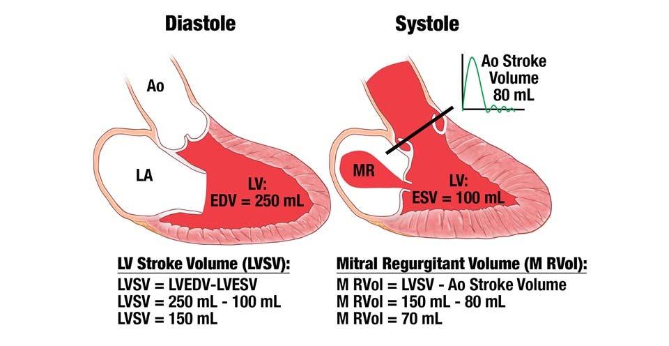


14 Quantitation of MR with CMR Quantification of MR with CMR LV stroke volume Aortic Stroke Volume Reg Volume = LV SV Ao SV 14 14


15 Aortic Regurgitation Aortic Regurgitation Color Doppler 15 15
 Reversal of flow in the aorta (EDVel >20 cm/s)")

16 Integrative Approach to AR Integrative approach should be used Supportive data LV/LA size PHT >500 Specific Data (>90% Sp) Reversal of flow in the aorta (EDVel >20 cm/s) Vena contracta > 0.6 cm % LVOT 65% Quantitative Data: 2D and 3D Regurgitant Volume Regurgitant Fraction EROA Attempt to understand discrepant measurements! Aortic Regurgitation 16 16


17 Aortic compliance The aortic reverse/forward flow ratio (35±10%) was positively associated with parameters of aortic stiffness Holodiastolic and rapid flow reversal can occur in elderly patients with stiff aorta, even in the absence of significant AR Hashimoto,H and Ito, S. Hypertension. 2013;62:


18 RF as PHT CircAdapt computational model of the human cardiovascular system Palau Caballero G et al. J Am Soc Echocardiogr 2016 Nov;29(11): LV Stiffness and PHT RF as PHT Inconsistent AR severity scores can result from variations in ventricular and aortic stiffness. A more rapid decay in the diastolic transvalvular pressure gradient due to ventricular or aortic stiffening can lead to overestimation of AR severity by PHT. Palau Caballero G et al. J Am Soc Echocardiogr 2016 Nov;29(11):


19 Chronic Aortic Regurgitation by Doppler Echocardiography * Specific Criteria for Mild AR VC width < 0.3 cm Central Jet, width < 25% of LVOT Small or no flow convergence Soft or incomplete jet by CW PHT > 500 ms Normal LV size Yes, mild Does AR meet specific criteria of mild or severe AR? Intermediate Values: AR Probably moderate 2-3 criteria 2-3 criteria Perform quantitative methods whenever possible to refine assessment Yes, severe * Specific Criteria for Severe AR Flail Valve VC width > 0.6 cm Central Jet, width 65% of LVOT Large flow convergence PHT < 200 ms Prominent holodiastolic flow reversal in the descending aorta Enlarged LV with normal function 4 criteria Definitively mild (quantitation not needed) RVol < 30 ml RF < 30% EROA <0.1 cm 2 AR Grade I RVol ml RF 30-39% EROA cm 2 AR Grade II RVol ml RF 40-49% EROA cm 2 AR Grade III RVol 60 ml RF 50% EROA 0.3 cm 2 AR Grade IV 4 criteria Definitively severe (may still quantitate) 3 specific criteria for severe AR Mild AR Moderate AR Severe AR Poor TTE quality or low confidence in measured Doppler parameters Discordant quantitative and qualitative parameters and/or clinical data Indeterminate AR Consider further testing: TEE or CMR for quantitation * Beware of limitations of color flow assessment in eccentric AR jets; volumetric quantitation and integration of other parameters is advised 19 19


20 CMR in Aortic Regurgitation Quantitation of AR with CMR 20 20


21 Tricuspid Valve Regurgitation TR & PR MR Zoghbi et al J Am Soc Echocardiogr April MR cho


22 The Spectrum of Tricuspid Regurgitation PISA in Tricuspid Regurgitation 22 22


 RT Hahn and JL Zamorano.")
23 PISA LIMITED FOR FUNCTIONAL TR! TETHERED DYNAMIC Volume overload is welltolerated for years No reduction in RV function Few symptoms of insidious onset Poor understanding about grading the severity of TR on Echo Patients present LATE!! NON CIRCULAR SEVERE x3 3D vena contracta area = 1.5 cm2 TR Grading: Work in Progress SCOUT 1 is the first tricuspid valve device trial to use Doppler quantitative measures of disease severity Hahn RT. Circ Cardiovasc Imaging Dec;9(12) RT Hahn and JL Zamorano. European Heart Journal - Cardiovascular Imaging (2017) 00, 1 2 doi: /ehjci/jex


24 Chronic Tricuspid Regurgitation by Doppler Echocardiography Yes, mild Specific Criteria for Mild TR Thin, small central color jet VC width <0.3 cm PISA Radius <0.4 cm at Nyquist cm/s Incomplete or faint CW jet Systolic dominant Hepatic vein flow Tricuspid A-wave dominant inflow Normal RV/RA Does TR meet most specific criteria for mild or severe TR? No Minority of criteria or Intermediate Values: TR Probably Moderate Perform VC measurement, and May perform quantitative PISA method, whenever possible* Yes, severe Specific Criteria for Severe TR Dilated annulus with no valve coaptation or flail leaflet Large central jet > 50% of RA VC width > 0.7 cm PISA radius > 0.9 cm at Nyquist 30-40cm/s Dense, triangular CW jet or sine wave pattern. Systolic reversal of Hepatic vein flow Dilated RV with preserved function VC width < 0.3 cm * EROA < 0.2 cm 2 * RVol < 30 ml VC width cm * EROA cm 2 * RVol = ml VC width > 0.7 cm * EROA > 0.4 cm 2 * RVol 45 ml Mild TR Moderate TR Severe TR Poor TTE quality or low confidence in measured Doppler parameters Discordant quantitative and qualitative parameters and/or clinical data Indeterminate TR Consider further testing: TEE or CMR for quantitation * Clinical experience in quantitation of TR is much less than that with mitral and aortic regurgitation CMR in TR and Right Heart Visualization & Quantitation In my limited experience, CMR UNDERESTIMATES TR 24 24


25 Pulmonic Regurgitation Chronic Pulmonic Regurgitation by Color Doppler Specific Criteria for mild PR Small Jet, with narrow width Soft or faint CW jet Slow deceleration time Normal RV Size Yes, mild Does PR meet most specific criteria for mild or severe PR? No Minority of criteria or Intermediate Values: PR Probably Moderate May Perform volumetric quantitative methods, if possible, whenever significant PR is suspected* Yes, severe Specific Criteria for Severe PR Jet width/annulus 70% Dense jet, PHT < 100 ms Early termination of PR flow Diastolic flow reversal in PA branches Dilated RV with NL function RF <20% RF 20-40% RF >40% Mild PR Moderate PR Severe PR Poor TTE quality or discordant parameters with clinical data, particularly when significant PR may be suspected Indeterminate PR Consider CMR for quantitation * Clinical experience in quantitation of PR is sparse


26 CMR in Pulmonic Regurgitation Quantitation of Rvol, RV Size & Function New ASE Valvular Regurgitation Guidelines- Endorsed by SCMR General Considerations What is New? Emphasis on identification of Etiology/Mechanism of regurgitation 2D/3D TTE an integrative approach & algorithms to assess severity When is TEE needed Important role of CMR & CMR methodology The challenge of co existing valvular lesions A clinical perspective Library of case studies on the web:
ASE Guidelines on Aortic Regurgitation What Do I Measure? Case Studies
 ASE Guidelines on Aortic Regurgitation What Do I Measure? Case Studies Mitral Regurgitation The New ASE Guidelines: Role of 2D/3D and CMR William A. Zoghbi MD, FASE, MACC Professor and Chairman, Department
Valvular Regurgitation: Can We Do Better Than Colour Doppler?
 Valvular Regurgitation: Can We Do Better Than Colour Doppler? A/Prof David Prior St Vincent s Hospital Melbourne Sports Cardiology Valvular Regurgitation Valve regurgitation volume loads the ventricles
New 3D Quantification of Mitral Regurgitation Severity. Judy Hung, MD Cardiac Ultrasound Laboratory Massachusetts General Hospital Boston, MA
 New 3D Quantification of Mitral Regurgitation Severity Judy Hung, MD Cardiac Ultrasound Laboratory Massachusetts General Hospital Boston, MA No Financial Disclosures No off label discussion of devices
Professors Carpentier and McGoon Mechanism, resulting from the disease Severity of regurgitation, resulting from the mechanism Echo
 Professors Carpentier and McGoon Mechanism, resulting from the disease Severity of regurgitation, resulting from the mechanism Echo define the mechanism, quantify the regurgitation severity CP1293058-3
Prof. JL Zamorano Hospital Universitario Ramón y Cajal
 Prof. JL Zamorano Hospital Universitario Ramón y Cajal Should we forget TR? Nath J et al. Impact of tricuspid regurgitation on long-term survival. J Am Coll Cardiol. 2004; 43:405-409 Why is it difficult
How to assess ischaemic MR?
 ESC 2012 How to assess ischaemic MR? Luc A. Pierard, MD, PhD, FESC, FACC Professor of Medicine Head, Department of Cardiology University Hospital Sart Tilman, Liège ESC 2012 No conflict of interest Luc
Quantification of Aortic Regurgitation
 Quantification of Aortic Regurgitation ASE Review 2018 Boston Susan E Wiegers, MD, FASE, FACC Professor of Medicine And thanks to Dr. Roberto Lang Disclosure None related to this presentation 1 Objectives
Quantifying Aortic Regurgitation
 Quantifying Aortic Regurgitation Linda D. Gillam, MD, MPH Morristown Medical Center Dorothy and Lloyd Huck Chair Cardiovascular Medicine Atlantic Health System No Disclosures 1 Valve Dysfunction Functional
Marti McCulloch, BS, MBA, RDCS, FASE Houston, Texas
 Marti McCulloch, BS, MBA, RDCS, FASE Houston, Texas Mitral Regurgitation What to Expect Review Specific Signs of Severity Supportive Signs of Severity Qualitative Parameters Structural Doppler Quantitative
What are the best diagnostic tools to quantify aortic regurgitation?
 What are the best diagnostic tools to quantify aortic regurgitation? Agnès Pasquet, MD, PhD Pôle de Recherche Cardiovasculaire Institut de Recherche Expérimentale et Clinique Université catholique de Louvain
EVALUATION OF CHRONIC MITRAL REGURGITATION: ASSESSING MECHANISMS AND QUANTIFYING SEVERITY 2018 STRUCTURAL HEART DISEASE CONFERENCE June 1, 2018
 1 EVALUATION OF CHRONIC MITRAL REGURGITATION: ASSESSING MECHANISMS AND QUANTIFYING SEVERITY 2018 STRUCTURAL HEART DISEASE CONFERENCE June 1, 2018 David A. Orsinelli, MD, FACC, FASE Professor, Internal
MITRAL REGURGITATION ECHO PARAMETERS TOOL
 Comprehensive assessment of qualitative and quantitative parameters, along with the use of standardized nomenclature when reporting echocardiographic findings, helps to better define a patient s MR and
MR echo case. N.Koutsogiannis Department of Cardiology University Hospital Of Patras
 MR echo case N.Koutsogiannis Department of Cardiology University Hospital Of Patras Case A 35 years old male came to the echo lab for a third opinion for his valvulopathy. He reports a long standing MR
Quantification of MR
 Valvular Regurgitation: Putting the New Guidelines into Practice James D. Thomas, MD, FACC, FASE, FESC Director, Center for Heart Valve Disease Bluhm Cardiovascular Institute Professor of Medicine, Feinberg
PISA Evaluation of Mitral Regurgitation. Raymond Graber, MD Cardiac Anesthesia Group University Hospitals Case Medical Center 4/07/2011
 PISA Evaluation of Mitral Regurgitation Raymond Graber, MD Cardiac Anesthesia Group University Hospitals Case Medical Center 4/07/2011 Introduction Evaluation of MR. What is PISA? Physiologic basis Issues
Quantitation of Aortic Regurgitation ASCeXAM / ReASCE Review Course
 Quantitation of Aortic Regurgitation 2017 ASCeXAM / ReASCE Review Course David Adams, ACS, RCS, RDCS, FASE Duke University Medical Center May 1, 2017 Disclosures None 1 Exam based on: Zoghbi et al. JASE
25 different brand names >44 different models Sizes mm
 Types of Prosthetic Valves BIOLOGIC STENTED Porcine xenograft Pericardial xenograft STENTLESS Porcine xenograft Pericardial xenograft Homograft (allograft) Autograft PERCUTANEOUS MECHANICAL Bileaflet Single
Tricuspid and Pulmonary Valve Disease
 Tricuspid and Pulmonary Valve Disease Lawrence Rudski MD FRCPC FACC FASE Professor of Medicine Director, Division of Cardiology Jewish General Hospital McGill University Right Sided Failure Edema Gut congestion
Diastolic Heart Function: Applying the New Guidelines Case Studies
 Diastolic Heart Function: Applying the New Guidelines Case Studies Mitral Regurgitation The New ASE William Guidelines: A. Zoghbi Role MD, of FASE, 2D/3D MACCand CMR Professor and Chairman, Department
Titel Kardiologie-SG.ch hot topics in heart failure and mitral regurgitation
 Titel Kardiologie-SG.ch hot topics in heart failure and mitral regurgitation where and how to quantify mitral regurgitation: Echolab, Cathlab or MRI? Philipp K. Haager, St. Gallen Measuring mitral regurgitation?
Tricuspid and Pulmonary Valve Disease
 Tricuspid and Pulmonary Valve Disease Lawrence Rudski MD FRCPC FACC FASE Professor of Medicine Director, Division of Cardiology Jewish General Hospital McGill University Question 1 All of the following
Ioannis Alexanian, MD, PhD Department of Cardiology General Hospital of Chest Diseases Sotiria Athens
 MITRAL REGURGITATION IN PATIENT WITH SEVERE AORTIC VALVE STENOSIS Ioannis Alexanian, MD, PhD Department of Cardiology General Hospital of Chest Diseases Sotiria Athens I HAVE NOTHING TO DECLARE Management
Hemodynamic Assessment. Assessment of Systolic Function Doppler Hemodynamics
 Hemodynamic Assessment Matt M. Umland, RDCS, FASE Aurora Medical Group Milwaukee, WI Assessment of Systolic Function Doppler Hemodynamics Stroke Volume Cardiac Output Cardiac Index Tei Index/Index of myocardial
CHAPTER 4 AN EFFICACIOUS APPROACH FOR THE QUANTIFICATION OF MITRAL REGURGITATION USING IMAGE PROCESSING AND PROXIMAL FLOW CONVERGENCE METHOD
 CHAPTER 4 AN EFFICACIOUS APPROACH FOR THE QUANTIFICATION OF MITRAL REGURGITATION USING IMAGE PROCESSING AND PROXIMAL FLOW CONVERGENCE METHOD 4.1. Introduction Mitral Regurgitation also called Mitral Insufficiency
The Doppler Examination. Katie Twomley, MD Wake Forest Baptist Health - Lexington
 The Doppler Examination Katie Twomley, MD Wake Forest Baptist Health - Lexington OUTLINE Principles/Physics Use in valvular assessment Aortic stenosis (continuity equation) Aortic regurgitation (pressure
Echocardiography. Guidelines for Valve and Chamber Quantification. In partnership with
 Echocardiography Guidelines for Valve and Chamber Quantification In partnership with Explanatory note & references These guidelines have been developed by the Education Committee of the British Society
TAVR: Echo Measurements Pre, Post And Intra Procedure
 2017 ASE Florida, Orlando, FL October 10, 2017 8:00 8:25 AM 25 min TAVR: Echo Measurements Pre, Post And Intra Procedure Muhamed Sarić MD, PhD, MPA Director of Noninvasive Cardiology Echo Lab Associate
LUST trial. Echocardiography USER S MANUAL
 LUST trial Echocardiography USER S MANUAL Rosa Sicari, Luna Gargani Ins1tute of Clinical Physiology Na1onal Council of Research, Pisa, Italy Parameters required (1) Aortic root Measurement of aortic root
The difficult patient with mitral regurgitation
 Clinical pathways The difficult patient with mitral regurgitation Stress echo can be the best tool Challenging cases Maria João Andrade, Lisbon PT Management of Severe Chronic Organic MR Echo Exercise
Comments restricted to Sapien and Corevalve 9/12/2016. Disclosures: Core Lab contracts with Edwards Lifesciences, Middlepeak, Medtronic
 Para-ValvularRegurgitation post TAVR: Predict, Prevent, Quantitate, Manage Linda D. Gillam, MD, MPH, FACC, FASE Chair, Department of Cardiovascular Medicine Morristown Medical Center/Atlantic Health System
Regurgitant Lesions. Bicol Hospital, Legazpi City, Philippines July Gregg S. Pressman MD, FACC, FASE Einstein Medical Center Philadelphia, USA
 Regurgitant Lesions Bicol Hospital, Legazpi City, Philippines July 2016 Gregg S. Pressman MD, FACC, FASE Einstein Medical Center Philadelphia, USA Aortic Insufficiency Valve anatomy and function LVOT and
Guidelines in perspective?
 EuroValve 2016 Challenges in the management Secondary MR Guidelines in perspective? Luc A. Pierard, MD, PhD Professor of Medicine Head of the Department of Cardiology, Liège, Belgium Heart Valve Clinic,
Imaging MV. Jeroen J. Bax Leiden University Medical Center The Netherlands Davos, feb 2015
 Imaging MV Jeroen J. Bax Leiden University Medical Center The Netherlands Davos, feb 2015 MV/MR: information needed on.. 1. MV anatomy 2. MR etiology - primary vs secondary 3. MR severity quantification
Echocardiography: Guidelines for Valve Quantification
 Echocardiography: Guidelines for Echocardiography: Guidelines for Chamber Quantification British Society of Echocardiography Education Committee Richard Steeds (Chair), Gill Wharton (Lead Author), Jane
ECHOCARDIOGRAPHY DATA REPORT FORM
 Patient ID Patient Study ID AVM - - Date of form completion / / 20 Initials of person completing the form mm dd yyyy Study period Preoperative Postoperative Operative 6-month f/u 1-year f/u 2-year f/u
Clinical Outcome of Tricuspid Regurgitation. David Messika-Zeitoun
 Clinical Outcome of Tricuspid Regurgitation David Messika-Zeitoun I have financial relationships to disclose Consultant for: Edwards, Symetis and Valtech Tricuspid Regurgitation is a Common Finding Tricuspid
CHAPTER 5 EFFICIENT APPROACHES FOR QUANTIFICATION OF AORTIC REGURGITATION USING PROXIMAL ISOVELOCITY SURFACE AREA PROCESS
 CHAPTER 5 EFFICIENT APPROACHES FOR QUANTIFICATION OF AORTIC REGURGITATION USING PROXIMAL ISOVELOCITY SURFACE AREA PROCESS 5.1. Introduction Aortic Regurgitation is also known as Aortic Insufficiency (AI).
JOINT MEETING 2 Tricuspid club Chairpersons: G. Athanassopoulos, A. Avgeropoulou, M. Khoury, G. Stavridis
 JOINT MEETING 2 Tricuspid club Chairpersons: G. Athanassopoulos, A. Avgeropoulou, M. Khoury, G. Stavridis Similarities and differences in Tricuspid vs. Mitral Valve Anatomy and Imaging. Echo evaluation
Primary Mitral Regurgitation
 EURO VALVE Madrid News from Valves Guidelines 2012: What s new and Why? Primary Mitral Regurgitation Luc A. Pierard, MD, PhD Professor of Medicine Head of the Department of Cardiology Heart Valve Clinic,
What echo measurements are key prior to MitraClip?
 APHP CHU Bichat - Claude Bernard What echo measurements are key prior to MitraClip? Eric Brochet,MD Cardiology Department Hopital Bichat Paris France No disclosure Conflict of interest Case 69 y.o man
Prosthesis-Patient Mismatch or Prosthetic Valve Stenosis?
 EuroValves 2015, Nice Prosthesis-Patient Mismatch or Prosthetic Valve Stenosis? Philippe Pibarot, DVM, PhD, FACC, FAHA, FASE FESC Canada Research Chair in Valvular Heart Diseases Université LAVAL Disclosure
Prosthetic valve dysfunction: stenosis or regurgitation
 Prosthetic valve dysfunction: stenosis or regurgitation Jean G. Dumesnil MD, FRCP(C), FACC, FASE(Hon) Quebec Heart and Lung Institute, Québec, Québec No disclosures Possible Causes of High Gradients in
What I Have Learned from 3D Imaging of Heart Valve Disease
 What I Have Learned from 3D Imaging of Heart Valve Disease Rebecca T. Hahn, MD Director of Interventional Echocardiography Columbia University Core Lab Director for multiple tricuspid device trials for
NEW GUIDELINES. A Guideline Protocol for the Assessment of Aortic Regurgitation From the British Society of Echocardiography Education Committee
 NEW GUIDELINES A Guideline Protocol for the Assessment of Aortic Regurgitation From the British Society of Echocardiography Education Committee Gill Wharton, Prathap Kanagala (Lead Authors) Richard Steeds
Aortic Stenosis: Spectrum of Disease, Low Flow/Low Gradient and Variants
 Aortic Stenosis: Spectrum of Disease, Low Flow/Low Gradient and Variants Martin G. Keane, MD, FASE Professor of Medicine Lewis Katz School of Medicine at Temple University Basic root structure Parasternal
MITRAL VALVE PATHOLOGY WITH TRICUSPID REGURGITATION (AND PHT)
") UNIVERSITY OF PADUA, SCHOOL OF MEDICINE Department of Cardiac,Thoracic and Vascular Sciences Padua, Italy MITRAL VALVE PATHOLOGY WITH TRICUSPID REGURGITATION (AND PHT) Luigi P. Badano**, MD, PhD, FESC,
UNIVERSITY OF PADUA, SCHOOL OF MEDICINE Department of Cardiac,Thoracic and Vascular Sciences Padua, Italy MITRAL VALVE PATHOLOGY WITH TRICUSPID REGURGITATION (AND PHT) Luigi P. Badano**, MD, PhD, FESC,
Uncommon Doppler Echocardiographic Findings of Severe Pulmonic Insufficiency
 Uncommon Doppler Echocardiographic Findings of Severe Pulmonic Insufficiency Rahul R. Jhaveri, MD, Muhamed Saric, MD, PhD, FASE, and Itzhak Kronzon, MD, FASE, New York, New York Background: Two-dimensional
Aortic Regurgitation & Aorta Evaluation
 VALVULAR HEART DISEASE Regurgitation Valvular Lessions 2017 Aortic Regurgitation & Aorta Evaluation Jorge Eduardo Cossío-Aranda MD, FACC Chairman of Outpatient Care Department Instituto Nacional de Cardiología
Echocardiographic Evaluation of Aortic Valve Prosthesis
 Echocardiographic Evaluation of Aortic Valve Prosthesis Amr E Abbas, MD, FACC, FASE, FSCAI, FSVM, RPVI Co-Director, Echocardiography, Director, Interventional Cardiology Research, Beaumont Health System
Echocardiographic evaluation of mitral stenosis
 Echocardiographic evaluation of mitral stenosis Euroecho 2011 Philippe Unger, MD, FESC Erasme Hospital, ULB, Brussels, Belgium I have nothing to declare EuroHeart Survey Etiology of single native left-sided
Dobutamine Stress testing In Low Flow, Low EF, Low Gradient Aortic Stenosis Case Studies
 Dobutamine Stress testing In Low Flow, Low EF, Low Gradient Aortic Stenosis Case Studies Mitral Regurgitation The New ASE Guidelines: Role of 2D/3D and CMR William A. Zoghbi MD, FASE, MACC Professor and
What Degree of MR Deserves Surgical or Transcatheter Intervention, and How Should It Be Assessed?
 What Degree of MR Deserves Surgical or Transcatheter Intervention, and How Should It Be Assessed? Robert J. Siegel, M.D., FACC Nov. 14-15, 2017, Beverly Hills Director, Cardiac Non-Invasive Laboratory
Aortic Regurgitation and Aortic Aneurysm - Epidemiology and Guidelines -
 Reconstruction of the Aortic Valve and Root - A Practical Approach - Aortic Regurgitation and Aortic Aneurysm Wednesday 14 th September - 9.45 Practice must always be founded on sound theory. Leonardo
When Does 3D Echo Make A Difference?
 When Does 3D Echo Make A Difference? Wendy Tsang, MD, SM Assistant Professor, University of Toronto Toronto General Hospital, University Health Network 1 Practical Applications of 3D Echocardiography Recommended
ICE: Echo Core Lab-CRF
 APPENDIX 1 ICE: Echo Core Lab-CRF Study #: - Pt Initials: 1. Date of study: / / D D M M M Y Y Y Y 2. Type of Study: TTE TEE 3. Quality of Study: Poor Moderate Excellent Ejection Fraction 4. Ejection Fraction
Back to Basics: Common Errors In Quantitation In Everyday Practice
 Back to Basics: Common Errors In Quantitation In Everyday Practice Deborah Agler, ACS, RDCS, FASE October 9, 2017 ASE: Echo Florida Rebecca T. Hahn, MD Director of Interventional Echocardiography Professor
Management of TR in Patients Undergoing Mitral Interventions
 Management of TR in Patients Undergoing Mitral Interventions Stephen H. Little, MD John S. Dunn Chair in Cardiovascular Research and Education, Associate professor, Weill Cornell Medicine shlittle@houstonmethodist.org
DOPPLER HEMODYNAMICS (1) QUANTIFICATION OF PRESSURE GRADIENTS and INTRACARDIAC PRESSURES
 QUANTIFICATION OF PRESSURE GRADIENTS and INTRACARDIAC PRESSURES") THORAXCENTRE DOPPLER HEMODYNAMICS (1) QUANTIFICATION OF PRESSURE GRADIENTS and INTRACARDIAC PRESSURES J. Roelandt DOPPLER HEMODYNAMICS Intracardiac pressures and pressure gradients Volumetric measurement
THORAXCENTRE DOPPLER HEMODYNAMICS (1) QUANTIFICATION OF PRESSURE GRADIENTS and INTRACARDIAC PRESSURES J. Roelandt DOPPLER HEMODYNAMICS Intracardiac pressures and pressure gradients Volumetric measurement
Direct Planimetry of Mitral Valve Regurgitation Orifice Area by Real-time 3D Transesophageal Echocardiography
 Direct Planimetry of Mitral Valve Regurgitation Orifice Area by Real-time 3D Transesophageal Echocardiography Ertunc Altiok, Sandra Hamada, Silke van Hall, Mehtap Hanenberg, Eva Grabskaya, Michael Becker,
ASCeXAM / ReASCE. Practice Board Exam Questions Monday Morning
 ASCeXAM / ReASCE Practice Board Exam Questions Monday Morning Ultrasound Physics Artifacts Doppler Physics Imaging, Knobology, and Artifacts Echocardiographic Evaluation of the RV Tricuspid and Pulmonary
AATS Annual Meeting Seattle, WA Irving Kron, M.D. Professor and Chairman Department of Surgery University of Virginia Hospital Charlottesville,
 AATS Annual Meeting Seattle, WA Irving Kron, M.D. Professor and Chairman Department of Surgery University of Virginia Hospital Charlottesville, Virginia AATS Ischemic MR Guideline Writing Group Roster
Prof. Patrizio LANCELLOTTI, MD, PhD Heart Valve Clinic, University of Liège, CHU Sart Tilman, Liège, BELGIUM
 The Patient with Aortic Stenosis and Mitral Regurgitation Prof. Patrizio LANCELLOTTI, MD, PhD Heart Valve Clinic, University of Liège, CHU Sart Tilman, Liège, BELGIUM Aortic Stenosis + Mitral Regurgitation?
Case Reviews: Hemodynamic Calculations in Valvular Regurgitation
 Case Reviews: Hemodynamic Calculations in Valvular Regurgitation Case 5 History: 69-year-old man with orthotopic heart transplant 15 years ago. Inferior MI several years ago. Recurrent CHF. Currently dyspneic
HOW IMPORTANT ARE THESE ECHO MEASUREMENTS ANYWAY?
 HOW IMPORTANT ARE THESE ECHO MEASUREMENTS ANYWAY? John D. Carroll, MD Professor, Director of Interventional Cardiology and Co-Medical Director of the Cardiac and Vascular Center, University of Colorado
3D Echo for Evaluation of Tricuspid Regurgitation Jong-Min Song, MD, PhD
 3D Echo for Evaluation of Tricuspid Regurgitation Jong-Min Song, MD, PhD Asan Medical Center University of Ulsan College of Medicine Seoul, Korea Causes of TR Primary causes (25%) Rheumatic Myxomatous
ECHO HAWAII. Role of Stress Echo in Valvular Heart Disease. Not only ischemia! Cardiomyopathy. Prosthetic Valve. Diastolic Dysfunction
 Role of Stress Echo in Valvular Heart Disease ECHO HAWAII January 15 19, 2018 Kenya Kusunose, MD, PhD, FASE Tokushima University Hospital Japan Not only ischemia! Cardiomyopathy Prosthetic Valve Diastolic
Doppler Basic & Hemodynamic Calculations
 Doppler Basic & Hemodynamic Calculations August 19, 2017 Smonporn Boonyaratavej MD Division of Cardiology, Department of Medicine Chulalongkorn University Cardiac Center, King Chulalongkorn Memorial Hospital
Functional Mitral Regurgitation
 Club 35 - The best in heart valve disease - Functional Mitral Regurgitation Steven Droogmans, MD, PhD UZ Brussel, Jette, Belgium 08-12-2011 Euroecho & other Imaging Modalities 2011 No conflicts of interest
Advanced Applica,on of Point- of- Care Echocardiography in Cri,cal Care. Dr. Mark Tutschka Dr. Rob ArnAield
 Advanced Applica,on of Point- of- Care Echocardiography in Cri,cal Care Dr. Mark Tutschka Dr. Rob ArnAield OBJECTIVES Provide an overview of common advanced echocardiographic techniques suitable for use
Bogdan A. Popescu. University of Medicine and Pharmacy Bucharest, Romania. EAE Course, Bucharest, April 2010
 Bogdan A. Popescu University of Medicine and Pharmacy Bucharest, Romania EAE Course, Bucharest, April 2010 This is how it started Mitral stenosis at a glance 2D echo narrow diastolic opening of MV leaflets
Imaging Assessment of Aortic Stenosis/Aortic Regurgitation
 Imaging Assessment of Aortic Stenosis/Aortic Regurgitation Craig E Fleishman, MD FACC FASE The Heart Center at Arnold Palmer Hospital for Children, Orlando SCAI Fall Fellows Course 2014 Las Vegas Disclosure
PROSTHETIC VALVE BOARD REVIEW
 PROSTHETIC VALVE BOARD REVIEW The correct answer D This two chamber view shows a porcine mitral prosthesis with the typical appearance of the struts although the leaflets are not well seen. The valve
Echo Doppler Assessment of Right and Left Ventricular Hemodynamics.
 Echo Doppler Assessment of Right and Left Ventricular Hemodynamics. Itzhak Kronzon, MD, FASE, FACC, FESC, FAHA, FACP, FCCP Northwell, Lenox Hill Hospital, New York Professor of Cardiology Hofstra University
imagine 2018 Echocardiography Today - Do, Learn, Do More, Learn More November 3-4, 2018 Georgia Technology Hotel and Conference Center
 Piedmont Heart presents imagine 2018 Echocardiography Today - Do, Learn, Do More, Learn More November 3-4, 2018 Georgia Technology Hotel and Conference Center Program Co-Directors Mani A. Vannan MBBS FASE
Organic mitral regurgitation
 The best in heart valve disease Organic mitral regurgitation Ewa Szymczyk Department of Cardiology Medical University of Lodz, Poland I have nothing to declare Organic mitral regurgitation leaflet abnormality
The FORMA Early Feasibility Study: 30-Day Outcomes of Transcatheter Tricuspid Valve Therapy in Patients with Severe Secondary Tricuspid Regurgitation
 The FORMA Early Feasibility Study: 30-Day Outcomes of Transcatheter Tricuspid Valve Therapy in Patients with Severe Secondary Tricuspid Regurgitation Susheel Kodali, MD Director, Structural Heart & Valve
HIGHLIGHT SESSION. Imaging. J. L. Zamorano Gomez (Madrid, ES) Disclosures: Speaker Philips
 Disclosures: Speaker Philips") Imaging. J. L. Zamorano Gomez (Madrid, ES) Disclosures: Speaker Philips Agenda ECHO Diagnosis & Prognosis : Functional MR Severity Aortic Stenosis CT How to select pts for TAVI Adding prognostic info to
Imaging. J. L. Zamorano Gomez (Madrid, ES) Disclosures: Speaker Philips Agenda ECHO Diagnosis & Prognosis : Functional MR Severity Aortic Stenosis CT How to select pts for TAVI Adding prognostic info to
How to Assess and Treat Obstructive Lesions
 How to Assess and Treat Obstructive Lesions Erwin Oechslin, MD, FESC, FRCPC, Director, Congenital Cardiac Centre for Adults Peter Munk Cardiac Centre University Health Network/Toronto General Hospital
Objectives. Considerations in management of multivalvular disease. Case Discussions. A Systematic Approach to Multivalve Disease.
 A Systematic Approach to Multivalve Disease James D. Thomas, MD, FACC, FASE Director, Center for Heart Valve Disease Bluhm Cardiovascular Institute Professor of Medicine, Feinberg School of Medicine, Northwestern
Quantification of Mitral Stenosis: Planimetry, pressure Half time, Continuity Common Errors
 Quantification of Mitral Stenosis: Planimetry, pressure Half time, Continuity Common Errors Christopher J Kramer RDCS Advanced Cardiovascular Services Aurora Health Care Milwaukee, WI No Disclosures Baumgartner,
Stress Testing in Valvular Disease
 2017 ASE Florida Orlando, FL October 10, 2017 2:40 2:50 PM 10 min Grand Harbor Ballroom South Stress Testing in Valvular Disease Muhamed Sarić MD, PhD, MPA Director of Noninvasive Cardiology Echo Lab Associate
Introduction RECOMMENDATIONS
 European Journal of Echocardiography (2010) 11, 223 244 doi:10.1093/ejechocard/jeq030 RECOMMENDATIONS European Association of Echocardiography recommendations for the assessment of valvular regurgitation.
3D Printing & Echocardiography
 ASE SOTA Feb 19, 2018 3D Printing & Echocardiography Stephen H. Little, MD John S. Dunn Chair in Cardiovascular Research and Education, Associate professor, Weill Cornell Medicine Disclosures Personal
P = 4V 2. IVC Dimensions 10/20/2014. Comprehensive Hemodynamic Evaluation by Doppler Echocardiography. The Simplified Bernoulli Equation
 Comprehensive Hemodynamic Evaluation by Doppler Echocardiography Itzhak Kronzon, MD North Shore LIJ/ Lenox Hill Hospital New York, NY Disclosure: Philips Healthcare St. Jude Medical The Simplified Bernoulli
Comprehensive Hemodynamics By Doppler Echocardiography. The Echocardiographic Swan-Ganz Catheter.
 Comprehensive Hemodynamics By Doppler Echocardiography. The Echocardiographic Swan-Ganz Catheter. Itzhak Kronzon, MD, FASE, FACC, FESC, FAHA, FACP, FCCP North Shore HS, LIJ/Lenox Hill Hospital, New York
Valve Disease Board Review Questions
 Valve Disease Board Review Questions Dennis A. Tighe, MD, FASE University of Massachusetts Medical School Worcester, MA Case 1 History A 61 year-old man Presents to hospital with worsening shortness of
Secondary MR joint with the mitral academy. What is new in our understanding of this disease? Luc Pierard University Hospital, Liège
 Secondary MR joint with the mitral academy What is new in our understanding of this disease? Luc Pierard University Hospital, Liège Faculty disclosure Luc Pierard I have no financial relationships to disclose.
Διαδερμική θεραπεία των παραβαλβιδικών διαφυγών Σ.Ράμμος
 ΔΜΙΝΑΡΙΟ ΔΠΙΣΗΜΟΝΙΚΩΝ ΔΝΩΔΩΝ ΔΛΛΗΝΙΚΟΤ ΚΟΛΛΔΓΙΟΤ ΚΑΡ ΙΟΛΟΓΙΑ 2-4.10.2015 Δπιζηημονική Ένωζη Δπεμβαηικής Καρδιολογίας Βαλβιδοπάθειες, Γιαδερμικές παρεμβάζεις Διαδερμική θεραπεία των παραβαλβιδικών διαφυγών
True morphology of mitral regurgitant flow assessed by three- dimensional transesophageal echocardiography
 DOI: 10.1111/echo.13395 ORIGINAL INVESTIGATION True morphology of mitral regurgitant flow assessed by three- dimensional transesophageal echocardiography Martin Lombardero M.D. Ruth Henquin D.L.S.H.T.M.,
Quantitative Assessment of Pulmonary Regurgitation by Echocardiography in Patients After Repaired TOF
 Quantitative Assessment of Pulmonary Regurgitation by Echocardiography in Patients After Repaired TOF 2013. 4. 20. 서울대학교어린이병원소아청소년과 권보상 W. B. TOF (large VSD, infundibular stenosis) 19 mo, 8.5 kg Indication
Ανεπάρκεια Τριγλώχινας Επιλογή Επεμβατικής Θεραπείας. Μ. Χρυσοχέρης Τμήμα Διαδερμικών Βαλβίδων ΔΘΚΑ ΥΓΕΙΑ
 Ανεπάρκεια Τριγλώχινας Επιλογή Επεμβατικής Θεραπείας Μ. Χρυσοχέρης Τμήμα Διαδερμικών Βαλβίδων ΔΘΚΑ ΥΓΕΙΑ Disclosures - Proctoring activities for Abbott Vascular, Edwards Lifesciences I and the HYGEIA Hospital
Federico M Asch MD, FASE MedStar Heart and Vascular Institute Georgetown University Washington, DC
 TAVR: When Things go Wrong Federico M Asch MD, FASE MedStar Heart and Vascular Institute Georgetown University Washington, DC Disclosures Academic Echo Core Lab Abbott / St Jude Medical Edwards Medtronic
HEMODYNAMIC ASSESSMENT
 HEMODYNAMIC ASSESSMENT INTRODUCTION Conventionally hemodynamics were obtained by cardiac catheterization. It is possible to determine the same by echocardiography. Methods M-mode & 2D echo alone can provide
Echocardiographic Evaluation of Aortic Valve Prosthesis
 Echocardiographic Evaluation of Aortic Valve Prosthesis Amr E Abbas, MD, FACC, FASE, FSCAI, FSVM, RPVI Co Director, Echocardiography, Director, Interventional Cardiology Research, Beaumont Health System
PARAVALVULAR LEAK POST TAVR. Elements of Follow-up Post TAVR
 PARAVALVULAR LEAK POST TAVR David S Rubenson MD FACC FASE Founding Director, Cardiac Non-Invasive Laboratory Scripps Clinic Medical Group number 1 Elements of Follow-up Post TAVR JACC CV Imag 2016;9:193
aortic regurgitation, vena contracta area, vena contracta width, live three-dimensional echocardiography
 RESEARCH FROM THE UNIVERSITY OF ALABAMA AT BIRMINGHAM Assessment of Aortic Regurgitation by Live Three-Dimensional Transthoracic Echocardiographic Measurements of Vena Contracta Area: Usefulness and Validation
Goals of Reporting. Background of the STS/ACC TVT Registry. TVT Registry TAVR and MitraClip 10/11/ /11/2016 9:10 9:30 AM
 10/11/2016 9:10 9:30 AM Rebecca T. Hahn, MD Director of Interventional Echocardiography Columbia University Medical College Background of the STS/ACC TVT Registry The STS/ACC TVT Registry is a benchmarking
Cover Page. The handle holds various files of this Leiden University dissertation.
 Cover Page The handle http://hdl.handle.net/1887/21650 holds various files of this Leiden University dissertation. Author: Shanks, Miriam Title: Evolving imaging techniques for the assessment of cardiac
Επιδιόπθωζη μιηποειδικήρ ζςζκεςήρ ζε ππόπηωζη ή πήξη γλωσίνων. Βαζίλειορ Σασπεκίδηρ Επιμεληηήρ Β Καπδιολογίαρ Γ.Ν. Παπαγεωπγίος
 Επιδιόπθωζη μιηποειδικήρ ζςζκεςήρ ζε ππόπηωζη ή πήξη γλωσίνων Βαζίλειορ Σασπεκίδηρ Επιμεληηήρ Β Καπδιολογίαρ Γ.Ν. Παπαγεωπγίος MV anatomy Otto C. NEJM 2001 Leaflets anatomy 2% of population Etiology Terminology
LV geometric and functional changes in VHD: How to assess? Mi-Seung Shin M.D., Ph.D. Gachon University Gil Hospital
 LV geometric and functional changes in VHD: How to assess? Mi-Seung Shin M.D., Ph.D. Gachon University Gil Hospital LV inflow across MV LV LV outflow across AV LV LV geometric changes Pressure overload