Light microscopic features of the rete testis, the vas efferens, the epididymis and the vas deferens in the adult rhesus monkey
|
|
|
- Owen Nicholas Thompson
- 5 years ago
- Views:
Transcription
1 J. Biosci., Vol. 1, Number 2, June 1979, pp Printed in India. Light microscopic features of the rete testis, the vas efferens, the epididymis and the vas deferens in the adult rhesus monkey ASHA PRAKASH, M. R. N. PRASAD and Τ. C. ANAND KUMAR* Department of Zoology, University of Delhi, Delhi *Neuroendocrine Research Laboratory, Department of Anatomy, All India Institute of Medical Sciences, New Delhi MS received 7 November 1978 Abstract. The present study was carried out to determine the detailed histological and cytological features of the excurrent ducts of the male reproductive system in the rhesus monkey. The excurrent ducts show a regional difference in their histological features. The use of some of these features as histological markers and their possible functional significance are discussed. The epithelial cells in the different components of the excurrent duct system possess cytological features which suggest their involvement in absorption and the secretion of different products into the lumen. Keywords. Rhesus monkey; rete testis; vas efferens; opididymis; vas deferens. Introduction The rete testis, the vas efferens, the epididymis and the vas deferens constitute a part of the excurrent ducts of the male reproductive system. These ducts not only subserve a simple function of conducting spermatozoa from the testis but they also play a more sophisticated role in altering the milieu of the spermatozoa (Hamilton, 1975). There is reason to believe that as a result of this latter function of the excurrent ducts, spermatozoa undergo a process of maturation and acquire the ability to fertilise ova. The microscopic structure of the excurrent ducts has been a subject of much interest and several studies have been carried out to identify the histological and cytological features associated with their diverse functions (Benoit, 1926; Reid and Cleland, 1957; Nicander, 1957a, b; Glover and Nicander, 1971; Hamilton, 1975; Hoffer, 1976; Kormano, 1977). Studies carried out so far have shown that although the general histological features of the excurrent ducts are essentially similar between species, there are subtle differences in their detailed cytological features. The rhesus monkey is one of the species of non-human primates widely used in experimental studies aimed at understanding reproductive processes occurring in Abbreviations : periodic acid Schiffs reagent, PAS. 185
2 186 Asha Prakash et al. man. All the same, little is known about the histological features of the excurrent ducts of the male reproductive system in this species, although, some histochemical (Arora Dinakar et al.,1977a, b, c) and ultrastructural (Dym, 1974; Ramos and Dym, 1977) studies have been carried out. The present investigation was undertaken to fill this gap and to form a basis for further structural studies on excurrent ducts in the rhesus monkey. Materials and methods Six adult, healthy male rhesus monkeys were used in the present study. After anaesthetising the animals with an intramuscular dose of Ketlar (5 mg/kg body wt., Parke-Davis), the testis along with the associated excurrent ducts were dissected out from one side of the animal and fixed by immersion in Bouin's fluid. Nicks were made in the tissues with a scalpel blade to facilitate penetration of the fixative. After fixation, small pieces of tissue were dissected out from the following regions: the cranial part of the testis which included a part of the rete testis, the vas efferens, the epididymis and representative pieces from the scrotal and abdominal portions of the vas deferens. The fixed material was processed and embedded in paraffin; representative sections (6 10 µm thick) from different portions of the excurrent duct system were stained with haematoxylin eosin or Periodic Acid Schiff's reagent (PAS). The epididymis was serially sectioned at a thickness of 10 µm. Regions corresponding to those tissues taken for paraffin sectioning were dissected out from the contralateral side of the animal, fixed in modified Karnovsky s fluid containing 4% paraformaldehyde and 1% glutaraldehyde, processed and embedded in araldite. The tissues were sectioned at 1 2 µm on a Reichert OMU 3 ultramicrotome and stained with 1% toluidine blue. Several photomicrographs of the excurrent ducts fixed in Bouin s fluid were prepared at a constant final magnification and the height of the epithelial cells and sterocilia and the diameter of the lumen were determined from these photomicrographs (table 1). Table 1. Dimensions of cell height, stereocilia height and lumen diameter in the vasa efferentia and different segments of the epididymis. * Proximal portion. ** Distal portion. Figures in parenthesis indicate number of observations made.
3 Microscopic features of monkey testis 187 Observations General The gross structural features of the testis and the associated excurrent ducts taken for the present study are shown in figure 1. The rete testis constitutes a system of interconnected channels extending from the mediastinum to the tunica albuginea. Twelve to 17 efferent ducts emerge from the cranial pole of the testis and become highly convoluted before individual ducts open into the epididymal duct. The epididymal duct is also highly convoluted and it continues into the vas deferens. The initial part of the vas deferens is mildly convoluted. The extratesticular portions of the excurrent ducts are surrounded by connective tissue comprising collagen, fibroblasts, macrophages, mast cells, nerve fibres and a network of blood and lymphatic vessels. The epithelium lining the lumen of the excurrent ducts lies over a basement membrane which is surrounded by a muscularis. Intra-epithelial lymphocytes occur throughout the excurrent ducts. These are pleomorphic cells with a clear cytoplasm and an intensely heterochromatic nucleus (figure 13; figure 16). Blood vessels are seen to lie close to the basement membrane. The luminal contents of the excurrent ducts show regional differences. The lumen in the rete testis, vas efferens and in the initial segment of the epididymis contains sparsely distributed spermatozo a and many immature germ cells (e.g., figure 4). Spermatozoal density increases progressively from the middle to the terminal segment where it is maximal. The proximal portion of the vas deferens also shows a high intraluminal concentration of spermatozoa. The number of immature germ cells is negligible; on the other hand, a large number of intraluminal macrophages are seen in areas of high spermatozoal density (figure 8). Rete testis The seminiferous tubules open into the rete testis through tubular extensions of the rete, the tubuli recti (figure 2). The junction between the seminiferous tubule and the tubulus rectus is distinguished by the presence of a knob-like protrusion composed of Sertoli cells (figure 2). It is not unusual to find spermatozoa associated with these Sertoli cells. The rete testis lying within the tunica albuginea opens into the proximal part of the efferent ducts (figure 3). The epithelial cells lining the rete show regional differences. Cells lining the tubuli recti are short columnar and possess an ovoid, indented nucleus. The cytoplasm of these cells is mostly homogenous but in the apical regions it has a vacuolated appearance (figure 11). The epithelium lining the rete in the mediastinum and in the tunica albuginea comprises mostly squam ous cells and a few large cuboidal cells which are distinguished by their hyaline cytoplasm and rounded nucleus (figure 12). In the region close to the junction between the rete and the vas efferens the epithelium is composed of short columnar cells whose apical regions contain prominent cytoplasmic vacuoles (figure 13).
4 188 Asha Prakash et al. Vas efferens The lumen of the vas efferens is lined by a pseudo-stratified columnar epithelium comprising ciliated and non-ciliated cells (figure 14). The height of the epithelial cells and the diameter of the lumen is indicated in table 1. The non-ciliated cell constitutes the predominant cell-type and it is distinguished by a basally situated, indented, euchromatic nucleus. Its cytoplasm contains a large number of granular inclusions which stain with PAS. The apical surface of this cell type shows features characteristic of a brush-border (figure 14). The ciliated cell has a pale cytoplasm containing a large number of mitochondria. The nucleus is round, centrally situated and contains euchromatin. The apical surface is lined by kinocilia associated with basal bodies which stain more deeply than the surrounding cytoplasm (figure 14). Epididymis The epididymis can be divided into an intial, a middle and a terminal segment on the basis of histological criteria defined by Glover and Nicander (1971). The dimensions of the epithelial cells, stereocilia and the lumen diameter in different segments of the epididymis are shown in table 1. The initial segment is characterised by a high epithelium with long stereocilia. The lumen is generally devoid of spermatozoa but, when presen t, they are very sparsely distributed (figure 5). In the middle segment the stereocilia are usually bent and spermatozoa are present in varying densities within the lumen (figure 6). In the terminal segment the epithelium is lower than in other regions and the stereocilia are also shorter. The lumen is packed with spermatozoa which exhibit a distinctly spiral arrangement. The peritubular mucularis shows considerable thickening (figure 8). Besides these histological differences, the lumen diameter of the epididymal duct increases progressively from the initial to the terminal segment (table 1). The epithelium in the distal part of the middle segment and in the proximal part of the terminal segment shows evaginations (figure 7) which in some sections appear as crypts. Three distinct cell types can be discerned in the epithelium lining the epididymal duct. These are: (1) the principal cell, which constitutes the predominant cell type, (2) the pale cell and (3) the basal cell. The principal cell extends from the basal lamina to the lumen. In the initial segment the principal cell is tall columnar and has an ovoid nucleus containing euchromatin (figure 5). The nuclear envelope has a regular outline which is a distinguishing feature of this segment. Cl ear vesicles and vacuoles fill the apical cytoplasm (figure 15). In the middle and terminal segments the principal cells become progressively shorter (figure 6 and figures 7 and 8) and the nuclear membrane displays indentations of increasing complexity (figure 16). Apical vesicles and vacuoles are few. The supranuclear region of principal cells shows negative images of the Golgi. Thecytoplasm cont ains well defined granules which stain deeply with toluidine blue. The granules are mostly distributed in the supranuclear region. Stereocilia extend into the lumen from the apical surface of principal cells (figures 15 and 16).
5 Microscopic features of monkey testis 189 The pale cell is distinguished by an apically situated nucleus which is round and contains much heterochromatin. The basal part of the cell is slender and elongated whilst the apical part is broad. The perinuclear cytoplasm in most cells contains thread-like inclusions which are presumably mitochondria. In some cells the supranuclear cytoplasm contains either vacuoles or large granules which stain intensely with PAS. The apical surface lacks stereocilia (figure 16). Pale cells are seen most frequently in the middle segment and least so in the terminal segment. The basal cell lies on the basement membrane and shows no connection with the lumen. The euchromatic nucleus is usually round and indented (figures 15 and 16). The cytoplasm of some basal cells contains granular material which stains metachromatically with toluidine blue. Vas deferens The pseudostratified epithelium is wavy and much taller in the scrotal (figure 9) than in the abdominal part of the vas deferens. In the abdominal portion of the vas deferens the epithelium forms deep projections into the lumen which show evidence of branching (figure 10). The epithelial cells of the vas deferens can be distinguished into Type I and Type II cells based on thei r cytological characteristics. The Type I cell is the predominant cell type. Cytological differences in this cell type are noticed between cells located in the proximal convoluted portion and in the distal straight portion of the vas deferens. The Type I cell in the former region (figure 17) exhibits extensions of the apical cytoplasm into the lumen. The nucleus is round or ovoid and the nuclear membrane is regular or mildly indented. In the distal straight segment of the vas deferens (figure 18), the Type I cell lacks apical extensions and the nucleus is ovoid and highly indented. The Type I cell in all regions of the vas deferens contains a vacuolated area above the nucleus which is distinct from the Golgi area. The Type II cell (figures 17 and 18) resembles the pale cell in the epididymal epithelium in that it possesses a round nucleus which is usually heterochromatic but may contain euchromatin, depending on the functional state of the cell. Moreover, the cytoplasm contains a perinuclear population of mitochondria. Unlike the epididymal pale cell, however, the Type II cell does not contain vacuoles and granules which stain with PAS. Basal cells resemble those present in the epididymal epithelium. However, basal cells containing metachromatic granules were not observed as frequently as in the epididymis. Discussion The present study has shown that the epithelium in the rete testis is of the simple type while in the vas efferens, epididymis and vas deferens the epithelium is pseudostratified. Besides a stable population of cells, the epithelium also contains a wandering population of intraepithelial lymphocytes which are presumed to be involved in preventing sperm antigens from reaching the blood stream and eliciting an putoimmune response (Dym and Romrell, 1975).
6 190 Asha Prakash et al. The possible functional significance of the histological and cytological features of the excurrent ducts can be better interpreted on the basis of what is already known of their physiology. Thus, the rete testis is not only an avenue through which spermatozoa traverse but a fairly substantial volume of fluid also courses through it (Tuck et al., 1970). It is therefore not surprising that only a few, sparsely distributed spermatozoa can be observed in histological sections of the rete testis. The rete testis fluid (RTF) is believed to be a mixture of fluids secreted by the seminiferous tubules and by the rete itself (Tuck et al., 1970). The cellular origin of the substances secreted by the rete has not yet been identified. The cytological features of the epithelial cells in the rete testis suggest that the cells having a hyaline cytoplasm and those having cytoplasmic vacuoles possibly contribute to the rete testis secretion; cells with such features occur in the tubuli recti, amidst the squamous cells in the mediastinum and intratunical portions of the rete testis and at the junction between the rete testis and th e vas efferens. Since the secretory cells in these regions of the rete are cytologically different from one another, it is likely that each of these 3 different cell types contribute different substances to the RTF. The vas efferens contains non-ciliated and ciliated cells. The non-ciliated cells constitute the predominant cell-type and they contain PAS positive granules as seen in other exocrine cells. The ciliated cell lacks such cytoplasmic inclusions but contains mitochondria which presumably constitute an energy source for ciliary movement. These cytological features are in consonance with a view that the nonciliated cells have a secretory role while ciliated cells subserve a function of transporting the constituents of the vas efferens by ciliary movement. The physiology of the epididymis has been more widely investigated than any other component of the excurrent ducts of the male reproductive system (Hamilton, 1975). Spermatozoa in the epididymis are known to undergo a process of maturation (Bedford, 1975). The epididymis is also known to function as a storehouse for spermatozoa (Glover and icander, 1971). Glover and Nicander (1971) suggested a nomenclature of initial and middle segments to such of the regions of the epididymis concerned with the maturation of spermatozoa and terminal segment to denote the region where spermatozoa are stored. The advantage of using this nomenclature against the classical terminology of caput, corpus and cauda to define the different regions of the epididymis has been discussed in detail by these authors. The present study has shown that it is also possible to distinguish an initial, a middle and a terminal segment in the epididymis of the rhesus monkey on the basis of histological criteria described by Glover and Nicander (1971). Apart from the presence of intraepithelial lymphocytes, the epididymal epithe lium of the rhesus monkey also contains 3 distinct cell types as has been found in the rat (Reid and Cleland, 1957). The cy tological features of the principal cell in the monkey epididymis are similar to those of the principal cell in the rate epididymis. The principal cell in the rat has been implicated with a function of fluid absorption (Hamilton, 1975) and it is likely that in the monkey also these cells have a similar role. The second cell type in the epididymal epithelium of the rat is the clear cell (Reid and Cleland, 1957) which is present only in the middle and terminal segments. These cells have been implicated with a secretory function and ultrastructural studies on the epididymis of the rat have shown that the clear cell in the middle segment is different from that found in the terminal segment (Anand Kumar et al., in press).
7 Microscopic features of monkey testis 191 In the rhesus monkey there is no cell type which is comparable to the clear cell of the rat epididymis. However, the second cell type in the epithelium of the monkey epididymis is the pale cell which is present in all the 3 segments. A similar cell type has been described in the human vas deferens (Hoffer, 1976). The cytological features of the pale cell show that they either contain an abundance of mitochondria, or PAS positive granules, or cytoplasmic vacuoles. These features suggest that pale cells may have a secretory function. The third cell type in the epididymal epithelium, the basal cell, is essentially similar in structure in the rhesus monkey and in the rat. The function of this cell type is not known. However, the presence of cytoplasmic droplets which stain metachromatically with toluidine blue in some cells suggests that these droplets may be lysosomes in which case it is likely that basal cells play a role in sequestering and disposing of the products absorbed by the epithelial cells. Further studies are needed to substantiate this view. The epithelium of the vas deferens comprises Type I and Type II cells and basal cells. The Type I cell in the proximal, convoluted portion of the vas deferens has apical extensions and a round to ovoid nucleus whose membrane may be smooth or mildly indented. However, in the more distal portions of the vas deferens, the Type I cell lacks apical extensions and possesses a highly indented, ovoid nucleus. The Type II cell resembles the pale cell in the epididymal epithelium. These cytological features of the epithelial cells of the vas deferens clearly suggest that the vas deferens in the rhesus monkey is not merely a simple conduit for the passage of spermatozoa, but it may also secrete substances which are possibly related to the survival of spermatozoa. Acknowledgement This work was supported by the Indian National Science Academy. The technical assistance of Shri S. C. P. Sharma is gratefully acknowledged. References Amann, R. P., Johnson, L., Thompson, D. L. and Pickett, B. W. (1976) Biol. Reprod., 15, 586. Arora Dinakar, R., Dinakar, N., Kumar, P., Talesara, C. L. and Prasad, Μ. R. Ν. (1977a) Indian J. Exp. Biol., 15, 859. Arora Dinakar, R., Dinakar, Ν., Kumar, P., Talesara, C. L. and Prasad, Μ. R. Ν. (1977b) Indian J. Exp. Biol., 15, 865. Arora Dinakar, R., Dinakar, N., Kumar, P., Talesara, C. L and Prasad, Μ. R. Ν. (1977c) Indian J. Exp. Biol., 15, 870. Bedford, J. M. (1976) in Handbook of Physiology, Endocrinology., ed E. B. Astwood and R. O. Greep, (Washington, DC: Physiological Society), 5, 303. Benoit, J. (1926) Arc. Anat. Histol. Embryol., 5, 173. Dinakar, N. (1976) Epididymal Function and Its Regulation in the Rhesus Monkey, Macaca mulatta., Ph.D. thesis, University of Delhi, Delhi. Dym, M. (1974) Am. J. Anat., 140, 1. Dym, M. and Romrell, L. J. (1976) J. Reprod. Fert., 42, 1. Glover, T. D. and Nicander, L. (1971) J. Reprod. Fert. Suppl., 13, 39 Hamilton, D. W., (1976) in Handbook of Physiology, Endocrinology., eds E. B. Astwood and R. O. Greep (Washington D C: Physiological Society)., 5, 269.
8 192 Asha Prakash et al. Hoffer, A. P. (1976) Biol. Reprod., 14, 425. Kormano, M. (1977) in The Testis eds A. D. Johnson, W. R. Gomes and N. L. Vandemark (New York Academic Press), 4, 461 Nicander, L. (1967a) Acta Morphol. Need. Scand., 1, 99. Nicander, L. (1967b) Acta Morphol. Neerl. Scand., 1, 337. Ramos, A. S. and Dym, M. (1977) Am. J. Anat., 149, 501. Reid, B. L. and Cleland, K. W. (1967) Aust. J. Zool., 5, 223. Tuck, R R., Setchell, B. P., Waites,G.M. H. and Young, J. A. (1970) Eur. J. Physiol., 318, 225. Figure 1. Gross morphology of the excurrent ducts of the male reproductive system in the rhesus monkey. Thirteen efferent ducts are seen to emerge from the cranial pole of the testis and join the epididymal duct in the initial segment of the epididymis (I). Middle segment (M); terminal segment (T); vas deferens (VD). Figure 2. Photomicrograph of the rete testis (RT) surrounded by fibrous tissue of the mediastinum (M). Tubular extensions of the rete, viz., the tubuli recti (TR) connect the seminiferous tubules (ST) with the RT. Note the knob-like protrusion of Sertoli cells (S) at the junction between the ST and TR 130.


9 Microscopic features of monkey testis 193 FIGS. 1 2


10 194 Asha Prakash et al. FIGS. 3 4
11 Microscopic features of monkey testis 195 Figure 3. Photomicrograph showing the junction between the intratunical portion of the rete testis (RT) and the vasa efferentia (VE) which is characterised by a wavy epithelium. Cytological features of circled area are shown in figure Figure 4. Photomicrograph of efferent ducts in cross-section. The epithelium comprises non-ciliated cells in which the nucleus is located basally and ciliated cells with a centrally located nucleus. The non-ciliated cells constitute the predominant cell type. The lumen contains spermatozoa and immature germ cells. 320.
12 196 Asha Prakash et al. Figure 5. Photomicrograph of the epididymal duct in the initial segment. The epithelial cells are tall and have long, straight stereocilia. The lumen is devoid of spermatozoa but contains some coagulum. The peritubular muscularis is only a few layers thick Figure 6. Photomicrograph of the epididymal duct in the middle segment of the epididymis. The height of the epithelial cells is considerably less than that in the initial segment. Stereocilia are bent and shorter than those present in the initial segment. Several spermatozoa are evenly dispersed in the lumen. The peritubular muscularis is a few layers thick. 300.


13 Microscopic features of monkey testis 197 FIGS 5 6


14 198 Asha Prakash et al. FIGS. 7 8
15 Microscopic features of monkey testis 199 Figure 7. Photomicrograph of the epididymal duct in the proximal portion of the terminal segment of the epididymis. A ductular evagination is also shown in this picture. The spermatozoa appear to be more concentrated than in the middle segment Figure 8. Photomicrograph of the epididymal duct in the terminal segment of the epididymis. Cell height and stereocilia height are much reduced as compared with the initial and middle segments. Intraluminal macrophages, distinguished by their large, round nuclei, are numerous (details are seen in enlargement in inset; section stained with toluidine blue; 1300). The peritubular muscularis is multi-layered J.B. 7
16 200 Asha Prakash et al. Figure 9. Photomicrograph of the scrotal portion of the vas deferens. The epithelium is wavy. The lumen contains spermatozoa and the muscular investment consists of many layers of smooth muscle cells Figure 10. Photomicrograph of the abdominal portion of the vas deferens. The epithelium forms deep projections into the lumen. Branching of the epithelium occurs at sites marked with an X. 300.


17 Microscopic features of monkey testis 201 FIGS. 9 10


18 202 Asha Prakash et al. FIGS
19 Microscopic features of monkey testis 203 Figure 11. Photomicrograph illustrating a cross-section of a tubulus rectus (TR) lying close to a seminiferous tubule (ST). The epithelium consists of short columnar cells with an indented, ovoid nucleus and a hyaline apical cytoplasm Figure 12. Photomicrograph illustrating squamous cells and a few large cells with round nuclei. These cell types are representative of the epithelium lining the rete testis lying in the mediastinum and in the tunica albuginea Figure 13. Photomicrograph illustrating the junctional area between the rete testis and the vas efferens (area encircled in figure 3). The short columnar cells of the rete at the junction possess an ovoid, indented nucleus and apically situated cytoplasmic vacuoles. The epithelium of the vas efferens has non-ciliated and ciliated cells. The rete epithelium lies towards the upper end of the photomicrograph. The epithelium of the vas efferens lies towards the lower end of the photomicrograph. Figure 14. Photomicrograph showing cytological features of the non-ciliated and ciliated cells. The non-ciliated cell contains a basally located, ovoid, indented, euchromatic nucleus and deeply staining cytoplasmic granules. The ciliated cell possesses a centrally placed, round, euchromatic nucleus. The cytoplasm contains numerous thread-like mitochondria. Dense granules are occasionally seen as in the ciliated cell marked with an arrow. 900.
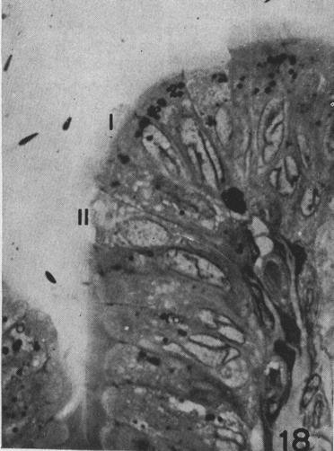
20 204 Asha Prakash et al. Figure 15. Photomicrograph of cell types in the initial segment of the epididymis. The principal cell (P) is tall with long stereocilia. The basally situated nucleus is ovoid and has a smooth nuclear membrane. Negative images of the Golgi apparatus are seen above the nucleus. Clear vesicles and vacuoles are prominent in the apical cytoplasm. Pale cells of two types, one containing an abundance of mitochondria (arrow-head) and the other containing vacuoles (arrow) and a basal cell (B) can be seen 900. Figure 16. Photomicrograph illustrating cell types in the terminal segment of the epididymis. Principal cells (P) possess a highly indented nucleus. Clear vesicles and vacuoles are not evident. The pale cell (arrow head) is seen to extend from the base of the epithelium to the lumen. A round heterochromatic nucleus lies in the apical part of the cell. An intraepithelial lymphocyte (L) is seen. Basal cell (Β) Figure 17. Photomicrograph illustrating cell types in the proximal convoluted portion of the vas deferens. The predominant Type I cell (I) in this region usually possesses slender extensions of the apical cytoplasm which show terminal expansions. The nucleus is devoid with few indentations. The Type II cell (II) resembles the pale cell containing mitochondria of the epididymal epithelium Figure 18. Photomicrograph of cell types in the distal straight portion of the vas deferens. The Type I cell (I) in this region lacks apical extensions; instead the luminal surface is lined by microvilli. The nucleus is highly indented. The cell possesses a vacuolated cytoplasm. Type II Cell (II). 900.


21 Microscopic features of monkey testis 205 FIGS
Ultrastructural studies on the epididymal spermatozoa in the rhesus monkey
 J. Biosci., Vol. 2, Number 3, September 1980, pp. 261-266. Printed in India. Ultrastructural studies on the epididymal spermatozoa in the rhesus monkey ASHA PRAKASH, M. R. N. PRASAD and T.C. ANAND KUMAR
Male Reproductive System
 Male Reproductive System Constitution of male reproductive system Genital gland ----testis Genital ducts epididymis / ductus deferens / urinary duct Accessory sex glands Penis prostate gland Seminal vesicle
Ultrastractural features of the principal cell in the epididymis of the rhesus monkey
 Biosci., Vol. 4, Number 4, December 1982, pp. 469-479. Printed in India. Ultrastractural features of the principal cell in the epididymis of the rhesus monkey T. C, ANAND KUMAR, ASHA PRAKASH and M. R.
MALE REPRODUCTIVE SYSTEM
 MALE REPRODUCTIVE SYSTEM The male reproductive system consists of primary sex organs (testes) and secondary or accessory sex organs. The secondary organs consist of a series of genital ducts (ductules
Histology of Male Reproductive system (1)
") Histology of Male Reproductive system (1) Prof. Dr. Malak A. Al-yawer Learning Objectives At the end of this lecture, the medical student will be able to: State the organization of the testis Define seminiferous
Histology of Male Reproductive system (1) Prof. Dr. Malak A. Al-yawer Learning Objectives At the end of this lecture, the medical student will be able to: State the organization of the testis Define seminiferous
Male Reproductive System
 Male Reproductive System organs that function in: gamete and hormone production not all in abdominal cavity paired testicles = controlled by LH & FSH duct systems accessory glands Testis: Gross Histology
A adipose cells. B capillary. C epithelium
 EPITHELIA Objective The objective of this class is to observe how different epithelia vary in terms of cell shape, size and number of cell layers enabling them to be well adapted for functions in different
Transport of Sperm. Endocrinology of the Epididymis and Sperm Maturation. Vas Efferentia. John Parrish Department of Animal Sciences
 Endocrinology of the Epididymis and Sperm Maturation John Parrish Department of Animal Sciences References: he Physiology of Reproduction, Knobil and Neill, 2006; Chapter on the Epididymis by Robaire ransport
Basic histology 5/4/2015
 Male reproductive system The male reproductive system is composed of the testes, genital ducts (the adjoining epididymis, and the vas deferens, a accessory sex glands (the seminal vesicles, the prostrate
The Male Reproductive System
 The Male Reproductive System YONG-MEI CHEN ( 陈咏梅 ) Dept. of Anatomy, Histology & Embryology Peking Union Medical College Tel:69156461 E-mail address: pumc_he@126.com Content Spermatogenesis Spermiogenesis
Pathology of Male Reproductive System 1
 Pathology of Male Reproductive System 1 Professor dr Ali Hassan Altimimi Professor of Pathology& Histology MSc, PHD, MD(UK) MALE REPRODUCTIVE SYSTEM The internal male genitalia consist of the testes with
THE EFFECTS OF LIGATION OF CAUDA EPIDIDYMIDIS ON THE DOG TESTIS
 Copyright 1974 The American Fertility Society FERTILITY AND STERILITY Vol. 25, No.3, March, 1974 Printed in U.S.A. THE EFFECTS OF LIGATION OF CAUDA EPIDIDYMIDIS ON THE DOG TESTIS A. M. VARE, M.B.B.S.,
18 Urinary system. 19 Male reproductive system. Female reproductive system. Blok 11: Genital and Urinary Tract Diseases
 Blok 11: Genital and Urinary Tract Diseases 18 Urinary System 19 Male Genital System 20 Female Genital System 18 Urinary system You should be able to: 1. Describe the structures and associated functions
Tissues. tissue = many cells w/ same structure and function. cell shape aids its function tissue shape aids its function
 Tissues tissue = many cells w/ same structure and function cell shape aids its function tissue shape aids its function Histology = study of tissues 4 types of tissues Epithelial coverings contact openings
Tissue: The Living Fabric: Part A
 PowerPoint Lecture Slides prepared by Janice Meeking, Mount Royal College C H A P T E R 4 Tissue: The Living Fabric: Part A Tissues Groups of cells similar in structure and function Types of tissues Epithelial
川北医学院讲稿. Under low power note the testis is enclosed by a strong fibrous. layer of serous epithelium. These fibrous tissue
 川北医学院讲稿 Experiment 5: Male and Female Reproductive System Hello, everybody, class is begin,keep quiet, please. And this is the last experimental class. Today we will learn 5 slices and review all structures
Regional Histology and HistoChemistry of the Ductus Epididymis in the Rhesus Monkey (Macaca mulatta)
") BIOLOGY OF REPRODUCTION 19, 1063-1069 (1978) Regional Histology and HistoChemistry of the Ductus Epididymis in the Rhesus Monkey (Macaca mulatta) DONALD J. ALSUM and ALAN G. HUNTER Department 0/Animal
BIOLOGY OF REPRODUCTION 19, 1063-1069 (1978) Regional Histology and HistoChemistry of the Ductus Epididymis in the Rhesus Monkey (Macaca mulatta) DONALD J. ALSUM and ALAN G. HUNTER Department 0/Animal
Efferent Ducts and Epididymis
 increase) the secretion of each of the androgen regulated proteins. Regulation of spermatogenesis is therefore an extremely complex cascade of cell-cell interactions with the Leydig cells supporting germ
Cell and Tissue Types. Epithelial, Connective, Muscle, Nerve
 Cell and Tissue Types Epithelial, Connective, Muscle, Nerve Objectives Explain the major stages of the cell cycle and cellular division (mitosis). Describe specific events occurring in each of the phases
The Fine Structure of the Epithelial Cells of the Mouse Prostate* II. Ventral Lobe Epithelium
 Published Online: 1 June, 1960 Supp Info: http://doi.org/10.1083/jcb.7.3.511 Downloaded from jcb.rupress.org on September 28, 2018 The Fine Structure of the Epithelial Cells of the Mouse Prostate* II.
Epithelial Tissue. Functions include: 1. Protection 4. Absorption 2. Secretion 5. Filtration 3. Sensory reception
 Tissues There are 4 primary tissue types in the human body: 1. Epithelial (covering/lining) 2. Connective (support) 3. Muscle (movement) 4. Nervous (control) Epithelium Epithelial Tissue Covers the surface
Medical School Histology Basics Male Reproductive System. VIBS 289 lab
 Medical School Histology Basics Male Reproductive System VIBS 289 lab Larry Johnson Texas A&M University OBJECTIVE To conduct a histologic examination of the testis (which produce spermatozoa), excretory
General Structure of Digestive Tract
 Dr. Nabil Khouri General Structure of Digestive Tract Common Characteristics: Hollow tube composed of a lumen whose diameter varies. Surrounded by a wall made up of 4 principal layers: Mucosa Epithelial
Dr. Abeer.c.Yousif. Histology -2 nd stage. What is histology?
 What is histology? Histology is the science of microscopic anatomy of cells and tissues, in Greek language Histo= tissue and logos = study and it's tightly bounded to molecular biology, physiology, immunology
Epithelium. Four primary tissue types:
 Epithelium Four primary tissue types: Epithelial (covering) Connective (support) Nervous (control) Muscular (movement) Smooth muscle Cardiac muscle Skeletal muscle 1 Epithelial Tissue Features Epithelial
Epithelia will be discussed according to the following scheme: Type Number of layers Shape Line drawing. Squamous Cuboidal Columnar
 Epithelia Epithelia will be discussed according to the following scheme: Type Number of layers Shape Line drawing Simple Squamous Cuboidal Columnar Covering and Lining epithelium Pseudostratified Stratified
MALE REPRODUCTIVE SYSTEM
 1 MALE REPRODUCTIVE SYSTEM SCPA 602 Anatomical Basis for Pathological Study Updated: 20.09.2018 Lect. Nisamanee Charoenchon, PhD nisamanee.cha@mahidol.ac.th Department of Pathobiology, Mahidol University
LABORATORY EXERCISES FOR MALE REPRODUCTIVE SYSTEM
 LABORATORY EXERCISES FOR MALE REPRODUCTIVE SYSTEM Slide #101 (1096). Testis, rat. sustentacular ( Sertoli ) cells Nuclei of Sustentacular cells Leydig cells Spermatogonia Spermatocytes Spermatids pale
Some Observations on the Fine Structure of the Goblet Cells. Special Reference to the Well-Developed Agranular Endoplasmic Reticulum
 Okajimas Folia Anat. Jpn., 58(4-6) : 583-594, March 1982 Some Observations on the Fine Structure of the Goblet Cells in the Nasal Respiratory Epithelium of the Rat, with Special Reference to the Well-Developed
Unit I Problem 9 Histology: Basic Tissues of The Body
 Unit I Problem 9 Histology: Basic Tissues of The Body - What is the difference between cytology and histology? Cytology: it is the study of the structure and functions of cells and their contents. Histology:
Epithelium tissue system
 Epithelium tissue system Histology : is the study of the microscopic anatomy (microanatomy) of cells and tissues of plants and animals. It is commonly performed by examining cells and tissues under a light
Epithelial Lecture Test Questions
 Epithelial Lecture Test Questions 1. Which of the following free surfaces lack(s) epithelia: a. lung alveoli (air sacs) b. hard palate c. joint cavities d. abdominal cavity e. salivary gland ducts 2. Which
Organs Histology D. Sahar AL-Sharqi. Respiratory system
 Respiratory system The respiratory system provides for exchange of O2 and CO2 to and from the blood. Respiratory organs include the lungs and a branching system of bronchial tubes that link the sites of
Tissues. tissue = many cells w/ same structure and function. cell shape aids function tissue shape aids function. Histology = study of tissues
 Tissues tissue = many cells w/ same structure and function cell shape aids function tissue shape aids function Histology = study of tissues 4 types of tissues Epithelial coverings contact openings Connective
Kidney Functions Removal of toxins, metabolic wastes, and excess ions from the blood Regulation of blood volume, chemical composition, and ph
 The Urinary System Urinary System Organs Kidneys are major excretory organs Urinary bladder is the temporary storage reservoir for urine Ureters transport urine from the kidneys to the bladder Urethra
Histology of Male Reproductive System
 Histology of Male Reproductive System Lecture Objectives Describe the histological features of the male reproductive system Male Reproductive System The male structures of reproduction include the: testes,
straight tubules Ultrastructural evidence for phagocytosis of spermatozoa in the bovine rete testis and testicular
 Ultrastructural evidence for phagocytosis of spermatozoa in the bovine rete testis and testicular straight tubules F. Sinowatz, K.-H. Wrobel, S. Sinowatz and P. Kugler Institutfür Anatomie der Universität
ADVERSE EFFECTS OF VASECTOMY: SPERM GRANULOMA OF EPIDIDYMIDES V. P. DIXIT
 ADVERSE EFFECTS OF VASECTOMY: SPERM GRANULOMA OF EPIDIDYMIDES V. P. DIXIT Reproduct ion Physiology Section, Department of Zoology, University of Rajasthan, Jaipur-302004 Summary: Rats and mice were vasectomized
The Male Reproductive System
 The Male Reproductive System The male reproductive system Testes Genital ducts Accessory sex glands: seminal vesicles prostate bulbourethral glands External genitalia: penis Structure of the Testis Tunica
Tissues. Tissues - Overview. Bio211 Laboratory 2. Epithelial and Connective Tissues
 Bio211 Laboratory 2 Epithelial and Connective Tissues 1 Tissues Tissues to be examined under the microscope Epithelial Tissue (p. 79 Lab Manual) [TODAY] Connective Tissue (p. 93 Lab Manual) [TODAY] Muscle/Nervous
يراهظلا( يئلاطلا جيسنلا
 Epithelium النسيج الطالئي )الظهاري( Features of Epithelium Epithelium occurs in the body as a sheet of cells that covers a body surface, lines a cavity, or forms a gland. Coverings, linings, glands. Derived
(Received 20th February 1974)
") INTRAEPITHELIAL LYMPHOCYTES IN THE MALE REPRODUCTIVE TRACT OF RATS AND RHESUS MONKEYS MARTIN DYM and LYNN J. ROMRELL Department of Anatomy, and Laboratory of Human Reproduction and Reproductive Biology,
INTRAEPITHELIAL LYMPHOCYTES IN THE MALE REPRODUCTIVE TRACT OF RATS AND RHESUS MONKEYS MARTIN DYM and LYNN J. ROMRELL Department of Anatomy, and Laboratory of Human Reproduction and Reproductive Biology,
Lab Activity 31. Anatomy of the Urinary System. Portland Community College BI 233
 Lab Activity 31 Anatomy of the Urinary System Portland Community College BI 233 Urinary System Organs Kidneys Urinary bladder: provides a temporary storage reservoir for urine Paired ureters: transport
Histology Notes -Part 1: Epithelial Tissues
 Introduction Group of cells w/ similar structure & function = TISSUE Four Basic Tissue Types 1. Epithelial-covers 2. Connective-supports 3. Muscular*-produces movement (will discuss in the muscular system
Epithelium Characteristics cont. 2. Apical Surface
 Epithelium Characteristics cont. 2. Apical Surface always has one exposed (apical) surface Some surfaces are smooth & slick, others may have: microvilli fingerlike extensions of the plasma membrane; increase
Histology = the study of tissues. Tissue = a complex of cells that have a common function
 { EPITHELIAL TISSUE Histology = the study of tissues Tissue = a complex of cells that have a common function The Four Primary Tissue Types: Epithelium (epithelial tissue) covers body surfaces, lines body
International Journal of Science, Environment and Technology, Vol. 7, No 5, 2018,
 International Journal of Science, Environment and Technology, Vol. 7, No 5, 2018, 1608 1614 ISSN 2278-3687 (O) 2277-663X (P) COMPARATIVE HISTOLOGICAL STUDIES OF DUEODENUM IN CATTLE SHEEP AND GOATS Thete
HISTOLOGY OF THE MALE REPRODUCTIVE SYSTEM
 HISTOLOGY OF THE MALE REPRODUCTIVE SYSTEM Learning Objectives: 1. Describe the histology of and identify, in order, the passageways through which sperm pass as they exit from the body. 2. Describe the
Histology Review Can you identify the Cell Structures? Can you identify the Stain? Can you identify the Cell type?
 Histology Review Can you identify the Cell Structures? Can you identify the Stain? Can you identify the Cell type? 2.01 Border of Epithelia (fluorescence) M A: lamina basalis B: epithelium C: other tissues
the structure of their ducts has been
 Tza JOURNAL 0? INVEa'riGATrVN DEBMATOLOOT Copyright t 1966 by The Williams & Wilkins Co. Vol. 46, No. I Printed in U.S.A. AN ELECTRON MICROSCOPIC STUDY OF THE ADULT HUMAN APOCRINE DUCT* KEN HASHIMOTO,
Histology Urinary system
 Histology Urinary system Urinary system Composed of two kidneys, two ureters, the urinary bladder, and the urethra, the urinary system plays a critical role in: 1- Blood filtration,(filtration of cellular
Prepared By Student. Dania Abed Al-majeed. Rahma Raad Hanna. Balqees Mohammed Aasim. Dania Hisham. Rasha Rafiee
 Prepared By Student Rahma Raad Hanna Balqees Mohammed Aasim Dania Hisham Dania Abed Al-majeed Rasha Rafiee Epithelia Epithelia can be derived from ectoderm, mesoderm or endoderm -ectoderm gives rise to
PRACTICAL HISTOLOGY LAB
 PRACTICAL HISTOLOGY LAB.1 ----------------------------------------------------------------------------- INTRODUCTION Cells are the smallest units of life, and are named according to their function. Cells
Seminiferous Tubules
 Testes The testes are compound tubular glands that lie within a scrotal sac, suspended from the body by a spermatic cord. The testes are dual organs that act as exocrine glands producing a holocrine secretion,
(b) Stomach s function 1. Dilution of food materials 2. Acidification of food (absorption of dietary Fe in small intestine) 3. Partial chemical digest
 Stomach s function 1. Dilution of food materials 2. Acidification of food (absorption of dietary Fe in small intestine) 3. Partial chemical digest") (1) General features a) Stomach is widened portion of gut-tube: between tubular and spherical; Note arranged of smooth muscle tissue in muscularis externa. 1 (b) Stomach s function 1. Dilution of food
(1) General features a) Stomach is widened portion of gut-tube: between tubular and spherical; Note arranged of smooth muscle tissue in muscularis externa. 1 (b) Stomach s function 1. Dilution of food
Lecture Overview. Chapter 4 Epithelial Tissues Lecture 9. Introduction to Tissues. Epithelial Tissues. Glandular Epithelium
 Visual Anatomy & Physiology First Edition Martini & Ober Chapter 4 Lecture 9 Lecture Overview Introduction to Tissues Location General characteristics Functions Classification Glandular Epithelium 2 Where
Histology. There are four basic tissue types in the body are :-
 Histology Lab.I There are four basic tissue types in the body are :- 1- Epithelial tissues (Epithelium) 2- Connective tissues 3- Muscular tissues 4- Nervous tissues 1-Epithelial tissues epithelial tissues
2. Epithelial Tissues Dr. Manal Othman
 Biology-232 GENERAL HISTOLOGY 2. Epithelial Tissues Dr. Manal Othman Anatomy Department CMMS, AGU HISTOLOGY: w Study of the structure and function of tissues and organs at the microscopic levels. w Tissues
General Human Histology. The Urinary System
 General Human Histology Lecture 8 Assist. Prof. Ahmed Anwar Albir The Urinary System Collecting Tubules & Ducts Urine passes from the distal convoluted tubules to collecting tubules that join each other
To General Embryology Dr: Azza Zaki
 Introduction To General Embryology The Human Development is a continuous process that begins when an ovum from a female is fertilized by a sperm from a male. Cell division, growth and differentiation transform
HISTOCHEMISTRY OF PHOSPHATASES IN THE EPIDIDYMIS OF RAM DURING POSTNATAL DEVELOPMENT*
 HISTOCHEMISTRY OF PHOSPHATASES IN THE EPIDIDYMIS OF RAM DURING POSTNATAL DEVELOPMENT* P.V.S. Kishore 1, Geetha Ramesh 2 and Sabiha Hayath Basha 3 Department of Veterinary Anatomy & Histology, Madras Veterinary
The Use of Rabbits in Male Reproductive Toxicology
 Environmental Health Perspectives Vol. 77, pp. 5-9, 1988 The Use of Rabbits in Male Reproductive Toxicology by Daniel Morton* The rabbit is the smallest and least expensive laboratory animal in which serial
Identification of the spermatogenic stages in living seminiferous tubules of man
 Identification of the spermatogenic stages in living seminiferous tubules of man V. Nikkanen, K.-O. S\l=o"\derstr\l=o"\m and M. Parvinen Department of Obstetrics and Gynecology, Turku University Central
Urinary system. Urinary system
 INTRODUCTION. Several organs system Produce urine and excrete it from the body Maintenance of homeostasis. Components. two kidneys, produce urine; two ureters, carry urine to single urinary bladder for
HISTOLOGY OF THE RESPIRATORY SYSTEM I. Introduction A. The respiratory system provides for gas exchange between the environment and the blood. B.
 HISTOLOGY OF THE RESPIRATORY SYSTEM I. Introduction A. The respiratory system provides for gas exchange between the environment and the blood. B. The human respiratory system may be subdivided into two
THE HOLOCRINE CELLS of the epithelium of the rat epididymis and vas
 Holocrine Cells of the Human Epididymis JAN MARTAN, Ph.D.,"' PAULL. RISLEY, Ph.D., and ZDENEK HRUBAN, M.D., Ph.D. t THE HOLOCRINE CELLS of the epithelium of the rat epididymis and vas deferens undergo
Basic Histology. By Mrs. Bailey
 Basic Histology By Mrs. Bailey Primary Tissues 1. Epithelial Tissue 2. Connective Tissue 3. Muscle Tissue 4. Nervous Tissue Very cellular Supported by underlying connective tissue Epithelial & connective
(Accepted 24 August 1981) (Vogimayr, Waites & Setchell, 1966; Setchell, Voglmayr & Waites, 1969; Tuck,
 (Vogimayr, Waites & Setchell, 1966; Setchell, Voglmayr & Waites, 1969; Tuck,") J. Anat. (1982), 135, 1, pp. 97-110 97 With 27 figures Printed in Great Britain The rete testis of birds T. A. AIRE Department of Veterinary Anatomy, University ofibadan, Nigeria. (Accepted 24 August 1981)
J. Anat. (1982), 135, 1, pp. 97-110 97 With 27 figures Printed in Great Britain The rete testis of birds T. A. AIRE Department of Veterinary Anatomy, University ofibadan, Nigeria. (Accepted 24 August 1981)
Urinary system. Urinary system
 Distal convoluted tubule (DCT) Highly coiled, ~ 5 mm in length Last part of the nephron. Wall; simple cuboidal epithelium Less metabolically active than the PCT no brush border light eosinophilic cytoplasm
Unit II: Tissues and Integumentary System
 Unit II: Tissues and Integumentary System 2.1 - Tissues Chapter 4 Written Response #1 1. What is a tissue? 2. What are four major types of tissues? Tissue Definition: a group or mass of similar cells working
Tissue: The Living Fabric
 PowerPoint Lecture Slide Presentation by Vince Austin Human Anatomy & Physiology FIFTH EDITION Elaine N. Marieb Chapter 4 Tissue: The Living Fabric Part A Tissues Groups of cells similar in structure and
Body Tissues Pearson Education, Inc.
 Body Tissues Tissues Groups of cells with similar structure and function Four primary types: Epithelial tissue (epithelium).1 Connective tissue.2 Muscle tissue.3 Nervous tissue.4 Epithelial Tissues Locations:
Epithelium-1. Hanan Jafar BDS.MSc.PhD
 Epithelium-1 Hanan Jafar BDS.MSc.PhD General features Epithelium is an avascular tissue composed of cells that cover the exterior body surfaces and line internal closed cavities and tubes. It also forms
Lab Animal Tissue. LEARNING OBJECTIVES: To understand the relationship between the structure and function of different animal tissues
 Name: Bio A.P. PURPOSE: HYPOTHESIS: NONE Lab Animal Tissue BACKGROUND: In animals, groups of closely related cells specialized to perform the same function are called tissues. There are four general classes
Chapter 1: Cells and Tissues
 Chapter 1: Cells and Tissues Cells and Tissues Carry out all chemical activities needed to sustain life Cells are the building blocks of all living things Tissues are groups of cells that are similar in
DIGESTIVE TRACT ESOPHAGUS
 DIGESTIVE TRACT From the lower esophagus to the lower rectum four fundamental layers comprise the wall of the digestive tube: mucosa, submucosa, muscularis propria (externa), and adventitia or serosa (see
THE EFFECTS OF REPEATED INJECTIONS OF CHORIONIC GONADOTROPIN ON THE TESTES OF THE LEOPARD FROG (RANA PIPIENS SCHREBER)
") THE EFFECTS OF REPEATED INJECTIONS OF CHORIONIC GONADOTROPIN ON THE TESTES OF THE LEOPARD FROG (RANA PIPIENS SCHREBER) ROBERT P. McCOURT Department of Zoology and Entomology, The Ohio State University,
THE EFFECTS OF REPEATED INJECTIONS OF CHORIONIC GONADOTROPIN ON THE TESTES OF THE LEOPARD FROG (RANA PIPIENS SCHREBER) ROBERT P. McCOURT Department of Zoology and Entomology, The Ohio State University,
CHAPTER 05 Histology: EPITHELIUM
 BIO 211: ANATOMY & PHYSIOLOGY I 1 CHAPTER 05 Histology: EPITHELIUM Part 01: Brief Introduction Part 02: Survey of Types Dr. Lawrence G. G. Altman www.lawrencegaltman.com Some illustrations are courtesy
TISSUES TYPES. CHAPTER 05 Histology: EPITHELIUM BIO 211: ANATOMY & PHYSIOLOGY I. HISTOLOGY = the study of tissues
 BIO 211: ANATOMY & PHYSIOLOGY I 1 CHAPTER 05 Histology: EPITHELIUM Part 01: Brief Introduction Part 02: Survey of Types Dr. Lawrence G. G. Altman www.lawrencegaltman.com Some illustrations are courtesy
Tissues 10/21/2016. Epithelial Tissue
 Tissues This is a generalized cell diagram. It shows the anatomy of a cell, but most cells do not actually look like this. Cells can have a wide variety of shapes and sizes, depending on their function.
Lecture Overview. Marieb s Human Anatomy and Physiology. Chapter 4 Tissues: The Living Fabric Epithelial Tissues Lecture 9. Introduction to Tissues
 Marieb s Human Anatomy and Physiology Marieb Hoehn Chapter 4 Tissues: The Living Fabric Epithelial Tissues Lecture 9 Lecture Overview Introduction to Tissues Epithelial Tissues Location General characteristics
Urinary System Laboratory
 Urinary System Laboratory 1 Adrenal gland Organs of The Urinary System Renal artery and vein Kidney Ureter Urinary bladder Figure 26.1 2 Urethra Functions of the urinary system organs: Urethra expels urine
Essentials of Anatomy and Physiology, 9e (Marieb) Chapter 3 Cells and Tissues. Short Answer. Figure 3.1
 Chapter 3 Cells and Tissues. Short Answer. Figure 3.1") Essentials of Anatomy and Physiology, 9e (Marieb) Chapter 3 Cells and Tissues Short Answer Figure 3.1 Using Figure 3.1, match the following: 1) The illustration of simple cuboidal epithelium is. Answer:
Essentials of Anatomy and Physiology, 9e (Marieb) Chapter 3 Cells and Tissues Short Answer Figure 3.1 Using Figure 3.1, match the following: 1) The illustration of simple cuboidal epithelium is. Answer:
A. cells that perform related functions and are similar in structure. B. extracellular material - made by cells and secreted into interstitial space
 I. tissue components A. cells that perform related functions and are similar in structure B. extracellular material - made by cells and secreted into interstitial space II. tissue types A. epithelium (e.)
The Repr duct ve System. Function: producing offspring
 The Repr duct ve System Function: producing offspring Anatomy of male reproductive system Location: The reproductive organs are classified as external and internal genitalia. The external genitalia are
Outline. Bio 105: Tissues Laboratory. Organization of the Human Body. Tissue - Epithelium. Tissues 3/2/ Copyright 2009 Pearson Education, Inc
 Outline Bio 105: Tissues Laboratory Laboratory 5 Reading: Chapter 4 I. Cell to cell contact II. Body Cavities III. Membranes IV. Homeostasis V. Integumentary System I. Includes skin, hair and nails 1 2
BIOH111. o Cell Biology Module o Tissue Module o Integumentary system o Skeletal system o Muscle system o Nervous system o Endocrine system
 BIOH111 o Cell Biology Module o Tissue Module o Integumentary system o Skeletal system o Muscle system o Nervous system o Endocrine system Endeavour College of Natural Health endeavour.edu.au 1 Textbook
Anatomy PHL 212. Dr. Dina A. A. Hassan. -
 Anatomy PHL 212 Dr. Dina A. A. Hassan Associate Professor College of Pharmacy (Female Section) Sattam Bin Abdulaziz University Al kharj / Kingdom of Saudi Arabia Email :- da.hassan@psau.edu.sa 1 Anatomy
Lec #2 histology. Bronchioles:
 Lec #2 histology. Last lecture we talked about the upper respiratory tract histology, this one is about the lower part histology. We will discuss the histology of: -bronchioles -respiratory bronchioles
Urinary System. Dr. Ahmed Maher Dr. Ahmed Manhal
 Urinary System Dr. Ahmed Maher Dr. Ahmed Manhal Presentation Map Kidney (cortex & medulla). Nephron. Duct system. Juxtaglomerular apparatus. Ureter, bladder & urethra. Definition & General Structure The
4. A phospholipid is an example of organization at the level.
 1. Physiology is the study of a. the structures of anatomical features. b. cellular metabolism. c. processes that allow organisms to function. d. how organ systems develop from the embryo. 2. Mary spends
POSTNATAL DIFFERENTIATION AND REGIONAL HISTOLOGICAL VARIATIONS IN THE DUCTUS EPIDIDYMIDIS OF RAMS*
 POSTNATAL DIFFERENTIATION AND REGIONAL HISTOLOGICAL VARIATIONS IN THE DUCTUS EPIDIDYMIDIS OF RAMS* P.V.S.Kishore 1, Geetha Ramesh 2 and Sabiha Hayath Basha 3 Department of Veterinary Anatomy and Histology,
Tissues Review 4 type
 Tissues Review 4 type Tissues Definition: a group of closely associated cells that perform related functions and are similar in structure Between cells: nonliving extracellular material Four basic types
CINtec p16 INK4a Staining Atlas
 CINtec p16 INK4a Staining Atlas Rating Rating Positive The rating positive will be assigned if the p16 INK4a -stained slide shows a continuous staining of cells of the basal and parabasal cell layers of
Ultrastructure of the Genital Duct Epithelium of the Male Port Jackson Shark, Heterodontus portusjacksoni
 Aust. J. Zool., 1992, 40, 257-66 Ultrastructure of the Genital Duct Epithelium of the Male Port Jackson Shark, Heterodontus portusjacksoni R. C. Jones and M. Lin Department of Biological Sciences, University
Histology / First stage The Urinary System: Introduction. Kidneys
 The Urinary System: Introduction The urinary system consists of the paired kidneys and ureters, the bladder, and the urethra. This system helps maintain homeostasis by a complex combination of processes
ESUR SCROTAL AND PENILE IMAGING WORKING GROUP MULTIMODALITY IMAGING APPROACH TO SCROTAL AND PENILE PATHOLOGIES 2ND ESUR TEACHING COURSE
 ESUR SCROTAL AND PENILE IMAGING WORKING GROUP MULTIMODALITY IMAGING APPROACH TO SCROTAL AND PENILE PATHOLOGIES 2ND ESUR TEACHING COURSE NORMAL ANATOMY OF THE SCROTUM MICHAEL NOMIKOS M.D. F.E.B.U. UROLOGICAL
All lecture of practical OSPE file
 All lecture of practical OSPE file Red: questions. Dark red: very important. Black: complete answers. Gray: notes extra. Editing File You should know before the exam: The diagrams in these slides are going
Ductus Deferens-a Comparative Histology in Mammals
 163 J Anat. Soc. India 52(2) 163-165 (2003) -a Comparative Histology in Mammals Khan, Aijaz, A., Zaidi, M.T; Faruqi, N.A. Department of Anatomy,J.N. Medical College,A.M.U. Aligarh, INDIA. Abstract. The
Biology. Dr. Khalida Ibrahim
 Dr. Khalida Ibrahim Biology Histology: Histology: is the study of the tissues of the body. Tissue: group of similar cells combined to perform a common function. The human body is composed of only 4 basic