Independent Ultrasound Reporting. BMUS UCD Study day 2018
|
|
|
- Stuart Knight
- 5 years ago
- Views:
Transcription
1 Independent Ultrasound Reporting BMUS UCD Study day 2018 Pamela Parker Consultant Sonographer Hull & East Yorkshire Hospitals Colin Griffin Lead Sonographer Royal Liverpool University Hospital
2 Report To give an account of the results of an investigation Collins Concise Dictionary

3 Image Reporting Consider: Report style construction language
4 Reports-writing style To ensure directness and clarity of style, it always helps to think of the people you re trying to communicate with through the report. Imagine them looking for answers to their questions. Readers are interested in clear, positive analysisnot in hedged statements and tentative conclusions. They respond to vitality in style.. Cambridge Guide to English Usage
5 Plain English Despite the name, Plain English is only partly about language. It also emphasises the importance of document design.any document needs clear layout, in sections and paragraphs that express the structure of the information and with effective headings and subheadings to identify local content. Adequate white space between sections also makes the information more accessible.
6 Plain English Despite the name, Plain English is only partly about language. It also emphasises the importance of document design. Any document needs clear layout, in sections and paragraphs that express the structure of the information and with effective headings and subheadings to identify local content. Adequate white space between sections also makes the information more accessible.
7 Plain English The most important principle of Plain English is to keep the reader in mind as you write. Think of yourself as communicating to someone across the table, and of how each sentence sounds. Plain English can clarify communication, it also reduces reading errors, reduces complaints and lawsuits related to official documents.
8 Picture This There is a double fronted two storey detached house with a single side garage. The garage is situated to the right side of the ground floor and appears attached There is a central front door with a small window directly above this on the first floor There are four more windows evident; two on the ground floor and two on the first floor. The windows are symmetrically placed either side of the front door The roof of the house is pitched and there are bilateral apex to the roofs directly above the first floor windows. Underneath the right sided ground floor window there is a garden bench There is a drive leading up to the garage and a lawn in front of the house
9 Picture This. There is a bilobular mass in the midline. The two lobes are sited one above the other in the LS plane. The lobules are ovoid, the superior one is marginally larger than the inferior lobule. Within the superior lobule there are two spherical cystic masses. These are equally spaced from the midline in the centre of the lobule. The cystic masses have concentric rings within with a hypoechoc central component Just inferior to the two cystic masses, situated in the midline, there is a triangular structure equal in size to one of the cyst Arising from the lateral borders of the triangular structure bilaterally there three fine septa extending beyond the outer margins of the lobular structure Arising from the superior border of the superior mass there are two inverted triangular structure equally spaced from the midline

10
11 Picture This. There is a bilobular mass in the midline. The two lobes are sited one above the other in the LS plane. The lobules are ovoid, the superior one is marginally larger than the inferior lobule. Within the superior lobule there are two spherical cystic masses. These are equally spaced from the midline in the centre of the lobule. The cystic masses have concentric rings within with a hypoechoc central component Just inferior to the two cystic masses, situated in the midline, there is a triangular structure equal in size to one of the cyst Arising from the lateral borders of the triangular structure bilaterally there three fine septa extending beyond the outer margins of the lobular structure Arising from the superior border of the superior mass there are two inverted triangular structure equally spaced from the midline Conclusion: There is a cute kitten present no suspicious features
12 Communication Communication problems are a causative factor in up to 80% of medical malpractice cases. Failure of radiological communication-appl Radiol
13 What is the question? Why is the examination being undertaken? Rule in/out an abnormality What is the clinical question? Specific question or a vague impression
14 The imaging examination Before the examination What is the clinical question During the examination Specific observations After the examination Judgement / conclusions / report
15 Making a decision Process of judgement : making a decision or conclusion on the basis of indications and probabilities Aim - to obtain information of a good predictive value

16 Ultrasound observations Location and size Internal characteristics Borders / outline Attenuating properties other features
17 Observations / conclusions Making a list of observations may be useful as a process, but conclusions are usually required in an ultrasound diagnosis

18 Professional Guidelines An ultrasound report may be defined as the recording and interpretation of observations from an ultrasound examination

19 Professional Guidelines The ultrasound report should be written by the person performing the ultrasound examination and should be viewed as an integral part of the whole examination.

20 Professional Guidelines The person issuing the report should take responsibility for the accuracy of the report.

21 Reporting error

22 Professional Guidelines Report style The practitioner should be aware of his/her limitations and consequently seek advice when necessary
 should be indicated The exclusion value and significance of the appearances should be stated where relevant.")
23 Professional Guidelines Limitations Any limitations should be stated and, if a relevant organ has not been fully examined, the reason(s) should be indicated The exclusion value and significance of the appearances should be stated where relevant.

24 Professional Guidelines Clinical implications The practitioner should be aware at all times of the implications for the client/patient of the contents of the report and act in accordance with local guidelines
25 Legal responsibility College of Radiographers Radiographers are legally accountable for their professional actions and for any negligence, whether by act or omission or injury. College of Radiographers 1994
26 Case 1 75 year old male GP Referral Clinical details: Recent weight loss with vague abdominal discomfort in the epigastrium. Vague mass? liver enlarged and nodular.
27 Report The liver is normal in size and appearance. No focal lesions or biliary dilatation. The CBD is of normal calibre. The gallbladder is normal with no evidence of calculi. Normal size and appearance of the spleen, pancreas and IVC. The known infrarenal abdominal aortic aneurysm measures 4.2cm. No large volume para-aortic lymphadenopathy demonstrated. Both kidneys are normal in size and appearance with good cortical preservation and no hydronephrosis. Normal bowel gas pattern seen with no obvious bowel mass demonstrated. No free fluid. Conclusion: No cause presenting symptoms demonstrated on this examination.
28 Case 2 85 year old male Nurse Specialist Referral Haematuria
29 Report US Urinary tract Unremarkable appearances of the kidneys which are normal in size. Difficult to appreciate the urinary bladder since it is partially distended. The prostate gland is not seen. Abdominal aorta is normal in calibre. Coincidental finding of multiple lesions seen in the liver. Post prandial collapsed gallbladder. No intrahepatic biliary duct dilatation. The CBD is not dilated. Pancreas obscured by bowel gas. Spleen appears normal. No abdominal or pelvic free fluid. Multiple cysts seen within the liver 12 October 2009 CT report. You may wish to further investigate the liver lesions with an ultrasound examination using contrast.

30 Reporting Guidance CP Griffin & P Parker

31 Ultrasound reporting There is no more difficult art to acquire the art of observation, and for some men it is quite as difficult to record an observation in brief and plain language Dr William Osler, Boston Medical Surgical Journal 1903
32 ASOUND_PRACTICE.pdf Nine steps to producing a report: Understand clinical information and aim of request and how the exam fits the clinical management of the patient Technical assessment of scan quality / limitations Observations : SSOTM. (size, shape, outline, texture and measurements) normal, unequivocally abnormal (expected/unexpected), equivocal, normal variants Analysis (observation & clinical reasoning) Clinical opinion and diagnostic implication Medical interpretation: wider implication of findings in whole patient context Advice: level of certainty and any further imaging/ix if needed Communication with referrer: adapt to different types of referring clinicians Take urgent action if necessary and document Communication with patient: if trained, appropriate knowledge to do so.
33 327 Reporting style: Brief, relevant description. Conclusion: answer the queries, most important issues first Include pertinent negatives. (ovarian mass, but NO liver metastases or ascites) Don t be vague, equivocal or hedge: answer the question clearly Avoid: sinister, concerning, large, worrying etc.. Imagine you are reading this about yourself/your loved ones If equivocal findings, indicate degree of uncertainty and advise how to resolve Beware the words: significant and non-specific
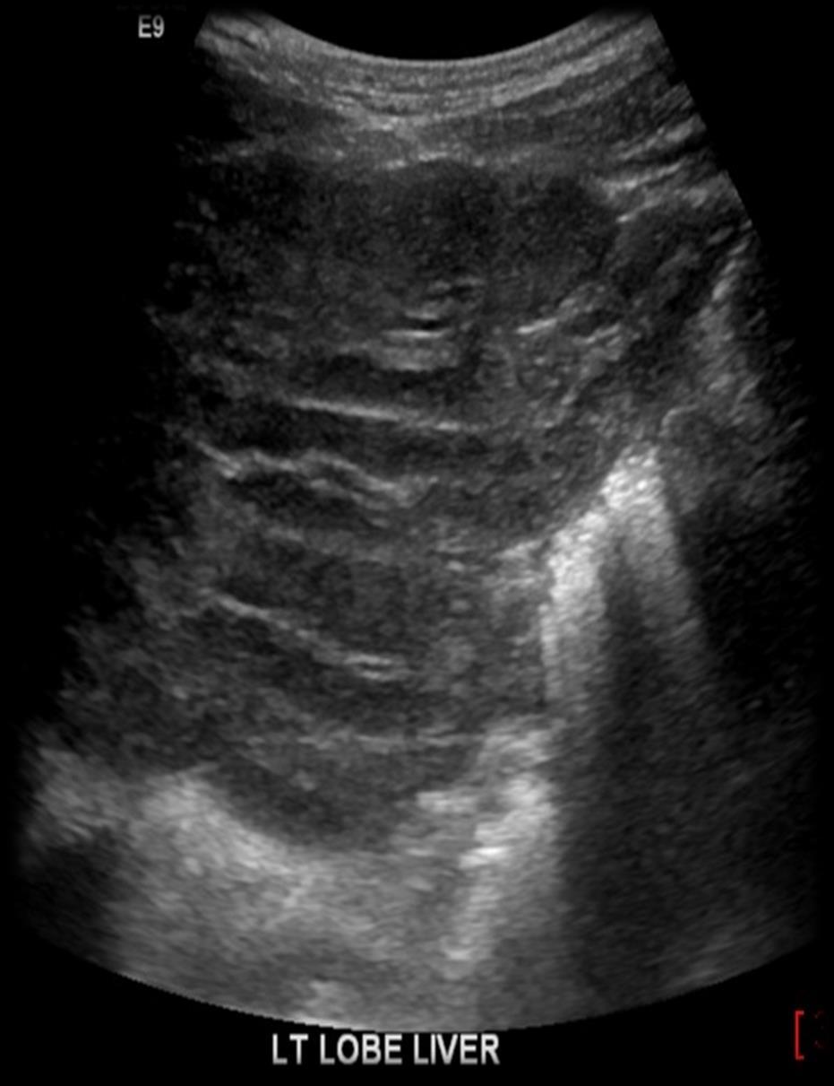
34 Case Example Clinical Information: 19yo male Hepatoma surveillance for PSC cirrhosis

35


36

37
38


39


40

41
42 Report? Tell me what you think?

43
44 Actual Ultrasound Report Comparison made with previous ultrasound scan performed 26/01/2018 and the recent MRCP performed 24/07/2018. The liver parenchyma is heterogenous in keeping with previous imaging and the patient's diagnosis. No obvious focal lesion is identified on today's scan. In the midline of the abdomen there are two rounded soft tissue lesions which appear to correspond to the previously identified splenunculi. The portal vein is patent with normal hepatopetal flow present, velocity 17 cm/s. Again there is splenomegaly present, with the spleen measuring in excess of 21 x 6.5 cm. No ascites within the upper abdomen. Conclusion No new features identified. No focal lesions identified.
45 Case Example Clinical Information: CT Please kindly image this 73 year old gentleman who is day 3 post reversal of ileostomy and ileocolic anastamosis. The patient has now deteriorated with significantly derranged inflammatory markers, a distended and tender abdomen and constipation. He is tachycardic We are concerned regarding a post operative anastamotic leak. Please may we request imaging to further investigate
46 CT Abdomen with contrast : IV contrast in portal venous phase. Comparison study: CT abdomen dated 23rd March Evidence of recent laparotomy and the ileo-colic anastomosis (2 205). Evidence of a previous and ileostomy tract within the subcutaneous tissue of the right iliac fossa. No peri-anastomotic free fluid or gas. No evidence of an anastomotic leak. Unremarkable appearances of the remaining colon. No small bowel obstruction. Small amount of free intra-abdominal fluid (HU 18) within the bilateral paracolic gutters, slightly more marked on the right. No free intra-abdominal gas. Similar appearance of a 19 mm focal thickening along the posterior wall of the gallbladder, this is likely due to a fold in the gallbladder wall although further characterisation should be discussed. No radiopaque gallstones. A 3 mm hypodense area demonstrated in segment 5 of the liver, which is too small to characterise which most likely represents a cyst. Unchanged appearances of a 12x10 mm and a 16x14 mm adrenal adenoma within the body and the medial limb of the left adrenal gland. Normal appearances of the pancreas, spleen, right adrenal gland and kidneys (unchanged large right lower pole renal cyst). Normal calibre of the abdominal aorta. Non-dependent gas demonstrated within the urinary bladder, consistent with recent urinary catheter insertion. Bibasal lower lobe consolidation/atelectasis with small bilateral pleural effusions. No destructive bone lesions.


47
48 US Request Clinical History : Area of thickening seen in GB on recent CT. Suggested u/s f/u/. Please do when pt is an inpt. Tx


49

50

51

52

53
54 Report? Tell me what you think?
55 Actual Ultrasound Report US Gallbladder : Comparison made with the recent CT performed 09/06/18. The defect identified on the CT has been identified today. The ultrasound features are suggestive of a gallbladder malignancy with local invasion into segment 4b of the liver. No calculi were identified within the gallbladder. Conclusion: Ultrasound evidence to suggest gallbladder malignancy with local invasion to the liver. MDT discussion is suggested to discuss management options.
56 No Feedback No CPD Cert Join us for BMUS ASM 4 6 th December 2018 Manchester
Guidelines, Policies and Statements D5 Statement on Abdominal Scanning
 Guidelines, Policies and Statements D5 Statement on Abdominal Scanning Disclaimer and Copyright The ASUM Standards of Practice Board have made every effort to ensure that this Guideline/Policy/Statement
Appendix 5. EFSUMB Newsletter. Gastroenterological Ultrasound
 EFSUMB Newsletter 87 Examinations should encompass the full range of pathological conditions listed below A log book listing the types of examinations undertaken should be kept Training should usually
Policies, Standards, and Guidelines. Guidelines for Abdominal Ultrasound Examination
 Policies, Standards, and Guidelines Guidelines for Abdominal Ultrasound Examination Approved by Council Feb 2018 Disclaimer and Copyright The ASUM Standards of Practice Board have made every effort to
Dr Claire Smith, Consultant Radiologist St James University Hospital Leeds
 Dr Claire Smith, Consultant Radiologist St James University Hospital Leeds Imaging in jaundice and 2ww pathway Image protocol Staging Limitations Pancreatic cancer 1.2.4 Refer people using a suspected
CT abdomen and pelvis
 CT abdomen and pelvis General indications: Assessment of vague abdominal symptoms (pain, colics,distenstion,...) Varifecation of a lesion discovered by other diagnostic modalities as US, barium,ivp, Staging
Abdomen and Retroperitoneum Ultrasound Protocols
 Abdomen and Retroperitoneum Ultrasound Protocols Reviewed By: Anna Ellermeier, MD Last Reviewed: March 2018 Contact: (866) 761-4200, Option 1 **NOTE for all examinations: 1. If documenting possible flow
Ovarian Lesion Benign vs Malignant?
 Ovarian Lesion Benign vs Malignant? Michele Keenan 1,2 Bernice Dunne 2 Mary Moran 1 Therese Herlihy 1 1. Radiography and Diagnostic Imaging, School of Medicine, University College Dublin, Ireland 2. Midland
Is Structured Reporting More Accurate Than Conventional Reporting in CT Reporting of the Abdomen and Pelvis?
 Is Structured Reporting More Accurate Than Conventional Reporting in CT Reporting of the Abdomen and Pelvis? A M Almuslim, MBBS; J G Ryan, MD; A Murtaza, MD Purpose The purpose of this research is to determine
Pediatric Hepatobiliary, Pancreatic & Splenic US
 Pediatric Hepatobiliary, Pancreatic & Splenic US Susan J. Back, MD Department of Radiology, The Children s Hospital of Philadelphia No Disclosures Objectives Normal Abnormal: cases and US advances Objectives
My Patient Has Abdominal Pain PoCUS of the Biliary Tract and the Urinary Tract
 My Patient Has Abdominal Pain PoCUS of the Biliary Tract and the Urinary Tract Objectives PoCUS for Biliary Disease PoCUS for Renal Colic PoCUS for Urinary Retention Biliary Disease A patient presents
Elastography in the. technically difficult patient. EPIQ ultrasound system. Ultrasound
 Ultrasound Elastography in the technically difficult patient EPIQ ultrasound system Chairman Department of Diagnostic Radiology Allegheny General Hospital Pittsburgh, PA, USA You can offer more information
Case Study: #3: Gallbladder Carcinoma?
 Case Study: #3: Gallbladder Carcinoma? By: Megan Wyatt K. SON Wyatt 225 2B1 RDMS, RVT Patient: Male 85 YOA Caucasian Indication: Elevated Alkaline Phosphatase History Annual physical showed elevated alkaline
Radiology of hepatobiliary diseases
 GI cycle - Lecture 14 436 Teams Radiology of hepatobiliary diseases Objectives 1. To Interpret plan x-ray radiograph of abdomen with common pathologies. 2. To know the common pathologies presentation.
The role for contrast-enhanced ultrasonography outside of focal liver lesions
 The role for contrast-enhanced ultrasonography outside of focal liver lesions Paul S. Sidhu King s College Hospital, London, UK Introduction Contrast-enhanced ultrasonography (US) of focal liver lesions
Abdominal ultrasound:
 Abdominal ultrasound: Non-traumatic acute abdomen Wittanee Na-ChiangMai, MD Department of Radiology ChiangMai University 26/04/2017 Contents Technique of examination Normal anatomy Emergency conditions
Body MRI from the Liver to the Bladder
 Body MRI from the Liver to the Bladder I Want You! Audience Participation Methodist Hospital Continuing Education Seminar Jordan Swensson, MD November 7, 2015 Objectives Observe the uses of MRI for organs
Normal Sonographic Anatomy
 hapter 2:The Liver DUNSTAN ABRAHAM Normal Sonographic Anatomy Homogeneous, echogenic texture (Figure 2-1) Measures approximately 15 cm in length and 10 12.5 cm anterior to posterior; measurement taken
Lab Monitor Images Dissection of the Abdominal Vasculature + Lower Digestive System
 Lab Monitor Images Dissection of the Abdominal Vasculature + Lower Digestive System Stomach & Duodenum Frontal (AP) View Nasogastric tube 2 1 3 4 Stomach Pylorus Duodenum 1 Duodenum 2 Duodenum 3 Duodenum
Abdominal Ultrasound : Aorta, Kidneys, Bladder
 Abdominal Ultrasound : Aorta, Kidneys, Bladder Nilam J. Soni, MD, MSc Associate Professor of Medicine Divisions of Hospital Medicine and Pulmonary/Critical Care Medicine Department of Medicine University
Appendix 9: Endoscopic Ultrasound in Gastroenterology
 Appendix 9: Endoscopic Ultrasound in Gastroenterology This curriculum is intended for clinicians who perform endoscopic ultrasonography (EUS) in gastroenterology. It includes standards for theoretical
Reporting Initiative: Motivations and Approach
 The RSNA Structured Reporting Initiative: Motivations and Approach Curtis P. Langlotz, MD, PhD Chair, RSNA Structured Reporting Committee Vice Chair for Informatics, Department of Radiology Professor of
Liver Ultrasound - Beyond the Basics. Pamela Parker Lead Sonographer
 Liver Ultrasound - Beyond the Basics Pamela Parker Lead Sonographer Aims Review what we know about the liver Reasons for imaging Focal lesions Diffuse disease Can we do more? The Liver The Liver The Liver
Question 1 History. Likely Diagnosis Differential. Further Investigation or Management. Requires Paediatric Surgical referral for laparotomy
 Question 1 Male newborn spilling green tinged vomit day 1 of life Imaging Abdominal X-Rays performed on 03/05/2012 Upper and lower gastrointestinal contrast studies performed on 03/05/2012 Abdominal X-Rays
Basic Abdominal Sonography
 24S Basic Abdominal Sonography Procedural Overview JOHN FATCHETT II, RDMS is provided. Patient preparation (i.e., fasting) scanning techniques, spleen, transducer. evaluation of abdominal anatomy in the
Biliary Tree Ultrasound - In a nutshell. Pamela Parker Lead Sonographer
 Biliary Tree Ultrasound - In a nutshell Pamela Parker Lead Sonographer Aims Review what we know about the biliary system Common pathologies Pitfalls Reporting tips The Nutshell Background Biliary examinations
What s your diagnosis?
 What s your diagnosis? Signalment: 9 year old MC 2.7 kg Papillion Presenting Complaint: Presented for work up of anorexia and vomiting History: He had presented to cardiology for work up of a grad IV/VI
Imaging Guided Biopsy. Edited & Presented by ; Hussien A.B ALI DINAR. Msc Lecturer,Reporting Sonographer
 Imaging Guided Biopsy Edited & Presented by ; Hussien A.B ALI DINAR. Msc Lecturer,Reporting Sonographer Objective By the End of this lessons you should : Define what biopsy Justify Aim to perform biopsy
ASSESSING THE PLAIN ABDOMINAL RADIOGRAPH M A A M E F O S U A A M P O F O
 ASSESSING THE PLAIN ABDOMINAL RADIOGRAPH M A A M E F O S U A A M P O F O Introduction The abdomen (less formally called the belly, stomach, is that part of the body between the thorax (chest) and pelvis,
Hilar cholangiocarcinoma. Frank Wessels, Maarten van Leeuwen, UMCU utrecht
 Hilar cholangiocarcinoma Frank Wessels, Maarten van Leeuwen, UMCU utrecht Content Anatomy Biliary strictures (Hilar) Cholangiocarcinoom Staging Biliary tract 1 st order Ductus hepatica dextra Ductus hepaticus
Pancreas & Biliary System. Dr. Vohra & Dr. Jamila
 Pancreas & Biliary System Dr. Vohra & Dr. Jamila 1 Objectives At the end of the lecture, the student should be able to describe the: Location, surface anatomy, parts, relations & peritoneal reflection
Newcastle HPB MDM updated radiology imaging protocol recommendations. Author Dr John Scott. Consultant Radiologist Freeman Hospital
 Newcastle HPB MDM updated radiology imaging protocol recommendations Author Dr John Scott. Consultant Radiologist Freeman Hospital This document is intended as a guide to aid radiologists and clinicians
Anatomy Jessica Ferguson Ashley Dobos May 31, 2006 LIVER
 Anatomy Jessica Ferguson Ashley Dobos May 31, 2006 LIVER 1) Other Names: Reidel s Lobe normal anatomic variant; projection of the right lobe that can extend as far as the iliac crest (Tempkin, p.54, Anatomy).
Hepatic Imaging: What Every Practitioner Should Know
 Hepatic Imaging: What Every Practitioner Should Know Shuchi K. Rodgers, MD Section Chief, Abdominal Imaging Director of Ultrasound Department of Radiology Einstein Medical Center rodgerss@einstein.edu
Biliary Ultrasonography Kathleen O Brien MD MPH RDMS Kaiser Permanente South Sacramento
 Biliary Ultrasonography Kathleen O Brien MD MPH RDMS Kaiser Permanente South Sacramento https://www.google.com/search?sa=g&hl=en&q=public+disclosure&tbm=isch&tbs=simg:caqsigeahwelekju2aqaaawlelcmpwgaygpgcamskpib_1qnza7ai
EFSUMB EUROPEAN FEDERATION OF SOCIETIES FOR ULTRASOUND IN MEDICINE AND BIOLOGY Building a European Ultrasound Community
 MINIMUM TRAINING REQUIREMENTS FOR THE PRACTICE OF MEDICAL ULTRASOUND IN EUROPE Appendix 9: Endoscopic Ultrasound in Gastroenterology This curriculum is intended for clinicians who perform endoscopic ultrasonography
Abdominal Ultrasound. Diane Hallinen, MD. Bloodroot
 Abdominal Ultrasound Diane Hallinen, MD Bloodroot Abdominal Ultrasound Vasculature Hepatobiliary Spleen Kidney Bladder Bowel Where to put the probe? Vasculature We are going to talk about Celiac Trunk
Imaging of liver and pancreas
 Imaging of liver and pancreas.. Disease of the liver Focal liver disease Diffusion liver disease Focal liver disease Benign Cyst Abscess Hemangioma FNH Hepatic adenoma HCC Malignant Fibrolamellar carcinoma
Nasogastric tube. Stomach. Pylorus. Duodenum 1. Duodenum 2. Duodenum 3. Duodenum 4
 Esophagus Barium Swallow Stomach and Duodenum 4 year old Upper GI Nasogastric tube Stomach and Duodenum 4 year old Upper GI Nasogastric tube Stomach Pylorus Duodenum 1 Duodenum 2 Duodenum 3 Duodenum 4
To describe the liver. To list main structures in porta hepatis.
 GI anatomy Lecture: 6 د. عصام طارق Objectives: To describe the liver. To list main structures in porta hepatis. To define portal system & portosystemic anastomosis. To list parts of biliary system. To
Ultrasound is not able to fully evaluate the stomach or bowel, as sound waves are unable to travel through gas and air.
 General Ultrasound What What is ultrasound? is Ultrasound? Ultrasound is safe and (usually) painless use of high frequency sound waves to produce diagnostic images of the internal organs and structures
Guide to Small Animal Reproductive Imaging using the Vevo 770
 Guide to Small Animal Reproductive Imaging using the Vevo 770 Course Objectives: After completion of this module, the participant will be able to accomplish the following: Recognize reproductive female
Radiological Investigations of Abdominal Trauma
 76 77 Investigations of Abdominal Trauma Introduction: Trauma to abdominal organs is a common cause of patient morbidity and mortality among trauma patients. Causes of abdominal trauma include blunt injuries,
4/9/2018 OBJECTIVES PANCREAOTO BILIARY ULTRASOUND: BEYOND CHOLECYSTITIS
 PANCREAOTO BILIARY ULTRASOUND: BEYOND CHOLECYSTITIS Jean Yves Sewah Kaiser Permanente West Los Angeles 1 OBJECTIVES Discuss the role of ultrasound in the evaluation of the gallbladder, biliary tree and
Abdominal Ultrasound
 Abdominal Ultrasound What is Ultrasound Imaging of the Abdomen? What are some common uses of the procedure? How should I prepare? What does the equipment look like? How does the procedure work? How is
Hepatobiliary Ultrasound Rimon Bengiamin, MD, RDMS Assistant Clinical Professor Director of Emergency Ultrasound UCSF Fresno. Objectives. Why?
 Hepatobiliary Ultrasound Rimon Bengiamin, MD, RDMS Assistant Clinical Professor Director of Emergency Ultrasound UCSF Fresno Objectives Discuss the goals of point-of-care biliary ultrasound Review the
Job Task Analysis for ARDMS Abdomen Data Collected: June 30, 2011
 Job Task Analysis for ARDMS Abdomen Data Collected: June 30, 2011 Reported: Analysis Summary for: Abdomen Examination Survey Dates 06/13/2011-06/26/2011 Invited Respondents 6,000 Surveys with Demographics
Abdominal Ultrasound
 Abdominal Ultrasound Imaging Control Buttons Depth The organ imaged should take up 3/4 of the screen Frequency = Penetration Use high frequencies (harmonics) for fluid filled and superficial structures
Autosomal Dominant Polycystic Kidney Disease
 Case Studies [1] July 01, 2014 By Amar Udare, MBBS [2] Case History: 45-year-old female with vague pain in the abdomen. Case History: A 45-year-old female presented with vague pain in the abdomen. A USG
Liver and Pancreatic Case discussion
 The Royal Marsden Liver and Pancreatic Case discussion Dr Ian Chau Consultant Medical Oncologist The Royal Marsden 77 year old gentleman with 2 months history of vague abdominal ache and clinically finding
Update on RECIST and Staging of Common Pediatric Tumors Ethan A. Smith, MD
 Update on RECIST and Staging of Common Pediatric Tumors Ethan A. Smith, MD Section of Pediatric Radiology C.S. Mott Children s Hospital University of Michigan ethans@med.umich.edu Disclosures No relevant
US Applications. Case Based Wrap-Up 1. Case 1 E-FAST. Case presentations E-FAST Abdominal. Pearls for each indication
 Case Based Wrap-Up 1 Stephanie J. Doniger MD RDMS FAAP FACEP Associate Director, Pediatric Emergency Ultrasound Stanford University Medical Center US Applications Case presentations E-FAST Abdominal Aorta
Imaging of common diseases of hepatobiliary and GI system
 Imaging of common diseases of hepatobiliary and GI system Natthaporn Tanpowpong, M.D. Diagnostic radiology Faculty of Medicine, Chulalongkorn University Normal plain radiograph A = Common bile duct
Abdomen and Pelvis CT (1) By the end of the lecture students should be able to:
 By the end of the lecture students should be able to:") RAD 451 Abdomen and Pelvis CT (1) By the end of the lecture students should be able to: State the common indications for Abdomen and pelvis CT exams Identify possible contra indications for Abdomen and
RAD 451 Abdomen and Pelvis CT (1) By the end of the lecture students should be able to: State the common indications for Abdomen and pelvis CT exams Identify possible contra indications for Abdomen and
CT WHOLE ABDOMEN AND PELVIS (PLAIN AND TRIPHASIC CONTRAST)
") CT WHOLE ABDOMEN AND PELVIS (PLAIN AND TRIPHASIC CONTRAST) Technique: Plain and triphasic intravenous contrast enhanced axial CT scan of abdomen and pelvis has been performed with retrospective 2D multiplanar
CT WHOLE ABDOMEN AND PELVIS (PLAIN AND TRIPHASIC CONTRAST) Technique: Plain and triphasic intravenous contrast enhanced axial CT scan of abdomen and pelvis has been performed with retrospective 2D multiplanar
objectives Pitfalls and Pearls in PET/CT imaging Kevin Robinson, DO Assistant Professor Department of Radiology Michigan State University
 objectives Pitfalls and Pearls in PET/CT imaging Kevin Robinson, DO Assistant Professor Department of Radiology Michigan State University To determine the regions of physiologic activity To understand
Abdomen Sonography Examination Content Outline
 Abdomen Sonography Examination Content Outline (Outline Summary) # Domain Subdomain Percentage 1 2 3 Anatomy, Perfusion, and Function Pathology, Vascular Abnormalities, Trauma, and Postoperative Anatomy
MANAGEMENT RECOMMENDATIONS
 1 MANAGEMENT RECOMMENDATIONS 1. Adrenal masses!!!!!!! page 2 2. Liver Masses!!!!!!! page 3 3. Obstetric US Soft Markers for Aneuploidy!! pages 4-6 4. Ovarian and Adnexal Cysts!!!!! pages 7-10 5. Pancreatic
Are extra-colonic findings on CT colonogram clinically significant? A review of 758 consecutive cases
 Are extra-colonic findings on CT colonogram clinically significant? A review of 758 consecutive cases Lynn WR 1, Vadhwana B 1, Borgstein R 2, Demetriou G 1, Nair MS 1, Meleagros L 1, Bell DJ 2 1 Department
The Spleen. Dr Fahad Ullah
 The Spleen BY Dr Fahad Ullah Spleen The spleen is an largest lymphoid organ shaped like a shoe that lies relative to the 9th and 11th ribs and is located in the left hypochondrium. Thus, the spleen is
Haematuria Clinic. Information for patients Urology PROUD TO MAKE A DIFFERENCE SHEFFIELD TEACHING HOSPITALS NHS FOUNDATION TRUST
 Haematuria Clinic Information for patients Urology PROUD TO MAKE A DIFFERENCE SHEFFIELD TEACHING HOSPITALS NHS FOUNDATION TRUST The Haematuria Clinic You have been referred to the Haematuria Clinic as
Tata Memorial Centre s opinion is summarized as follows: 1. Given the type 1 stricture (as mentioned in the structured summary), assessment
, assessment") March 5 th 2016 Dear Ms. Malti Sinha, Thank you for reaching out to Tata Memorial Centre for an expert opinion in regard to assessing your treatment options. Navya Network is pleased to offer this online
March 5 th 2016 Dear Ms. Malti Sinha, Thank you for reaching out to Tata Memorial Centre for an expert opinion in regard to assessing your treatment options. Navya Network is pleased to offer this online
ULTRASOUND AND ABDOMINAL MASSES
 Med. J. Malaysia Vol. 37 No. I March 1982. ULTRASOUND AND ABDOMINAL MASSES AHMAD KAMAL BIN MD ALIF INTRODUCTION It is approximately 30 years since ultrasound was first introduced into the field of medicine,
Excretory urography (EU) or IVP US CT & radionuclide imaging
 or IVP US CT & radionuclide imaging") Excretory urography (EU) or IVP US CT & radionuclide imaging MRI arteriography studies requiring catherization or direct puncture of collecting system EU & to a lesser extent CT provide both functional
Excretory urography (EU) or IVP US CT & radionuclide imaging MRI arteriography studies requiring catherization or direct puncture of collecting system EU & to a lesser extent CT provide both functional
Plain abdomen The standard films are supine & erect AP views (alternative to erect, lateral decubitus film is used in ill patients).
.") Plain abdomen The standard films are supine & erect AP views (alternative to erect, lateral decubitus film is used in ill patients). The stomach can be readily identified by its location, gastric rugae
Plain abdomen The standard films are supine & erect AP views (alternative to erect, lateral decubitus film is used in ill patients). The stomach can be readily identified by its location, gastric rugae
Long Case Set 02. Dr Raviraj Uppoor. Dr Sameer Shamshuddin. Consultant Radiologist Cumberland Infirmary, Carlisle, UK
 Long Case Set 02 www.frcrtutorials.com Dr Raviraj Uppoor MBBS, DMRD, DNB, FRCR Consultant Radiologist Cumberland Infirmary, Carlisle, UK Dr Sameer Shamshuddin MBBS, DMRD, FRCR Consultant Radiologist Royal
Rad Lab 4 Unknowns: Genitourinary!
 Rad Lab 4 Unknowns: Genitourinary! Peter Clarke MD! Don Di Salvo, MD! Clerkship Directors for Radiology! Harvard Medical School! Brigham and Women s Hospital! Dana Farber Cancer Institute! Case 1: 69 year
Heme Database Exercise Use the Hematopoietic Database to answer the following questions
 Heme Database Exercise Use the Hematopoietic Database to answer the following questions http://seer.cancer.gov/seertools/hemelymph/ 1. Assign a topography and histology code to polycythemia vera 2. List
Biliary Tree Ultrasound - In a nutshell. Pamela Parker Lead Sonographer
 Biliary Tree Ultrasound - In a nutshell Pamela Parker Lead Sonographer Aims Review what we know about the biliary system Common pathologies Pitfalls Reporting tips The Nutshell Background Biliary examinations
Uroradiology For Medical Students
 Uroradiology For Medical Students Lesson 4: Cystography & Urethrography - Part 2 American Urological Association Review Cystography is useful in evaluating the bladder, the urethra and the competence of
Obstetrics Content Outline Obstetrics - Fetal Abnormalities
 Obstetrics Content Outline Obstetrics - Fetal Abnormalities Effective February 2007 10 16% renal agenesis complete absence of the kidneys occurs when ureteric buds fail to develop Or degenerate before
Peritoneum: Def. : It is a thin serous membrane that lines the walls of the abdominal and pelvic cavities and clothes the viscera.
 Peritoneum: Def. : It is a thin serous membrane that lines the walls of the abdominal and pelvic cavities and clothes the viscera. Layers of the peritoneum: 1. Outer Layer ( Parietal Peritoneum) : lines
Cervix Carcinoma Case: Metastatic
 Cervix Carcinoma Case: Metastatic Dr Mariza Tunmer Radiation Oncologist Johannesburg Cervix Carcinoma: Metastatic Ms TL 32yr 17 March 2016 Presented to local hospital casualty with nausea & vomiting, abd
IT 의료융합 1 차임상세미나 복부질환초음파 이재영
 IT 의료융합 1 차임상세미나 2013-4-3 복부질환초음파 이재영 나는오늘누구를위하여 종을울리나? 전통적의료 의사 공학설계자 의사 최첨단진단장비들 USG, CT, MRI 환자 환자 현대의료 사용자중심의사고 US in the Abdomen Detection DDx Look Behavior Response by external stimuli Guiding Tool
Abdominal Ultrasonography
 Abdominal Ultrasonography David A. Masneri, DO, FACEP, FAAEM Assistant Professor of Emergency Medicine Assistant Director, Emergency Medicine Residency Medical Director, Operational Medicine Division Center
Patient Information. Age: 8 y/o Sex: Female. Date of Admission: Date of Discharge:
 Patient Information Age: 8 y/o Sex: Female Date of Admission: 92-10-08 Date of Discharge: 92-10-18 Chief Complaint Severe admominal pain and vomiting with dysuria since last afternoon Present Illness Lower
Accessory Glands of Digestive System
 Accessory Glands of Digestive System The liver The liver is soft and pliable and occupies the upper part of the abdominal cavity just beneath the diaphragm. The greater part of the liver is situated under
Evaluation of Suspected Pancreatic Cancer
 Evaluation of Suspected Pancreatic Cancer October 15, 2015 If you experience technical difficulty during the presentation: Contact WebEx Technical Support directly at: US Toll Free: 1-866-779-3239 Toll
Biliary tree dilation - and now what?
 Biliary tree dilation - and now what? Poster No.: C-1767 Congress: ECR 2012 Type: Educational Exhibit Authors: I. Ferreira, A. B. Ramos, S. Magalhães, M. Certo; Porto/PT Keywords: Pathology, Diagnostic
Inserting a percutaneous biliary drain and biliary stent (a tube to drain bile)
") Patient information - Radiology Unit Tel 0118 322 7991 Inserting a percutaneous biliary drain and biliary stent (a tube to drain bile) Introduction This leaflet tells you about the procedures known as
Patient information - Radiology Unit Tel 0118 322 7991 Inserting a percutaneous biliary drain and biliary stent (a tube to drain bile) Introduction This leaflet tells you about the procedures known as
CLINICAL PRESENTATION AND RADIOLOGY QUIZ QUESTION
 Donald L. Renfrew, MD Radiology Associates of the Fox Valley, 333 N. Commercial Street, Suite 100, Neenah, WI 54956 6/23/2012 Radiology Quiz of the Week # 78 Page 1 CLINICAL PRESENTATION AND RADIOLOGY
IMAGING OF LIVER, BILIARY TREE, PANCREAS
 IMAGING OF LIVER, BILIARY TREE, PANCREAS Department of Radiology West China Hospital, Sichuan University Yao Jin Learning Points The methodology for imaging the LBP (liver, biliary tree, and pancreas )
Direct Access Community Ultrasound Service (Non-Obstetric) Referrer Information. Providing services on behalf of the NHS
 Referrer Information. Providing services on behalf of the NHS") Direct Access Community Ultrasound Service (Non-Obstetric) Referrer Information Providing services on behalf of the NHS The Service Physiological Measurements Ltd work in partnership with the NHS to provide
Direct Access Community Ultrasound Service (Non-Obstetric) Referrer Information Providing services on behalf of the NHS The Service Physiological Measurements Ltd work in partnership with the NHS to provide
ISUOG Basic Training. Distinguishing between Normal & Abnormal Appearances of the Urinary Tract. Seshadri Suresh, India
 ISUOG Basic Training Distinguishing between Normal & Abnormal Appearances of the Urinary Tract Seshadri Suresh, India Learning objectives 13 & 14 At the end of the lecture you will be able to: describe
US LI-RADS v2017 CORE
 US LI-RADS v2017 CORE Screening or surveillance US in patient at high risk for HCC US category US-1 US-2 US-3 Negative Subthreshold Positive Category Concept Definition US-1 Negative US-2 Subthreshold
Q129. Which of the following is NOT true about lymph node?
 Q129. Which of the following is NOT true about lymph node? (1). Normal lymph node is not seen in the ultrasound image (2). It is general that high frequency probe is used due to normal lymph node is located
Biliary MRI w Eovist
 Biliary MRI w Eovist Is there any added value? Elmar M. Merkle, MD Director of MR Imaging Duke University Medical Center elmar.merkle@duke.edu Declaration of Conflict of Interest or Relationship Research
Basics of Interventional Radiology Coding 2017
 Basics of Interventional Radiology Coding 2017 Prepared and Published By: MedLearn Publishing A Division of Panacea Healthcare Solutions, Inc. 287 East Sixth Street, Suite 400 St. Paul, MN 55101 1-800-252-1578
Looking Outside the Box: Incidental Extracardiac Finding in Echo
 Looking Outside the Box: Incidental Extracardiac Finding in Echo Dr. Aijaz Shah Head of Division, Adult Echocardiography Laboratory Prince Sultan Cardiac Centre Riyadh Case 1 17 year old boy presented
Boot Camp Case Scenarios
 Boot Camp Case Scenarios Case Scenario 1 Patient is a 69-year-old white female. She presents with dyspnea on exertion, cough, and right rib pain. Patient is a smoker. 9/21/12 CT Chest FINDINGS: There is
Pelvic Ultrasound.
 Pelvic Ultrasound Before Your Exam: Drink 32 oz. of water one hour before your examination time. Try to drink all the liquid within 30 minutes. Do not urinate before the exam. Arrive for your exam with
The following are the objectives to be successfully completed by the IR fellow at the completion of training.
 GOALS and OBJECTIVES The following are the objectives to be successfully completed by the IR fellow at the completion of training. I. Patient Care Fellows must be able to provide patient care that is compassionate,
1. Long images of aorta (prox, mid, and dist) with AP measurements. 2. Trans images of aorta (prox, mid, and dist) with R/L measurements.
 with AP measurements. 2. Trans images of aorta (prox, mid, and dist) with R/L measurements.") Aorta 1. Long images of aorta (prox, mid, and dist) with AP measurements. 2. Trans images of aorta (prox, mid, and dist) with R/L measurements. 3. Long images of R/L common iliac arteries with AP measurements.
Aorta 1. Long images of aorta (prox, mid, and dist) with AP measurements. 2. Trans images of aorta (prox, mid, and dist) with R/L measurements. 3. Long images of R/L common iliac arteries with AP measurements.
A patient with an unusual congenital anomaly of the pancreaticobiliary tree
 A patient with an unusual congenital anomaly of the pancreaticobiliary tree Thomas Hocker, HMS IV BIDMC Core Radiology Case Presentation September 17, 2007 Review of Normal Pancreaticobiliary Tract Anatomy
Case Discussion Splenic Abscess
 Case Discussion Splenic Abscess Personal Data Gender: male Birth Date: 1928/Mar/06th Allergy: Mefenamic Smoking: 0.5 PPD for 55 years Alcohol: negative (?) 4 Months Ago Abdominal pain: epigastric area
Urinary incontinence. Urology Department. Patient Information Leaflet
 Urinary incontinence Urology Department Patient Information Leaflet Introduction This leaflet is for people who have been diagnosed with urinary incontinence. It contains information about the bladder,
Sex: 女 Age: 51 Occupation: 無 Admission date:92/07/22
 Sex: 女 Age: 51 Occupation: 無 Admission date:92/07/22 Chief complaint Unknown fever for one month Hand tremor and left huge renal tumor was noted Present illness Suffered from fever for one month, hand
Gastro system. Examination
 Gastro system Examination 1. INSPECTION: Skin lesions- scars Blood vessels: ABDOMEN Nine regions Inf vena cava Obstruction shows veins in flanks and emptying from distal to proximal SVC Portal vein Obstruction
Bladder Case # 1. Principal Diagnosis: Bladder Tumor, Suspect Transitional Cell Carcinoma. Secondary Diagnoses: 1. Hypertension. 2. Hyperlipidemia.
 DISCHARGE SUMMARY Bladder Case # 1 Date: 04/22/2010 Principal Diagnosis: Bladder Tumor, Suspect Transitional Cell Carcinoma. Secondary Diagnoses: 1. Hypertension. 2. Hyperlipidemia. Hospital Course: Mr.
Certificate in Clinician Performed Ultrasound (CCPU) Syllabus. Hepatic Procedural
 Syllabus. Hepatic Procedural") Certificate in Clinician Performed Ultrasound (CCPU) Syllabus Hepatic Procedural Page 1 of 6 12/18 Hepatic Procedural Syllabus Purpose: This unit is designed to cover the theoretical and practical curriculum
Certificate in Clinician Performed Ultrasound (CCPU) Syllabus Hepatic Procedural Page 1 of 6 12/18 Hepatic Procedural Syllabus Purpose: This unit is designed to cover the theoretical and practical curriculum
What s Your Diagnosis?
 What s Your Diagnosis? Signalment: 5 year old MC Belgian Malinois Presenting Complaint: Perineal hernia as well as not eating or defecating History: The patient presented to the KSU VHC on 7/28/2018 for