Reporting Initiative: Motivations and Approach
|
|
|
- Maurice Farmer
- 6 years ago
- Views:
Transcription
1 The RSNA Structured Reporting Initiative: Motivations and Approach Curtis P. Langlotz, MD, PhD Chair, RSNA Structured Reporting Committee Vice Chair for Informatics, Department of Radiology Professor of Radiology and Informatics (in Epidemiology and Biostatistics) Medical Director, Information Services University of Pennsylvania Health System Intersociety Conference: Summary Conclusions 2007 Structured reporting is the optimal reporting method, provided that structured reporting tools do not impede radiologist productivity Reporting tools should enable a hybrid of speech recognition and structured reporting Radiology professional organizations should create a repository of exemplary reports based on RadLex and other standard terminologies Dunnick & Langlotz, J Am Coll Radiol 5:626,


2 Forces Driving Change Consistency of report format and content Compliance with accreditation requirements Compensation from pay for reporting incentives Continuous quality improvement programs Tradeoffs of Radiology Reporting Payers Knight & Reiner Imaging Economics, 2004 Referring providers Practice managers Radiologists Patients 2
3 Problems with Information Extraction: Why not Google? Pertinent negatives There is no evidence of free air Automatic detection: sens 82%; spec 96%* Synonyms kidney stone vs. urolithiasis vs. renal calculus Hierarchical relationships cancer AND lung vs. adenocarcinoma AND lingula *Chapman et al. J Biomed Informatics 34: , 2001 The RSNA Reporting Initiative Goal: Create an on-line library of best-practices radiology report templates t for key clinical i l scenarios Based on standard terminology, including RadLex Developed by consensus in collaboration with professional organizations and standards bodies Available as text report templates, speech recognition macros, and true structured reports Adapted by radiology practices based on local practice patterns 3
4 Structured Report Format PA and Lateral Chest X-Ray PA and Lateral Chest X-Ray HISTORY: HISTORY: Positive PPD Positive PPD IMPRESSION: No active cardiopulmonary disease COMMENT: COMMENT: PA and lateral views of the chest exposed at 13:45 hours PA and on lateral June views 10 th are of reviewed the chest without exposed prior at 13:45 exams. hours on The June lungs 10 th are clear. reviewed The without heart is prior normal in exams. size. The mediastinal lungs are clear. contours The are heart normal. is normal There in size. is The no evidence mediastinal of tuberculosis. contours are normal. There is no evidence of tuberculosis. Consistent Report Organization: Macros and Templates LIVER: [..] GALLBLADDER: [..] BILIARY: [..] PANCREAS: [..] SPLEEN: [..] KIDNEYS: [..] VASCULAR: [..] OTHER FINDINGS: [..] IMPRESSION: [..] Sistrom & Langlotz, J Am Coll Radiol 2: ,

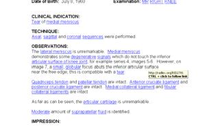
5 Consistent Report Organization LIVER: Demonstrates diffuse increased echogenicity, likely due to fatty infiltration. There are no focal lesions. GALLBLADDER: Normally distended with no gallstones. There is no pericholecystic fluid, wall thickening, or sonographic Murphy's sign. BILIARY: No intrahepatic ductal dilatation is identified. The common duct measures 6 mm at the porta hepatis. PANCREAS: Limited visualization due to gas in the stomach and colon. SPLEEN: Measures 9.9 cm in length and is normal. KIDNEYS: The right kidney measures 11.9 cm. The there is an echogenic structure within the inferior pole of the right kidney with posterior shadowing, likely a renal stone. It measures 8 mm. There is no right hydronephrosis or hydroureter. The left kidney measures 12.3 cm. and is normal. VASCULAR: The abdominal aorta is non aneurysmal. OTHER FINDINGS: The bladder was empty and not evaluated. IMPRESSION: No gallstones and no evidence of cholecystitis. There is an 8mm. stone within the inferior pole of the right kidney without evidence of hydronephrosis. Sistrom & Langlotz, J Am Coll Radiol 2: , 2005 Standard Report Language MRI Knee Medial meniscus: [normal]. tear intersubstance tear flap tear radial tear meniscal cyst degenerative change 5
6 The Interactive Radiology Report McCauley, T. R. et al. Am. J. Roentgenol. 2002;179: Copyright 2008 by the American Roentgen Ray Society 6


7 Benefits of a Library of Report Templates Starting ti point for practices adopting structured reporting Standardizes and improves the quality of radiology reports Enables data mining and quality measurement Fosters the development of new and better reporting systems The End Curtis P Langlotz MD PhD Curtis P. Langlotz, MD, PhD Chair, RSNA Structured Reporting Committee Vice Chair for Informatics, Department of Radiology Professor of Radiology and Informatics (in Epidemiology and Biostatistics) Medical Director, Information Services University of Pennsylvania Health System 7
Is Structured Reporting More Accurate Than Conventional Reporting in CT Reporting of the Abdomen and Pelvis?
 Is Structured Reporting More Accurate Than Conventional Reporting in CT Reporting of the Abdomen and Pelvis? A M Almuslim, MBBS; J G Ryan, MD; A Murtaza, MD Purpose The purpose of this research is to determine
Abdomen and Retroperitoneum Ultrasound Protocols
 Abdomen and Retroperitoneum Ultrasound Protocols Reviewed By: Anna Ellermeier, MD Last Reviewed: March 2018 Contact: (866) 761-4200, Option 1 **NOTE for all examinations: 1. If documenting possible flow
Background. Structured Thyroid Ultrasound Reports 1/8/2018. Clear Communication Improves Management
 Thyroid Ultrasound Reports Clear Communication Improves Management Corey J. Hiti, MD Wendy Yang, MD 2 Michael Campbell, MD FACS 2 Thomas Loehfelm, MD PhD UC Davis Medical Center Department of Radiology,
Abdominal ultrasound:
 Abdominal ultrasound: Non-traumatic acute abdomen Wittanee Na-ChiangMai, MD Department of Radiology ChiangMai University 26/04/2017 Contents Technique of examination Normal anatomy Emergency conditions
Elastography in the. technically difficult patient. EPIQ ultrasound system. Ultrasound
 Ultrasound Elastography in the technically difficult patient EPIQ ultrasound system Chairman Department of Diagnostic Radiology Allegheny General Hospital Pittsburgh, PA, USA You can offer more information
My Patient Has Abdominal Pain PoCUS of the Biliary Tract and the Urinary Tract
 My Patient Has Abdominal Pain PoCUS of the Biliary Tract and the Urinary Tract Objectives PoCUS for Biliary Disease PoCUS for Renal Colic PoCUS for Urinary Retention Biliary Disease A patient presents
Guidelines, Policies and Statements D5 Statement on Abdominal Scanning
 Guidelines, Policies and Statements D5 Statement on Abdominal Scanning Disclaimer and Copyright The ASUM Standards of Practice Board have made every effort to ensure that this Guideline/Policy/Statement
Abdominal Ultrasound. Diane Hallinen, MD. Bloodroot
 Abdominal Ultrasound Diane Hallinen, MD Bloodroot Abdominal Ultrasound Vasculature Hepatobiliary Spleen Kidney Bladder Bowel Where to put the probe? Vasculature We are going to talk about Celiac Trunk
Table of Contents Chapter 1: Perfect Pictures, Imperfect Prose...13 Part I: Practical Advice...21 Chapter 2: Expressing an Imaging Observation...
 Table of Contents Chapter 1: Perfect Pictures, Imperfect Prose...13 The Earliest Radiology Reports... 15 Radiology Communication a Century Later... 15 The Radiology Report at a Turning Point... 17 A Guide
Biliary Ultrasonography Kathleen O Brien MD MPH RDMS Kaiser Permanente South Sacramento
 Biliary Ultrasonography Kathleen O Brien MD MPH RDMS Kaiser Permanente South Sacramento https://www.google.com/search?sa=g&hl=en&q=public+disclosure&tbm=isch&tbs=simg:caqsigeahwelekju2aqaaawlelcmpwgaygpgcamskpib_1qnza7ai
Policies, Standards, and Guidelines. Guidelines for Abdominal Ultrasound Examination
 Policies, Standards, and Guidelines Guidelines for Abdominal Ultrasound Examination Approved by Council Feb 2018 Disclaimer and Copyright The ASUM Standards of Practice Board have made every effort to
Peer Review in Radiology
 Department of Radiology Massachusetts General Hospital Harvard Medical School Peer Review in Radiology Hani Abujudeh MD, MBA Associate Professor of Radiology Harvard Medical School Massachusetts General
US Applications. Case Based Wrap-Up 1. Case 1 E-FAST. Case presentations E-FAST Abdominal. Pearls for each indication
 Case Based Wrap-Up 1 Stephanie J. Doniger MD RDMS FAAP FACEP Associate Director, Pediatric Emergency Ultrasound Stanford University Medical Center US Applications Case presentations E-FAST Abdominal Aorta
Hepatobiliary Ultrasound Rimon Bengiamin, MD, RDMS Assistant Clinical Professor Director of Emergency Ultrasound UCSF Fresno. Objectives. Why?
 Hepatobiliary Ultrasound Rimon Bengiamin, MD, RDMS Assistant Clinical Professor Director of Emergency Ultrasound UCSF Fresno Objectives Discuss the goals of point-of-care biliary ultrasound Review the
Appendix 5. EFSUMB Newsletter. Gastroenterological Ultrasound
 EFSUMB Newsletter 87 Examinations should encompass the full range of pathological conditions listed below A log book listing the types of examinations undertaken should be kept Training should usually
Gallbladder & Pancreas Ultrasonography
 복부초음파 : 담낭과췌장 Gallbladder & Pancreas Ultrasonography 김정훈 Department of Radiology 1 Interaction of sound with matter (1) 반사 (Reflection) (2) 굴절 (Refraction) (3) 흡수 (Absorption) (4) 산란 (Scattering) 음향저항
1. Long images of aorta (prox, mid, and dist) with AP measurements. 2. Trans images of aorta (prox, mid, and dist) with R/L measurements.
 with AP measurements. 2. Trans images of aorta (prox, mid, and dist) with R/L measurements.") Aorta 1. Long images of aorta (prox, mid, and dist) with AP measurements. 2. Trans images of aorta (prox, mid, and dist) with R/L measurements. 3. Long images of R/L common iliac arteries with AP measurements.
Aorta 1. Long images of aorta (prox, mid, and dist) with AP measurements. 2. Trans images of aorta (prox, mid, and dist) with R/L measurements. 3. Long images of R/L common iliac arteries with AP measurements.
Anatomy Jessica Ferguson Ashley Dobos May 31, 2006 LIVER
 Anatomy Jessica Ferguson Ashley Dobos May 31, 2006 LIVER 1) Other Names: Reidel s Lobe normal anatomic variant; projection of the right lobe that can extend as far as the iliac crest (Tempkin, p.54, Anatomy).
Documentation Dissection
 History of Present Illness: Documentation Dissection The patient is a 50-year-old male c/o symptoms for past 4 months 1, severe 2 bloating and stomach cramps, some nausea, vomiting, diarrhea. In last 3
Sonography of Gall Bladder
 Sonography of Gall Bladder Vikram Dogra,MD Professor of Radiology, Urology and BME Director of Ultrasound Associate Chair of Education and Research University of Rochester, NY Objectives Describe the Congenital
Incidental findings: A retrospective analysis of management
 Incidental findings: A retrospective analysis of management Authors and disclosures Authors: Steven Boe, Dana Boe, Jeffrey Kaye, Anu Bansal, Marc Glickstein Disclosures: None Purpose Determine if appropriate
Radiology of hepatobiliary diseases
 GI cycle - Lecture 14 436 Teams Radiology of hepatobiliary diseases Objectives 1. To Interpret plan x-ray radiograph of abdomen with common pathologies. 2. To know the common pathologies presentation.
ULTRASOUND AND ABDOMINAL MASSES
 Med. J. Malaysia Vol. 37 No. I March 1982. ULTRASOUND AND ABDOMINAL MASSES AHMAD KAMAL BIN MD ALIF INTRODUCTION It is approximately 30 years since ultrasound was first introduced into the field of medicine,
Case Study: #3: Gallbladder Carcinoma?
 Case Study: #3: Gallbladder Carcinoma? By: Megan Wyatt K. SON Wyatt 225 2B1 RDMS, RVT Patient: Male 85 YOA Caucasian Indication: Elevated Alkaline Phosphatase History Annual physical showed elevated alkaline
Basic Abdominal Sonography
 24S Basic Abdominal Sonography Procedural Overview JOHN FATCHETT II, RDMS is provided. Patient preparation (i.e., fasting) scanning techniques, spleen, transducer. evaluation of abdominal anatomy in the
US in non-traumatic acute abdomen. Lalita, M.D. Radiologist Department of radiology Faculty of Medicine ChiangMai university
 US in non-traumatic acute abdomen Lalita, M.D. Radiologist Department of radiology Faculty of Medicine ChiangMai university Sagittal Orientation Transverse (Axial) Orientation Coronal Orientation Intercostal
Emergency Ultrasound Standard Reporting Guidelines
 Emergency Ultrasound Standard Reporting Guidelines October 2011 *heterogenous corrected to heterogeneous on pages 9 & 11, January 2016 Emergency Ultrasound Standard Reporting Guidelines: Introduction and
Pocket-sized versus standard ultrasound machines in abdominal imaging
 Singapore Med J 2014; 55(6): 325-333 doi: 10.11622/smedj.2014078 CMEArticle Pocket-sized versus standard ultrasound machines in abdominal imaging Ka Hei Tse 1, MBChB, Wing Hang Luk 1, FRCR, FHKAM, Mau
Bedside RUQ Ultrasound. Replace Formal ULS? Why Bedside ULS RUQ? RUQ Ultrasound. Bedside ULS is Limited, Goal-Directed
 Bedside RUQ Ultrasound RUQ Ultrasound Why do it How to do it Elizabeth Kwan UCSF Emergency Ultrasound Fellow Why Bedside ULS RUQ? Dx or Rule Out Acute Cholecystitis Cholelithiasis, Choledocolithiasis Earlier
Comparative Effectiveness of a Pilot Patient-Centered Ultrasound Report for Hydronephrosis Management
 Comparative Effectiveness of a Pilot Patient-Centered Ultrasound Report for Hydronephrosis Management Geolani W. Dy, MD Society of Women in Urology 6 th Annual Winter Meeting January 21, 2017 Cultural
Abdominal Ultrasound : Aorta, Kidneys, Bladder
 Abdominal Ultrasound : Aorta, Kidneys, Bladder Nilam J. Soni, MD, MSc Associate Professor of Medicine Divisions of Hospital Medicine and Pulmonary/Critical Care Medicine Department of Medicine University
Heme Database Exercise Use the Hematopoietic Database to answer the following questions
 Heme Database Exercise Use the Hematopoietic Database to answer the following questions http://seer.cancer.gov/seertools/hemelymph/ 1. Assign a topography and histology code to polycythemia vera 2. List
Biliary Tree Ultrasound - In a nutshell. Pamela Parker Lead Sonographer
 Biliary Tree Ultrasound - In a nutshell Pamela Parker Lead Sonographer Aims Review what we know about the biliary system Common pathologies Pitfalls Reporting tips The Nutshell Background Biliary examinations
Hilar cholangiocarcinoma. Frank Wessels, Maarten van Leeuwen, UMCU utrecht
 Hilar cholangiocarcinoma Frank Wessels, Maarten van Leeuwen, UMCU utrecht Content Anatomy Biliary strictures (Hilar) Cholangiocarcinoom Staging Biliary tract 1 st order Ductus hepatica dextra Ductus hepaticus
CT abdomen and pelvis
 CT abdomen and pelvis General indications: Assessment of vague abdominal symptoms (pain, colics,distenstion,...) Varifecation of a lesion discovered by other diagnostic modalities as US, barium,ivp, Staging
Imaging of Biliary Tract Emergencies in Jorge A. Soto, MD Professor of Radiology Boston University Medical Center.
 Imaging of Biliary Tract Emergencies in 2011 Jorge A. Soto, MD Professor of Radiology Boston University Medical Center Introduction Biliary emergencies are: Common Come in many flavors Deceiving: frequent
Independent Ultrasound Reporting. BMUS UCD Study day 2018
 Independent Ultrasound Reporting BMUS UCD Study day 2018 Pamela Parker Consultant Sonographer Hull & East Yorkshire Hospitals Colin Griffin Lead Sonographer Royal Liverpool University Hospital Report To
Objectives. Hepatobiliary Ultrasound: Anatomy, Technique, Pathology. RUQ: Normal Anatomy. Emergency Ultrasound: Gallbladder Location
 Hepatobiliary Ultrasound: Anatomy, Technique, Pathology Laleh Gharahbaghian, MD FAAEM Associate Director, EM Ultrasound Co-Director, EM Ultrasound Fellowship Stanford University Medical Center Seric Cusick,
Looking Outside the Box: Incidental Extracardiac Finding in Echo
 Looking Outside the Box: Incidental Extracardiac Finding in Echo Dr. Aijaz Shah Head of Division, Adult Echocardiography Laboratory Prince Sultan Cardiac Centre Riyadh Case 1 17 year old boy presented
Are extra-colonic findings on CT colonogram clinically significant? A review of 758 consecutive cases
 Are extra-colonic findings on CT colonogram clinically significant? A review of 758 consecutive cases Lynn WR 1, Vadhwana B 1, Borgstein R 2, Demetriou G 1, Nair MS 1, Meleagros L 1, Bell DJ 2 1 Department
Pancreas & Biliary System. Dr. Vohra & Dr. Jamila
 Pancreas & Biliary System Dr. Vohra & Dr. Jamila 1 Objectives At the end of the lecture, the student should be able to describe the: Location, surface anatomy, parts, relations & peritoneal reflection
ASSESSING THE PLAIN ABDOMINAL RADIOGRAPH M A A M E F O S U A A M P O F O
 ASSESSING THE PLAIN ABDOMINAL RADIOGRAPH M A A M E F O S U A A M P O F O Introduction The abdomen (less formally called the belly, stomach, is that part of the body between the thorax (chest) and pelvis,
Anatomy: Know Your Abdomen
 Anatomy: Know Your Abdomen Glossary Abdomen - part of the body below the thorax (chest cavity); separated by the diaphragm. Anterior - towards the front of the body. For example, the umbilicus is anterior
Biliary Tree Ultrasound - In a nutshell. Pamela Parker Lead Sonographer
 Biliary Tree Ultrasound - In a nutshell Pamela Parker Lead Sonographer Aims Review what we know about the biliary system Common pathologies Pitfalls Reporting tips The Nutshell Background Biliary examinations
Imaging of common diseases of hepatobiliary and GI system
 Imaging of common diseases of hepatobiliary and GI system Natthaporn Tanpowpong, M.D. Diagnostic radiology Faculty of Medicine, Chulalongkorn University Normal plain radiograph A = Common bile duct
Expected and unexpected gallstones in primary care
 Expected and unexpected gallstones in primary care 7 Speets AM, Van der Graaf Y, Hoes AW, Kalmijn S, De Wit NJ, Mali WPThM. Expected and unexpected gallstones in primary care. Submitted. CHAPTER 7 Abstract
Abdominal radiology 腹部放射線學
 Abdominal radiology 腹部放射線學 台北醫學大學 - 市立萬芳醫院 留偉順 laowilson@hotmail.com The Normal Abdominal Series Chest Supine abdomen Erect abdomen Left lateral decubitus abdomen Learning objectives Understanding normal
Improving Patients' Understanding of Radiology Reports: Comparing Coverage of a Lay-Language Radiology Glossary to MedlinePlus
 Improving Patients' Understanding of Radiology Reports: Comparing Coverage of a Lay-Language Radiology Glossary to MedlinePlus American College of Radiology National Meeting May 2017 Teresa Martin-Carreras,
Focused Assessment Sonography of Trauma (FAST) Scanning Protocol
 Scanning Protocol") Focused Assessment Sonography of Trauma (FAST) Scanning Protocol Romolo Gaspari CHAPTER 3 GOAL OF THE FAST EXAM Demonstrate free fluid in abdomen, pleural space, or pericardial space. EMERGENCY ULTRASOUND
Focused Assessment Sonography of Trauma (FAST) Scanning Protocol Romolo Gaspari CHAPTER 3 GOAL OF THE FAST EXAM Demonstrate free fluid in abdomen, pleural space, or pericardial space. EMERGENCY ULTRASOUND
List by Terms Visceral anomalies
 1 List by Terms Visceral anomalies Dilated 10128 Dilated cerebral ventricle 11 7 2 0 20,00 10201 Dilated aorta 9 8 2 1 5,26 10207 Dilated aortic arch 9 8 3 0 5,00 10213 Dilated carotid 3 12 4 1-47,37 10218
Contrast enhanced ultrasound (CEUS) in gallbladder and bile duct pathology: technique, interpretation and clinical applications
 in gallbladder and bile duct pathology: technique, interpretation and clinical applications") Contrast enhanced ultrasound (CEUS) in gallbladder and bile duct pathology: technique, interpretation and clinical applications Poster No.: C-2099 Congress: ECR 2011 Type: Scientific Exhibit Authors: E.
Contrast enhanced ultrasound (CEUS) in gallbladder and bile duct pathology: technique, interpretation and clinical applications Poster No.: C-2099 Congress: ECR 2011 Type: Scientific Exhibit Authors: E.
Refractory anemia without Primary refractory anemia. sideroblasts RA
 Heme Database Exercise Use the Hematopoietic Database to answer the following questions http://seer.cancer.gov/seertools/hemelymph/ 1. Assign a topography and histology code to polycythemia vera C42.1
Hepatic Imaging: What Every Practitioner Should Know
 Hepatic Imaging: What Every Practitioner Should Know Shuchi K. Rodgers, MD Section Chief, Abdominal Imaging Director of Ultrasound Department of Radiology Einstein Medical Center rodgerss@einstein.edu
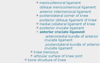
Knee: Meniscus Back to Basics
 Knee: Meniscus Back to Basics Kyung Jin Suh kyungjin.suh@gmail.com Doctor Radiology, Daegu, KOREA Medial Lateral 7.7 10.2 11.6 9.6 10.6 mm Posterior > Anterior horn 10.6 mm Posterior = Anterior horn Medial
What s your diagnosis?
 What s your diagnosis? Signalment: 9 year old MC 2.7 kg Papillion Presenting Complaint: Presented for work up of anorexia and vomiting History: He had presented to cardiology for work up of a grad IV/VI
Comparative Study between Plain Radiography and Ultrasound Abdomen in Non Traumatic Surgical Acute Abdominal Conditions
 ORIGINAL ARTICLE Comparative Study between Plain Radiography and Ultrasound Abdomen in Non Traumatic Surgical Acute Sharma P 1, Sidharth 2, Singh BP 3, Singh D 3, Gupta A 4 1 Department of Radiology and
Intraductal papillary mucinous neoplasm of the bile ducts: a rare form of premalignant lesion of invasive cholangiocarcinoma
 Intraductal papillary mucinous neoplasm of the bile ducts: a rare form of premalignant lesion of invasive cholangiocarcinoma Authors: R. Revert Espí, Y. Fernandez Nuñez, I. Carbonell, D. P. Gómez valencia,
Job Task Analysis for ARDMS Abdomen Data Collected: June 30, 2011
 Job Task Analysis for ARDMS Abdomen Data Collected: June 30, 2011 Reported: Analysis Summary for: Abdomen Examination Survey Dates 06/13/2011-06/26/2011 Invited Respondents 6,000 Surveys with Demographics
Plain Radiographs in Non-Traumatic Abdominal Pain. Plain Radiographs in Non-Traumatic Abdominal Pain
 Jake Block, MD Associate Professor Associate Vice-Chairman for Clinical Operations Director, Musculoskeletal and Emergency Radiology Department of Radiology and Radiological Sciences Vanderbilt University
L o o k L i s t e n F e e l S c a n. Your Pocus Cards For Your Every Day Scanning.
 L o o k L i s t e n F e e l S c a n Your Pocus Cards For Your Every Day Scanning E-FAST Extended Focused Assessment by Sonography in Trauma Subcostal Heart View Pleural Sliding on M-mode (Sea-shore sign)
Appendix 9: Endoscopic Ultrasound in Gastroenterology
 Appendix 9: Endoscopic Ultrasound in Gastroenterology This curriculum is intended for clinicians who perform endoscopic ultrasonography (EUS) in gastroenterology. It includes standards for theoretical
Use of Ultrasound in NAFLD
 Institute for Liver and Digestive Health Use of Ultrasound in NAFLD Dr. Davide Roccarina Specialist in General Medicine Specialist Doctor in Clinical Ultrasound and non-invasive liver assessment Hepatology
Acute renal colic Radiological investigation in patients with renal colic
 Acute renal colic Radiological investigation in patients with renal colic Mikael Hellström Professor Department of Radiology Sahlgrenska University Hospital Göteborg University 0.9-1.8/1.000 inhabitants
An abdominal ultrasound produces a picture of the organs and other structures in the upper abdomen.
 Scan for mobile link. Ultrasound - Abdomen Ultrasound imaging of the abdomen uses sound waves to produce pictures of the structures within the upper abdomen. It is used to help diagnose pain or distention
Urinary system Ultrasound (Renal & Urinary bladder)
") Urinary system Ultrasound (Renal & Urinary bladder) Edited & Presented by ; Hussien A.B ALI DINAR. Msc.Phd ISRRT Associate Member Lecturer (National university) Reporting Sonographer (PHC) Objective By
Urinary system Ultrasound (Renal & Urinary bladder) Edited & Presented by ; Hussien A.B ALI DINAR. Msc.Phd ISRRT Associate Member Lecturer (National university) Reporting Sonographer (PHC) Objective By
Pediatric Hepatobiliary, Pancreatic & Splenic US
 Pediatric Hepatobiliary, Pancreatic & Splenic US Susan J. Back, MD Department of Radiology, The Children s Hospital of Philadelphia No Disclosures Objectives Normal Abnormal: cases and US advances Objectives
Magnetic Resonance Cholangiopancreatography (MRCP) in a District General Hospital
 in a District General Hospital") Magnetic Resonance Cholangiopancreatography (MRCP) in a District General Hospital Poster No.: C-1790 Congress: ECR 2012 Type: Authors: Scientific Exhibit J. A. Maguire 1, H. Kasem 2, M. Akhtar 2, M. Strauss
Magnetic Resonance Cholangiopancreatography (MRCP) in a District General Hospital Poster No.: C-1790 Congress: ECR 2012 Type: Authors: Scientific Exhibit J. A. Maguire 1, H. Kasem 2, M. Akhtar 2, M. Strauss
Body MRI from the Liver to the Bladder
 Body MRI from the Liver to the Bladder I Want You! Audience Participation Methodist Hospital Continuing Education Seminar Jordan Swensson, MD November 7, 2015 Objectives Observe the uses of MRI for organs
Abdomen Sonography Examination Content Outline
 Abdomen Sonography Examination Content Outline (Outline Summary) # Domain Subdomain Percentage 1 2 3 Anatomy, Perfusion, and Function Pathology, Vascular Abnormalities, Trauma, and Postoperative Anatomy
Case Scenario 1. Discharge Summary
 Case Scenario 1 Discharge Summary A 69-year-old woman was on vacation and noted that she was becoming jaundiced. Two months prior to leaving on that trip, she had had a workup that included an abdominal
Pre-operative prediction of difficult laparoscopic cholecystectomy
 International Surgery Journal http://www.ijsurgery.com pissn 2349-3305 eissn 2349-2902 Research Article DOI: http://dx.doi.org/10.18203/2349-2902.isj20151083 Pre-operative prediction of difficult laparoscopic
Navigating the Biliary Tract with CT & MR: An Imaging Approach to Bile Duct Obstruction
 Navigating the Biliary Tract with CT & MR: An Imaging Approach to Bile Duct Obstruction Ann S. Fulcher, MD Medical College of Virginia Virginia Commonwealth University Richmond, Virginia Objectives To
Evaluation of Suspected Pancreatic Cancer
 Evaluation of Suspected Pancreatic Cancer October 15, 2015 If you experience technical difficulty during the presentation: Contact WebEx Technical Support directly at: US Toll Free: 1-866-779-3239 Toll
In this edition we will take a look at Cholelithiasis diagnoses and illustrate the increased specificity under the ICD-10-CM nomenclature.
 On October 1, 2015, the ICD-9-CM code set that is used to report medical diagnoses in the United States will be replaced with the ICD-10-CM code set. The new code set provides more than 68,000 codes, compared
Ex. 1 :Language of Anatomy
 Collin College BIOL 2401 : Human Anatomy & Physiology Ex. 1 :Language of Anatomy The Anatomical Position Used as a reference point when referring to specific areas of the human body Body erect Head and
List by Region - Visceral Anomalies
 1 List by Region - Visceral Anomalies General Terms 10127 Situs inversus 80,00 10125 Aneurysm 68,42 10126Fluid-filled abdomen -35,00 Brain 10131 Hydrocephaly 10128 Dilated cerebral ventricle 20,00 10132
Diagnostic Imaging
 www.fisiokinesiterapia.biz Diagnostic Imaging Diagnostic Imaging is no longer limited to radiography. Major technological advancements have lead to the use of new and improved imaging technologies. The
EFSUMB EUROPEAN FEDERATION OF SOCIETIES FOR ULTRASOUND IN MEDICINE AND BIOLOGY Building a European Ultrasound Community
 MINIMUM TRAINING REQUIREMENTS FOR THE PRACTICE OF MEDICAL ULTRASOUND IN EUROPE Appendix 9: Endoscopic Ultrasound in Gastroenterology This curriculum is intended for clinicians who perform endoscopic ultrasonography
Ultrasound and CT in lymphoma: Bring out the particulars
 Ultrasound and CT in lymphoma: Bring out the particulars Poster No.: C-1774 Congress: ECR 2012 Type: Educational Exhibit Authors: A. Anton, S. M. Petrescu, C. ZAHARIA, C. A. Minoiu ; Ploiesti/ 1 2 2 3
Abdominal Ultrasound
 Abdominal Ultrasound What is Ultrasound Imaging of the Abdomen? What are some common uses of the procedure? How should I prepare? What does the equipment look like? How does the procedure work? How is
Table E1. Standardized Mortality Ratios for Total and Specific Causes of Death Parameter Radiologists Psychiatrists No. of Deaths
 RSNA, 2016 10.1148/radiol.2016152472 Table E1. Standardized Mortality Ratios for Total and Specific Causes of Death Parameter Radiologists Psychiatrists No. of Deaths Observed/Expected No. of Deaths Observed/Expected
Do you want to be an excellent Radiologist? - Focus on the thoracic aorta on lateral chest image!!!
 The lateral chest radiograph: Challenging area around the thoracic aorta!!! Do you want to be an excellent Radiologist? - Focus on the thoracic aorta on lateral chest image!!! Dong Yoon Han 1, So Youn
Alice Fung, MD Oregon Health and Science University
 Alice Fung, MD Oregon Health and Science University Disclosure Comments The speaker Alice Fung, MD Has relevant financial relationships to disclose. Received honorarium from (Guerbet). This individual
Lab Monitor Images Dissection of the Abdominal Vasculature + Lower Digestive System
 Lab Monitor Images Dissection of the Abdominal Vasculature + Lower Digestive System Stomach & Duodenum Frontal (AP) View Nasogastric tube 2 1 3 4 Stomach Pylorus Duodenum 1 Duodenum 2 Duodenum 3 Duodenum
Normal Sonographic Anatomy
 hapter 2:The Liver DUNSTAN ABRAHAM Normal Sonographic Anatomy Homogeneous, echogenic texture (Figure 2-1) Measures approximately 15 cm in length and 10 12.5 cm anterior to posterior; measurement taken
Anatomy II ANAT 302. Course Description
 Anatomy II ANAT 302 Course Description This course provides the students with lectures and comprehensive overview of the gross anatomy of the components of the respiratory, cardiovascular, digestive and
Plain abdomen The standard films are supine & erect AP views (alternative to erect, lateral decubitus film is used in ill patients).
.") Plain abdomen The standard films are supine & erect AP views (alternative to erect, lateral decubitus film is used in ill patients). The stomach can be readily identified by its location, gastric rugae
Plain abdomen The standard films are supine & erect AP views (alternative to erect, lateral decubitus film is used in ill patients). The stomach can be readily identified by its location, gastric rugae
Obstetrics Content Outline Obstetrics - Fetal Abnormalities
 Obstetrics Content Outline Obstetrics - Fetal Abnormalities Effective February 2007 10 16% renal agenesis complete absence of the kidneys occurs when ureteric buds fail to develop Or degenerate before
Intrahepatic Cholangiocarcinoma (ICC) Detected by Sonography
 Detected by Sonography") 661245JDMXXX10.1177/8756479316661245Journal of Diagnostic Medical SonographyHamer research-article2016 Case Study Intrahepatic Cholangiocarcinoma (ICC) Detected by Sonography Journal of Diagnostic Medical
661245JDMXXX10.1177/8756479316661245Journal of Diagnostic Medical SonographyHamer research-article2016 Case Study Intrahepatic Cholangiocarcinoma (ICC) Detected by Sonography Journal of Diagnostic Medical
International Journal of Case Reports and Images (IJCRI)
") www.edoriumjournals.com clinical images PEER REVIEWED OPEN ACCESS Is it just another case of acute uncomplicated cholecystitis? A case of emphysematous cholecystitis an uncommon complication and associated
www.edoriumjournals.com clinical images PEER REVIEWED OPEN ACCESS Is it just another case of acute uncomplicated cholecystitis? A case of emphysematous cholecystitis an uncommon complication and associated
Structured reports: 10 years experience in private radiology clinics
 Structured reports: 10 years experience in private radiology clinics Poster No.: C-2028 Congress: ECR 2014 Type: Educational Exhibit Authors: T. Laswad, D. Fournier, J. Moreau, H. Brat, M. Deac, T. 1 1
In any operation. Indications. Anaesthesia. Position of the patient. Incision. Steps of the operation. Complications.
 In any operation Indications. Anaesthesia. Position of the patient. Incision. Steps of the operation. Complications. Abdominal operation I position for operation Supine Abdominal operation I position for
Special Instructions
 FDA and ACR guidelines are as follows: Special Instructions Safety concerning NSF and gadolinium-based contrast agents (GBCA) Prior to administering MRI contrast (GBCA), any patient who answers yes to
1 Right & left Hepatic ducts Gastric Impression of spleen
 Pancreatic Model 1 Right & left Hepatic ducts 14 Gastric Impression of spleen 2 Common hepatic duct 15 Renal Impression of spleen 3 Cystic Duct 16 Colic Impression of spleen 4 Common Bile Duct 17 Splenic
ROTATION REPORT OUTLINE
 ROTATION REPORT OUTLINE 1. Introduction and Thanks 2. Selection criteria 3. Timing, Rotations, Academic activities report 4. Cost 5. Comments and suggestions Introduction and thanks My name is Dr. Edelson
Appropriate Imaging Tests Lead to Meaningful Results. Dr. Richard Wasley May 2011
 Appropriate Imaging Tests Lead to Meaningful Results Dr. Richard Wasley May 2011 Summarize the advantages and limitations of specific imaging tests and why clinical information is so important to radiologists
Ultrasound in the ICU
 Ultrasound in the ICU Kristine E. W. Breyer, MD Assistant Professor Anesthesia & Critical Care Medicine UCSF DISCLOSURES: NONE Definition The Ultrasound Exam Types & Uses Training Clinical Examples Objectives
Original Research Article
 Original Research Article Role of (Non Gynaecological Causes) Kaleem Ahmad 1, Rishav Kumar Jain 2, Ashok Yadav 3, Shilpa Vahikar 4 1 Associate Professor, Department of Radiodiagnosis, 2 Professor, Department
US LI-RADS v2017 CORE
 US LI-RADS v2017 CORE Screening or surveillance US in patient at high risk for HCC US category US-1 US-2 US-3 Negative Subthreshold Positive Category Concept Definition US-1 Negative US-2 Subthreshold
Certificate in Clinician Performed Ultrasound (CCPU) Syllabus. Biliary
 Syllabus. Biliary") Certificate in Clinician Performed Ultrasound (CCPU) Syllabus Biliary Page 1 of 6 12/18 Biliary Syllabus Purpose: This unit is designed to cover the theoretical and practical curriculum for basic ultrasound
Certificate in Clinician Performed Ultrasound (CCPU) Syllabus Biliary Page 1 of 6 12/18 Biliary Syllabus Purpose: This unit is designed to cover the theoretical and practical curriculum for basic ultrasound