75% Nephrectomy and the Disposition of Inorganic Mercury In DMSA-Treated Rats Lacking Functional MRP2
|
|
|
- Johnathan Hamilton
- 5 years ago
- Views:
Transcription
1 JPET Fast This Forward. article has not Published been copyedited on and December formatted. The 23, final 2009 version as may DOI: /jpet differ from this version. JPET # % Nephrectomy and the Disposition of Inorganic Mercury In -Treated Rats Lacking Functional MRP2 Rudolfs K. Zalups and Christy C. Bridges Division of Basic Medical Sciences Mercer University School of Medicine Macon, Georgia Copyright 2009 by the American Society for Pharmacology and Experimental Therapeutics.
2 This article has not been copyedited and formatted. The final version may differ from this version. JPET # Running Title: MRP2 and Hg 2+ Handling in 75% NPX Rats Corresponding Author: Dr. Rudolfs K. Zalups Division of Basic Medical Sciences Mercer University School of Medicine 1550 College Street Macon, GA (912) phone (912) fax Number of text pages (Abstract - Index Terms) = 31 Number of Figures = 8 Number of Tables = 4 Number of Words in Abstract = 249 Number of Words in Introduction = 712 (Including References) Number of Words in Discussion 1375 References = 37 Abbreviations: ABC, ATP-binding cassette;, meso 2,3-Dimercaptopropanesunccinc acid; DMPS, 2,3-Dimercaptopropanesulfonic acid; HgCl 2, mercuric chloride; HgO, mercuric oxide; Hg 2+, inorganic mercury; MRP2, multidrug-resistance protein 2; MURR, Missouri University Research Reactor; NPX, nephrectomized; OAT1, organic anion transporter 1; PAH, para aminohippuric acid; SO, sham-operated; TR, transport-deficient. 2
3 ABSTRACT In the present study, we evaluated the disposition of inorganic mercury (Hg 2+ ) in sham- operated (SO) and 75% nephrectomized (NPX) Wistar and transport-deficient (TR ) rats treated with saline or the chelating agent meso-2,3-dimercaptosuccinic acid (). Based on previous studies, and TR rats were used as tools to examine the potential role of MRP2 in the disposition of inorganic mercury (Hg 2+ ) during renal insufficiency. All animals were treated i.v. with a low (0.5-µmol/kg) dose of mercuric chloride (HgCl 2 ). At 24 and 28 h after exposure to HgCl 2, matched groups of Wistar and TR rats received normal saline or (i.p.). Fortyeight h after exposure to HgCl 2, the disposition of Hg 2+ was examined. A particularly notable effect of 75% nephrectomy in both strains of rats was enhanced renal accumulation of Hg 2+, specifically in the outer stripe of the outer medulla. In addition, hepatic accumulation, fecal excretion, and blood levels, of Hg 2+ were enhanced in rats after 75% nephrectomy, especially in the TR rats. Treatment with increased both the renal tubular elimination and urinary excretion of Hg 2+ in all rats. did not, however, affect hepatic content of Hg 2+, even in the 75% NPX TR rats. We also show with real-time PCR that following 75% nephrectomy and compensatory renal growth, expression of MRP2 (only in Wistar rats) and OAT1 is enhanced in the remaining functional proximal tubules. We conclude that MRP2 does play a significant role in the renal and corporal disposition of Hg 2+ following a 75% reduction of renal mass. 3
4 INTRODUCTION Under homeostatic conditions, renal proximal tubular epithelial cells (in rats) do not, or cannot, secrete efficiently mercuric ions into the tubular lumen following exposure to a nonnephrotoxic dose of inorganic mercury (Hg 2+ ). The low level of secretion of Hg 2+ under these circumstances likely relates to significant binding of mercuric ions to protein-thiols in the intracellular milieu. However, following treatment with the dithiol chelating agents 2,3- Dimercaptopropane-1-sulfonic acid (DMPS) or meso-2,3-dimercaptosuccinic acid (), mobilization and secretory-elimination of Hg 2+ along the proximal tubule has been shown to be great enough to reduce the renal burden of Hg 2+ in normal and uninephrectomized (50% NPX) rats by as much as 80-85% within a 24-h period (Zalups, 1993). In a series of recent studies, we demonstrated that the ATP-binding cassette (ABC) protein, MRP2, plays a significant role in the secretion of Hg 2+ at the luminal plasma membrane of proximal tubular epithelial cells, especially following treatment with DMPS or (Bridges et al., 2008a; Bridges et al., 2008b; Zalups and Bridges, 2008). Interestingly, when 50% of the functional renal mass is removed surgically in rats, and then the rats are exposed to Hg 2+ or methylmercury (CH 3 Hg + ), enhanced uptake and retention of mercuric ions occurs along the pars recta of renal proximal tubules, especially in the portion present in the outer stripe of the outer medulla (Zalups et al., 1987; Zalups and Lash, 1990; Zalups, 1991b; Zalups, 1991a; Zalups et al., 1992; Zalups and Cherian, 1992a; Zalups and Cherian, 1992b; Zalups, 1993). This enhanced uptake appears to be causally linked to profound structural and functional adaptive changes associated with compensatory renal growth, which occurs after renal mass is reduced significantly. Moreover, the enhanced accumulation of mercuric ions that occurs in the pars recta following a 50% reduction of renal mass also appears to increase the severity of the 4
5 nephropathy induced by lower nephrotoxic doses of Hg 2+ (Zalups and Diamond, 1987; Zalups et al., 1987; Houser and Berndt, 1988). At present, the mechanisms responsible for the enhanced net accumulation of mercuric ions along the pars recta of proximal tubules after a 50% reduction of renal mass remain not fully defined, although an amplification of transport mechanisms and increases in the intracellular contents of the thiols glutathione (GSH) and metallothionein (MT) have been suggested (Zalups and Lash, 1990; Zalups et al., 1995). Experimental evidence indicates that both luminal and basolateral mechanisms participate in the accumulation of mercury following uninephrectomy (Zalups, 1997), and that a p-aminohippuric acid (PAH)-dependent mechanism is active. PAH has a particularly high affinity for the organic anion transporter 1 (OAT1), which in the kidneys is present almost exclusively in the basolateral membrane of proximal tubular epithelial cells. We have recently demonstrated in vitro that OAT1, which has a broad substrate-specificity, is capable of transporting the most likely mercuric conjugates of non-protein thiols (such as cysteine, homocysteine and N- acetylcysteine) believed to be present in blood (Aslamkhan et al., 2003; Zalups and Ahmad, 2004; Zalups and Ahmad, 2005). When functional renal mass is reduced by approximately 75-80% of normal (as a result of disease, trauma or surgery), the hypertrophic changes that occur along the remaining functional proximal tubules are greater than those engendered by a 50% reduction of renal mass. However, the diminished capacity for ultrafiltration combined with the imposed increased workload on the hypertrophied remaining nephrons becomes too great for them to maintain normal fluid and electrolyte homeostasis; and chronic renal failure ensues. We postulate that increased expression of certain membrane transporters constitutes a significant part of the adaptive structural and functional responses that occur in hypertrophied proximal tubular epithelial cells after renal mass has been reduced by 75% nephrectomy. In particular, we hypothesize that expression of transporters involved in tubular secretion (such as 5
6 OAT1 in the basolateral membrane and MRP2 in luminal membrane) is enhanced. In the present investigation, we studied the disposition of an i.v. administered low dose (0.5-µmol/kg) dose of mercuric chloride (HgCl 2 ) in 75% nephrectomized (75% NPX) and sham- operated (SO), Wistar and transport deficient (TR ) rats treated with normal saline or (an FDA-approved chelating agent that mobilizes Hg 2+ in renal proximal tubular epithelial cells). Treatment with was used as a tool to assess the potential role of MRP2 in the renal disposition of Hg 2+ in rats that had undergone a sham-operation or a 75% reduction of renal mass. The primary aims of the present study were to determine if 75% nephrectomy: 1) affects the expression of OAT1 and MRP2 in the remaining functional proximal tubular epithelial cells; and 2) affects the role of MRP2 in the renal and corporal disposition of Hg 2+. 6
7 MATERIALS AND METHODS Animals: Male TR and normal (control) Wistar rats weighing g were purchased from Harlan Laboratories (Indianapolis, IN). All animals were provided a commercial laboratory diet (Tekland 6% rat diet, Harlan Laboratories) and water ad libitum throughout all aspects of animal experimentation. In the first phenotypic characterization of TR rats, Jansen and colleagues (Jansen et al., 1985) demonstrated that the rats had hereditary conjugated hyperbilirubinemia, which was subsequently attributed to a mutation in the mrp2 gene (Mayer et al., 1995; Paulusma et al., 1996). As a result of this mutation, the presence Mrp2 mrna is low, while the presence of Mrp2 protein is absent, in the tissues of these rats (Mayer et al., 1995). TR rats represent a reliable model to study the hepatic and renal secretion of various substrates of MRP2 (de Vries et al., 1989; Masereeuw et al., 2003; Smeets et al., 2004). Groups: Four groups of four TR from the pool of purchased TR rats and four groups of four Wistar rats were selected at random and Wistar rats. Two groups of TR rats and two groups of Wistar rats underwent 75% nephrectomy, while the remaining four corresponding groups of rats underwent a sham operation. Experimental Design Fourteen days after surgery, all rats were injected intravenously with a non-toxic 0.5 µmol/kg dose of mercuric chloride (HgCl 2 ; in 2 ml/kg normal saline), containing 203 HgCl 2 (1 µci/rat). The nephrotoxic nature of the 0.5 µmol/kg dose of HgCl 2 in the 75% NPX rats was 7
8 determined previously with histopathological analyses of renal slices (unpublished findings). Twenty-four and 48 h after exposure to HgCl 2, one group SO and one group of 75% NPX rats of both strains (Wistar and TR ) received an intraperitoneal, 100-mg/kg dose of (in 2 ml/kg normal saline). At the same time, one group of SO and one group of 75% NPX rats of both strains received an intraperitoneal injection of normal saline (2 ml/kg). Forty-eight hours after the injection of HgCl 2, all groups of rats were sacrificed and samples of blood, liver, kidneys, urine and feces were collected for analysis of Hg 2+ content. Surgery: After anesthesia was induced with 70 mg/kg ketamine and 6 mg/kg xylazine (i.m.); a midline incision was made through the skin and musculature of the abdomen with a scalpel. Subsequently, the right and left kidneys of each animal were isolated from the perinephric fascia and fat, without damaging the liver or corresponding adrenal glands. For the groups that underwent 75% nephrectomy, the right renal artery and vein and right ureter were ligated with a single sterile 1-0 silk suture. Then the right kidney was excised distal to the ligature, following which; the left kidney was exteriorized from the body through the mid-line incision. Subsequently, it was placed in a Lucite cup in a manner that exposed the posterior surface of the organ. Using a dissecting microscope, a sterile 4-0 silk suture was threaded between the renal vein and posterior branch of the renal artery. When the ligature was in place, it was tied tightly. Approximately one half of the left kidney would generally turn to a darker color, indicating a cessation, or great reduction of blood flow to that portion of the kidney. SO animals were treated similarly, except that their right kidney was not excised and the left renal artery was not ligated. Based on numerous findings from our laboratory, the removal of the right kidney and the tying off of the posterior branch of the left renal artery is sufficient to induce early systemic 8
9 This article has not been copyedited and formatted. The final version may differ from this version. JPET # changes associated with chronic renal failure (Zalups, 1989; Zalups and Henderson, 1992; Zalups, 1995). With the ligature tied, the left kidney was removed from the Lucite cup and was placed back into its normal retroperitoneal position. The abdominal muscles were sewn together using sterile 4-0 silk suture and the opposite ends of the incised skin were approximated using sterile 9-mm stainless steel wound clips. Recovery from Surgery: A period of 14 days was allowed for recovery from surgery and to allow for the completion of the rapid phase of compensatory renal growth in the 75% NPX rats. Injection of Hg 2+ : Animals were first anesthetized lightly with ether. Then, a small incision was made through the skin in the mid-ventral region of the thigh to expose the femoral vein and artery. A non-nephrotoxic 0.5 µmol/kg dose of HgCl 2 (in 2 ml normal saline) (containing 1 µci of 203 Hg 2+ ) was administered into the vein. The wound was closed using sterile, 9-mm, stainless steel wound clips. Animals were then placed individually in plastic metabolic cages, in which water and food were provided ad libitum. Injections of meso-2,3-dimercaptosuccinic Acid (): At both 24 and 28 h after exposure to HgCl 2, corresponding groups of Wistar and TR rats received an intraperitoneal injection of either normal saline (2 ml/kg) or a 100-mg/kg dose of (in 2 ml/kg normal saline). Forty-eight hours after receiving the non-nephrotoxic dose of HgCl 2, the rats were anesthetized deeply (with 70 mg/kg ketamine and 6 mg/kg xylazine) and blood, liver, kidneys, urine and feces were collected for analysis of Hg 2+ content. 9
10 Collection of Tissues, Organs, Urine and Feces Forty-eight h after the injection of Hg 2+, rats were anesthetized with ketamine and xylazine (70/6 mg/kg). Once anesthetized, two 1-mL samples of blood were obtained from the inferior vena cava. One of the samples was placed in a polystyrene tube for determination of 203 Hg 2+ content, while the other sample was placed in a Microtainer tube (Becton Dickenson and Co., Franklin Lakes, NJ), which was centrifuged at 21,000 x g for 90 seconds. Subsequently, the cellular and plasma fractions were removed and placed in separate polystyrene tubes for estimation of Hg content. The kidney(s) was/were also removed from each animal. After each kidney was trimmed of fascia and fat, it was weighed and cut in half along the mid-transverse plain. From one half of the left kidney, a 3-mm transverse slice was utilized for separation of cortex, outer stripe of outer medulla, inner stripe of outer medulla and inner medulla. Each zone of the kidney was weighed and placed in a polystyrene tube for estimation of 203 Hg 2+ content. Following removal of the kidneys, the liver was excised carefully, weighed, and a 1-g section was removed for determination of 203 Hg 2+ content. Urine and feces were collected for 24-h intervals throughout the duration of the study. Each 24-h collection was mixed by vortexing and a 1-mL sample was weighed and placed in a polystyrene tube for estimation of 203 Hg 2+ content. All of the feces excreted by each animal during the final 24-h period were counted to determine the content of 203 Hg 2+ excreted in the feces. Determination of Hg 2+ Content in Samples of Tissue, Organs, Urine, and Feces All samples were placed in 12 x 75 mm polystyrene tubes, which were sealed 10
11 immediately to prevent evaporation or desiccation. The content of 203 Hg 2+ in each sample was determined by counting the samples in a Wallac Wizard 3 automatic gamma counter (Perkin Elmer, Boston, MA). The total content of Hg 2+ in the entire kidney, liver and blood volume is expressed as percent of administered dose. Concentrations of Hg 2+ in the renal samples are expressed as percent of dose per gram of tissue. Total blood volume was estimated to be 6% of body weight. Urinary and fecal excretion of Hg 2+ is expressed as percent of administered dose per the last 24 h of study. Real-time PCR Cortex and outer stripe were obtained from each set of animals. Tissues were frozen immediately in liquid nitrogen. Frozen tissues were then ground up and total RNA was isolated using TRIzol Reagent (Invitrogen) according to the manufacturer s protocol. Reverse transcription of one microgram of RNA was carried out using reverse transcriptase and random hexamers (Applied Biosystems, Foster City, CA). Real-time PCR analyses of Mrp2 and Oat1 expression were performed using an ABI Prism 7000 sequence detection system and commercially available gene expression assays (Mrp2: Rn_ ; Oat1: Rn_ ; Applied Biosystems). Analyses were designed according to manufacturer s recommendation. GAPDH was used as a reference gene. Generation of 203 Hg Hg 2+ was generated by neutron activation of a target of mercuric oxide (HgO) at the Missouri University Research Reactor (MURR) and subsequent chemical handling and analyses by the method described previously (Belanger, 2001; Bridges et al., 2004; Bridges and Zalups, 2004). The specific activities of the 203 Hg 2+ ranged from 6 to12 mci/mg. 11
12 Data Analyses: Each set of respective data were analyzed first with the Kolmogorov-Smirnov to test for normality, and then with Levene s test for homogeneity of variances. Differences among means were then analyzed using a two-way analysis of variance (ANOVA). When statistically significant F-values were obtained, the data were analyzed further using Tukey s post hoc multiple comparison test. A p-value of < 0.05 was considered statistically significant. 12
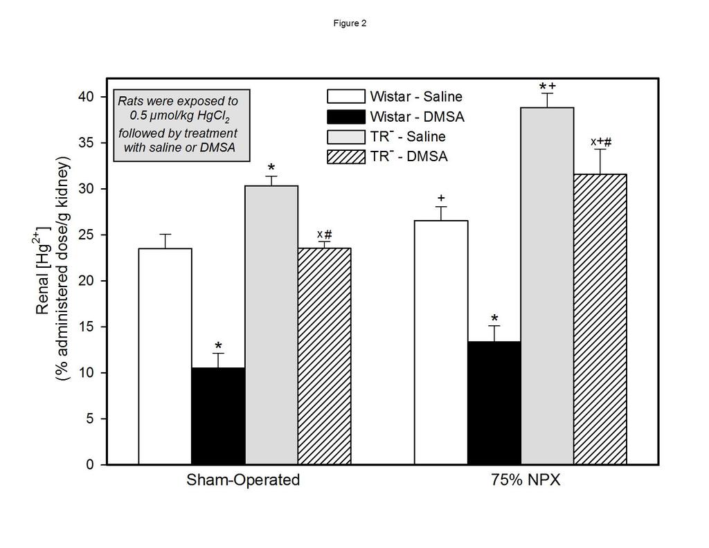
13 RESULTS Renal Expression of Mrp2 and Oat1: Real-time PCR analyses were performed on renal tissue 14 days after surgery. In the Wistar rats, expression of Mrp2 was enhanced approximately 3.5-fold, while expression of Oat1 was increased approximately 2.5-fold (Figure 1). Transcripts encoding Mrp 2 were not detected in SO or 75% NPX TR rats. A similar finding has also been reported recently (Oswald et al., 2006). Additionally, the expression of Oat1 in the 75% NPX TR fourfold greater than that in corresponding SO rats. Renal Concentration of Hg 2+ : rats was amplified almost In the group of SO Wistar rats exposed to mercuric chloride and then treated with normal saline, the renal concentration of Hg 2+ was approximately 23% of the dose/g tissue 48 h after exposure to Hg 2+ (Figure 2). In the corresponding group of Wistar rats treated with, the renal concentration of Hg 2+ was about 55% less than that in the saline-treated group (Table 1). Interestingly, the renal concentration of Hg 2+ in the SO TR rats treated with saline was approximately 28% greater than that in the corresponding group of saline-treated Wistar rats. However, the renal concentration of Hg 2+ in the group of TR rats treated with was, on average, only about 22% less than that in the corresponding group of saline-treated TR rats. The overall renal concentration of Hg 2+ in the group of 75% NPX Wistar rats treated with normal saline was approximately 13% greater than that in the corresponding group of SO Wistar rats treated with normal saline (Figure 2 and Table 1). By contrast, the renal concentration of Hg 2+ in the 75% NPX Wistar rats treated with was nearly 51% less than that in 75% NPX Wistar rats treated with normal saline. In the 75% NPX TR rats treated with saline, the renal 13
14 concentration of Hg 2+ was approximately 46% greater than that in the group of 75% NPX Wistar rats treated with saline. Additionally, the renal concentration of Hg 2+ in the 75% NPX TR rats treated with saline was about 28% greater than that in the corresponding SO group treated with saline. In the group of 75% NPX TR rats treated with DMPS, the renal concentration of Hg 2+ was approximately only about 19% less than that in the 75% NPX rats treated with saline. Concentration of Hg 2+ in the Renal Cortex: Among the SO rats, the renal cortical concentration of Hg 2+ in the -treated group was approximately 56% less than that in the SO rats treated with saline (Figure 3 and Table 2). By contrast, there was no significant difference in the renal cortical concentration of Hg 2+ between the two groups of SO TR groups of SO TR rats. The renal cortical concentration of Hg 2+ in the two rats was significantly greater than that in the corresponding SO group of Wistar rats, the greatest difference detected between the two groups of -treated rats. The renal cortical concentration of Hg 2+ in the 75% NPX Wistar rats treated with was about 49% less than that in the 75% NPX Wistar rats treated with saline. In the 75% TR rats treated with saline, the renal cortical concentration of Hg 2+ was approximately 96% greater than that in the corresponding group of 75% NPX Wistar rats treated with saline. Between the two groups of 75% NPX rats treated with, the renal cortical concentration of Hg 2+ was approximately 187% greater in the 75% NPX TR rats. In the two groups of 75% NPX Wistar rats treated with saline or, the renal concentration of Hg 2+ was not significantly different from that in the corresponding group SO Wistar rats treated in the same manner. Only in the group of 75% TR 14 rats treated with saline was the renal cortical concentration significantly greater than that in the corresponding group of SO rats treated in the same manner. Concentration of Hg 2+ in the Renal Outer Stripe of the Outer Medulla:
15 The patterns for the accumulation of inorganic Hg 2+ in the outer stripe of the outer medulla were considerably different from those detected in the renal cortex (Figure 4 and Table 3). Among the four groups of SO rats, the concentration of Hg 2+ in the outer stripe of the outer medulla was significantly less in the -treated Wistar rats and the saline-treated TR rats relative to that in the Wistar rats treated with saline. The only other significant difference in the outer stripe of the outer medulla among the SO rats was detected between the two groups of rats treated with. There was no significant difference in the concentration of Hg 2+ in the outer stripe of the outer medulla between the two groups of TR Significant differences in the concentration of Hg 2+ rats. in the outer stripe of the outer medulla were also detected among the four groups of 75% NPX rats. In both groups of 75% NPX rats treated with, the average concentration of Hg 2+ in the outer stripe of the outer medulla was significantly less than that in the corresponding group of 75% NPX rats treated with saline. In addition, the mean concentration of Hg 2+ in the outer stripe of the outer medulla of the 75% NPX Wistar treated with was greater than that in the 75% NPX TR The most marked differences in the average concentration of Hg 2+ in the outer stripe of the outer medulla were detected between corresponding groups of 75% NPX rats and SO rats. The greatest difference in the concentration of Hg 2+ in the outer stripe of the outer medulla was detected between the 75% NPX Wistar rats treated with saline and the corresponding SO rats. Wistar rats treated with saline. Values were approximately 125% greater in the corresponding NPX rats. Accumulation of Hg 2+ in the outer stripe of the outer medulla was also significantly greater in the 75% NPX rats between the paired groups of Wistar rats treated with and the TR rats treated with saline. Interestingly, there was no significant difference in the average concentration of Hg 2+ in the outer stripe of the outer medulla between the 75% NPX and SO groups of TR rats treated with. 15
16 Urinary Excretion of Hg 2+ : Profound changes in the urinary excretion of Hg 2+ were detected among the groups of SO and 75% NPX Wistar and TR rats during the second 24-hour period of study (Figure 5 and Table 4). Among the four groups of SO rats, urinary excretion of Hg 2+ was greatest in the Wistar rats treated with. In fact, the amount of Hg 2+ excreted in 24 h by the Wistar rats treated with was on average 394% greater than that in Wistar rats treated with saline. Also, the average amount of Hg 2+ excreted in 24 h in the TR greater than that in the corresponding saline-treated TR Hg 2+ in the TR rats treated with was about 363% rats. Interestingly, urinary excretion of rats treated with saline was about 76% less than that in the corresponding group of Wistar rats treated with saline. Moreover, urinary excretion of Hg 2+ in the TR was about 78% less than that in the Wistar rats treated with. rats treated with Urinary excretion of Hg 2+ among the four groups of 75% NPX rats was also greatest in the group of Wistar rats treated with. In this group, the mean urinary excretion of Hg 2+ over 24 h was approximately 372% greater than that in the corresponding group of Wistar rats treated with saline. In addition, the mean urinary excretion of Hg 2+ in the 75% NPX TR treated with was significantly greater than that in the 75% NPX TR saline. Furthermore, urinary excretion of Hg 2+ in the group of 75% NPX TR rats rats treated with rats treated with was less than that in the 75% NPX Wistar rats treated with DMPS. Differences in the urinary excretion of Hg 2+ were detected between corresponding groups of 75% NPX rats and SO rats, except that there was no significant difference between the two groups of saline-treated Wistar rats. Hepatic Content of Hg 2+ : 16
17 Approximately 4% of the administered dose of Hg 2+ was present in the liver of the SO Wistar rats during the last 24h of study (Figure 6). In both groups of SO TR rats, the hepatic content of Hg 2+ was approximately 9-10% of the administered dose. Among the four groups of SO rats, hepatic content of Hg 2+ was over twofold greater in both groups of TR rats relative to that in the corresponding groups of Wistar rats. A similar pattern in the hepatic disposition of Hg 2+ was also detected among the four groups of 75% NPX rats. The hepatic content of Hg 2+ in the group of 75% NPX saline-treated Wistar rats was significantly different from that in the corresponding SO saline-treated Wistar rats, although the difference in hepatic content was not profound. Hepatic content of Hg 2+ in the two groups of 75% NPX TR rats was as a great as 14-15% of the administered dose at the end of 48h of study, which was the greatest hepatic burden of Hg 2+ detected among the eight groups of rats studied. Fecal Excretion of Hg 2+ : Among the four groups of SO rats and four groups of 75% NPX rats, the most profound differences in the fecal excretion of Hg 2+ during the second 24 hours of study were detected in the corresponding groups of TR rats (Figure 7). Both groups of SO Wistar rats excreted approximately 10% of the dose in the feces in 24 h. By contrast the two groups of SO TR rats excreted only between 3-4% of the dose of Hg 2+ in the feces in 24h. Both groups of 75% NPX Wistar rats excreted approximately 16% of the administered dose of Hg 2+ in the feces in 24h. This level of fecal excretion of Hg 2+ was significantly different from that in the two corresponding groups of SO rats. Additionally, both groups of 75% NPX TR rats excreted more Hg 2+ in the feces than the corresponding groups of SO TR rats treated in the same manner. 17
18 Content of Hg 2+ in Blood: Only about 1% of the administered dose was present in the estimated blood volume of the SO saline-treated Wistar rats 48h after the administration of the 0.5 µmol/kg dose of Hg 2+ (Figure 8). In the SO -treated Wistar rats the content of Hg 2+ in blood was only about 0.6% of the administered dose. Approximately % of the dose of Hg 2+ was present in the blood of the two groups of SO TR rats. The content of Hg 2+ in the blood of each of the four groups of 75% NPX rats was greater than that in the corresponding group of SO rats. By far, the greatest levels of Hg 2+ in blood among the all of the groups were detected in the two groups TR rats. In these two groups, the levels of Hg 2+ in blood, 48h after injection of mercuric chloride, averaged between 2.0 and 2.4% of the dose. 18
19 DISCUSSION We along with other investigators have shown that performing a 75% nephrectomy induces hypertrophic changes along the remaining functional nephrons and metabolic changes consistent with early chronic renal failure (Kunau and Whinnery, 1978; Zalups et al., 1985; Zalups, 1989; Zalups and Henderson, 1992). Most notably, it has been demonstrated that the concentration of creatinine in the plasma and the fractional excretion of potassium increase in response to a diminution in whole animal glomerular filtration rate (GFR). Moreover, selective cellular hypertrophy occurs along remaining functional nephrons, especially along the proximal tubule (Taal et al., 2004). It has been well established that significant reductions in functional renal mass result in increased transcriptional and translational activity, which ultimately results in cellular hypertrophy. One theory for this hypertrophic response purports that putative humoral mediators known as renotropins are important factors underlying the renal hypertrophic changes that occur after renal mass is reduced significantly (Taal et al., 2004). In addition to the aforementioned changes, our present findings show that 75% nephrectomy induces significant changes in the expression of two important secretory transporters in the remaining hypertrophied functional proximal tubules. In Wistar rats, the expression of Oat1 and Mrp2 increased greatly following 75% nephrectomy. Interestingly, the renal expression of Oat1 was greater in the 75% NPX TR rats than in the 75% Wistar rats, although the reason for this pattern of expression is not clear at this time. Enhanced accumulation of Hg 2+ in the renal outer stripe of the outer medulla was a prominent finding following 75% nephrectomy. This was especially true in the 75% NPX Wistar rats treated with saline. Increased accumulation and/or retention of Hg 2+ in this zone of the kidney is an established response found in uninephrectomized rats treated with non-toxic doses 19
20 of Hg 2+ (Zalups and Lash, 1990; Zalups, 1991b; Zalups, 1991a; Zalups and Cherian, 1992a; Zalups and Cherian, 1992b; Zalups, 1993). The altered accumulation of mercuric ions in the outer stripe of the outer medulla likely represents the product of a number of biochemical and physiological changes that occur during compensatory cellular hypertrophy along segments of the proximal tubule. As mentioned above, the expression of both Oat1 and Mrp2 were enhanced in the Wistar rats following 75% nephrectomy. We hypothesize that enhanced expression of these two transporters provide a more efficient secretory route for the elimination of Hg 2+ and other xenobiotics in hypertrophied proximal tubular epithelial cells, particularly those lining the pars recta, where tubular secretion is most prominent. Additional findings from Zhang and colleagues support our current molecular findings (Zhang et al., 2008). They demonstrated that expression of OAT1 and OAT3 proteins was enhanced in proximal tubular cells in an ischemic/re-perfusion rat model in which the animals underwent a right-sided uninephrectomy followed by compression of the left renal pedicle for 50 min. Moreover, recent findings demonstrate enhanced expression of P-glycoprotein in the luminal membrane of proximal tubular epithelial cells in Wistar rats with reduced renal mass and diabetes (Amaral et al., 2009). Previous findings from our laboratories demonstrate that intracellular concentrations of the thiols, glutathione (GSH) and metallothionein (MT) 1 and 2, increase significantly in the renal cortex and outer stripe of the outer medulla of rats days after uninephrectomy (Zalups and Lash, 1990; Zalups and Cherian, 1992a; Zalups and Cherian, 1992b; Zalups et al., 1995). The enhanced thiol content in hypertrophied proximal tubular epithelial cells, especially those present in the outer stripe of the outer medulla, likely plays an important role in the intracellular accumulation, retention and luminal elimination of mercuric ions in pars recta segments of hypertrophied renal proximal tubules in 75% NPX rats. Renal, hepatic, urinary and fecal data from SO TR rats and 75% NPX TR rats provide further support for our hypothesis implicating MRP2 in the luminal secretion and elimination of 20
21 Hg 2+ (Bridges et al., 2008a; Bridges et al., 2008b). In summary, renal and hepatic contents of Hg 2+ were significantly greater and the rates of urinary and fecal excretion of Hg 2+ were significantly less in the TR rats than in corresponding control Wistar rats. Treatment with decreased the renal concentrations of Hg 2+ (specifically in both the cortex and outer stripe of the outer medulla), and enhanced the urinary excretion of Hg 2+ in both control and 75% NPX Wistar and TR rats. No significant differences in the overall renal concentration of Hg 2+ were detected between the groups of 75% NPX and SO Wistar rats treated with. However, at the level of the outer stripe of the outer medulla, treatment with diminished the concentration of Hg 2+ to a much greater degree in 75% NPX Wistar rats than in SO Wistar rats. Interestingly, treatment with had no significant effect on the concentration of Hg 2+ in the renal outer stripe of the SO TR Hg 2+ rats, although net accumulation of in the renal outer stripe of the medulla of the saline-treated, 75% NPX TR significantly greater than that in corresponding SO TR rats. rats was The fact that the accumulation of Hg 2+ in the outer stripe of the outer medulla was enhanced in both strains of 75% NPX rats suggests that the rates of uptake of Hg 2+ in the hypertrophied proximal tubular cells are enhanced in this renal zone secondary to enhanced activity of and/or amplification in the number of membrane transporters capable of taking up mercuric species (such as cysteine and homocysteine S-conjugates of Hg 2+ ). An additional factor that may have contributed to the enhanced accumulation of Hg 2+ in the outer stripe of the outer medulla of the 75% NPX rats is an increase in thiol-containing proteins and other molecules capable of retaining mercuric ions in the hypertrophied proximal tubular epithelial cells. Based on our molecular findings, amplification in the number and activity of OAT1 transporters along the pars recta of proximal tubules is a potential factor that contributed to the enhanced accumulation of Hg 2+ detected in the renal outer stripe of the outer medulla after 75% 21
22 nephrectomy. It is well-established that there is a specific amplification in surface density of both luminal and basolateral membranes in proximal tubular epithelial cells subsequent to significant reductions in renal mass (Taal et al., 2004). Moreover, as a result of the profound effects of in the Wistar rats, but not the TR rats, one is led to suggest that the enhanced elimination of Hg 2+ detected in the 75% Wistar rats is due largely to the export of intracellular species of Hg 2+ (in the form of a conjugate) by MRP2. Both of the aforementioned hypotheses are supported further by the demonstration of an amplification of OAT1 and MRP2 subsequent to 75% nephrectomy. 22
23 Cumulative fecal excretion of Hg 2+ was increased significantly in all groups of NPX rats relative to that in corresponding SO rats. Enhanced fecal excretion of Hg 2+ in NPX rats relative to that in control rats has also been documented in a couple of recent studies (Zalups, 1993; Zalups et al., 1987). Moreover, fecal excretion of Hg 2+ was several-fold less in TR rats than in corresponding Wistar rats. Interestingly, the patterns for fecal excretion of Hg 2+ correspond (for the most part) to the patterns for the magnitude of hepatic retention of Hg 2+. Due to the inherent genetic defect in the TR rats, hepatocytes lack the ability to eliminate Hg 2+ into the biliary system, which strongly support a role of MRP2 in the biliary secretion of mercuric species from within hepatocytes. Estimated contents of Hg 2+ in blood were also greater in 75% NPX rats than in corresponding SO rats, with the greatest effects of 75% nephrectomy seen in the TR Increased levels of Hg 2+ clearance of Hg 2+. rats. in blood likely reflect the effects of diminished renal and hepatic In summary, the present findings show for the first time that 75% nephrectomy greatly affects the disposition of Hg 2+ in the kidneys, liver and blood of both Wistar rats and rats lacking a functional form of the export-transporter MRP2. In the kidneys, 75% nephrectomy caused the accumulation of Hg 2+ to increase greatly in the outer stripe of the outer medulla, which is likely due to enhanced retention of Hg 2+ in the S3 segment of proximal tubules. This enhanced accumulation of Hg 2+ in the outer stripe correlates, in part, to our findings showing that 75% nephrectomy induces enhanced expression of the secretory transporters OAT1 and MRP2. Moreover, the enhanced urinary excretion of Hg 2+ in 75% NPX TR rats treated with either saline or indicates that the expression of some additional transporter (other than MRP2), in the luminal plasma membrane of proximal tubules in the TR rats, which is capable of exporting mercuric species, is amplified in a compensatory manner due to factors associated with proximal tubular hypertrophy. 23
24 24
25 REFERENCES Amaral JS, Pinho MJ and Soares-da-Silva P (2009) Regulation of amino acid transporters in the rat remnant kidney. Nephrol Dial Transplant 24: Aslamkhan AG, Han YH, Yang XP, Zalups RK and Pritchard JB (2003) Human renal organic anion transporter 1-dependent uptake and toxicity of mercuric-thiol conjugates in Madin- Darby canine kidney cells. Mol Pharmacol 63: Belanger M, Westin, A., Barfuss, D.W. (2001) Some health physics aspects of working with 203 Hg in university reasearch. Health Phys. (Suppl. 1) 80:S29-S30. Bridges CC, Bauch C, Verrey F and Zalups RK (2004) Mercuric conjugates of cysteine are transported by the amino acid transporter system b(0,+): implications of molecular mimicry. J Am Soc Nephrol 15: Bridges CC, Joshee L and Zalups RK (2008a) MRP2 and the DMPS- and -mediated elimination of mercury in TR(-) and control rats exposed to thiol S-conjugates of inorganic mercury. Toxicol Sci 105: Bridges CC, Joshee L and Zalups RK (2008b) Multidrug resistance proteins and the renal elimination of inorganic mercury mediated by 2,3-dimercaptopropane-1-sulfonic acid and meso-2,3-dimercaptosuccinic acid. J Pharmacol Exp Ther 324: Bridges CC and Zalups RK (2004) Homocysteine, system b0,+ and the renal epithelial transport and toxicity of inorganic mercury. Am J Pathol 165: de Vries MH, Redegeld FA, Koster AS, Noordhoek J, de Haan JG, Oude Elferink RP and Jansen PL (1989) Hepatic, intestinal and renal transport of 1-naphthol-beta-Dglucuronide in mutant rats with hereditary-conjugated hyperbilirubinemia. Naunyn Schmiedebergs Arch Pharmacol 340:
26 Houser MT and Berndt WO (1988) Unilateral nephrectomy in the rat: effects on mercury handling and renal cortical subcellular distribution. Toxicol Appl Pharmacol 93: Jansen PL, Peters WH and Lamers WH (1985) Hereditary chronic conjugated hyperbilirubinemia in mutant rats caused by defective hepatic anion transport. Hepatology 5: Kunau RT, Jr. and Whinnery MA (1978) Potassium transfer in distal tubule of normal and remnant kidneys. Am J Physiol 235:F Masereeuw R, Notenboom S, Smeets PH, Wouterse AC and Russel FG (2003) Impaired renal secretion of substrates for the multidrug resistance protein 2 in mutant transport-deficient (TR-) rats. J Am Soc Nephrol 14: Mayer R, Kartenbeck J, Buchler M, Jedlitschky G, Leier I and Keppler D (1995) Expression of the MRP gene-encoded conjugate export pump in liver and its selective absence from the canalicular membrane in transport-deficient mutant hepatocytes. J Cell Biol 131: Oswald S, Westrup S, Grube M, Kroemer HK, Weitschies W and Siegmund W (2006) Disposition and sterol-lowering effect of ezetimibe in multidrug resistance-associated protein 2-deficient rats. J Pharmacol Exp Ther 318: Paulusma CC, Bosma PJ, Zaman GJ, Bakker CT, Otter M, Scheffer GL, Scheper RJ, Borst P and Oude Elferink RP (1996) Congenital jaundice in rats with a mutation in a multidrug resistance-associated protein gene. Science 271: Smeets PH, van Aubel RA, Wouterse AC, van den Heuvel JJ and Russel FG (2004) Contribution of multidrug resistance protein 2 (MRP2/ABCC2) to the renal excretion of p- aminohippurate (PAH) and identification of MRP4 (ABCC4) as a novel PAH transporter. J Am Soc Nephrol 15: Taal MW, Luyckx VA and Brenner BM (2004) Adaptation to Nephron Loss, in The Kidney 26
27 (Brenner BM ed) pp , Saunders, Philadelphia, Pa. Zalups RK (1989) Effect of dietary K+ and 75% nephrectomy on the morphology of principal cells in CCDs. Am J Physiol 256:F Zalups RK (1991a) Autometallographic localization of inorganic mercury in the kidneys of rats: effect of unilateral nephrectomy and compensatory renal growth. Exp Mol Pathol 54: Zalups RK (1991b) Renal accumulation and intrarenal distribution of inorganic mercury in the rabbit: effect of unilateral nephrectomy and dose of mercuric chloride. J Toxicol Environ Health 33: Zalups RK (1993) Influence of 2,3-dimercaptopropane-1-sulfonate (DMPS) and meso-2,3- dimercaptosuccinic acid () on the renal disposition of mercury in normal and uninephrectomized rats exposed to inorganic mercury. J Pharmacol Exp Ther 267: Zalups RK (1995) Progressive losses of renal mass and the renal and hepatic disposition of administered inorganic mercury. Toxicol Appl Pharmacol 130: Zalups RK (1997) Enhanced renal outer medullary uptake of mercury associated with uninephrectomy: implication of a luminal mechanism. J Toxicol Environ Health 50: Zalups RK and Ahmad S (2004) Homocysteine and the renal epithelial transport and toxicity of inorganic mercury: role of basolateral transporter organic anion transporter 1. J Am Soc Nephrol 15: Zalups RK and Ahmad S (2005) Handling of cysteine S-conjugates of methylmercury in MDCK cells expressing human OAT1. Kidney Int 68: Zalups RK, Barfuss DW and Kostyniak PJ (1992) Altered intrarenal accumulation of mercury in uninephrectomized rats treated with methylmercury chloride. Toxicol Appl Pharmacol 27
28 115: Zalups RK and Bridges CC (2008) MRP2 involvement in renal proximal tubular elimination of methylmercury mediated by DMPS or. Toxicol Appl Pharmacol. Zalups RK and Cherian MG (1992a) Renal metallothionein metabolism after a reduction of renal mass. I. Effect of unilateral nephrectomy and compensatory renal growth on basal and metal-induced renal metallothionein metabolism. Toxicology 71: Zalups RK and Cherian MG (1992b) Renal metallothionein metabolism after a reduction of renal mass. II. Effect of zinc pretreatment on the renal toxicity and intrarenal accumulation of inorganic mercury. Toxicology 71: Zalups RK and Diamond GL (1987) Mercuric chloride-induced nephrotoxicity in the rat following unilateral nephrectomy and compensatory renal growth. Virchows Arch B Cell Pathol Incl Mol Pathol 53: Zalups RK, Fraser J and Koropatnick J (1995) Enhanced transcription of metallothionein genes in rat kidney: effect of uninephrectomy and compensatory renal growth. Am J Physiol 268:F Zalups RK and Henderson DA (1992) Cellular morphology in outer medullary collecting duct: effect of 75% nephrectomy and K+ depletion. Am J Physiol 263:F Zalups RK, Klotzbach JM and Diamond GL (1987) Enhanced accumulation of injected inorganic mercury in renal outer medulla after unilateral nephrectomy. Toxicol Appl Pharmacol 89: Zalups RK and Lash LH (1990) Effects of uninephrectomy and mercuric chloride on renal glutathione homeostasis. J Pharmacol Exp Ther 254: Zalups RK, Stanton BA, Wade JB and Giebisch G (1985) Structural adaptation in initial collecting tubule following reduction in renal mass. Kidney Int 27: Zhang R, Yang X, Li J, Wu J, Peng WX, Dong XQ, Zhou SF and Yu XQ (2008) Upregulation of 28
29 rat renal cortical organic anion transporter (OAT1 and OAT3) expression in response to ischemia/reperfusion injury. Am J Nephrol 28:
30 FOOTNOTES The current body of research was supported in part by grants (ES05980 (RKZ) and ES (CCB)) from the National Institute of Environmental Health Sciences (NIEHS). 30
31 This article has not been copyedited and formatted. The final version may differ from this version. JPET # FIGURE LEGENDS Figure 1: Results from real-time PCR analyses of the expression of Mrp2 and Oat1 in samples of combined renal cortex and outer stripe of the outer medulla from sham-operated and 75% nephrectomized (75% NPX) Wistar and TR rats 14 days after surgery. * = significantly different (p<0.05) from corresponding control. + = significantly different (p<0.05) from the corresponding mean obtained from Wistar rats. Figure 2: Renal concentration of inorganic mercury ([Hg 2+ ]) in sham-operated and 75% nephrectomized (75% NPX) Wistar and TR rats, treated with an intraperitoneal, 100 mg/kg dose of 2,3-Dimercaptosuccinic acid () or saline 24 and 28 hours after being injected intravenously with a 0.5 µmol/kg dose of mercuric chloride. Animals were sacrificed 48h after being injected with Hg 2+. Each value represents the mean ± SE from four animals. * = significantly different (p<0.05) from the corresponding group of Wistar rats treated with saline. X = significantly different (p<0.05) from the corresponding group of TR rats treated with saline. # = significantly different (p<0.05) from the corresponding group of Wistar rats treated with. + = significantly different (p<0.05) from the corresponding group of sham-operated rats treated in the same manner. Figure 3: Concentration of inorganic mercury ([Hg 2+ ]) in the renal cortex of sham-operated and 75% nephrectomized (75% NPX) Wistar and TR 31 rats, treated with an intraperitoneal 100 mg/kg dose of 2,3-Dimercaptosuccinic acid () or saline 24 and 28 hours after being injected intravenously with a 0.5 µmol/kg dose of mercuric chloride. Animals were sacrificed 48h after being injected with Hg 2+. Each value represents the mean ± SE from four animals. * =
32 This article has not been copyedited and formatted. The final version may differ from this version. JPET # significantly different (p<0.05) from the corresponding group of Wistar rats treated with saline. X = significantly different (p<0.05) from the corresponding group of TR rats treated with saline. # = significantly different (p<0.05) from the corresponding group of Wistar rats treated with. + = significantly different (p<0.05) from the corresponding group of sham-operated rats treated in the same manner. Figure 4: Concentration of inorganic mercury ([Hg 2+ ]) in the renal outer stripe of the outer medulla of sham-operated and 75% nephrectomized (75% NPX) Wistar and TR rats, treated with an intraperitoneal 100 mg/kg dose of 2,3-Dimercaptosuccinic acid () or saline 24 and 28 hours after being injected intravenously with a 0.5 µmol/kg dose of mercuric chloride. Animals were sacrificed 48h after being injected with Hg 2+. Each value represents the mean ± SE from four animals. * = significantly different (p<0.05) from the corresponding group of Wistar rats treated with saline. X = significantly different (p<0.05) from the corresponding group of TR rats treated with saline. # = significantly different (p<0.05) from the corresponding group of Wistar rats treated with. + = significantly different (p<0.05) from the corresponding group of sham-operated rats treated in the same manner. Figure 5: Amount of inorganic mercury (Hg 2+ ) excreted in the urine in 24 h by sham-operated and 75% nephrectomized (75% NPX) Wistar and TR rats, treated with an intraperitoneal 100 mg/kg dose of 2,3-Dimercaptosuccinic acid () or saline 24 and 28 hours after being injected intravenously with a 0.5 µmol/kg dose of mercuric chloride. Animals were sacrificed 48h after being injected with Hg 2+. Each value represents the mean ± SE from four animals. * = significantly different (p<0.05) from the corresponding group of Wistar rats treated with saline. X = significantly different (p<0.05) from the corresponding group of TR 32 rats treated with saline. # = significantly different (p<0.05) from the corresponding group of Wistar rats treated with.
33 This article has not been copyedited and formatted. The final version may differ from this version. JPET # = significantly different (p<0.05) from the corresponding group of sham-operated rats treated in the same manner. Figure 6: Hepatic content of inorganic mercury (Hg 2+ ) in sham-operated and 75% nephrectomized (75% NPX) Wistar and TR rats, treated with an intraperitoneal 100 mg/kg dose of 2,3-Dimercaptosuccinic acid () or saline 24 and 28 hours after being injected intravenously with a 0.5 µmol/kg dose of mercuric chloride. Animals were sacrificed 48h after being injected with Hg 2+. Each value represents the mean ± SE from four animals. * = significantly different (p<0.05) from the corresponding group of Wistar rats treated with saline. # = significantly different (p<0.05) from the corresponding group of Wistar rats treated with. + = significantly different (p<0.05) from the corresponding group of sham-operated rats treated in the same manner. Figure 7: Amount of inorganic mercury (Hg 2+ ) excreted in the feces in 24 h by sham-operated and 75% nephrectomized (75% NPX) Wistar and TR rats, treated with an intraperitoneal 100 mg/kg dose of 2,3-Dimercaptosuccinic acid () or saline 24 and 28 hours after being injected intravenously with a 0.5 µmol/kg dose of mercuric chloride. Animals were sacrificed 48h after being injected with Hg 2+. Each value represents the mean ± SE from four animals. * = significantly different (p<0.05) from the corresponding group of Wistar rats treated with saline. # = significantly different (p<0.05) from the corresponding group of Wistar rats treated with. + = significantly different (p<0.05) from the corresponding group of sham-operated rats treated in the same manner. Figure 8: Estimated content of inorganic mercury (Hg 2+ ) in the total blood volume of sham- operated and 75% nephrectomized (75% NPX) Wistar and TR rats, treated with an 33
34 This article has not been copyedited and formatted. The final version may differ from this version. JPET # intraperitoneal 100 mg/kg dose of 2,3-Dimercaptosuccinic acid () or saline 24 and 28 hours after being injected intravenously with a 0.5 µmol/kg dose of mercuric chloride. Animals were sacrificed 48h after being injected with Hg 2+. Blood volume was estimated to be 6% of body weight. Each value represents the mean ± SE from four animals. * = significantly different (p<0.05) from the corresponding group of Wistar rats treated with saline. # = significantly different (p<0.05) from the corresponding group of Wistar rats treated with. + = significantly different (p<0.05) from the corresponding group of sham-operated rats treated in the same manner. 34
35 This article has not been copyedited and formatted. The final version may differ from this version. JPET #
36 JPET # Table 1 Groups Relative Differences in Mean Values for Renal [Hg 2+ ] - 55% 28% - 13% % % - NSD % % % % 22% % 13% % 46% - - NSD % % % - 46% % % - 136% 19% - SO = Sham-Operated; 75% NPX = 75% Nephrectomized; TR - = Transport Deficient Rats; = Rats received 0.9% normal saline (2 ml kg - 1 ); = Rats received 100 mg kg -1 meso-2, 3-Dimercaptosuccinic acid 24 and 28 h after surgery; - = Not Applicable; NSD = Not Significantly (P>0.05) Different. Each directional arrow indicates the relative change of a group identified by a row-label with respect to a group identified by a column-label. JPET Fast Forward. Published on December 23, 2009 as DOI: /jpet This article has not been copyedited and formatted. The final version may differ from this version. 36 Downloaded from jpet.aspetjournals.org at ASPET Journals on December 29, 2018
37 JPET # Table 2 Relative Differences in Mean Values for [Hg 2+ ] in the Renal Cortex Groups - 56% 52% - NSD % % - NSD % - - NSD % % NSD NSD NSD % 96% - - NSD % % % - 96% % NSD - 187% 25% - SO = Sham-Operated; 75% NPX = 75% Nephrectomized; TR - = Transport Deficient Rats; = Rats received 0.9% normal saline (2 ml kg - 1 ); = Rats received 100 mg kg -1 meso-2, 3-Dimercaptosuccinic acid 24 and 28 h after surgery; - = Not Applicable; NSD = Not Significantly (P>0.05) Different. Each directional arrow indicates the relative change of a group identified by a row-label with respect to a group identified by a column-label. JPET Fast Forward. Published on December 23, 2009 as DOI: /jpet This article has not been copyedited and formatted. The final version may differ from this version. 37 Downloaded from jpet.aspetjournals.org at ASPET Journals on December 29, 2018
38 JPET # Table 3 Relative Differences in Mean Values for [Hg 2+ ] in the Renal Outer Stripe of the Outer Medulla Groups - 45% 20% - 125% % % - 94% % - - NSD % - NSD 64% NSD NSD 125% % 34% % % - - NSD % - 34% % NSD - NSD 49% - SO = Sham-Operated; 75% NPX = 75% Nephrectomized; TR - = Transport Deficient Rats; = Rats received 0.9% normal saline (2 ml kg - 1 ); = Rats received 100 mg kg -1 meso-2, 3-Dimercaptosuccinic acid 24 and 28 h after surgery; - = Not Applicable; NSD = Not Significantly (P>0.05) Different. Each directional arrow indicates the relative change of a group identified by a row-label with respect to a group identified by a column-label. JPET Fast Forward. Published on December 23, 2009 as DOI: /jpet This article has not been copyedited and formatted. The final version may differ from this version. 38 Downloaded from jpet.aspetjournals.org at ASPET Journals on December 29, 2018
39 JPET # Table 4 Relative Differences in Mean Values for Hg 2+ Excretion in Urine Groups - 394% 76% - NSD % % - 18% % % % % 363% % NSD % NSD % % % % - NSD % % - 42% 135% - SO = Sham-Operated; 75% NPX = 75% Nephrectomized; TR - = Transport Deficient Rats; = Rats received 0.9% normal saline (2 ml kg - 1 ); = Rats received 100 mg kg -1 meso-2, 3-Dimercaptosuccinic acid 24 and 28 h after surgery; - = Not Applicable; NSD = Not Significantly (P>0.05) Different. Each directional arrow indicates the relative change of a group identified by a row-label with respect to a group identified by a column-label. JPET Fast Forward. Published on December 23, 2009 as DOI: /jpet This article has not been copyedited and formatted. The final version may differ from this version. 39 Downloaded from jpet.aspetjournals.org at ASPET Journals on December 29, 2018

40
41

42

43

44

45

46

47
Rudolfs K. Zalups,* Delon W. Barfuss, and Lawrence H. Lash
 Toxicology and Applied Pharmacology 154, 135 144 (1999) Article ID taap.1998.8562, available online at http://www.idealibrary.com on Disposition of Inorganic Mercury Following Biliary Obstruction and Chemically
MRP2 and the DMPS- and DMSA-Mediated Elimination of Mercury in TR and Control Rats Exposed to Thiol S-Conjugates of Inorganic Mercury
 TOXICOLOGICAL SCIENCES 105(1), 211 220 (2008) doi:10.1093/toxsci/kfn107 Advance Access publication May 28, 2008 MRP2 and the DMPS- and DMSA-Mediated Elimination of Mercury in TR and Control Rats Exposed
Christy C. Bridges, Lucy Joshee, and Rudolfs K. Zalups. Division of Basic Medical Sciences, Mercer University School of Medicine, Macon, Georgia
 0022-3565/08/3241-383 390$20.00 THE JOURNAL OF PHARMACOLOGY AND EXPERIMENTAL THERAPEUTICS Vol. 324, No. 1 Copyright 2008 by The American Society for Pharmacology and Experimental Therapeutics 130708/3288879
DEPLETION OF GLUTATHIONE IN THE KIDNEY AND THE RENAL DISPOSITION OF ADMINISTERED INORGANIC MERCURY
 0090-9556/97/2504-0516 523$02.00/0 DRUG METABOLISM AND DISPOSITION Vol. 25, No. 4 Copyright 1997 by The American Society for Pharmacology and Experimental Therapeutics Printed in U.S.A. DEPLETION OF GLUTATHIONE
RUDOLFS K. ZALUPS, LISA D. PARKS, VERNON T. CANNON, and DELON W. BARFUSS
 0026-895X/98/020353-11$3.00/0 Copyright by The American Society for Pharmacology and Experimental Therapeutics All rights of reproduction in any form reserved. MOLECULAR PHARMACOLOGY, 54:353 363 (1998).
Molecular Interactions with Mercury in the Kidney
 0031-6997/113/5201-0113$03.00/0 PHARMACOLOGICAL REVIEWS Vol. 52, No. 1 Copyright 20113 by The American Society for Pharmacology and Experimental Therapeutics Printed in U.S.A. Molecular Interactions with
Autometallographic Localization of Inorganic Mercury in the Kidneys of Rats: Effect of Unilateral Nephrectomy and Compensatory Renal Growth
 EXPERIMENTAL AND MOLECULAR PATHOLOGY 54, 10-21 (191) Autometallographic Localization of Inorganic Mercury in the Kidneys of Rats: Effect of Unilateral Nephrectomy and Compensatory Renal Growth RUDOLFS
Relationships between the Renal Handling of DMPS and DMSA and the Renal Handling of Mercury
 pubs.acs.org/crt Relationships between the Renal Handling of DMPS and DMSA and the Renal Handling of Mercury Rudolfs K. Zalups* and Christy C. Bridges Division of Basic Medical Sciences, 1550 College Street,
Pretreatment with p-aminohippurate inhibits the renal uptake and accumulation of injected inorganic mercury in the rat
 ELSEVIER Toxicology 103 (1995) 23-35 Pretreatment with p-aminohippurate inhibits the renal uptake and accumulation of injected inorganic mercury in the rat Rudolfs K. Zalups* a, Delon W. Barfussb Division
The OS/+ mouse: a genetic animal model of reduced renal mass
 The OS/+ mouse: a genetic animal model of reduced renal mass RUDOLFS K. ZALUPS Division of Basic Medical Sciences, Mercer University School of Medicine, Macon, Georgia 31207 Zalups, Rudolfs K. The OS/+
Nephron Structure inside Kidney:
 In-Depth on Kidney Nephron Structure inside Kidney: - Each nephron has two capillary regions in close proximity to the nephron tubule, the first capillary bed for fluid exchange is called the glomerulus,
Homocysteine and the Renal Epithelial Transport and Toxicity of Inorganic Mercury: Role of Basolateral Transporter Organic Anion Transporter 1
 J Am Soc Nephrol 15: 2023 2031, 2004 Homocysteine and the Renal Epithelial Transport and Toxicity of Inorganic Mercury: Role of Basolateral Transporter Organic Anion Transporter 1 RUDOLFS K. ZALUPS and
Urinary System. consists of the kidneys, ureters, urinary bladder and urethra
 Urinary System 1 Urinary System consists of the kidneys, ureters, urinary bladder and urethra 2 Location of Kidneys The kidneys which are positioned retroperitoneally lie on either side of the vertebral
Osmotic Regulation and the Urinary System. Chapter 50
 Osmotic Regulation and the Urinary System Chapter 50 Challenge Questions Indicate the areas of the nephron that the following hormones target, and describe when and how the hormones elicit their actions.
TITLE PAGE. Transport of N-Acetylcysteine S-Conjugates of Methylmercury in. MDCK Cells Transfected Stably with hoat1
 JPET This Fast article Forward. has not been Published copyedited and on formatted. May 20, The 2005 final as version DOI:10.1124/jpet.105.086645 may differ from this version. TITLE PAGE Transport of N-Acetylcysteine
RENAL SYSTEM 2 TRANSPORT PROPERTIES OF NEPHRON SEGMENTS Emma Jakoi, Ph.D.
 RENAL SYSTEM 2 TRANSPORT PROPERTIES OF NEPHRON SEGMENTS Emma Jakoi, Ph.D. Learning Objectives 1. Identify the region of the renal tubule in which reabsorption and secretion occur. 2. Describe the cellular
Urinary system. Lab-7
 Urinary system Lab-7 Excretion: processes that remove wastes and excess materials from the body Urinary system (kidneys): excretes nitrogenous wastes, excess solutes, and water The Kidneys Regulate Water
The Urinary System 15PART A. PowerPoint Lecture Slide Presentation by Patty Bostwick-Taylor, Florence-Darlington Technical College
 PowerPoint Lecture Slide Presentation by Patty Bostwick-Taylor, Florence-Darlington Technical College The Urinary System 15PART A Functions of the Urinary System Elimination of waste products Nitrogenous
Excretory System 1. a)label the parts indicated above and give one function for structures Y and Z
label the parts indicated above and give one function for structures Y and Z") Excretory System 1 1. Excretory System a)label the parts indicated above and give one function for structures Y and Z W- X- Y- Z- b) Which of the following is not a function of the organ shown? A. to produce
Excretory System 1 1. Excretory System a)label the parts indicated above and give one function for structures Y and Z W- X- Y- Z- b) Which of the following is not a function of the organ shown? A. to produce
Molecular and ionic mimicry and the transport of toxic metals
 Toxicology and Applied Pharmacology 204 (2005) 274 308 Review Molecular and ionic mimicry and the transport of toxic metals Christy C. Bridges, Rudolfs K. Zalups* Division of Basic Medical Sciences, Mercer
Physio 12 -Summer 02 - Renal Physiology - Page 1
 Physiology 12 Kidney and Fluid regulation Guyton Ch 20, 21,22,23 Roles of the Kidney Regulation of body fluid osmolarity and electrolytes Regulation of acid-base balance (ph) Excretion of natural wastes
PHGY210 Renal Physiology
 PHGY210 Renal Physiology Tomoko Takano, MD, PhD *Associate Professor of Medicine and Physiology McGill University *Nephrologist, McGill University Health Centre Lecture plan Lecture 1: Anatomy, basics
Human Urogenital System 26-1
 Human Urogenital System 26-1 Urogenital System Functions Filtering of blood, Removal of wastes and metabolites Regulation of blood volume and composition concentration of blood solutes ph of extracellular
Temporal Changes in Metallothionein Gene Transcription in Rat Kidney and Liver: Relationship to Content of Mercury and Metallothionein Protein 1
 0022-3565/00/2951-0074$03.00/0 THE JOURNAL OF PHARMACOLOGY AND EXPERIMENTAL THERAPEUTICS Vol. 295, No. 1 Copyright 2000 by The American Society for Pharmacology and Experimental Therapeutics 2604/849232
The Urinary System PART A
 15 The Urinary System PART A PowerPoint Lecture Slide Presentation by Jerry L. Cook, Sam Houston University ESSENTIALS OF HUMAN ANATOMY & PHYSIOLOGY EIGHTH EDITION ELAINE N. MARIEB Functions of the Urinary
EXCRETION QUESTIONS. Use the following information to answer the next two questions.
 EXCRETION QUESTIONS Use the following information to answer the next two questions. 1. Filtration occurs at the area labeled A. V B. X C. Y D. Z 2. The antidiuretic hormone (vasopressin) acts on the area
NOTES: CH 44 Regulating the Internal Environment (Homeostasis & The Urinary System)
") NOTES: CH 44 Regulating the Internal Environment (Homeostasis & The Urinary System) HOMEOSTASIS **Recall HOMEOSTASIS is the steady-state physiological condition of the body. It includes: 1) Thermoregulation:
NOTES: CH 44 Regulating the Internal Environment (Homeostasis & The Urinary System) HOMEOSTASIS **Recall HOMEOSTASIS is the steady-state physiological condition of the body. It includes: 1) Thermoregulation:
Urinary system. Urinary system
 INTRODUCTION. Several organs system Produce urine and excrete it from the body Maintenance of homeostasis. Components. two kidneys, produce urine; two ureters, carry urine to single urinary bladder for
PARTS OF THE URINARY SYSTEM
 EXCRETORY SYSTEM Excretory System How does the excretory system maintain homeostasis? It regulates heat, water, salt, acid-base concentrations and metabolite concentrations 1 ORGANS OF EXCRETION Skin and
April 08, biology 2201 ch 11.3 excretion.notebook. Biology The Excretory System. Apr 13 9:14 PM EXCRETORY SYSTEM.
 Biology 2201 11.3 The Excretory System EXCRETORY SYSTEM 1 Excretory System How does the excretory system maintain homeostasis? It regulates heat, water, salt, acid base concentrations and metabolite concentrations
URINARY SYSTEM I. Kidneys II. Nephron Unit and Urine Formation
 URINARY SYSTEM I. Kidneys A. Location and Structure 1. Retroperitoneal 2. Between T12 and L3 3. Rt. kidney slightly lower 4. Two bean shaped organs 5. Adrenal gland 6. Internal construction a. Renal cortex
BIOLOGY - CLUTCH CH.44 - OSMOREGULATION AND EXCRETION.
 !! www.clutchprep.com Osmoregulation regulation of solute balance and water loss to maintain homeostasis of water content Excretion process of eliminating waste from the body, like nitrogenous waste Kidney
Figure 26.1 An Introduction to the Urinary System
 Chapter 26 Figure 26.1 An Introduction to the Urinary System Components of the Urinary System Kidney Produces urine Ureter Transports urine toward the urinary bladder Urinary Bladder Temporarily stores
Chapter 23. The Nephron. (functional unit of the kidney
 Chapter 23 The Nephron (functional unit of the kidney Renal capsule The Nephron Renal cortex Nephron Collecting duct Efferent arteriole Afferent arteriole (a) Renal corpuscle: Glomerular capsule Glomerulus
Urinary bladder provides a temporary storage reservoir for urine
 Urinary System Organs Kidney Filters blood, allowing toxins, metabolic wastes, and excess ions to leave the body in urine Urinary bladder provides a temporary storage reservoir for urine Paired ureters
BCH 450 Biochemistry of Specialized Tissues
 BCH 450 Biochemistry of Specialized Tissues VII. Renal Structure, Function & Regulation Kidney Function 1. Regulate Extracellular fluid (ECF) (plasma and interstitial fluid) through formation of urine.
clearing activity is produced and destroyed in the rat. Both the
 THE SITES AT WHICH PLASMA CLEARING ACTIVITY IS PRODUCED AND DESTROYED IN THE RAT. By G. H. JEFFRIES. From the Sir William Dunn School of Pathology, Oxford. (Received for publication 25th June 1954.) CLEARING
Physiology Lecture 2. What controls GFR?
 Physiology Lecture 2 Too much blood is received by the glomerular capillaries, this blood contains plasma, once this plasma enters the glomerular capillaries it will be filtered to bowman s space. The
Urinary System. Chapter 17 7/19/11. Introduction
 7/19/11 Chapter 17 Urinary System Introduction A. The urinary system consists of two kidneys that filter the blood, two ureters, a urinary bladder, and a urethra to convey waste substances to the outside.
Chapter 44. Osmoregulation and Excretion
 Chapter 44 Osmoregulation and Excretion Overview: A Balancing Act Physiological systems of animals operate in a fluid environment Relative concentrations of water and solutes must be maintained within
Kidneys and Homeostasis
 16 The Urinary System The Urinary System OUTLINE: Eliminating Waste Components of the Urinary System Kidneys and Homeostasis Urination Urinary Tract Infections Eliminating Waste Excretion Elimination of
12/7/10. Excretory System. The basic function of the excretory system is to regulate the volume and composition of body fluids by:
 Excretory System The basic function of the excretory system is to regulate the volume and composition of body fluids by: o o removing wastes returning needed substances to the body for reuse Body systems
S.N.KANSAGRA SCHOOL BIOLOGY DEPARTMENT. 1. Fibrous connective tissue covering the kidneys.
 Name Q1. Name the following: S.N.KANSAGRA SCHOOL Date 1. Fibrous connective tissue covering the kidneys. 2. The deep notch present on the inner surface of the kidney. 3. The peripheral dark reddish brown
Bio 322 Human Anatomy Objectives for the laboratory exercise Urinary System Filtration Reabsorption Secretion Concentration
 Bio 322 Human Anatomy Objectives for the laboratory exercise Urinary System Required reading before beginning this lab: Saladin, KS: Human Anatomy 5 th ed (2017) Chapter 25 For this lab you will use parts
Urine Formation by the Kidneys: I. Glomerular Filtration, Renal Blood Flow and Their Control.
 Urine Formation by the Kidneys: I. Glomerular Filtration, Renal Blood Flow and Their Control. Chapter 26 Yanal A Shafagoj. MD. PhD Lecture-1 Introduction 31/3/2015 1 University of Jordan Faculty of Medicine
Unit 2b: EXCRETION OF DRUGS. Ms.M.Gayathri Mpharm (PhD) Department of Pharmaceutics Krishna Teja Pharmacy college Subject code: 15R00603 (BPPK)
 Department of Pharmaceutics Krishna Teja Pharmacy college Subject code: 15R00603 (BPPK)") Unit 2b: EXCRETION OF DRUGS By Ms.M.Gayathri Mpharm (PhD) Department of Pharmaceutics Krishna Teja Pharmacy college Subject code: 15R00603 (BPPK) Excretion, along with metabolism and tissue redistribution,
Unit 2b: EXCRETION OF DRUGS By Ms.M.Gayathri Mpharm (PhD) Department of Pharmaceutics Krishna Teja Pharmacy college Subject code: 15R00603 (BPPK) Excretion, along with metabolism and tissue redistribution,
28/04/2013 LEARNING OUTCOME C13 URINARY SYSTEM STUDENT ACHIEVEMENT INDICATORS STUDENT ACHIEVEMENT INDICATORS URINARY SYSTEM & EXCRETION
 LEARNING OUTCOME C13 Analyse the functional interrelationships of the structures of the urinary system Learning Outcome C13 URINARY SYSTEM STUDENT ACHIEVEMENT INDICATORS Students who have fully met this
Urinary System Laboratory
 Urinary System Laboratory 1 Adrenal gland Organs of The Urinary System Renal artery and vein Kidney Ureter Urinary bladder Figure 26.1 2 Urethra Functions of the urinary system organs: Urethra expels urine
A&P 2 CANALE T H E U R I N A R Y S Y S T E M
 A&P 2 CANALE T H E U R I N A R Y S Y S T E M URINARY SYSTEM CONTRIBUTION TO HOMEOSTASIS Regulates body water levels Excess water taken in is excreted Output varies from 2-1/2 liter/day to 1 liter/hour
11/05/1431. Urine Formation by the Kidneys Tubular Processing of the Glomerular Filtrate
 Urine Formation by the Kidneys Tubular Processing of the Glomerular Filtrate Chapter 27 pages 327 347 1 OBJECTIVES At the end of this lecture you should be able to describe: Absorptive Characteristics
CONTROLLING THE INTERNAL ENVIRONMENT
 AP BIOLOGY ANIMAL FORM & FUNCTION ACTIVITY #5 NAME DATE HOUR CONTROLLING THE INTERNAL ENVIRONMENT KIDNEY AND NEPHRON NEPHRON FUNCTIONS Animal Form & Function Activity #5 page 1 NEPHRON STRUCTURE NEPHRON
1. a)label the parts indicated above and give one function for structures Y and Z
label the parts indicated above and give one function for structures Y and Z") Excretory System 1 1. Excretory System a)label the parts indicated above and give one function for structures Y and Z W- renal cortex - X- renal medulla Y- renal pelvis collecting center of urine and then
Excretory System 1 1. Excretory System a)label the parts indicated above and give one function for structures Y and Z W- renal cortex - X- renal medulla Y- renal pelvis collecting center of urine and then
Urinary System. Genitourinary System (Part A-1) Module 10 -Chapter 11. Overview 1/4/2013
 Module 10 -Chapter 11. Overview 1/4/2013") Genitourinary System (Part A-1) Module 10 -Chapter 11 Overview Urinary system Kidneys Ureter Bladder Nephron Male reproductive system Testes Accessory Structures Susie Turner, M.D. 71/3/13 Major homeostatic
Genitourinary System (Part A-1) Module 10 -Chapter 11 Overview Urinary system Kidneys Ureter Bladder Nephron Male reproductive system Testes Accessory Structures Susie Turner, M.D. 71/3/13 Major homeostatic
Biology Slide 1 of 36
 Biology 1 of 36 38 3 The Excretory System 2 of 36 Functions of the Excretory System 1.Function: process which eliminates metabolic wastes 3 of 36 Functions of the Excretory System (The skin excretes excess
Chapter 25: Urinary System
 Chapter 25: Urinary System I. Kidney anatomy: retroperitoneal from 12 th thoracic to 3 rd lumbar area A. External anatomy: hilus is the indentation 1. Adrenal gland: in the fat at the superior end of each
URINARY SYSTEM ANATOMY
 URINARY SYSTEM ANATOMY Adapted from Human Anatomy & Physiology Marieb and Hoehn (9 th ed.) OVERVIEW Metabolism of nutrients by the body produces wastes that must be removed from the body. Although excretory
Lithium-induced Tubular Dysfunction. Jun Ki Park 11/30/10
 Lithium-induced Tubular Dysfunction Jun Ki Park 11/30/10 Use of Lithium Mid 19 th century: treatment of gout Late 19 th century: used for psychiatric disorders Early 20 th century: sodium substitute to
014 Chapter 14 Created: 9:25:14 PM CST
 014 Chapter 14 Created: 9:25:14 PM CST Student: 1. Functions of the kidneys include A. the regulation of body salt and water balance. B. hydrogen ion homeostasis. C. the regulation of blood glucose concentration.
Renal Quiz - June 22, 21001
 Renal Quiz - June 22, 21001 1. The molecular weight of calcium is 40 and chloride is 36. How many milligrams of CaCl 2 is required to give 2 meq of calcium? a) 40 b) 72 c) 112 d) 224 2. The extracellular
describe the location of the kidneys relative to the vertebral column:
 Basic A & P II Dr. L. Bacha Chapter Outline (Martini & Nath 2010) list the three major functions of the urinary system: by examining Fig. 24-1, list the organs of the urinary system: describe the location
The Urinary System. BIOLOGY OF HUMANS Concepts, Applications, and Issues. Judith Goodenough Betty McGuire
 BIOLOGY OF HUMANS Concepts, Applications, and Issues Fifth Edition Judith Goodenough Betty McGuire 16 The Urinary System Lecture Presentation Anne Gasc Hawaii Pacific University and University of Hawaii
Urinary System and Excretion. Bio105 Lecture 20 Chapter 16
 Urinary System and Excretion Bio105 Lecture 20 Chapter 16 1 Outline Urinary System I. Function II. Organs of the urinary system A. Kidneys 1. Function 2. Structure III. Disorders of the urinary system
THE EFFECT OF ASCORBIC ACID ON RENAL FUNCTION IN DOGS WITH ISCHEMIA REPERFUSION INJURY
 Nigerian Veterinary Journal 2010 Vol 31(1):66-70 THE EFFECT OF ASCORBIC ACID ON RENAL FUNCTION IN DOGS WITH ISCHEMIA REPERFUSION INJURY KISANI 1 A.I* and AKINRIMADE 2 J.F 1 Department of Veterinary Surgery
A. Correct! Flushing acids from the system will assist in re-establishing the acid-base equilibrium in the blood.
 OAT Biology - Problem Drill 16: The Urinary System Question No. 1 of 10 1. Which of the following would solve a drop in blood ph? Question #01 (A) Decreased retention of acids. (B) Increased excretion
1. Anatomy / Vascularisation. 2. Urine concentration. 3. Axial heterogeneity of some segments
 Lise BANKIR 1. Anatomy / Vascularisation 2. Urine concentration 3. Axial heterogeneity of some segments Rat kidney. Arterial filling with Microfil silicone rubber Alcian Blue staining Filling of arterial
Online publication date: 25 June 2010
 This article was downloaded by: [informa internal users] On: 28 July 2010 Access details: Access Details: [subscription number 755239602] Publisher Taylor & Francis Informa Ltd Registered in England and
Outline Urinary System. Urinary System and Excretion. Urine. Urinary System. I. Function II. Organs of the urinary system
 Outline Urinary System Urinary System and Excretion Bio105 Chapter 16 Renal will be on the Final only. I. Function II. Organs of the urinary system A. Kidneys 1. Function 2. Structure III. Disorders of
Bladder Schistosomes. Normally, urine is sterile. Presence of blood may indicate an infection.
 Bladder Schistosomes Normally, urine is sterile. Presence of blood may indicate an infection. 17.1 Introduction -Cells produce waste that can become toxic if they accumulate Functions the urinary system
Outline Urinary System
 Urinary System and Excretion Bio105 Lecture Packet 20 Chapter 16 Outline Urinary System I. Function II. Organs of the urinary system A. Kidneys 1. Function 2. Structure B. Urine formation 1. Hormonal regulation
BIOH122 Human Biological Science 2
 BIOH122 Human Biological Science 2 Session 16 Urinary System 1 The Kidneys Bioscience Department Endeavour College of Natural Health endeavour.edu.au Session Plan o Functions of Urinary system o The Kidneys:
Renal-Related Questions
 Renal-Related Questions 1) List the major segments of the nephron and for each segment describe in a single sentence what happens to sodium there. (10 points). 2) a) Describe the handling by the nephron
19. RENAL PHYSIOLOGY ROLE OF THE URINARY SYSTEM THE URINARY SYSTEM. Components and function. V BS 122 Physiology II 151 Class of 2011
 19. RENAL PHYSIOLOGY THE URINARY SYSTEM Components and function The urinary system is composed of two kidneys, the functionally filtering apparatus, which connect through two tubular structures called
P-aminohippurate Accumulation in Kidney Cortex Slices: Stimulation by Dicarboxylates, Amino Acids and Their Oxoanalogues
 Physiol. Res. 40:339-344, 1991 P-aminohippurate Accumulation in Kidney Cortex Slices: Stimulation by Dicarboxylates, Amino Acids and Their Oxoanalogues R. DZIJRIK, M. GERYKOVA, V. SPUSTOVA Center of Clinical
The Urinary System. Copyright 2003 Pearson Education, Inc. publishing as Benjamin Cummings
 The Urinary System Functions of the Urinary System Elimination of waste products Nitrogenous wastes Toxins Drugs Functions of the Urinary System Regulate aspects of homeostasis Water balance Electrolytes
Excretory System. Biology 2201
 Excretory System Biology 2201 Excretory System How does the excretory system maintain homeostasis? It regulates: Body heat Water-salt concentrations Acid-base concentrations Metabolite concentrations ORGANS
Excretory System. Excretory System
 Excretory System Biology 2201 Excretory System How does the excretory system maintain homeostasis? It regulates: Body heat Water-salt concentrations Acid-base concentrations Metabolite concentrations 1
A. Incorrect! The urinary system is involved in the regulation of blood ph. B. Correct! The urinary system is involved in the synthesis of vitamin D.
 Human Anatomy - Problem Drill 22: The Urinary System Question No. 1 of 10 1. Which of the following statements about the functions of the urinary system is not correct? Question #01 (A) The urinary system
The Weight of the Evidence: The Role of Chelation in the Treatment of Arsenic and Mercury Poisoning
 The Weight of the Evidence: The Role of Chelation in the Treatment of Arsenic and Mercury Poisoning Michael J. Kosnett, MD, MPH Associate Clinical Professor Division of Clinical Pharmacology & Toxicology
Ch17-18 Urinary System
 Ch17-18 Urinary System Main Function: Filter the blood Other Functions: maintain purity and consistency of internal fluids eliminates nitrogenous wastes, toxins, and drugs from the body regulates blood
Salt and Water Balance and Nitrogen Excretion
 Announcements Exam is in class on WEDNESDAY. Bring a #2 pencil and your UFID. You must come to your registered class section (except those with DRC accommodations). Office hours Mon 1-3 pm. Teaching evals:
The functions of the kidney:
 The functions of the kidney: After reading this lecture you should be able to.. 1. List the main functions of the kidney. 2. Know the basic physiological anatomy of the kidney and the nephron 3. Describe
EXCRETORY SYSTEM E. F. G. H.
 XRTORY SYSTM 1. Label the following parts of the nephron in the diagram below:..... F. G. H. I. J. K. L. 2. Identify the following as either True or False: There is a greater osmotic concentration in the
Excretion and Water Balance
 Excretion and Water Balance In the body, water is found in three areas, or compartments: Plasma, the liquid portion of the blood without the blood cells, makes up about 7 percent of body fluid. The intercellular
UNIT 3 Conditions supporting life
 Biology Form 4 Page 32 Ms. R. Buttigieg UNIT 3 Conditions supporting life In this unit we shall be seeing how an important condition that supports life is the ability of the organism to maintain a constant
Use the following diagram to answer the next question. 1. In the diagram above, pressure filtration occurs in a. W b. X c. Y d. Z
 Part A: Multiple Choice Questions Value: 32 Marks Suggested time: 40 minutes Instructions: For each question select the best answer and record your choice on the Scantron card provided. Using an HB pencil,
Chapter 13 The Urinary System
 Biology 12 Name: Urinary System Per: Date: Chapter 13 The Urinary System Complete using BC Biology 12, page 408-435 13.1 The Urinary System pages 412-413 1. As the kidneys produce urine, they carry out
Nephrology - the study of the kidney. Urology - branch of medicine dealing with the male and female urinary systems and the male reproductive system
 Urinary System Nephrology - the study of the kidney Urology - branch of medicine dealing with the male and female urinary systems and the male reproductive system Functions of the Urinary System 1. Regulation
Human Anatomy Unit 3 URINARY SYSTEM
 Human Anatomy Unit 3 URINARY SYSTEM In Anatomy Today Components Kidneys Ureters Urinary bladder Urethra Functions Storage of urine Bladder stores up to 1 L of urine Excretion of urine Transport of urine
BIPN100 F15 Human Physiology (Kristan) Lecture 18: Endocrine control of renal function. p. 1
 Lecture 18: Endocrine control of renal function. p. 1") BIPN100 F15 Human Physiology (Kristan) Lecture 18: Endocrine control of renal function. p. 1 Terms you should understand by the end of this section: diuresis, antidiuresis, osmoreceptors, atrial stretch
BIPN100 F15 Human Physiology (Kristan) Lecture 18: Endocrine control of renal function. p. 1 Terms you should understand by the end of this section: diuresis, antidiuresis, osmoreceptors, atrial stretch
The functional anatomy of the urinary system. Human Anatomy Department Dr. Anastasia Bendelic
 The functional anatomy of the urinary system Human Anatomy Department Dr. Anastasia Bendelic Plan Development of the kidneys and their abnormalities Development of the urinary ways and their abnormalities
Copyright 2009 Pearson Education, Inc. Copyright 2009 Pearson Education, Inc. Figure 19-1c. Efferent arteriole. Juxtaglomerular apparatus
 /6/0 About this Chapter Functions of the Kidneys Anatomy of the urinary system Overview of kidney function Secretion Micturition Regulation of extracellular fluid volume and blood pressure Regulation of
Kidney Functions Removal of toxins, metabolic wastes, and excess ions from the blood Regulation of blood volume, chemical composition, and ph
 The Urinary System Urinary System Organs Kidneys are major excretory organs Urinary bladder is the temporary storage reservoir for urine Ureters transport urine from the kidneys to the bladder Urethra
Excretion and Water Balance
 Excretion and Water Balance 1. Osmoregulation (water balance) a. Most marine invertebrates are osmoconformers in which the concentration of solutes in their body fluid is equal to that of their environment.
Excretion Chapter 29. The Mammalian Excretory System consists of. The Kidney. The Nephron: the basic unit of the kidney.
 Excretion Chapter 29 The Mammalian Excretory System consists of The Kidney 1. Vertebrate kidneys perform A. Ion balance B. Osmotic balance C. Blood pressure D. ph balance E. Excretion F. Hormone production
The Urinary System. Chapter 17
 The Urinary System Chapter 17 Quick Overview of the Urinary System Bladder Schistosomes Normally, urine is sterile. Presence of blood may indicate an infection. 17.1 Introduction -Cells produce waste that
EXCRETION IN HUMANS 31 JULY 2013
 EXCRETION IN HUMANS 31 JULY 2013 Lesson Description In this lesson we: Discuss organs of excretion Look at the structure of the urinary system Look at the structure and functioning of the kidney Discuss
Appropriate Laboratory Testing for Toxic Metals
 Appropriate Laboratory Testing for Toxic Metals David Quig, PhD Doctor s Data, Inc. Exposure to Toxic Metals Metals Adversely Affect Every Organ System Neurotoxic (ASD, ADHD, PD, Alzhiemers) Cardiovascular
Chapter 26: Urinary System By: Eddie Tribiana and Piers Frieden
 Chapter 26: Urinary System By: Eddie Tribiana and Piers Frieden The urinary system is important because it performs vital excretory functions Takes blood from renal arteries into the kidney to filtrate
Nephron Anatomy Nephron Anatomy
 Kidney Functions: (Eckert 14-17) Mammalian Kidney -Paired -1% body mass -20% blood flow (Eckert 14-17) -Osmoregulation -Blood volume regulation -Maintain proper ion concentrations -Dispose of metabolic