PAPAYA FRUIT XYLANASE: TRANSLATION AND ACTIVITY DURING FRUIT SOFTENING
|
|
|
- Christian Grant
- 5 years ago
- Views:
Transcription

1 PAPAYA FRUIT XYLANASE: TRANSLATION AND ACTIVITY DURING FRUIT SOFTENING A DISSERTATION SUBM ITTED TO THE GRADUATE DIVISION OF THE UNIVER SITY OF HAW AI 1 IN PARTIAL FULFILLM ENT OF THE REQUIREM ENTS FOR THE DEGREE OF DOCTOR OF PHILOSOPHY IN HORTICULTURE AUGUST 2005 By Ashariya Manenoi Dissertation Committee: Robert E. Pauli, Chairperson Mitiku D. Habte Richard M. Manshardt Chung-Shin Tang Henrik H. Albert

2


3 ACKNOWLEDGEMENTS I truly appreciate Dr. Robert E. Pauli, my major advisor, for his support, guidance, and encouragement throughout the Ph.D. program. I would like to thank you to all committee members. Dr. Henrik H. Albert, Dr. Mitiku Habte, Dr. Richard M. Manshardt, and Dr. Chung-Shin Tang for their valuable comments on my dissertation. I would like to extend my gratitude to Dr. Nancy Jung Chen for her supervision, technical help. I also thank you to Dr. Helen Turano for her guidance and valuable comments on my dissertation. I also thank you to Dr. Yong Soo Kim for allowing me using the instron machines. My special thanks to Susan Magita and her staffs at UH Poamoho Experimental station for good maintenance of papaya trees. I would like to thanks to all my friends for their help and support throughout the years. I deeply appreciate to Mr. George McClosky and Dr. Apiradee Uthairatanakij for encouragement throughout my study. Finally, I would like to express appreciation to my parents and sisters for encouragement and understanding while I am studying at University of Hawaii at Manoa.


4 ABSTRACT Fruit softening is a significant event during ripening and has been widely accepted to be the result of the alteration of cell wall components mediated by cell wall hydrolases. In papaya, cell wall-hemicelluloses are extensively modified as fruit start to soften. Endoxylanase has been found to be a prominent hemicellulose modifyingenzyme during papaya fruit ripening and may play a role in the softening process. Studies on three papaya cultivars; Line 8, Sunset, and Line 4-16 showed differences in softening behavior. Each cultivar showed a unique relationship between softening, skin color changes, ethylene production and respiration rate. Endoxylanase activity was detected in all papaya cultivars. The relationship study showed that when papaya fruit started to soften, endoxylanase gene expression was highly related with endoxylanase protein accumulation and activity. This relationship pattern was continued to the end of ripening and was found consistent in all papaya cultivars tested. The effect of MCP (1-methylcyclopropene) as an ethylene action inhibitor for delaying papaya fruit ripening was also investigated in this work. Papaya fruit following MCP treatment showed a delay in climacteric respiration, ethylene production, skin color development and softening. MCP had more effect in delaying papaya fruit ripening at earlier stage of ripening (less than 30% skin yellowing stage). Fruit after treatment with MCP at less than 10% skin yellowing stage showed a significant delay in softening and caused the rubbery texture when ripened. Papaya fruit that had the rubbery texture showed markedly suppressed in endoxylanase gene, protein and enzymatic activity. Endoxylanase gene expression, protein accumulation and activity might be interrupted and an incomplete recovery was in papaya fruit following MCP treatment. The effect of MCP on the specific cell wall IV

5 enzymes such as endoxylanase, indicated ethylene regulation contributed toward fruit softening.


6 TABLE OF CONTENTS ACKNOWLEDGEMENTS... ABSTRACT... LIST OF TABLES... LIST OF FIGURES... LIST OF ABBREVIATIONS... iii iv xi xii xv CHAPTER 1 INTRODUCTION... 1 CHAPTER 2 LITERATURE REVIEWS Papaya fruit development and ripening Introduction Botany and fruit morphology Importance Papaya fruit development and ripening Fruit growth Fruit ripening Physiology and biochemistry of fruit softening Cellular characteristics Cell wall modification and fruit softening Plant cell wall Cell wall metabolism Cell wall hydrolases Non-enzymatic mechanisms Papaya fruit softening vi

 and ethylene (C2H4) production... 39 4.3 Results... 40 4.3.1 Skin and flesh color development... 40 4.3.2 Respiration and ethylene production.")
7 2.3.1 Papaya fruit cell wall materials Papaya fruit wall enzymes Endoxylanase Papaya fruit endoxylanase Fruit ripening and MCP Mode of action of MCP Physiological responses to MCP CHAPTER 3 HYPOTHESES AND OBJECTIVES Hypotheses Objectives CHAPTER 4 THE SOFTENING BEHAVIORS OF PAPAYA: LINES', 'SUNSET A N D LINE 4-16 DURING RIPENING Introduction Materials and Methods Plant Materials Skin color and flesh color determination Firmness determination Carbon dioxide (CO2) and ethylene (C2H4) production Results Skin and flesh color development Respiration and ethylene production Firmness Discussion VII

8 CHAPTER 5 THE RELATIONSHIP BETWEEN PAPAYA FRUIT SOFTENING AND ENDOXYLANASE GENE EXPRESSION, PROTEIN ACCUMULATION, AND A C TIV ITY Introduction Materials and Methods Papaya tissues Firmness determination Endoxylanase activity Gene construct and cloning Expression the recombinant protein Pilot expression Preparation the cell lysate at denature and non-denaturing conditions Non-denaturing conditions Denaturing conditions Protein purification Non-denaturing conditions Denaturing conditions Preparation of the protein for polyclonal antibody production RNA extraction Non-radioactive RNA probe preparation RNA blot analysis Protein extraction and protein blot analysis Results V III


9 5.3.1 Fruit firmness at different stages of papaya fruit ripening Endoxylanase activity Endoxylanase {CpaEXYl) mrna expression The endoxylanase recombinant protein expression Endoxylanase protein accumulation Discussion CHAPTER 6 EFFECT OF MCP (1-METHYLCYCLOPROPENE) ON PAPAYA FRUIT RIPENING AND THE RELATIONSHIP BETWEEN PAPAYA FRUIT SOFTENING TO ENDOXYLANASE GENE EXPRESSION, PROTEIN AND ACTIVITY Introduction Materials and Methods Papaya fruit MCP application Fruit evaluation Weight loss Skin and flesh color development Firmness Incidence or severity of disease Total soluble solids Carbon dioxide (CO2) and ethylene (C2H4) production Data analysis Endoxylanase activity RNA extraction IX
... 91 6.3.5 Effect of MCP on disease and disorder... 97 6.3.6 Effect of MCP on respiration and ethylene production.")

10 6.2.6 Non-radioactive RNA probe preparation RNA blot analysis Protein extraction and protein blot analysis Results Effect of MCP on color development Effect of MCP on softening Effect of MCP on weight loss Effect of MCP on total soluble solids (TSS) Effect of MCP on disease and disorder Effect of MCP on respiration and ethylene production Effect of MCP on fruit softening at different ripening stages Effect of MCP on Sunset papaya fruit softening and endoxylanase activity Effect of MCP on the endoxylanase {CpaEXYl) mrna expression in Sunset papaya Effect of MCP on the endoxylanase protein accumulation in Sunset papaya Discussion... I l l CHAPTER 7 SUMMARY REFERENCES
 of Line 4-16 papaya fruit during ripening... 96 6.3 Effect of MCP on disease incidence of Rainbow papaya fruit during ripening... 101 6.")
11 Table LIST OF TABLES Page 6.1 Effect of MCP on TSS (total soluble solids) of Rainbow papaya fruit during ripening Effect of MCP on TSS (total soluble solids) of Line 4-16 papaya fruit during ripening Effect of MCP on disease incidence of Rainbow papaya fruit during ripening Effect of MCP on disease incidence of Line 4-16 papaya fruit during ripening Days to ripen, firmness and weight loss of Rainbow papaya fruit after MCP treatment at different stages of ripening XI


12 LIST OF FIGURES Figure Page 4.1 Flesh color development of papaya fruit three cultivars during storage at 22 C Skin color development of papaya fruit three cultivars during storage at 22 C Respiration of papaya fruit three cultivars during storage at 22 C Ethylene production of papaya fruit three cultivars during storage at 22 C Firmness of papaya fruit three cultivars during storage at 22 C A. The amino acid sequence of CpaEXYH that corresponds to the presumed catalytic domain (Met 283 to Gly 532) B. The entry vector, pent/sd/d-topo-xyl-cat C. The destination vector, pet-pdest 42-Xyl-Cat Endoxylanase activity and firmness of three papaya cultivars: A Line 8, B Sunset, and C Line 4-16 at different stages of ripening Endoxylanase gene expression by RNA blot analysis at different stages during papaya fruit ripening: A. 'Line 8, B. Sunset, and C. Line SDS-Polyacrylamide gel electrophoresis of the pilot expression of the recombinant protein pet-pdest 42-Xyl-Cat xii


13 5.5 SDS-Polyacrylamide gel electrophoresis of the large scale expression and protein blot analysis of the recombinant protein 71 pet-pdest 42-Xyl-at SDS-Polyacrylamide gel electrophoresis of the recombinant protein petpdest 42-Xyl-Cat purified under non-denaturing condition SDS-Polyacrylamide gel electrophoresis of the recombinant protein petpdest 42-Xyl-Cat purified under denaturing condition Endoxylanase protein in three papaya cultivars; A and B, Line 8 ; C and D, Sunset ; E and F, Line 4-16 at different stages of ripening Effect of MCP on skin color development of papaya fruit during ripening of A. Rainbow, and B. Line Effect of MCP on flesh color development of papaya fruit during ripening of A. Rainbow, and B. Line Effect of MCP on firmness of papaya fruit during ripening of A. Rainbow, and B. Line Effect of MCP on weight loss of papaya fruit during ripening of A. Rainbow, and B. Line Effect of MCP on respiration rate of papaya fruit during ripening of A. Rainbow, B. Sunset, and C. Line Effect of MCP on ethylene production of papaya fruit during ripening of A. Rainbow, B. Sunset, and C. Line Firmness of Sunset papaya fruit after MCP treatment compared to the non-mcp treated fruit X III

14 6.8 Endoxylanase activity of Sunset papaya fruit after MCP treatment compared to the non-mcp treated fruit Relationship between firmness and endoxylanase activity of Sunset papaya fruit after treatment with 100 nl L'^ MCP for 12 hours Endoxylanase gene (C pae XY l) expression of Sunset papaya after MCP treatment Endoxylanase protein (CpaEXYl) of Sunset papaya fruit after MCP treatment X IV

15 LIST OF ABBREVIATIONS AGO ACS CBD cdna CDTA CSPD 1-Aminocyclopropane-1-carboxylic acid oxidase 1-Aminocyclopropane-1-carboxylic acid synthase Carbohydrate binding domain Complementary DNA Trans-1,2-diaminocyclo-hexane-N,N,N, N -tetraacetic acid Disodium3-(4-methoxyspiro{1,2-dioxetane-3,2 -(5 -chloro) [ ^'^]decan}-4-yl0phenyl phosphate DIG HRP Hyb IPTG kda MCP mrna RNase SDS-PAGE Digoxigenin Horseradish-peroxidase Hybridization Isopropyl-U-thiogalactopyranoside Kilo Dalton 1-Methylcylcopropene Messenger Ribonucleic acid Ribonuclease Sodium dedecyl sulphate polyacrylamide gel electrophoresis X V
.")
.")
16 CHAPTER 1 INTRODUCTION Papaya is one of the most important tropical fruit crops that is widely grown throughout the world. Hawaiian papaya is highly regarded as fruit having good quality and long postharvest shelf life. There is a potential to expand the market to the US mainland, Europe and Japan (Radi et al., 1994). However, it is difficult to maintain papaya quality after harvest due to ripening and susceptibility to disease and mechanical injury. Quality losses are often associated with poor ripening conditions, and incorrect storage during postharvest handling and transportation (Pauli et al., 1997). Papaya fruit are harvested when they show the first sign of skin color change and subsequently show dramatic changes in flesh and skin color, softening, flavor and aroma during ripening. Fruit softening is a major limiting physiological factor in maintaining quality and shelf life (Zhang and Pauli, 1990; Pauli, et al., 1999). Fruit softening is associated with the alteration of cell wall components caused by the action of wall hydrolase enzymes (Brady, 1987). In papaya fruit, wall pectin and hemicelluloses show modification during ripening (Pauli, et al., 1999). A range of cell wall enzyme activities are detected and associated with climacteric respiration, ethylene production, and skin color changes (Pauli and Chen, 1983). The increase in fi-galactosidase, endo- and exopolygalacturonase (PG), pectin methylesterase (PME), glucanase and endoxylanase activities are detected at different levels throughout ripening (Pauli and Chen, 1983; Lazan et al., 1995). In Sunset papaya, endo-pg and endoxylanase markedly increase at the 40% to 60% yellow skin stage and then decline to the end of ripening (Pauli and Chen, 1983). Three isoforms of R>-galactosidase are detected in Eksotika papaya and may be involved in softening related changes in ripening (Lazan et al., 1995; 2004).

 is classified in the group of glycosyl-hydrolyzing enzymes (Familly 10) that hydrolyzes (2.-1,4-xylan linkages of cell wall hemicellulose (Henrissat, 1991).")
17 Papaya fruit softening is a continuous process and closely correlated with the depolymerization of hemicellulose throughout the process (Pauli et al., 1999). It is suggested that the changes in hemicellulose related hydrolysis enzymes might be necessary for fruit softening process including papaya (Huber, 1983; Tong and Gross, 1988; Pauli et al., 1999; Brummell et al., 2004). Endoxylanase is a predominant cell wall hydrolase in papaya and may play an important role in wall modification during papaya fruit softening. Endoxylanase (Endo-R>- 1,4-xylanase) is classified in the group of glycosyl-hydrolyzing enzymes (Familly 10) that hydrolyzes (2.-1,4-xylan linkages of cell wall hemicellulose (Henrissat, 1991). Endoxylanase has been found in many organisms such as bacteria, fungi, and higher plants. The activity of endoxylanase has been detected in fruit such as Japanese pear (Yamaki and Kajiura, 1983), avocado (Ronen et al., 1991), bell pepper (Picha Sethu et al., 1996), and banana (Prabha and Bhagyalakshmi, 1998). In papaya fruit, an endoxylanase with a molecular weight of 32.5 kda was partially purified (Chen and Pauli, 2003). An endoxylanase cdna clone {CpaEXYl) was isolated containing of coding sequence with 1,752 nucleotides and codes for a 584 amino acid polypeptide having the molecular weight of kda. The identity of endoxylanse sequence gene between papaya and Arabidopsis ranges from 32% to 6 8 %. CpaEXYl endoxylanase has homology with the barley aleurone and corn tapetum endoxylanase at approximately 40% and 54%, respectively. CpaEXYl mrna expression is present during papaya fruit ripening. The accumulation of mrna is detected as the fruit begin to soften (Chen and Pauli, 2003). This suggests that endoxylanase may contribute to papaya fruit softening.


18 The purpose of this research was to investigate the role of endoxylanase during papaya fruit ripening. In the first stage, three papaya cultivars with differences in softening behaviors were investigated. The relationship between ripening characteristics: respiration, ethylene, skin color development and softening were determined. In the second stage, the relationship between papaya fruit softening, endoxylanase gene expression, protein accumulation and activity were determined for the three different cultivars. The mrna was determined by Northern blot hybridization to the specific probe. The protein level was determined by western blot analysis. The endoxylanase specific antibody was produced to the hydrolytic domain of CpaEXYl recombinant protein from E. coli expression system. In the third stage of this research, the effect of MCP (1-methylcyclopropene) on papaya fruit ripening was determined. MCP is a non toxic gas that acts as a noncompetitive ethylene action inhibitor (Sisler and Serek, 1997). MCP was introduced commercially as a tool for extending postharvest shelf life and improving fruit quality. Numerous fruit when treated with MCP show altered ripening physiologies (Blankenship and Dole, 2003). Treatment with MCP causes substantial retention of firmness in apple (Weis and Bramlage, 2002). Treatment of Sunrise solo papaya fruit with MCP delays ripening and alters softening during ripening (Jacomino et al., 2002; Ergun and Huber 2004). The MCP- induced modification of papaya fruit softening was used to investigate endoxylanase role in ripening-related softening. The relationship between the softening in MCP-treated papaya fruit and endoxylanase gene expression, protein accumulation and activity was determined.


19 CHAPTER 2 REVIEW LITERATURE 2.1 Papaya fruit development and ripening Introduction Botany and fruit morphology Papaya {Carica papaya. L.) is the only member of the genus Carica in the family Caricaceae that originated from central tropical America. Papaya is an important economic crop grown in central and South America, throughout tropical Asia and tropical Africa. Papaya is a herbaceous plant having a single main stem that branches when injured (Nakasone, 1986). The cylindrical papaya stem is hollow and straight with prominent leaf scars. The leaves are palmately lobed and found in bunched form at the apex. Flowers occur on the modified cymose inflorescences in the leaf axils. Flowers can be of three main types: male, female and hermaphrodites (Nakasone, 1986). The sex of hermaphrodite flower can be altered by season. High temperature in the summer often leads to functional female flowers (Nakasone, 1986). The papaya fruit can be spherical, pyriform, oval, or elongate in shape (Nakasone 1986). Fruit from hermaphroditic trees have a cylidrical or pyriform shape with a small seed cavity. This shape and size is required to meet international standards (Nakasone and Pauli, 1998). Fruit from a female tree are nearly round and oval and have a thin wall. Each fruit has five carpels and seed cavity that can be either star or round shaped with the seed attached to the placenta. Immature seed are white and turn black at maturation. During ripening, the edible flesh color of immature fruit which is white turns yellow or red outwardly from the seed cavity (Nakasone and Pauli, 1998). 4
.")
.")
20 The Solo papaya cultivar was introduced to Hawaii in 1911 from Babados (Yee et al., 1970). The Solo cultivar has become the parent line of many commercial cultivars such as Kapoho, Sunrise, Sunset and Waimanalo (Nakasone and Pauli, 1998). In Hawaii, the new commercial transgenic ringspot virus-resistant cultivars, Rainbow and Sunup, are of the Solo types (Gonsalves, 2004) Importance Papaya is an important tropical fruit crop. In 2003, world production was estimated at billion pounds (Food and Agriculture Organization, 2003). In South America, Brazil has the largest papaya production following tropical Asia having the second largest. In Hawaii, papaya has been an important crop for more than 80 years. In 2003, Hawaii produced about 40.8 million pounds for the fresh market (Hawaii Agriculture Statistics, 2004). The fruit is most commonly consumed fresh. After peeling and deseeding, the flesh can be either eaten alone or mixed with fruit salad. The green fruit is also consumed as a salad and vegetable or canned with syrup. The papaya juice and puree can be prepared from peeled or unpeeled fruit (Nakasone and Pauli, 1998). In some countries, the young leaves, stems and flowers are cooked and consumed like vegetables (Nakasone and Pauli, 1998). The papaya fruit is regarded as an excellent vitamin C source from 3.55 to 7.15 mg g"'' of edible portion (Wenkam, 1990). The papaya also contains vitamins A and B, including iron and calcium. The latex from the plant and the green fruit contains the proteolytic enzyme papain. The best-known use of papain is as meat tenderizers. Other 5
. 2.")

21 uses for papain include clarifying beer, de-hairing hides before tanning, and as an adjunct in rubber manufacturing. Papain is also used in the manufacturing of cosmetics and pharmaceutical products (Nakasone and Pauli, 1998) Papaya fruit development and ripening Fruit growth After pollination and fruit set, the fruit continues to grow and develop to maturity approximately 22 to 24 weeks (Pauli, 1993). The pattern of fruit growth can be divided into two stages. The first stage takes place In the first 11 weeks after anthesis, and involves cell division. During this stage, the total fruit starch declines from 0.4% to less than 0.1%. In the second growth stage, the pericarp is expanded and the placenta develops. Mesocarp growth parallels seeds and total fruit growth. Sugar begins to accumulate about 16 weeks after anthesis as the fruit reaches full size (Pauli, 1993). Many factors effect fruit growth and development from pollination until fruit maturation such as cultivars, age of bearing trees, and time of year (Nakasone, 1986). Field temperature can significantly affect fruit development. Cold temperature can considerably delay maturation by 2 to 4 weeks (Allan et al., 1987; Khune and Allan, 1970) Fruit ripening After the maturation stage, the papaya fruit start to ripen. The mesocarp begins to develop flesh color around the seed cavity and progressively ripens outwards. The first skin color change occurs on the stigma end. The degree of skin color development has been used for the harvesting index (Pauli et al., 1997). 6
. The CO2 inside the fruit cavity increases from 1.5% to 13.")
.")
22 Papaya is a climacteric fruit. The respiration rate at room temperature (20 C) is between 9 to 18 mg CO2 kg'^ h'^ at the color break and up to 70 to 90 mg CO2 kg''' h' when ripen. Ethylene production is 7 to 10 pg kg h''' of the maximum rate (Pauli, 1993). The CO2 inside the fruit cavity increases from 1.5% to 13.5% from the mature green stage to full yellow, and O2 declines from 17.5% to 3.5% (Jones and Kubota, 1940). No difference has been found between the fruit attached or detached to the tree in the pattern of respiration and ethylene production in the fruit cavity (Akamine and Goo, 1979). The papaya fruit contain very low starch and low acidity (Selvaraj et al., 1982). The total soluble solids and the reducing sugar in papaya fruit slightly increase from the mature green to the full ripe papaya (Jones and Kubota, 1940). Sucrose, glucose and fructose are major soluble sugars in the papaya fruit with sucrose as the most prominent sugar at the ripening stage (Chen, 1964; Chan, et al., 1979). During ripening, papaya fruit produce a number of different volatile compounds including linalool, ethyl acetate, phenyl acetonitrile, and benzyl isothiocyanate (Pauli, 1993). The softening of papaya fruit increases in parallel with its ripening (Qui et al., 1995). The rate of fruit softening is different among cultivars (Zhang and Pauli, 1990). The changes in cell wall polymers and related enzymatic activities of papaya fruit softening are reviewed below.
.")
.")
23 2.2 Physiology and biochemistry of fruit softening Cellular Characteristics Fruit are mainly composed of parenchyma cells, which have large vacuoles. Each cell has the distinct middle lamella between the individual cell wall (Huber, 1983). The characteristics of each cell such as cell size, volume, shape, packing, and wall thickness determined the texture of a cell, which varies between fruit (Harker et al., 1997). Softened fruit has increased in cell separation, rounder shape and larger size of intercellular space than the non-softened tissue (Lapsley et al., 1992; Harker et al., 1997; De Smedt et al., 1998). In some fruit such as the banana, the loss of starch grains is obvious during softening (Prabha and Bhagyalakshmi, 1998). During fruit softening, cell wall thickness becomes uneven and the cell walls are changed due to the cellulose microfibril disorganization and the dissolution of the middle lamella (Luza and Lizana, 1992; Seymour and Gross, 1996). The cell walls of wild type tomato are thicker than the colorless non-ripening (Cnr) mutant that is firmer at the fully ripe stage (Orfila et al., 2001). Wall thickness increases as a result of swelling of the parenchyma cell in ripening fruit. Swelling of the cell walls leads to softening in some fruit, such as blackberry and avocado. Cell wall swelling is not found in the fruit that have a crisp texture such as apple, pear, and watermelon (Redgwell et al., 1997). Cell strength is determined by the mechanical and physical properties of the cell wall, the internal pressure (turgor) and the bonding between neighboring cells (Harker et al., 1997). In softened tissues, turgor pressure is reduced by the alteration of membrane permeability function that causes an increase in solutes concentration in the cell wall, and cell wall become more loosening (Shakel et al., 1991; Brady, 1987). The adhesion 8

.")
24 and separation between cells also declines due to the breakdown and loss of pectins from the middle lamella (Crookes and Grieson, 1983), and increase in pectin hydration (Jarvis, 1984), Cell wall modification and fruit softening Piant Celi Wali Plant cell walls are a complex structure consisting of the building blocks of cellulose, hemicellulose, pectin and protein (Keestra et al., 1973; Abersheim, 1976; Carpita and Gibeaut, 1993; Carpita and McCann, 2000). Cellulose is compose of fl-1,4- D-glucan chains organized into microfibrils that exhibit a paracrystalline structure (Carpita and McCann, 2000). Hemicelluloses or the cross-linking glycans consist of xyloglucans and glucuronoarabinoxylans (Carpita and McCann, 2000). Xyloglucans consist of the linear chain of (i-1,4-d-glucan having a-d-xylose unit on the 6 -position of glucose unit. This contrasts with glucuronoarabinoxylans that have a xylan backbone with arabinose and glucose unit as side chains. Galacturonic acids are major components of the fi-1,4-d-galacturonosyl-linked residues in the pectin polymers. Pectins have two types of homogalacturonans (HGAs) and rhamnogalacturonan (RG I) polymers. HGAs also have two modified structures of xylogalacturonan and rhamnogalacturonan II (Carpita and McCann, 2000). Structural proteins in the cell wall consist of three unique amino acids: hydroxy-proline-rich glycoproteins (HRGP), prolinerich-proteins (PRPs) and glycine-rich proteins (GRPs) (Carpita and McCann, 2000). The most accepted primary cell wall architecture is a network of cellulose microfibrils cross linked by hydrogen bonding to hemicelluloses and embedded in the pectin polysaccharides, gel-like matrix. The cell walls also have an independent network 9

. The primary cell walls are mainly found during cell growth and development.")
25 of the distinct structural proteins (Carpita and Gibeaut, 1993). In most dicot plants, the amount of cellulose and hemicelluloses-xyloglucan are approximately equal and are grouped as Type I cell walls. These differ from Type II cell walls, which are predominant in hemicelluloses-glucuronoarabinoxylans cross-linking with cellulose. Type II cell walls are mostly found in monocots (Carpita and McCann, 2000). The primary cell walls are mainly found during cell growth and development. Research in cell wall structure has been primarily conducted on non-fruit plant tissues. However, fruit cell walls have been determined to have a similar structure to those tissues that are rich in pectin and high in galacturonic acid, galactose and arabinose (Seymour et al., 1993) Cell wall metabolism During fruit ripening, the cell wall undergoes considerable modification. The nature and extent of these changes vary between species. Fruit softening is a direct consequence of the ripening-associated changes of the cell wall polysaccharides (Ahmed and Labavitch, 1980). The investigations of particular wall components have led to the determination of the neutral sugars composition as related to their cell wall structural changes. The cell wall metabolism analyzed from the sequential extraction has been reported for virtually every ripening fruit (Ahmed and Labavitch, 1980). Cell wall materials from sequential extraction are present in the water-soluble and the chelating- soluble (CDTA/Na CO ) fraction including weak and strong alkaline (KOH) fractions, 2 3 respectively. These results have lead to speculation about the enzymes that are responsible for the loss of neutral sugars. 10
 and pear (Yoshioka et al., 1992) start softening.")
.")
26 The neutral sugar analyses of the cell wall demonstrate the major changes present in fruit pectins and hemicelluloses occur during ripening. Depolymerized pectins are observed in the water-soluble fraction when fruit such as a tomato (Smith et al., 1988) and pear (Yoshioka et al., 1992) start softening. The soluble pectins bound with ionic and covalent bonds are released by extraction with the chelating agent and Na^COg, respectively. The hemicelluloses are differentially bound to the cell walls. The extraction with weak and strong alkali releases hemicelluloses of glucomannan, xylan and xyloglucan from the cell walls (Brummell and Harpster, 2001). During fruit ripening, the loss of galactose and arabinose, has been observed (Gross and Sams, 1984). Redgwell et al. (1997) examined the monosaccharide compositions in cell wall materials from different fruit species at the unripe and ripe stages. Galactose is the predominant neutral sugar observed in tomato, kiwifruit, and plum, while galactose and arabinose are found in equal amount in avocado and strawberry. In blackberry, xylose is the predominant neutral sugar. In apple, the high level of arobinose may be associated with the extensive loss of the highly branched form of arabinan and debranching of the rhamnogalacturonan I during fruit softening (Pena and Carpita, 2004) Cell wall hydrolases The disassembly of cell walls during ripening is thought to be a major contributor to fruit softening caused by the action of wall modifying enzymes (Huber, 1983). Many different enzymes are present in the cell walls during fruit development and ripening. The most substantial degradation occurs in pectins and hemicelluloses due to hydrolytic enzymatic activities. Intensive studies on the physiology, biochemistry and molecular control of these cell wall hydrolases have been reported in various fruit. 11
. The endo-pg catalyzes the internal 1,4-l3.-D-galacturonide linkage in the homogalacturonan pectins.")
.")
27 Polygalacturonase Polygalacturonase (PG) was the first cell wall hydrolyzing enzyme that was thought to play a major role in fruit softening. PG functions as either an endo-and exotype activities (Huber, 1983). The endo-pg catalyzes the internal 1,4-l3.-D-galacturonide linkage in the homogalacturonan pectins. The exo-pg types remove the galacturonic acid units from the non-reducing terminal end. PG shows increased activity during fruit ripening and its activity level is varied among different fruit (Hobson, 1962). High PG activity is found in tomato and avocado (Huber and O Donoghue, 1993) whereas very low activity has been reported in melon, apple and strawberry (Hadfield and Bennett, 1998). PG activity is detected at the color break stage or when the tomato fruit start to ripen and continue into the late ripening stage. The PG mrna and protein appear as ripening occurs (Della Penna et al., 1986). Expression of PG mrna in the antisense ACC synthase tomato fruit is also regulated by ethylene at low level (0.1-1 ul L'^) (Sitrit and Bennett, 1998). The high PG activity in tomato and other crops with significant commercial value has lead to extensive research on its role in fruit softening. To determine PG role, the antisense approach was first examined in tomato. Transgenic fruit have inhibited pectin degradation, and a reduced amount of water-soluble polyuronides couple to a dramatic decrease in PG mrna expression and PG activity to less than 1% of the controls. However, there is no significant difference in fruit softening in the transgenic fruit compared to the non-transgenic fruit. In addition, a greater amount of high molecular weight pectin is found that correlates with the lack of solubilization of chelate-extractable polyuronide in the transgenic fruit (Smith et al., 1988; 1990). To re-examine the role of PG, Giovannoni et al., (1989) and Knapp et al., (1989) introduced a PG gene into the 12
.")
, and peach (Lester et al., 1996). The PG mrna level is correlated with the temporal cell wall changes in the rapidly ripening Charentais type melon.")
28 non-softening, ripening impaired rin mutant tomato. The transgenic fruit of this mutant has an increased PG activity to about 60% of the wild type. However, the depolymerization extent is not altered and the rin tomato does not soften (Della Penna et al., 1990). PG activity is responsible for pectin solubilization and depolymerization in the late stage of ripening (Hadfield and Bennett, 1998) of tomato (Langley et al., 1994), melon (Hadfield et al., 1998), and peach (Lester et al., 1996). The PG mrna level is correlated with the temporal cell wall changes in the rapidly ripening Charentais type melon. This evidence supports the conclusion that PG activity is important at the late ripening stage that is characterized by the depolymerization of polyuronides (Rose et al., 1998). PG has many isoforms that have different roles during fruit ripening. In tomato fruit, there is an increase in two isoforms of PG: PG1 and PG2 (PG2A and PG2B). PG2 contains only single catalytic polypeptide but PG1 has at least one catalytic polypeptide of PG2 and a glycoprotein, the (i-subunit (Tucker et al., 1981). The strong binding of the (i-subunit to PG2 regulates the catalytic activity of PG (Knegt et al ). In in vitro study, the PG (i-subunit or a converter increases the heat stability of the PG and changes other characteristic of PG1, such as ph optimum (Tucker et al., 1981; Pressey, 1984; Knegt et al., 1988). During tomato fruit ripening, the (i-subunit provides the specific binding site for enhancing the activity of PG2 subunit (Barry and Brady, 1993). The (i-subunit mrna and protein are present before fruit ripening (Zheng et al., 1992). The antisense PG (i-subunit in tomato fruit shows more softening than the control during ripening (Chun and Huber, 2000). 13
.")

29 The role of PG in the ripening of other fruit such as strawberry is less clear. PG is found in the cell wall at the first stage of ripening and subsequent wall degradation is due to other enzymes such as pectin lyase (Medina-Escobar et al., 1997). However, the transgenic approach by introducing sense and antisense PG is insufficient to explain the mechanism for ripening-related tissue softening. Since the plant cell wall is a complex structure, other enzymes potentially related to fruit softening and cell wall metabolism have been widely investigated. Pectin methylesterase Pectin methylesterase (PME) is secreted into the cell wall where it has a role in deesterification of polygalactuonan. The removal of methyl groups on galacturonic acid by PME, leads to negatively charged pectin. This charged pectin can be cross-linked via divalent cations such as calcium to form a gel in the cell wall. The degree and pattern of methyl esterification is important as it impacts the rate at which other hydrolase enzymes like PG and pectate lyase can cleave pectin (Pressey and Avants, 1982). Numerous isoforms of PME are found in many plant tissues (Pressey and Avants, 1972; Tucker et al., 1982). PME is present during early fruit development and during ripening (Harriman et al., 1991; Tieman et al., 1992). The decrease in PME activity has been observed during the late ripening of many fruits, such as strawberry (Barnes and Patchett, 1976), banana (Kanellis et al., 1989), melon (Fils-Lycaon and Buret, 1991) and peach (Glover and Brady, 1994). Tomato fruit from antisense PME transgenic plants have a lower enzymatic activity and an undetectable level of PME protein and mrna. This low activity is 14


30 associated with an increase in the molecular weight (MW) and methyl-esterification of pectins resulting in a decrease in level of chelator-soluble pectins and total polyuronides (Tieman et al., 1992). The high amount of methyl-esterified pectins in transgenic fruit is due to their resistance to the PG activity. The decrease in the amount of pectin ionically bound to the cell wall in transgenic fruit is presumed to be due to a reduction in crosslinked calcium (Tieman and Handa, 1994). Antisense PME fruit show little effect on fruit softening during normal ripening but has substantial effect on tissue integrity at the overripe stage leading to an increase viscosity during processing (Tieman and Handa, 1994). li-galactosidase fi-galactosidase acts on the terminal galactosyl residues of galactan that has rhamnose or arabinose side chains. The polymers are mainly found in rhamnogalacturonan I (RGI) (Carpita and Gibeaut, 1993). Increased galactosidase activitiy is correlated with the free galactosyl contents as tomato fruit start to ripen and the activity continues to increase through ripening. Three isoforms of (i-galactosidase (I, II and III) have been purified from ripening tomato (Pressey, 1983), pear (Tateishi et al., 2001), avocado (Tateishi et al, 2002), and persimmon (Nakamura et al., 2003). During tomato fruit ripening, only (i-galactosidase II activity significantly increases and closely related to softening (Pressey, 1983; Carey et al., 1995; Carrington and Pressey, 1996). Seven different genes of fl-galactosidase, TBG1-TBG7, have been isolated from fruit. TBG4 is predicted to be the galactosidase II and it shows a higher level of gene expression in fruit at the early color changing stage. The suppression of TBG4 in antisense plant results in one red ripe fruit that was 40% firmer than the control (Smith et al., 2002). The increase in firmness is correlated to a lower level of TBG 4 mrna and 15
.")
 reduced in firmness and exhibited in fruit cracking and presence of a double cuticle in the early stages of fruit development (Moctezuma et al., 2003).")
31 exo-galactanase activity and a higher galactosyl content at the early stages of ripening (Smith et al., 2002). The higher galactosyl-containing side chains might decrease wall porosity and prevent the entry of other hydrolase enzymes resulting in wall modification (Carpita and McCann, 2000). In contrast, the antisense suppression of (i-galactosidase gene {TBG6) in some tomato lines (No. 6-2 and 6-10) reduced in firmness and exhibited in fruit cracking and presence of a double cuticle in the early stages of fruit development (Moctezuma et al., 2003). (i-galactosidase isoform III has been shown to be an important isoform in other fruit such as Japanese pear and avocado. In Japanese pear fruit, the mrna expression and activity of (i-galactosidase III (JP-GAL) has been detected during softening of (Tateishi et al., 2001). In avocado fruit, the peak (i-galactosidase {AV- G allll) activity is found approximately three days after AV- G a llll mrna Is detected. This peak suggests that a post-transcriptional modification may occur (Tateishi et al., 2002). Xyloglucan endotransglycosylase/hydrolase (XTH) Xyloglucan endotransglycosylase (XET) is now known as xyloglucan endotransglycosylase/hydrolase (XTH) (Rose et al., 2002). XTH has many roles on plant cell growth and development, especially in the cotyledon and germinating seeds. The function of this enzyme is to cleave the internal linkages in the glucan backbone and transfer the cleaved end to another non-reducing end of a xyloglucan polymer (Fry et al., 1992). XTH might therefore have the same role in synthesis of new wall xyloglucan during growth (Campbell and Braam, 1996). XTH has been detected in many fruit, such as tomato (Maclachlan and Brady, 1992; 1994), apple, and kiwifruit (Redgwell and Fry, 1993; Percy et al., 1996). Young expanding fruit have high XTH activity that declines 16

, kiwifruit (Redgwell and Fry, 1993), and persimmon (Cutillas Iturralde et al.")
32 during fruit maturation and then rises slightly during ripening (Maclachlan and Brady, 1992; 1994). In kiwifruit, XTH activity increases at the same time as swelling of the cell wall becomes visible in the tissue. The swelling is caused by the expansion of the matrix microfibrils resulting in an increased susceptibility of the xyloglucan polymers to XTH (Redgwell and Fry, 1993). The high level of XTH gene expression is related to softening during ripening in tomato (Arrowsmith and de Silva, 1995), kiwifruit (Redgwell and Fry, 1993), and persimmon (Cutillas Iturralde et al., 1994). The tomato fruit of antisense LeEXGTI, which encodes XTH, are smaller in size whereas over-expressed transgenic fruit have a larger fruit size (Asada et al., 1999). However, the suppression of LeEXTB^ in transgenic tomato is not correlated to the fruit softening (De Silva et al., 1994). XTH activity has a clear role in plant cell growth but its role on fruit softening is unclear. Glucanase Glucanase (EGase) hydrolyzes the linkage between 1,4-(i-D-glycans of xyloglucan, glucomannan and other polysaccharides having the same linkage (Carpita and Gibeaut, 1993). This enzyme is involved in diverse processes such as cell elongation, abscission and fruit softening (Brummell et al., 1994). Plant EGase is found in a wide range of species including bean (Tucker et al., 1987), avocado (Tucker and Milligan, 1991), tomato (Lashbrook et al., 1994), and strawberry (Harpster et al., 1998). Many isoforms has been found throughout fruit development and ripening (Lashbrook et al., 1994; Brummell et al., 1997). 17
. This suggests that EGase may have different regulatory control genes between climacteric and non-climacteric fruit (Abeles and Takada, 1990; Harpster et al., 1998). Two EGase genes [LeCeH and LeCel2) have been characterized in tomato.")
.")
33 EGase gene expression is positively regulated by ethylene in climacteric fruit, such as tomato (Gonzalez-Bosch et al., 1996) but not in non-climacteric fruit, such as strawberry (Harpster et al., 1998). This suggests that EGase may have different regulatory control genes between climacteric and non-climacteric fruit (Abeles and Takada, 1990; Harpster et al., 1998). Two EGase genes [LeCeH and LeCel2) have been characterized in tomato. Both are highly expressed at the early tomato fruit ripening. LeCeH mrna declines later in ripening whereas the LeCel2 is progressively expressed and more pronounced during fruit softening (Lashbrook et al., 1994). In strawberry, high level of FaEG1 and FaEG3 mrna has been observed during the ripening process. FaEG3 is highly expressed when fruit start to soften (Llop-Tous et al., 1999). The distinct protein sequence of FaEG3 contains a putative cellulose binding domain (CBD) (Trainotti et al., 1999a) that may be involved in the mechanism of cell wall weakening through the loosening and disorganizing cellulose microfibrils (Trainotti et al., 1999b). The suppression of LeCe/2 in transgenic tomato lines significantly increases the force to break fruit abscission zones but does not affect fruit softening (Brummell at al., 1999a). The over-expression of pepper EGase (CaCeH) in transgenic tomato alters in other polymers of matrix-glycan other than in xyloglucan. The over-expressed transgenic fruit are slightly firmer than the control at all stages of skin color development (Harpster et al., 2002a). When pepper EGase gene is suppressed, there is a reduced CMCase activity in ripe mature red pepper fruit. However, depolymerization of matrix-glycans still occurs in these CaCeH suppressed lines. (Harpster et al., 2002b). 18
 are highly evident in strawberry and banana fruit while PG expression and activity does not have a clear role in their softening (Marin-Rodriguez et")
.")
34 other cell wall hydrolases Changes in the cell wall components during fruit ripening indicate a role for a number of hydrolases. Pectic degrading enzymes like pectate lyase {Pel) are highly evident in strawberry and banana fruit while PG expression and activity does not have a clear role in their softening (Marin-Rodriguez et al., 2003). Pectate lyase cleaves the (i- 1,4-linkage between galatorunosyl residues giving the 4,5-unsaturated oligogalacturonates by fl-elimination (Yoder et al., 1993). Firmness is increased in strawberry fruit with antisense Pel (Jimenez-Bermudez et al., 2002). a-l-arabinofuranosidase (a-af) catalyze the hydrolysis of terminal non-reducing arabionosyl residues of various pectic and hemicellulosic polymers. This enzyme exhibits a similar pattern to li-galactosidase. a-af is detectable in both the control of and the antisense ACC synthase tomato fruit during ripening (Sozzi et al., 2002). Mannan transglycosylase activity is responsible for the transglycosylation of glucomannan and galactoglucomannan. Activity is observed in ripening tomato and might be involved in softening (Schroder et al., 2004). The presence of fi-hexosaminidase and a- mannosidase has been reported in bell capsicum softening related ripening (Jagadeesh, et al., 2004) Non-enzymatic mechanisms Expansin Expansins are novel wall proteins that are present in the plant cell wall with no hydrolysis or transferase activity. Their functions are proposed to disrupt the hydrogen bond between cellulose and hemicellulose polymers (Cosgrove, 1999). Expansins assist cell wall extension during plant growth and development (McQueen-Mason and 19
, leaves (Keller and Cosgrove, 1995), internodes (Cho and Kende, 1997), and fruit (Rose et al., 1997). Expansin gene expression and protein are detected in ripening fruit.")

35 Cosgrove, 1994). Expansin gene has been found in many plant tissues: hypocotyls (McQueen-Mason et al., 1992), coleoptiles (Li et al., 1993), leaves (Keller and Cosgrove, 1995), internodes (Cho and Kende, 1997), and fruit (Rose et al., 1997). Expansin gene expression and protein are detected in ripening fruit. Several ripening related-expansin genes have been identified. These are LeExpl in tomato (Rose et al., 1997), FeExp2 in strawberry (Civello et al., 1999), and PpExp in peach (Hayama et al., 2000). The abundance of expansin mrna accumulation during ripening suggests a role in fruit softening. Suppression of expansin gene {Exp1) in tomato results in reduced polyuronide depolymerization and an increase in fruit firmness. The level of polyuronide depolymerization is greater than that observed in PG suppressed tomato line. In contrast, the over-expression of expansin {Exp1) in tomato results in an increase in the hemicellulose depolymerization (Brummell and Labavitch, 1997), promotion of fruit softening and an increases in paste and juice viscosity compared to that of the control lines (Brummell et al., 1999b). The results have lead to three conclusions as to e xpa nsin s involvement fruit softening. First, expansin is responsible for the loosening of the linkage between hemicelluloses and cellulose (McQueen-Mason and Cosgrove, 1995). Second, expansin is not required for hemicelluloses breaking down but is involved in the polyuronide depolymerization. Third, wall loosening is due to expansin allowing other hydrolases to continue the softening process (Brummell et al., 1999c). Studies of transgenic tomato lines with either PG and expansin gene suppression (LePG and LeE xpl) were further investigated by crossing over until to the third generation. The progeny of the homozygous lines are found a significant increase in firmness and longer-shelf life. Additionally, the suppressed lines have an increase in the tomato juices viscosity (Powell et al., 2003). 20
 expression is largely detected during fruit ripening and not altered when fruit treated with auxin or de-achened.")
. In peach, three isoforms of the expansin gene are found.")
36 In non-climacteric fruit, the expansin gene in strawberry shows another assumption of gene regulation and might not be related to softening. Expansin gene (FeExp2) expression is largely detected during fruit ripening and not altered when fruit treated with auxin or de-achened. In contrast, the EGase gene (Face/7) of strawberry is negatively regulated by auxin (Harpster et al., 1998). Thus, FeExp2 is not regulated by auxin but may have other internal regulations (Civello et al., 1999). In peach, three isoforms of the expansin gene are found. During storage, PpExp3 mrna is more pronounced and plays a greater role in fruit softening than P pexpl and PpExp2 (Hayama et al., 2003). In addition, other cell wall structural proteins, such as the proline-rich extensin has been found. Extensin maintains the fruit cell wall growth rate and is involved in the softening phase of grape berry. The high level of extension like-protein transcription is observed in ripening berry (Davies and Robinson, 2000). Other mechanisms Alternative mechanisms on fruit softening have been suggested. The decline in cell wall ph and changes in ionic composition during fruit ripening may affect enzymatic activities (Ugalde et al., 1988; Chun and Huber, 1998; Almeida and Huber, 1999). The highly reactive hydroxyl radicals (*0H) from oxidative mechanisms have been proposed to cause cell wall breakdown (Schweikert et al., 2000). The conversion of oxygen (O2) to superoxide (0 2 ') and hydrogen peroxide (H2O2) may produce *0H and have a predominant role in wall polysaccharide degradation leading softening (Fry et al., 2001; Dunville and Fry, 2003). The copper ion (Cu^*) that is bound to cell wall may also help wall loosening by acting as a specific site for pro-oxidants such as H2O2. 21

.")
37 Action by pro-oxidants leads to the cleavage of the nearby polysaccharide chains (Fry et al., 2002). In in vitro study, the endogenous H2O2 can also be generated from ascorbic acid oxidation and potentially promotes the solubilizatiion and depolymerization of wall polysaccharide (Dumville and Fry, 2003). Oxygen has also been found to accelerate the tissue softening. Incubation of pieces of red bell pepper in the solution at ph 3.5 and storage in air results in rapid softening compared to anaerobic condition. This softening can be prevented by the addition of sulfite, which acts as an oxygen scavenger (McFeeters et al., 2004). In the presence of H2O2. sulfite is oxidized to sulfate. Therefore, the high level of H2 0 2 from ascorbic acid in red bell pepper may act to ameliorate effect of sulfites on tissue softening (McFeeters et al., 2004). Cell wall biosynthesis There is evidence that new cell wall synthesis occurs during fruit maturation and softening. The early studies suggested that cell wall synthesis occurs at the same time as degradation and may be involved in fruit softening (Knee et al., 1977). The synthesis of pectin, hemicellulose and cellulose are detected as fruit softening occurs during ripening (Mitcham et al., 1989). The cellulose synthase catalytic subunit and sucrose synthase gene has been observed to show concomitant activity during early stages of fruit ripening. Cellulose could be synthesized from sucrose synthase (Delmer, 1999). Two distinct genes, a glucosyltransferase and UDP-glucose transferase, may be involved in polysaccharide synthesis during peach fruit softening (Trainotti et al., 2003). 22
.")

38 2.3 Papaya fruit softening Papaya fruit cell wall materials In papaya, the modification of cell wall components parallels the softening related-ripening process. As the fruit starts to ripen, cell wall modifications begin from the inner mesocarp and move toward the outer mesocarp during ripening (Lazan et al., 1995). Alterations in wall pectins and hemicelluloses have been observed throughout papaya fruit ripening (Pauli et al., 1999). Pectins with a low degree of branching in the middle lamella and high branching in primary cell walls decline in molecular weight. This is associated with an increase in the pectin solubilization and depolymerization (Lazan et al., 1995; Pauli et al., 1999; Manrique and Lajolo, 2004, AN et al., 2004). The hemicelluloses in the papaya fruit cell wall are extensively modified during ripening. This is indicated by a decline in molecular mass and an increase in the solubility of highalkaline extractable fraction (Pauli et al., 1999; Lazan et al., 2004). Alteration of cell wall hemicelluloses have been related to fruit softening in a number of other fruit species. When the firmness begins to decline in papaya, the depolymerization of hemicellulose is detected and is followed by depolymerization of pectin during the late stage of fruit ripening (Pauli et al., 1999). Glucose and xylose are the major neutral sugars found in the ripened fruit at 50% skin yellow stage (Pauli et al., 1999). Mannose and galactose are present as minor constituents, followed by trace amounts of rhamnose and arabinose (Pauli et al., 1999). The high glucose and xylose in the wall fractions may reflect the composition of the hemicellulose-xyloglucan (Pauli et al., 1999; Manrique and Lajolo, 2004). 23
.")
. Two of these esterases are also detected at early ripening.")
39 2.3.2 Papaya fruit wall enzymes In papaya fruit, a wide range of wall degrading enzymes have been detected during ripening. Close relationships exist between cell wall hydrolases and other ripening events such as ethylene production, respiration, skin color development and softening (Pauli and Chen, 1983). PME (pectin methylesterase) is detectable in mature green fruit and in the early ripe stage. Its activity then increases throughout the ripening process (Pauli and Chen, 1983; AN et al., 2004). Four isoforms of papaya esterase have been detected in developing fruit (Tan and Weinheimer, 1976). Two of these esterases are also detected at early ripening. Only one esterase (predicted to be PME) continues to increase as ripening continues (Pauli and Chen, 1983; Pal and Selvaraj, 1987). PG (polygalacturonase) activity has been detected in papaya fruit (Pauli and Chen, 1983). Fruit at the mature green stage has a low level of PG activity. Higher activity is detected when the fruit starts to ripen. Papaya shows it s highest PG activity at the same time as fruit have maximum of respiration rate and ethylene production (Pauli and Chen, 1983; AN et al., 2004). Papaya fruit shows a gradient of PG activity with the highest activity occurring in the endocarp and the lowest in the exocarp (Chan et al., 1981; Lazan et al., 1991). The purified PG from papaya has been identified as an endoand exo-type with a molecular weight of 164 kda and 34 kda, respectively (Chan and Tam, 1982). A high level of a and l3.-galactosidase activity has been detected throughout papaya fruit ripening (AN et al., 1998). The (2>-galactosidases were purified from Eksotika 24
.")
. Endoxylanase activity is detected during papaya fruit ripening. Fruit increase in endoxylanase activity at the same time as they have an increased respiration rate and ethylene production.")
40 papaya and have three isoforms (AN et al., 1998; Lazan et al., 2004). (i-galactosidase I and III protein are found in dimer isoform having molecular weight of 67 and 55 kda, respectively, whereas fi-galactosidase II is monomer of 67 kda (AN et al., 1998). The figalactosidase I activity increases 4 to 8 fold during ripening. fi-galactosidase II and III are detected in developing fruit and decline in activity during the ripening process (AN et al., 1998). Endoxylanase activity is detected during papaya fruit ripening. Fruit increase in endoxylanase activity at the same time as they have an increased respiration rate and ethylene production. This coincides with an increase in fruit softening (Pauli and Chen, 1983). Endoxylanase and PG show similar patterns which are probably related to changes in fruit texture during ripening. Endoglucanase activity in papaya fruit starts to increase during the ripening process and declines during the late ripening stage. The pattern of endoglucanase activity is not correlated with the increase in respiration rate, ethylene production and softening (Pauli and Chen, 1983; AN et al., 2004). The other glycosidase enzymes such as mannosidase, glucosidase and arabinosidase are detectable in papaya fruit. However, their activities are very low compared to galactosidase and endoxylanase (AN etal., 1998). The differences of pectin esterase and PG have been examined between papaya that have a normal texture and papaya that have a rubbery texture (Jiang et al., 2003). The pectin esterase had a lower activity in rubbery textured fruit than in the normal fruit. The rubbery texture is assumed to be due to pectin esterase inhibition (Jiang, et al., 25
.")

41 2003). The activity of some papaya cell wall hydrolases: PG, a- and (3>-galactosidase are also enhanced by wounding of fresh cut fruit after storage (Karakurt and Huber, 2004) Endoxylanase Hemicelluloses or matrix glycans are major components of the cell wall and constitute up to 30% to 35% of its dry weight (Carpita and Gibeaut, 1993). Hemicelluloses form hydrogen bonds to the cellulose microfibrlls (Carpita and McCann, 2000). The primary cell walls of flowering plants have 2 major hemicellulose groups: xyloglucans (XyGs) and glucoronoarabinoxylans (GAXs). The XyGs groups are mostly found in dicots and in some monocots. XyGs consist of a linear chain of 1,4-(i-D-glucan linked with a-d-xylose units at the 0-6 position of the glucose unit. Some xyloxyl units are substituted further with a-l-arabinose or fi-d-galactose. The GAXs groups have (i-1,4-d-xylosyl residue as the main backbone polymer with arabinose and glucose as side chains (Carpita and McCann, 2000). GAXs are mainly found in the bromeliads, palms, gingers, cypresses and grasses. The polysaccharides in this group are xylan, glucoronoxylan, arabinoxylan, mannan, glucomanan and galactoglucomanan (Carpita and McCann, 2000). Hemicelluloses undergo substantial depolymerization in many ripening fruits such as the tomato (Maclachlan and Brady, 1994), melon (Rose et al., 1998) and pear (Martln-Cabrejas et al., 1994). The decrease in the molecular mass of the tightly bound hemicellulose fractions occurs at the same time as the onset of fruit softening (Rose et al., 1998). Work on the muskmelon (Doux-Gayat et al., 1978) and olive fruit (Vierhuis et al., ) suggests that a large proportion of hemicelluloses in the cell wall materials may be related to the high amount of xylans. Large amount of xylans have been found in 26

. Endoxylanase is classified in the family 10 glycosyl-hydrolyzing enzymes.")
42 the distinct stone cells of the pear (Labavitch and Greve, 1983), guava fruit (Marcelin et al., 1993), and pineapple (Smith and Harris, 1995). Many enzymes correlated to the modification of hemicelluloses have been investigated. The glucan backbone of xyloglucan groups are likely hydrolyzed by endoglucanases. The xylan groups are hydrolyzed by endo-li-1,4-xylanase (endoxylanase) to xylooligosccharides which would be further hydrolyzed by (i-xylosidase at the non-reducing ends to D-xylosyl unit (Biely, 1985; Cleemput et al., 1997). Endoxylanase is classified in the family 10 glycosyl-hydrolyzing enzymes. This family is based on the comparison of the primary structure of the catalytic domains and related sequences of these enzymes (Henrissat, 1991). Endoxylanase has been found in many organisms including bacteria, fungi, and higher plants. It is most likely that many endoxylanase have not yet been identified. Endoxylanase enzymes can be found in both glycosidase families 10 and 11 (Henrissat and Bairoch, 1996). The family 10 is approximately 40 kda in size have, low isoelectric point (p/) and cleave a wide range of xylooligosaccharide and xylan substrates. Family 11 is smaller in size about 20 kda, high p /, and hydrolyse on the (i-1,4-xylose side chain (Charnock et al., 2000). Glycosyl hydrolases such as endoxylanase have a modular structure that includes carbohydrate binding domains (CBD) in addition to the catalytic domains. The two domains are spatially separated by a flexible linker sequence (Gilkes et al., 1991). The CBD may promote the activity of catalytic domain by mediating contact between the enzyme and its substrate (Black et al., 1996). The CBD may also restrict the substrate by anchoring the enzymes to a fixed location within cell wall. The linker may increase the number of glycosidic bonds available to the enzyme s active sites (Black et al., 1997). 27
.")
.")
43 Thus, the removal of either the CBD or the linker sequence may reduce the enzymatic activity against insoluble cellulose-hemicellulose complexes in plant cell walls. However, the removal of the CBD or linker sequence has no effect on the enzyme s activity against soluble substrate (Ferreira et al., 1990). Endoxylanases have been identified in higher plants (Slade et al., 1989; Banik et al., 1997; Caspers et al., 2001; Susuki et al., 2002). Twelve putative xylanase-like genes have been predicted for Arabidopsis (Henrissat and Bairoch, 1996). One of these putative endoxylanases, which is located in the developing vascular bundles, and might be involved in secondary cell wall metabolism. Base on their sequences, the CBDs of the Arabidopsis endoxylanases have similar structures to that of CBDs of microorganisms (Suzuki et al., 2002). Endoxylanases have been intensively studied in cereal crops such as maize and wheat. During seed germination, endoxylanases in the aleurone layer hydrolyze the cell wall and allow the other hydrolytic enzymes access to the endosperm (Banik et al., 1996, 1997; Rhodes and Stone, 2002).The endoxylanases are also a predominant protein in the pollen coat and are secreted by the maize tapetum. They may serve in the cell wall hydrolysis necessary for the initial penetration to the stigma surface (Bih et al., 1999). The structure and the function of endoxylanase in cereal crops and other plants are reviewed by Simpson et al.(2003). Endoxylanase activity has been detected in fruits such as, pear (Yamaki and Kajiura, 1983), avocado (Ronen et al., 1991), bell pepper (Picha Sethu et al., 1996), banana (Prabha and Bhagyalaksmi, 1998), and papaya (Chen and Pauli, 2003). 28


44 Papaya fruit endoxylanase The endoxylanase activity is detected in the papaya fruit mesocarp as it starts to soften (Pauli and Chen, 1983). The activity of the endoxylanase is consistent with changes in the cell wall hemicelluloses that occurs during ripening. The endoxylanase was first purified from the mesocarp of ripe papaya mesocarp by Chen and Pauli (2003) using an S-Sepharose column. The partially purified endoxylanase has a ph optimum of 5.5. Its activity is highest between ph 5 and 7 at 45 C. The endoxylanase protein is approximately 32.5 kda size on the SDS-PAGE. The amino acid sequences of selected tryptic peptides show a high homology to the endo-1,4-fi-xylanase. A putative full length cdna endoxylanase CpaEXyl clone was subsequently isolated from a papaya fruit cdna library. CpaEXyl contains a 1752 nucleotides open reading frame and codes for a 584 amino acid protein having a molecular weight of kda. The endoxylanase cdna sequence of papaya has homology to the Arabidopsis sequences that ranges from 27% to 69%. CpaEXyH has homology with the barley aleurone and corn tapetum endoxylanase cdna sequence at approximately 40% and 54%, respectively. The CpaEXyl protein has a predicted signal peptide, four potential N- glycosylation sites, and a predicted carbohydrate binding module (CBM) in addition to the catalytic module. This structure has been widely observed in the glycosyl hydrolase enzymes in numerous microorganisms and Arabidopsis (Simpson et al., 2003). Proteolytic post translational processing has been suggested for the papaya endoxylanase (Chen and Pauli, 2003). Similar processes have been observed for barley aleurone (Caspers et al., 2001) and maize tapetum (Wu et al., 2002) endoxylanases. 29
.")
, silver thiosulphate (STS), aminoethyoxyvinylglycine (AVG), 2,5-norbornardiene (2,5-NBD), and diazocyclopentadiene (DACP) have long been known to be effective ethylene inhibitors")
45 2.4 Fruit ripening and MCP Climacteric fruit shows a rise in the respiration rate and ethylene production during the ripening process (Brady, 1987). The sharp increase in ethylene has been credited with controlling the changes in many processes such as color, aroma, flavor, and texture (Lelievre et al., 1997). A number of approaches for delaying ripening in many different fruits have involved inhibiting ethylene production and the responses to ethylene. Carbon dioxide (CO^), silver thiosulphate (STS), aminoethyoxyvinylglycine (AVG), 2,5-norbornardiene (2,5-NBD), and diazocyclopentadiene (DACP) have long been known to be effective ethylene inhibitors (Burg and Burg, 1967; Veen, 1986; Sisler and Serek, 1999). Recently, the ethylene inhibitor, 1-methylcyclopropene (MCP), which is a novel gas, was developed by Edward Sisler and Sylvia Blankenship (Sisler et al., 1996; Blankenship and Dole, 2003). Comparison of the inhibiting activity of MCP and other inhibiting compounds: cyclopropene, 3-methylcyclopropene, and 3,3- dimethylcyclopropene shows that MCP has a higher ethylene inhibiting activity in a number of plants (Sisler et al., 1996; Sisler and Serek, 1997; Sisler et al., 1999). In addition, two structural analogues of MCP: 1-ethylcyclopropene (1-ECP) and 1-propylcyclopropene (1-PCP) are less effective than MCP in inhibiting the ethylene action (Feng et al., 2004). MCP has been found to be the most effective chemical for ethylene inhibition and is approved for using in commercial application. MCP is a gas that can be used effectively at very low concentrations, and is non-toxic to humans and the environment (Environmental Protection Agency, 2002). MCP was first used on ornamental crops and 30

.")
46 subsequently on edible crops (Blankenship and Dole, 2003). Moreover, MCP is used widely in research programs on a number of flowers, fruits and vegetables and provides valuable insight into the mechanism of ethylene actions and responses at the physiological and biochemical and molecular levels Mode of action of MCP The responses of plant to growth regulators vary by their sensitivity of a particular tissue and organ (Klee, 2002). Ethylene is one of the essential plant hormones which regulates a number of physiological and biochemical processes (Abeles et al., 1992). The mechanisms of ethylene action have been widely studied with respect to the number of ethylene receptor functions (Kieber, 1997). The response to the ethylene is negatively regulated at the ethylene receptors level (Kieber et al., 1993; Ciardi and Klee, 2001). MCP (CH^) is one of many compounds that interact with the ethylene receptors in preventing the ethylene responses (Sisler et al., 1996). To compete with the ethylene, MCP presumably binds to the metal ion in the ethylene receptor and thus prevents ethylene from binding to that receptor (Sisler and Serek, 1997). This binding is assumed to be non-competitive (Sisler and Serek, 1999). The structure of MCP that acts as an ethylene antagonist is presumably mediated by the electron-repelling property of its methyl group (Sisler and Yang, 1984). The inhibitory action of MCP may be explained based on the proposed model of ethylene binding receptors. Kieber et al., (1993) has shown that ethylene binds to a copper ion, which is held by two receptor proteins. The copper ions are coordinated by 31

.")
47 the amino acid in the receptor domain that mediates ethylene binding receptor (Rodriguez et al., 1999). Therefore, the binding of MCP to the copper ion in the ethylene receptor likely results in the rearrangement of the receptor ligan and thereby preventing ethylene action. This assumption is based on the receptor function in Arabidopsis. It has been found that the ligand is bound to amino acids: cysteine and histidine in the receptor protein (Bleecker, 1999). When these amino acids are substituted by other genes, plants do not respond to ethylene (Schaller and Bleecker, 1995; Hall et al., 2000). The inhibitory action of MCP may also be explained utilizing the negative regulator model for ethylene receptor function. In the absence of ethylene, the receptors are active and act to inhibit the ethylene responsive pathway. In the presence of ethylene, a conformation change occurs in the copper-bound receptors that results in the receptors being unable to act negatively. This has been referred to as the off state for the receptors (Hua and Meyerowitz, 1998). MCP may act to bind the copper ion in the receptor and thus prevent ethylene from binding the receptor or causing the conformation change in the receptors (Rodriguez et al., 1999). The result would mean that the receptors remain in the active state due to stearic hindrance by MCP. This MCP inhibitory action is supported by the evidence obtained for receptor cofactor silver ion (Binder and Bleecker, 2003). The silver ion can replace the copper ion as the receptor cofactor. However, unlike the copper ion, binding of ethylene to the silver does not result in activation of the ethylene dependent response pathway, presumably due to stearic hindrance, through alternative explanation may apply. However, no clear mechanism has been proposed as to how MCP prevents ethylene action at the receptor level and how ethylene recovery from MCP effect occurs. 32
, apple (Fan et al.")
 fruit shows a significant delay in the rise of ethylene production.")
48 Ethylene responses may return after MCP treatment when the tissues develop new receptor sites or after MCP disassociates from the receptor (Sisler and Serek, 1997; Blankenship and Dole, 2003) Physiological responses to MCP A number of climacteric fruit following MCP treatment have shown a reduction of respiration and ethylene production such as plum (Abdi et al., 1998), apple (Fan et al., 1999), apricot (Fan et al., 2000), and pear (Trinchero et al., 2004). MCP treatment of avocado (Jeong et al., 2002) and persimmon (Harima et al., 2003) fruit shows a significant delay in the rise of ethylene production. After treatment with MCP following storage at a chilling temperature of La France pear has suppressed ethylene, respiration rate and has delayed softening as compared to the untreated control (Kubo et al., 2003). In contrast, the non-climacteric pineapple fruit produced more ethylene after MCP treatment (Selvarajah et al., 2001). In strawberry, when treated with MCP had a significant increase in respiration rate after application of ethylene to the fruit in its early and late harvest stage (Tian et al., 2000). The effect of MCP and the role of ethylene biosynthesis by ACC synthase (ACS) and ACC oxidase (ACO) gene expression have been determined in many fruit. MCP treatment of peach fruit has no effect on ACS gene expression or activity but inhibits ACO gene expression relative to untreated control (Mathooko et al., 2001). In banana, no transcript accumulation of ACS is observed at any time following MCP treatment, but ACO transcription is detected (Pathak et al., 2003). Tomato fruit treated with MCP shows parallel positive and negative feedback regulation (Nakasutka et al., 1997; 1998). In avocado, wound-induced PA-ACS1 mrna accumulation in ethylene biosynthesis is 33
. Ripening related color changes have been observed in fruit by MCP treatment.")
; banana (Sisler et al, 1996) and apple (Fan and Mattheis, 1999). However, MCP has no effect on de-greening of the Oroblanco pummelo grapefruit (Porat et al., 2001).")
49 enhanced by MCP. This result suggests the negative feedback regulation by ethylene. In contrast, MCP does not affect the accumulation of wound induced PA-ACO mrna. Thus, ethylene exhibits a positive feedback regulation on PA-ACO (Owino et al., 2002). Ripening related color changes have been observed in fruit by MCP treatment. MCP treatment results in a delay in skin color changes in avocado (Feng et al., 2000). Treatment with MCP effectively inhibits the ethylene degreening of Shamouti orange (Porat et al., 1999); banana (Sisler et al, 1996) and apple (Fan and Mattheis, 1999). However, MCP has no effect on de-greening of the Oroblanco pummelo grapefruit (Porat et al., 2001). The impact of MCP on volatile production has been detected in many apple varieties. Production of volatile alcohols and esters are reduced following MCP treatment (Fan and Mattheis, 1999). The aroma of a ripening plum is reduced with MCP treatment but can be restored by treatment with propylene (Abdi et al., 1998). In contrast, MCP treatment has no effect on ripening and volatile production in d Anjou pear (Argenta et al., 2003). The effect of MCP treatment on fruit firmness has been observed in softening retention for many fruit such as avocado (Feng et al., 2000), persimmon (Nakano et al., 2001), apple (Fan et al., 1999), and pear (Trinchero et al., 2004). Queen Cox and Bramley apple fruit can retain firmness for up to 2 to 3 months after pre-storage with MCP application (Dauny and Joyce, 2003). The softening of a plum is also significantly retarded by MCP treatment after storage at low temperature (Menniti et al., 2004). The softening of melting flesh peach and plum is delayed by MCP treatment (Liguori et al., 2004). In banana, applying MCP following heat treatment results in a delay fruit softening (Jiang et al., 2002). Rapid flesh softening of a Saijo persimmon is retarded by 34
 and strawberry (Tian et al., 2000).")

50 MCP after the removal of astringency by dry ice (Xu et al., 2004). However, MCP treatment has no effect on the retention of fruit firmness in some fruit species such as orange (Porat et al., 1999) and strawberry (Tian et al., 2000). In addition, when strawberry fruit are treated with a higher concentration of MCP, they show a greater degree of softening than the non-mcp treated control fruit (Tian et al., 2000). The effect of MCP on wall degrading enzymes shows differences in their activity patterns and varies between fruit species. In avocado, treatment with MCP for 24 hours and followed by ethylene application for 24 hours results in a reduction of PG and endoglucanase activity during storage (Feng et al., 2000). In Red Rosa plum fruit, PG and EGase have reduced in activity during ripening but both of endo-pg and PME are increased following MCP treatment (Dong et al., 2001a). In avocado fruit, MCP markedly suppresses PG throughout ripening. In contrast, activity a- and (l-galactosidase in MCPtreated fruit is only slightly changed during storage (Jeong et al., 2002). li-larabinofuranosidase {learfi) highly accumulates during tomato fruit ripening after treatment with MCP. This is not found in the untreated fruit, which is presumably due to the negative regulation of ethylene. Some wall enzymes are suggested to be ethylene independent. For example, tomato fruit when treated with MCP do not show alteration in (i-d-xylosidases {LeXYLl and LeXYL2) mrna level (Itai et al., 2003). It is also found in persimmon fruit after MCP treatment. The activity of PG, fi-d-galactosidase and (i-dxylosidase are not effected by MCP which does not parallel softening related-ethylene production (Xu et al., 2004). The effect of MCP on fruit quality during ripening has been studied in many fruit. The parameters measured include tritratable acid (TA), total soluble solids (TSS), starch 35
, apricot, plum (Dong et al.")
 and tomato (Diaz et al, 2002).")
51 content (SC) and soluble pectin (SP) content. For example, McIntosh, and Law Rome apples have higher soluble solids than the untreated controls. The opposite result is found for the Delicious and Empire apple. In contrast, the soluble solids in MCP-treated fruit remain constant in the orange (Porat et al., 1999), apricot, plum (Dong et al., 2002) and banana (Jiang et al., 2004). The effect of MCP treatment on diseases and disorders has been reported in many fruit. Increased disease incidence is observed in MCP-treated strawberry (Ku et al., 1999) and tomato (Diaz et al, 2002). In orange, MCP caused more chilling injures, and higher rot incidence than the control, untreated fruit (Porat et al., 1999). Similar results are found in Elberta peach when treated with MCP. The fruit develops a greater number of chilling injury symptoms following internal and external browning and mealiness during storage at 5 C (Fan et al., 2002). Some MCP treatments suppress superficial scald in many apple varieties such as McIntosh and Delicious (Rupasinghe et al., 2000), Granny Sm ith apple (Fan et al., 1999), Delicious and Law Rome (Watkins et al., 2000). MCP also prevents the deleterious effect of water-soaking in watermelon (Mao et al., 2004) but not in melon (Du Chatenet et al., 2000). 36


52 CHAPTER 3 HYPOTHESES AND OBJECTIVES 3.1 Hypotheses Fruit softening is thought to be the result of the alteration of cell walls mediated by cell wall hydrolases. In papaya fruit, endoxylanase is a prominent enzyme during fruit ripening and may play a role in its softening. The hypotheses were: (1) A high level of endoxylanase gene expression, protein accumulation and activity correlate to papaya fruit softening. This relationship is consistent in ail papaya cultivars that have differences in softening behaviors. (2) The endoxylanase gene expression, protein accumulation, and activity are correlated to the disruption of papaya fruit softening following MCP treatment. 3.2 Objectives I. Determine the softening behavior of different papaya cultivars during ripening. II. Determine the relationship between endoxylanase gene expression, protein accumulation and activity to papaya fruit softening. III. Investigate the effect of MCP on delaying the ripening of papaya fruit IV. Determine the relationship between endoxylanase gene expression, protein accumulation and activity to papaya fruit softening modifying MCP treatment. 37


53 CHAPTER 4 THE SOFTENING BEHAVIORS OF PAPAYA LINE 8, SUNSET AND LINE 4-16 DURING RIPENING 4.1 Introduction Softening is a major event during fruit ripening and is related to other ripening characteristics such as the climacteric respiratory rise, ethylene production, skin color and flesh color development, flavor, and aroma production (Brady, 1987). The relationship between these parameters has been used to characterize fruit's ripening changes. Differences in these ripening events are observed between species and cultivars. The cultivar variation in ripening behaviors has been studies in a number of fruit such as tomato (Grument et al., 1981), nectarine (Brecht et al., 1984), melon (Aggelis et al., 1997), and papaya (Zhang and Pauli, 1990). The fruit of three wild tomato species exhibited the typical climacteric respiration and ethylene production. All three species show differences in the pattern of respiration and ethylene production. These differences are correlated with other ripening events such as fruit softening and color changes (Grumet et al., 1981). In papaya, the ripening characteristics of two papaya cultivars Sunset and Kapoho and two slow ripening lines RL-1-3 and RL-1-12 has been studied during storage at room temperature (22 C) and they show differences in respiration, ethylene production, color development and fruit softening (Zhang and Pauli, 1990). The objective of this study was to characterize the softening behavior of papaya fruit during ripening. The patterns of skin color development, respiration, ethylene production and 38


54 firmness were compared between the three cultivars: Sunset, Line 8, and Line Materials and Methods Plant Materials The papaya fruit 3 cultivars, Line 8, Sunset and Line 4-16 were harvested from the trees at the Poamoho Experimental Station in central Oahu, Hawaii. Fruit at the first sign of color break (<10% yellow skin) were harvested and used in all experiment. After transfer to the lab, fruit were soaked in 650 ppm of 2,4-Thiazolylbenzimidazole (TBZ) (Couey and Farias, 1979) for 20 seconds and allowed to ripen at room temperature (21 to 22 "C) Skin color and flesh color determination The degree of skin yellowing was subjectively determined by visual estimation (0 to 100% yellow). Flesh color development was determined by the percentage of the internal flesh that showed color development Firmness determination The deformation force (hardness) was measured by penetrometer (LKG-14, Amtek, Largo, Florida) fitted with a 1.5 cm disc and a force required to depress the disc 1 mm into the fruit was used Carbon dioxide (CO^) and ethylene (C^H^) production Five fruit of each variety were individually placed in 2,500 ml jar. The jars were sealed daily for one.hour and a one ml gas sample was taken from the head-space for CO^ and C^H^ measurement. The CO^ concentration was detected and determined by an 39
 fitted with an alumina column, injection temperature of 80 C and photoionization detector (detector temperature")

55 infrared CO^ gas analyzer (LiCor 6251) with a 30 ml min nitrogen gas flow rate (Bassi and Spencer, 1985). Ethylene was determined by a gas chromatography (GC-8A, Shimadzu Corp, Kyoto, Japan) fitted with an alumina column, injection temperature of 80 C and photoionization detector (detector temperature 120 C) and 30 ml min '' helium flow rate. 4.3 Results Skin and flesh color development All three cultivars showed differences in a number of storage days from the color break (first sign of yellow) to full yellow skin (>90% yellow). Line 8 papaya fruit ripened faster than Sunset and Line Fruit developed from <10% yellow to full color took 5 to 7 days, 10 to 12 days, and 12 to 15 days for Line 8, Sunset, and Line 4-16, respectively (Figure 4.2). Line 8 fruit showed slight skin yellow development in the first few days and then developed more advanced skin color (30% to 70% yellow) during 4 to 6 days after storage. In the Sunset fruit, the rate of skin color development took longer than Line 8 and only showed slight coloring in the first 5 days. Fruit then rapidly developed skin color during 40% to 70% yellow stage. In Line 4-16, fruit gradually reached full color during the first 12 to 15 days of storage. Line 8 and Line 4-16 fruit had yellow flesh color and only Sunset fruit had redorange flesh color. The rate of internal color development was slightly different between to three cultivars (Figure 4.1). Line 8 fruit at the color break stage had 80 to 90% of the flesh color and took approximately 1 to 3 days to develop the full yellow flesh. Sunset fruit were 80 to 90% red flesh color at the color break stage and developed to full color after 4 days. The Line 4-16 had approximately 80% flesh yellow at <10% skin yellowing stage and slowly developed to 100% yellow after 5 to 7 days of storage. 40
. The ethylene production had a maximum of 43.")
. Ethylene production started to increase to the maximum of -1-1 49.")
56 4.3.2 Respiration and ethylene production All three cultivars of papaya fruit exhibited a peak in respiration rate and ethylene production but showed different patterns. Line 8 fruit showed a gradual increase in -1-1 respiration and reached a maximum rate of approximately 24.1 ml kg h after 5 days -1-1 (Figure 4.3). The ethylene production had a maximum of 43.1 nl g h after 4 days and thereafter declined during a further 6 to 7 days of storage (Figure 4.4). Sunset fruit showed -1-1 a gradual increase in respiration rate and reached a maximum rate of 23.7 ml kg h after 8 days of storage (Figure 4.3). Ethylene production started to increase to the maximum of nl g h after 7 days of storage and then declined through the end of storage (Figure 4.4). Line 4-16 fruit respiration and ethylene production was different from the other two -1-1 cultivars. The respiration rate for the first 7 days was about 10 ml kg h and then gradually -1-1 increased to a maximum rate of 17.0 ml kg h after 14 days of storage followed by a slight decline toward the end of the storage period (Figure 4.3). Ethylene production in Line 4-16 did not start until 5 days after storage and increase gradually to the maximum of nl g h after 14 days of storage (Figure 4.4). Thereafter, ethylene production declined through the end of storage Firmness All three cultivars of papaya fruit showed a decline in firmness during storage at 22 C. The average the initial firmness were 150.4, 167.7, and N cm'^ (Newton) for Line 8, Sunset, and Line 4-16 fruit, respectively (Figure 4.5). Line 8 fruit started to soften after 3 days and thereafter markedly decrease in firmness from 99.4 N cm'^ to 19.5 N cm'^ 41
 toward the end of ripening which was after 9 to 11 days. Line 4-16 fruit showed a delay in softening and slight declined in firmness (134.")

57 during 5 days of storage. Sunset fruit slightly decreased in firmness during the first 7 days and then markedly declined in firmness (from N cm'^ to 28.6 N cm'^) toward the end of ripening which was after 9 to 11 days. Line 4-16 fruit showed a delay in softening and slight declined in firmness (134.7 N cm'^ ) during the first 10 days of storage and then the softening rate increased from 10 to 14 days (20.01 to N cm'^). The fruit then continued to soften at the same rate as Sunset until to the end of storage. 4.4 Discussion The ripening behavior of all three varieties showed differences in time course during storage at room temperature. Softening was correlated with other ripening characteristics and our results agreed with published studies (Zhang and Pauli, 1990). Fruit softening did not parallel skin color development in the three cultivars (Figure 4.2). The rate of fruit softening in Line 8 and Sunset started to increase when fruit was at 30% to 40% and 60% to 70% skin yellow, respectively. The softening of Line 4-16 were not detected until the fruit developed approximately 80% skin yellow stage and then significantly declined after the 100% skin yellow stage. The softening pattern based on the skin color development in this experiment was similar to report for other cultivars such as Kapoho, Sunrise, and slow ripening line R1-13 (Zhang and Pauli, 1990). The relationship between the pattern of softening, respiration, and ethylene production in the three cultivars was similar during storage (Figure 4.3 and 4.4). The decline in firmness coincided with the onset of climacteric respiration and ethylene production. Fruit exhibited the climacteric peak when fruit markedly increase in softening rate. Based on skin color determination, the maximum rate of respiration and ethylene production of Line 8 was detected when fruit had the 40% skin yellow and showed softening. 42


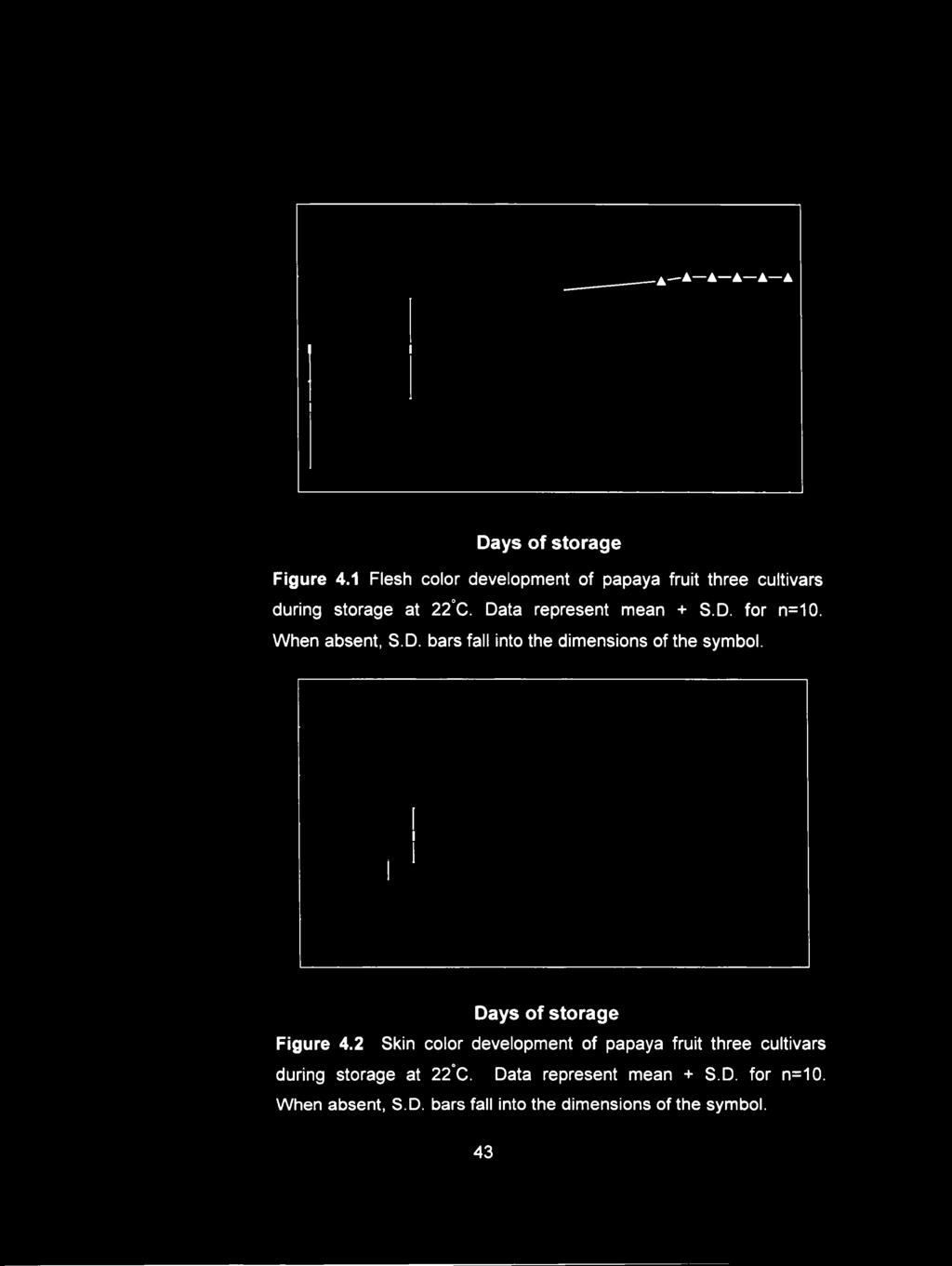
58 Days of storage Figure 4.1 Flesh color development of papaya fruit three cultivars during storage at 22 C. Data represent mean + S.D. for n=10. When absent, S.D. bars fall into the dimensions of the symbol. Days of storage Figure 4.2 Skin color development of papaya fruit three cultivars during storage at 22 C. Data represent mean + S.D. for n=10. When absent, S.D. bars fall into the dimensions of the symbol. 43

 >» 10 JC UJ 0 'Lines' O 'Sunset' - 4 - 'Line4-16' 1 2 3 4 5 6 7 8 9 10 11 12 13 14 15 16 17 18 19 20 Days of storage Figure 4.")
59 30 'Lines' o 'Sunset' - A - 'Line4-16' I I I I J 1 I I I I I L J L Days of storage Figure 4.3 Respiration of papaya fruit three cultivars during storage at 22 C. Data represent mean + S.D. for n=5. When absent, S.D. bars fall into the dimensions of the symbol. JC 70 l i 60 _l c 50 c o 40 u 3 o 30 2 a. o 20 c 0) >» 10 JC UJ 0 'Lines' O 'Sunset' 'Line4-16' Days of storage Figure 4.4 Ethylene production of papaya fruit three cultivars during storage at 22 C. Data represent mean + S.D. for n=5. When absent, S.D. bars fall into the dimensions of the symbol. 44


60 <M E o (A (A 0) C E Days of storage Figure 4.5 Firm ness of papaya fruit three cultivars during storage at 22 C. Data represent mean + S.D. for n=10. When absent, S.D. bars fall into the dimensions of the symbol. (N= Newton) 45

61 Sunset fruit showed a similar pattern that fruit had 60% to 70% skin yellowing at the peak of respiration and ethylene production and there was a decline in firmness. The slow ripening Line 4-16 fruit had peak of respiration rate when the fruit had developed to 80 to 90% of skin color. Fruit had the maximum ethylene production after the 90% skin yellowing stage. Papaya fruit softening showed a correlation between respiration and ethylene production in all cultivars in that softening paralleled skin color development in each cultivar. However, the degree of skin yellowing development was not always correlated to the papaya fruit firmness (Manrique and Lajolo, 2004). This has been observed to be true for kiwifruit where softening is not associated with the color development but is more closely related with respiratory climacteric and ethylene production (Wang et al., 2000). 46

.")

62 CHAPTER 5 THE RELATIONSHIP BETWEEN PAPAYA FRUIT SOFTENING AND ENDOXYLANASE ACTIVITY, GENE EXPRESSION AND PROTEIN AMOUNT 5.1 Introduction Softening is a major event during fruit ripening and largely contributes to the deterioration of postharvest quality and shelf life. The alteration of cell wall components is mainly responsible for softening and textural changes. A significant increase in depolymerization and solubility of pectin and hemicellulose results in wall loosening and disintegration during softening (Brady, 1987; Fischer and Bennett, 1991). The cell wall modifying enzymes play an essential role in the softening processes in a number of fruit (Brady, 1987). Many different enzymes are present throughout fruit ripening and have different activity levels and patterns against different substrates. Polygalacturonase (PG), pectin methylesterase (PME) and fi-galactosidase have been studied in tomato due to their involvement in pectin degradation (Pressy, 1983; Hobson and Grierson, 1993). The endoglucanase and xyloglucan endotransglycosylase/hydrolase (XTH) might also be involved in the considerable changes in the hemicellulose-glycan matrix (Maclachlan and Brady, 1992; 1994). The amount and activity of these enzymes show good correlation with the expression of their respective genes during softening. Numerous enzymes are present in the wall that might have a role in softening that have to be characterized in the fruit. In addition, there are differences in the wall composition and enzymes related-wall modifications during softening that are variable between species and cultivars (Brummell and Harpster, 2001). 47
. The activities of endo-and exo-pg, PME, glucanase, galactosidase and xylanase have been detected in papaya fruit during ripening (Pauli and Chen, 1983; Lazan et al., 1991, 1995).")
.")
.")
63 During papaya {Carica papaya L.) fruit ripening, the cell walls show an increase in the solubilization and decline in the molecular weight of both pectin and hemicellulose (Lazan et al.,1995; Pauli 1999). The activities of endo-and exo-pg, PME, glucanase, galactosidase and xylanase have been detected in papaya fruit during ripening (Pauli and Chen, 1983; Lazan et al., 1991, 1995). It has been reported that changes in hemicellulose coincide with a marked increase of endoxylanase activity. These changes are more pronounced as the fruit soften (Pauli et al., 1999). Several published studies suggest that enzymes that specifically modified hemicellulose may trigger fruit softening (Maclachlan and Brady, 1994; Rose et al., 1998; Brummel et al., 2004). Endoxylanase randomly catalyzes the endohydrolysis of (i-1,4-d-xylosyl linkages of xylan. Xylan is a constituent of hemicellulose in cell walls that have other complex structures: xyloglucan, glucomannan, and arabinoxylan (Carpita and Gibeaut, 1993). Xylan has been found in the secondary cell wall of most plants and is also found in the primary cell wall of monocots. Highly and minimally branched xylans are observed that vary in structure between plant species. Unbranched xylan has been found in the tobacco pith cell wall (Eda et al., 1976), muskmelon (Doux-Gayat et al., 1978) and Barletf pear (Ahmed and Labavitch, 1980). Endoxylanase activity has been investigated in many fruit such as pear (Yamaki and Kakiuchi, 1979), tomato (Barka et al., 2000), avocado (Ronen et al., 1991), carambola (Chin et al., 1999), mango (Bhagyalakshmi et al., 2002), and papaya (Pauli and Chen, 1983). Genes encoding endoxylanase have been extensively identified in various microorganisms. These endoxylanases have diverse functional characteristics (Collins et al., 2005). In plants, Arabidopsis has 12 predicted endoxylanase-like genes (Henrissat 48
.")
.")

64 and Bairoch, 1996), of which only one is known to be expressed in the vascular bundle (Suzuki et al., 2002). In barley, endoxylanase has an important role in the degradation of the aleurone layer (Banik et al., 1996). In maize, endoxylanase is important during pollen release in tapetum and germination of the pollen tube (Bih et al., 1999). Papaya endoxylanase protein was first purified from ripened fruit and has a MW of 32.5 kda (Chen and Pauli, 2003). It has been identified as the product of an ORF (open reading frame) of 1752 nucleotides {CpaEXYl). The ORF encodes a protein with high homology to the endoxylanase family 10 (Henrissat, 1991). The encoded protein has a predicted MW of 65 kda. However, the purified endoxylanase from papaya has molecular weight of 32.5 kda, which may be the result of posttranslation modification (Chen and Pauli, 2003). The CpaEXYl mrna is accumulated during papaya fruit ripening and increases in abundance as the fruit softens (Chen and Pauli, 2003). However, it is unclear how CpaEXYl mrna levels, protein amount and activity are related to papaya fruit softening. The role of the endoxylanase whether it triggers the cascade of events in the fruit softening remained to be determined. The objective of the current study was to determine the relationship between papaya fruit softening and endoxylanase gene expression, protein level and activity. In order to investigate the role of endoxylanase in papaya, three cultivars having different patterns of softening were examined. To investigate the protein level, specific primary antibody to papaya endoxylanase was produced from a recombinant protein. coli expression system was used for producing a recombinant protein. 49
 were harvested to be used in the experiment and allowed to ripen at room temperature (21")
, fitted with a 1.5 cm disc. The hardness force required to depress the 1.")

65 5.2 Materials and Methods Papaya tissues Three different cultivars of papaya fruit, Line 8, Sunset and Line 4-16 were harvested from the trees at the Poamoho Experimental Station in central Oahu, Hawaii. Fruit at the first sign of color break (<10% skin yellow stage) were harvested to be used in the experiment and allowed to ripen at room temperature (21 to 22 C) Firmness determination Papaya fruit at different skin color development: <10%, 25%, 50%, 75%, and 100% yellow were examined for firmness. The fruit firmness was determined using a penetrometer (LKG-14, Amtek, Largo, Florida), fitted with a 1.5 cm disc. The hardness force required to depress the 1.5 cm disc 1 mm into the fruit was measured Endoxylanase activity Mesocarp tissue from papaya fruit at different stages of ripening, as determined by the skin color development, was cut into small pieces and frozen in liquid nitrogen. Two grams of the frozen tissue were thawed and ground on ice with three volumes of pre-chilled extraction buffer; 0.2 M sodium acetate buffer ph 4.5, with 1 mm dithiopyridine, and 1 mm phenyl methyl-sulfonyl fluoride (PMSF). The tissues were subsequently homogenized in the homogenizer for one minute and incubated on ice in the 1 C cold room for 15 min. The homogenate was centrifuged at 10,321 xg for 15 min. to remove the cell debris and the supernatant was passed through a Whatman No.1 filter paper. To determined activity, the RBB-xylan (Remazol Brilliant Blue) was used as the substrate. The RBB-xylan solution was prepared in 0.1% RBB-xylan in 50 mm mopholino-ethanesulfonic acid (MES) containing 0.01% timersol with ph adjusted to
. The precipitate was pelleted down by centrifugation for ten min.")

 encoding endoxylanase gene {CpaEXYl) was previously isolated and cloned into pbluescript II SK (Stratagene, La Jolla, California).")
66 The reaction was started by adding 0.1 ml of enzyme supernatant to 0.4 ml of the substrate and incubated at 37 C for 20 hours. After incubation, the reaction was terminated by adding two volumes of absolute ethanol (-20 C). The precipitate was pelleted down by centrifugation for ten min. at 10,321 xg and the blue color of supernatant was used for determination enzyme activity by reading the absorbance at 590 nm. The boiled enzyme supernatant at 100 C for five min. was used as the control to eliminate background dye intensity. The results were expressed as the activity units per gram fresh weight. One unit of endoxylanase activity was determined as the activity required to liberate one nmol of RBB to the supernatant from RBB-xylan reaction mixture Gene construct and cloning An ORF (open reading frame) encoding endoxylanase gene {CpaEXYl) was previously isolated and cloned into pbluescript II SK (Stratagene, La Jolla, California). The gene construct and cloning were performed according the Gateway system s protocol (Invitrogen, Carlsbad, California). The nucleotide 850 to 1596 region of the large ORF C paex Yl corresponding to the presumed catalytic domain (Met265- Glysss) (Figure 5.1 A) was amplified by the polymerase chain reaction method (PCR) with Taq polymerase. The sequences of gene-specific oligonucleotides were used for PCR primers as follow: foha/ard primer 5 CACC ATG GTT CAA TTC GCT AAG 3 and reverse primer 5 ACC ATT CTG ATG TTT GCC 3. The underlined sequences do not have homology to the gene in catalytic domain. They were added to facilitate the binding to the vector following the Gateway system (Invitrogen, Carlsbad, California). The PCR conditions were used as follow: step 1 initial denature at 95 C for 2 min; step 2 denature at 95 C for 30 s., step 3 annealing at 55 C for 1 min; step 4 extension at 72 C for 2 min; 51
.")
 and incubated for 15 min. at room temperature (22 C).")

67 step 5 final extension at 72 C for 3 min. PCR at step 2 to 4 was repeated for 30 cycles. The PCR product was cloned into the directional TOPO pentr vector, pentr/sd/d-topo by cloning reaction for producing the entry vector (Figure 5.1B). The TOPO cloning reaction was performed following the manufacturing procedure. The reaction was gently mixed between the PCR product and pentr/sd/d-topo vector (1:1 molar ratio) and incubated for 15 min. at room temperature (22 C). The ligated DNA was transformed into the competent TOPO10 cell following the standard chemical transformation protocol (Invitrogen, Carlsbad, California). Briefly, after adding the TOPO cloning reaction to the competent TOPO10 cell, the mixture was incubated for 30 min. on ice and heat shock induced at 42 C for 30 second. The SOC medium was added to cell mixture and shaken at 37 C at 200 rpm for one hour and then cultured on the selective LB agar containing 50 pg ml'^ kanamycin. The plasmid DNA was extracted from the selected transformants and was analyzed for the insertion by restriction enzyme. A positive clone was analyzed by DNA sequencing to confirm its identity with the DNA sequence of CpaEXYl corresponding to the presumed catalytic domain via Greenwood Molecular Biology Facility, University of Hawaii at Manoa. The plasmid DNA from the positive entry clone was used for the next step. The plasmid DNA from the entry clone containing the insertion was inserted into the destination vector, pet-pdest42 by the LR clonase reaction according to the manufacturer s procedures (Invitrogen, Carlsbad, California). The destination vector pet-pdest42 contains specific features of T7 lac promoter (for high level IPTGinducible expression of the gene of interest in. coll.), V5 epitope and 6 XHis tag for detection and purification. 52


68 MVQFAKKNGIAlRGHNVFWDDPKYQSGWVSSLSPNDLNAAAT KRINSVMNRYKGQVIGWDWNENLHFSFFESKLGANASAVFY GEAHKTDPSTTLFMNEYNTVEDSRDGQATPAKYLEKLRSIQS LPGNGNMGIGLESHFSSSPPNIPYMRSAIDTLAATGLPVWLT EVDVQSGGNQAQSLEQILREAHSHPKVRGIVIWSAWSPNGCY RMCLTDNNFHNLPTGDWDKLLREWGGGATVKGKTDQNG pbr322 origin rrnb T2 term Figure 5.1 A. The amino acid sequence of CpaEXY 1 that corresponds to the presumed catalytic domain (M etjgjto Gly532). B. The entry vector, pentr/sd/d-topo-xyl-cat. 53


69 T7 promoter Xyl-Cat attb2 V5 tag 6xHis tag Amp Amp pbr322 origin Figure 5.1 C. The destination vector, pet-pdest42-xyl-cat. The plasmid drawing used PlasMapper web server program (Dong et al., 2004). 54

.")
.")
70 Approximately 300 ng of each entry plasmid DNA and destination vector DNA were mixed together before adding the LR clonase enzyme. The reaction was incubated at 25 C for 60 min. and the reaction was stopped by adding 2 pl of 2 pg pl"'' proteinase K. The resulting DNA construct was transformed into competent TOPO10 cells by standard chemically induced transformation as outlined above. Transformants were grown on LB agar containing 50 pg ml'^ carbenicillin. The resulting plasmid was given the destination vector name, pet-pdest42-xyl-cat (Figure 5.1 C). The positive clone was analyzed with the restriction enzyme and confirm by sequencing at Greenwood Molecular Biology Facility, University of Hawaii at Manoa Expression of the recombinant protein Piiot expression The pet-pdest42-xyl-cat plasmid was transformed into the E. coli strain BL21 (DE3). The transformation was performed following the manufacturer s protocol (Invitrogen, Carlsbad, California). Cell transformed with the plasmid were selected on LB agar supplemented with 50 pg ml'^ carbenicillin. Cells from a single colony were cultured in 10 ml of LB containing 50 pg ml'^ carbenicillin at 37 C and shaken at 250 rpm overnight. The 500 pi of overnight culture was transferred and further cultured in 10 ml LB containing 50 pg ml''' carbenicillin at 37 C and shaken at 250 rpm until the culture reached an optical density (O.D.) of 0.6 to 0.8. The expression of the recombinant protein was induced by adding IPTG (isopropyl-(i-thiogalactopyranoside) to a final concentration one mm. The cells were cultured for an additional four hours. A 500 pl sample of the culture was collected before induction and every hour after IPTG induction 55

, 1 0 % glycerol, 2 % fi-mercaptoethanol, 0.")
. The gel was stained with 0.1% coomassie blue mixed with 40% Methanol and 10% acetic acid for 30 min.")
71 The sample was centrifuged at the 16,000 g in a microcentrifuge for 30 s. at room temperature. The supernatant was removed from the pellet by aspiration at each time point. The sample pellet was stored at -80 C for protein analysis. Protein samples were analyzed by SDS-PAGE (Sodium dodecyl sulphate- Polyacrylamide Gel Electrophoresis) following the mini protein II dual slab gel's protocol (Bio Rad, Richmond, California). Briefly, all pellet samples were thawed and resuspended in 80 pl of IX sample buffer (0.5M Tris-HCI ph 6.8, 2% SDS (sodium dodecyl sulphate), 1 0 % glycerol, 2 % fi-mercaptoethanol, 0.1 % bromophenol blue) and boiled for five min. Each protein sample (5 pl) was loaded and separated on the 12% of SDS-PAGE (30% acrylamide mix, 1.5 M Tris ph 8.8, 10% SDS, 10% ammonium persulphate, and 0.04% TEMED (N,N,N,N -tetramethylethylenediamine). The gel was stained with 0.1% coomassie blue mixed with 40% Methanol and 10% acetic acid for 30 min. Gel was then destained with 40% methanol and 10% acetic acid for three hours to overnight. The gel was evaluated for the protein band at the expecting size range for the recombinant protein. To confirm the over-expression of the recombinant protein, protein western blot analysis using Anti-V5-HRP antibody was performed according to the manufacturing instructions (Invitrogen, Carlsbad, California). The Anti-V5 antibody recognizes the 14 amino acid sequence Gly-Lys-lle-Pro-Asn-Leu-Leu-Gly-Leu-Asp-Ser-Thr present at the C-terminal of the recombinant protein. For western blot analysis, approximately 30 pg of the protein sample from the pilot expression was separated on the 12% SDS- PAGE. Protein was electrophoretically transferred to nitrocellulose membrane (Bio-Rad, Hercules, California) in transferring buffer (lomm NaHCOs, 3mM NaCOa, 20% methanol, 56
 for either one hour (at room temperature) or overnight (at 4 C). The membrane was then washed with the PBST buffer (PBS plus 0.05% Tween-20) twice for five min.")
 twice for five min. The protein blotting was detected using a chemiluminescent HRP reaction development protocol (Amersham Pharmacia Biotech, Piscataway, New Jersey).")

72 ph 9.9) at 100 V for one hour. The membrane was then incubated in blocking buffer (PBS buffer: 137 mm NaCI, 2.7 mm KCI, 10 mm Na2HP04, 1.8 mm KH2 PO4 ph 7.4, plus 5% nonfat dry milk) for either one hour (at room temperature) or overnight (at 4 C). The membrane was then washed with the PBST buffer (PBS plus 0.05% Tween-20) twice for five min. and then incubated in the blocking buffer having the Anti-V5-HRP antibody at working dilution of 1:5,000. The membrane was washed again with the PBST buffer (PBS plus 0.05% Tween-20) twice for five min. The protein blotting was detected using a chemiluminescent HRP reaction development protocol (Amersham Pharmacia Biotech, Piscataway, New Jersey). The membrane was exposed on the X-ray film for approximately five second to develop the desired signal. To isolate enough recombinant protein, the initial culture and subsequent reactions were scaled up to large volume of bacterial culture. Four ml of an overnight culture of the recombinant construct pet-pdest42-xyl-cat was cultured into 200 ml LB media containing 50 pg ml'^ carbenicillin. The procedure used above in the pilot scale production was utilized and the pellet of the 200 ml culture was collected and prepared for protein extraction Preparation of the cell lysate under denature and non-denaturing conditions The bacteria lysate was prepared under both denaturing and non-denaturing conditions according to the manufacturer s protocols (Invitrogen, Carlsbad, California) Non-denaturing conditions For non-denaturing conditions, the pellet was dissolved in 40 ml of native binding buffer (50 mm NaPO 4 ph 8.0, 0.5 M NaCI, 8 mg of lysozyme) and incubated 57
 and endonuclease (0.5 U ml'^) on ice for 15 min.")

 and gently shaken for ten min. at room temperature.")
73 on ice for 30 min. The cells were then lysed by incubation in the water bath sonicator for three min., flash frozen in liquid nitrogen and quickly thawed at 37 C. The viscous lysate was incubated with RNase A (10 pg ml'^) and endonuclease (0.5 U ml'^) on ice for 15 min. The lysate was homogenized by repeated pass through a 18-guage syringe needle several times, and centrifuged at 10,321 xg at 4 C for 15 min. to pellet the cellular debris. A five pl supernatant aliquot was investigated on SDS-PAGE to evaluate protein presence as previously describe Denaturing conditions The protein that remained in the insoluble pellet from the non-denaturing condition step was recovered for the denatured lysate. The lysate was resuspended in 8 ml guanidium lysis buffer (6 M guanidium HCI, 20 mm NaP0 4 ph 7.8, 500 mm NaCI) and gently shaken for ten min. at room temperature. The lysate was then sonicated using water bath sonicator for five min. The pellet debris was removed by centrifugation at 5,000xg for 15 min. at 4 C. A five pl supernatant aliquot was run on SDS-PAGE to evaluate protein presence as previously describe Protein purification The soluble recombinant protein from the non-denaturing and denaturing conditions was purified using the affinity tag method (ProBond Purification System, Invitrogen, Carlsbad, California). The protein that contained 6 X His-tagged at C- terminal was purified utilizing the chelating ligand iminodiacetic acid (IDA), attached to a highly cross linked agarose matrix. IDA binds Ni^^ ions at three coordination sites. 58


74 Non-denaturing conditions The protein lysate from the previously non-denaturing extraction was purified in the two ml of IDA resin following the manufacture s procedures (Invitrogen, Carlsbad, California). The recombinant protein with the histidine tags was bound to the resin that was already equilibrated with four volumes of native buffer (50 mm NaP0 4 ph 8.0, 0.5 M NaCI, 3 M imidazole). The protein in the column was gently mixed on a rotator at 21 to 22 C for 60 min., and the unbound protein was removed with four volumes of native washing buffer (50 mm NaP0 4 ph 8.0, 0.5 M NaCI, 20 mm imidazole). The bound recombinant protein was eluted from the resin with four volumes native elution buffer (50 mm NaP0 4 ph 8.0, 0.5 M NaCI, 250 mm imidazole). A one ml aliquot was collected and analysis by SDS-PAGE Denaturing conditions The protein lysate from the previous denaturing extraction was purified in the IDA resin column following the manufacturer s procedure (Invitrogen, Carlsbad, California). The protein with histidine tag was bound to the resin a the column that was pre-incubated with four volumes of denaturing binding buffer (6 M Urea, 20 mm NaPO 4 ph 7.8, 500 mm NaCI). The protein in the column was gently rotated at room temperature for 30 min. The unbound protein was washed twice with four volumes of denaturing buffer at ph 7.8 and was following washed twice with the same buffer at ph 6.0. The unbound protein was then washed three times with four volumes of denaturing wash buffer ph 5.3. The bound recombinant protein was eluted with four volumes of the denaturing eluted buffer (6 M Urea, 20 mm NaPO 4, 500 mm NaCI) using ph gradient from 4.0 to 3.0. One ml fractions was collected and their monitored ato.d
 at 100 V. for one hour.")

 was determined by comparison with the retention times of known PTH-amino acid standards. S.2.7.")
75 The recombinant protein was confirmed by N-terminal sequence analysis. The protein sample from the pilot expression was separated on 12% SDS-PAGE and electroblotted on PVDF membrane (Bio-Rad, Flercules, California) at 100 V. for one hour. The expected protein band was stained with 0.1% Ponceau S and cut from the membrane and submitted to the Greenwood Molecular Biology Facility, University of Hawaii at Manoa. The N-terminal amino acid sequence was performed on an Applied Biosystems Precise 494 automated sequencer. The instrument performs pulsed-liquid or gas-phase Edman chemistry. The N-terminal amino acids were coupled, cleaved and converted to the PTH-amino acids, which are then analyzed on an ABI 140C HPLC system. The identity of each successive amino acid residue (of the sequence) was determined by comparison with the retention times of known PTH-amino acid standards. S Preparation of the protein for poiycionai antibody production The protein was run on 12% SDS-PAGE and the desired band was excised from the gels. Approximately two mg of protein were excised from the gel. The protein gel was subsequently freeze-dried and crushed into small pieces. For polyclonal antibody production in rabbits, the protein gel was sent to Sigma-Genosys (1442 Lake Front Circle, The Woodlands, Texas 77380). The immunization and bleeding schedules in the rabbits were for a total of 77 days. The pre-immune bleed was at day 0. The first, second and third production bleed were at day 49, 63, and 77, respectively. All samples were shipped to the lab in Honolulu and stored at -20 C for papaya protein analysis. 60
. Approximately ten grams of mesocarp tissues were ground to a powder with a coffee grinder in liquid nitrogen.")
 and vigorously shaken for one min at room temperature. Pre-chilled absolute ethanol (0.25 volumes) and 5 M potassium acetate (0.")

76 5.2.8 RNA extraction RNA was isolated from papaya fruit: Line 8, Line 4-16 and Sunset at different stage of the skin color development. The RNA extraction procedure was performed as described in Mason and Botella (1997). Approximately ten grams of mesocarp tissues were ground to a powder with a coffee grinder in liquid nitrogen. The powder was extracted with extraction buffer (150 mm Tris Base, 2% (w/v) SDS, 50 mm EDTA (ethylenediaminetetra acetic acid), and 1% (v/v) fi-mercaptoethanol, with ph adjusted to 7.5 with boric acid) and vigorously shaken for one min at room temperature. Pre-chilled absolute ethanol (0.25 volumes) and 5 M potassium acetate (0.11 volumes) were added to the mixture and shaken for one min at room temperature. One volume of chloroformisoamyl alcohol (24:1) was added and shaken for three min. The homogenate was then centrifuged at 23,300 g for 40 min at 4 C. The upper phase was removed and extracted with water-equilibrated phenol (ph 4.5)-chloroform-isoamyl alcohol (25:24:1) mixture three times or until no interphase layer appeared. The last upper phase layer was precipitated with 2.25 volumes of absolute alcohol at -20 C for two hours or overnight. The nucleic acid pellet was collected after centrifugation at 23,300 g for 40 min and washed again with 80% ethanol and centrifuged at 23,300xg for 15 min. The pellet was redissolved in RNase-free water. To the precipitate RNA, lithium chloride was added to give a final concentration of 2 M and incubated at -20 C overnight. The RNA was pelleted down by centrifugation at 27,300xg for 30 min at 4 C and the pellet was washed with 80% ethanol and recentrifuged at 27,300xg for 15 min. at 4 C. The RNA pellet was redissolved in 300 pl of RNase-free water and again precipitated with 2.5 volumes of absolute ethanol and 0.1 volume of sodium acetate at -80 C for 30 min. The RNA solution was centrifuged at 23,300xg for 30 min. at 4 C to pellet the RNA. The pellet was washed twice with 80% ethanol and was redissolved in a small amount of 61

. The pbluescript II KS vector that contains ORF of endoxylanase {CpaEXYl) and T3 RNA promoter upstream of ORF was used for RNA probe preparation.")

77 RNase-free water. The RNA solution was stored at -80 C until use Non-radioactive RNA probe preparation DIG-labeled RNA probes using the in-vitro transcription labeling was performed according to the manufacturer s instructions (Roche Applied Science, Indianapolis, Illinois). The pbluescript II KS vector that contains ORF of endoxylanase {CpaEXYl) and T3 RNA promoter upstream of ORF was used for RNA probe preparation. Plasmid DNA was extracted according to the HiSpeed Midi kit s instructions (Qiagen, Valencia, California). The plasmid was linearized with the restriction enzyme Xbal. The in vitro transcription labeling was conducted with one pg of linearized plasmid DNA and mixed with the nucleotide labeling D1G-11-UTP, by adding T3 RNA polymerase. The mixture was incubated at 42 C for one hour and 0.2 M EDTA was added to stop the reaction. The yield of the newly synthesized DIG-labeled-RNA probe of approximately size of 900 nucleotides was determined the yield by serial dilution according to the instructions of DIG Northern Starter Kit (Roche Applied Science, Indianapolis, Illinois) RNA blot analysis A 30 pg of total RNA from each papaya cultivar at different stages of ripening (from step 5.2.8) was run on 1% agarose gel containing 0.66 M formaldehyde as described in Fourney et al. (1988). After electrophoresis, the RNA gel was rinsed, prior to blotting in 20X SSC (3 M N ac I, 300 mm Sodium citrate ph 7.0) twice for five min. The RNA was fixed by UV-cross linking and the membrane was briefly rinsed in distilled water and allowed to air dry. The membrane was prehybridized with the hybridization buffer, DIG Easy Hyb, in a hybridization tube and gently incubated at 6 8 C for two hours. 62

 using the CSPD chemiluminescent as the substrate for alkaline")
 at different stages of skin color development were used for")
78 The probe RNA labeling was denatured by boiling for five min. and rapidly cooled on ice. The hybridization was performed by adding the denatured DlG-labeled RNA probe at a concentration of ng ml ^ to pre-warm hybridization buffer and then incubated overnight at 6 8 C. The membrane was washed at room temperature in 2X SSC and 0.1% SDS twice for 15 min, and the washing was continued with 0.5X SSC and 0.1% SDS at 6 8 C for twice for 15 min. The detection was carried out as described in a guideline of the DIG Northern Starter Kit (Roche Applied Science, Indianapolis, Illinois) using the CSPD chemiluminescent as the substrate for alkaline phosphatase. The membrane was initially exposed on the X-ray film for two hours the first time. Multiple exposures were taken to achieve the desired signal strength Protein extraction and protein blot analysis The papaya fruit mesocarp from three cultivars of papaya: Line 8, Line 4-16, and Sunset ) at different stages of skin color development were used for protein extraction. Two grams of frozen papaya mesocarp tissues were ground into powder with a coffee grinder. The papaya protein extraction followed the method of Schuster and Davies (1983). The tissue powder was extracted with two volumes of the extraction buffer (0.1 M Tris-HCL ph 8.8, 10 mm EDTA, 0.9 M sucrose) and two volumes of watersaturated phenol. The homogenate was vigorously shaken at 4 C for 15 min. and then centrifuged at 10,321 xg for 15 min. to separate the phases. The upper phase containing the protein in the phenol solution was re-extracted with an equal volume of the extraction buffer and centrifuged at 10,321 xg for 15 min. The upper layer of protein solution was precipitated with five volumes of 0.1 M ammonium acetate in 100% methanol at -20 C overnight. The precipitate was washed three times with 0.1 M ammonium acetate in 100% methanol and once with 100% acetone. The pellet was air dried and 63

) three times for 10 min at room temperature.")

79 re-suspended in 500 pi of 50 mm Tris-HCL ph 6.8 and 1.5% SDS buffer. The protein was denatured in a 6 X sample buffer prior to investigation by SDS-PAGE. An equal amount of protein at different stages of papaya fruit ripening was then separated by SDS-PAGE and blotted onto the nitrocellulose membrane at room temperature in 10 mm NaHCOa, 3 mm NaCOa, and 20% methanol. The membrane was incubated in a PBS blocking buffer (137mM NaCI, 2.7 mm KCI, lomm Na2HP0 4, 1.8 mm KH2PO4 ph 7.4, plus 5% nonfat dry milk) at 4 C overnight. The membrane was washed with the PBST buffer (PBS buffer plus 0.05% Tween-20 (v/v)) three times for 10 min at room temperature. To detect the endoxylanase protein, the membrane was incubated with anti-cat-cpa XYf-antiserum as the primary antibody dilution of 1:5,000 in blocking solution for one hour. The membrane was washed again three times with the PBST buffer for ten min. Polypeptides that cross react with the endoxylanase antiserum was bound with rabbit IGG (whole molecule) coupled to horse-radish peroxidase (Sigma-Genosys, The Woodlands, Texas). The conjugate was diluted 1:10,000 in a blocking solution and incubated for one hour at room temperature. The membrane was then washed with PBST buffer for 3X10 min at room temperature. The blots were detected using a chemiluminescent HRP reaction development kit (Amersham Pharmacia Biotech, Piscataway, New Jersey). The membrane was first exposed to the x-ray film for approximately five second to develop the desired signal. 64


80 5.3 Results Fruit firmness at different stages of papaya fruit ripening Line 8, Sunset, and Line 4-16 papaya fruit showed a difference in softening patterns during storage at 21 to 22 C. Line 8 fruit started to soften at the 25% skin yellow stage. Fruit showed a rapid softening (from N cm'^ to 32.1 N cm'^) when the skin developed from the 25% to the 50% skin yellow stage and its softening then slightly increase to the end of ripening (Figure 5.2A ). Sunset fruit showed a rapid softening (from N cm'^ to N cm'^) when skin color was developed from the 50% to the 75% yellow and then softening slightly increased at the end of ripening (Figure 5.2B). Line 4-16 fruit showed a slow softening pattern. Firmness was maintained until skin colors developed to the 75% skin yellow stage. Fruit had an increased softening rate at the 100% skin yellow stage (firmness declined from N cm'^ to 59.8 N cm'^) and then slightly declined after the 100% skin yellow stage for 5 to 7 days (Figure 5.2C) Endoxylanse activity Endoxylanase activity was detected in the Line 8, Sunset and Line 4-16 papaya fruit during ripening. Activity was first detected when Line 8 and Sunset papaya fruit were at the 25% skin yellowing stage (Figure 5.2A and 5.2B). Activity in Line 8 and Sunset fruit then dramatically increased to the 100% skin yellow stage. In Line 4-16 fruit, endoxylanase activity was first detected at the 75% skin yellow stage (Figure 5.2C). Activity then increased at the 100% skin color and continued at a constant level for 7 days (Figure 5.2C). 65
 Firm ness 2 ^ Endoxylanase activity Firm ness o - -")


81 40 Endoxylanase activity A Line 8' »Endoxylanase activity E u OT in 0) Firm ness 2 ^ Endoxylanase activity Firm ness o d 100 7d Fruit stage (% Skin yellow ing) Figure 5.2 Endoxylanase activity and firmness of three papaya cultivars: A. Line 8, B. Sunset, and C. Line 4-16 at different stages of ripening. N = Newton; d = days after full ripening; g = gram; Fw = Flesh weight 66

. In Sunset fruit, the CpaEXYl mrna expression can be detected at the 50% yellow skin stage to the full ripe stage (100% skin yellowing) (Figure 5.3B).")
. 5.3.4 The endoxylanase recombinant protein expression The catalytic domain of endoxylanase was over-expressed in the pet-pdest 42 using the Gateway system.")
82 5.3.3 Endoxylanase {CpaEXYl) mrna expression The expression of the gene for CpaEXYl was investigated by RNA blot analysis in the different cultivars of papaya fruit (Figure 5.3). An approximately single 1.7 kb band of mrna from papaya mesocarp tissues was detected when hybridization was performed with the specific RNA probe. The CpaEXYl mrna in Line 8 fruit was first detected at the 25% skin yellow stage and increased abundantly at the 50% to the 75% skin yellowing and then slightly declined at the full yellow (Figure 5.3A). In Sunset fruit, the CpaEXYl mrna expression can be detected at the 50% yellow skin stage to the full ripe stage (100% skin yellowing) (Figure 5.3B). Papaya Line 4-16 fruit showed CpaEXYl mrna expression that was first detected when the fruit reached 100% yellow skin and continued as the fruit increased softening (Figure 5.3C) The endoxylanase recombinant protein expression The catalytic domain of endoxylanase was over-expressed in the pet-pdest 42 using the Gateway system. In an initial attempt to express catalytic domain of CpaEXYl for the pilot expression, it was shown that the expected size of MW 32 kda of the recombinant protein can be induced with 0.5 mm and 1.0 mm IPTG (Figure 5.4). The recombinant protein was later expressed in a larger volume of bacterial culture (Figure 5.5A) and investigated by protein blot analysis (Figure 5.5B). The protein was extracted and subsequently purified using the affinity fusion system at non-denaturing and denaturing conditions. The results showed that the recombinant protein was soluble in the native extraction buffer (Figure 5.6). The protein was also in the insoluble form as inclusion bodies and was dissolved in the denaturing buffer of urea and guanidium HCI buffer (Figure 5.7). The SDS-PAGE electrophoresis was carried out to investigate the recombinant protein. The results showed that the catalytic domain of CpaEX Yl protein 67
 were also found after purification and presented in the protein blotting analysis.")


83 was over-expressed under these conditions. However, this study noted that two recombinant protein bands were found in the gel after induced expression. The two distinct bands (which are close in size) were also found after purification and presented in the protein blotting analysis. Two protein bands were separated by N- terminal sequencing and the lower band had identity to the catalytic domain of CpaEXYl protein Endoxylnase protein accumulation To determine endoxylanase protein during papaya fruit ripening, protein from fruit mesocarp at different skin color stage were extracted and subjected to SDS-PAGE analysis. Immunoblotting assay was performed using the polyclonal antibody raised against the endoxylanase protein. The polyclonal antibody raised against endoxylanase bound a protein of approximately 32 kda during papaya fruit ripening. The 32 kda endoxylanase protein of Line 8 and Sunset fruit showed a strong cross reaction when the fruit reached the 50% to the 100% skin yellow stage (Figure 5.8A and 5.8B). Line 4-16 fruit had a strong cross reaction to the endoxylanase protein when the fruit developed a 100% skin yellow stage and continued as fruit increased softening (Figure 5.8C). Moreover, longer exposure than 5 second showed that the presumed the target endoxylanase protein in Line 8 and Sunset fruit was present at 25% skin yellow stage. The band was also detected in Line 4-16 fruit at the 75% skin yellowing with longer exposure. In addition, there was a cross reaction from primary endoxylanase antibody with other high molecular proteins, at approximately 65 kda, in all three papaya cultivars. 68

 The endoxylanase mrna expression after exposure on the X-ray film for 2 hours.")
84 A Line 8 ««( r * B Sunset U M 1 C Line Fruit stage (% Skin yeiiowing) Figure 5.3 Endoxylanase gene expression by RNA blot analysis at different stages of papaya fruit ripening; A. Line 8, B. Sunset, and C. Line (1) The endoxylanase mrna expression after exposure on the X-ray film for 2 hours. (2) The ribosomal RNA was stained with ethidium bromide. d= Days after full ripening 69

: Lane 1: Protein before induction Lane 2, 3, and")

85 8 kda Recombinant Xyl-Cat protein 20 t'.i si n -'f# m 14 Figure 5.4 SDS-Polyacrylamide gel electrophoresis of the pilot expression of recombinant protein pet-pdest42-xyl-cat. The recombinant protein samples were resuspended in IX sample buffer. The gel was stained with Coomassie Brilliant Blue. The samples were loaded on the gel in the following order (from left to right): Lane 1: Protein before induction Lane 2, 3, and 4; Protein after induction with 0.5 mm IPTG for 2, 3, and 4 hrs, respectively Lane 5, 6, and 7: Protein after induction with 1.0 mm IPTG for 2, 3, and 4 hrs, respectively Lane 8 : Protein molecular weight standards (molecular weights are shown on the right) The arrow indicates the recombinant Xyl-Cat protein of approximately 32 kda. 70

: Lane 1: Protein molecular weight Lane 2: Protein before induction")

86 kda ^Recom binant Xyl-C at protein Figure 5.5 SDS-polyarylamide gel electrophoresis of the large scale expression and protein blot analysis of the recombinant protein pet-pdest42-xyl-cat. A. The recombinant protein samples were resuspended in 1X sample buffer. The gel was stained with Coomassie Brilliant Blue. The samples were loaded on a gel in the following order (from left to right): Lane 1: Protein molecular weight Lane 2: Protein before induction Lane 3, 4, and 5: Protein after induction with 0.5 mm IPTG for 2, 3, and 5 hours, respectively B. The protein blot was analyzed for investigation of the recombinant protein expression. Lane 1: Protein before induction Lane 2: Protein after induction with 0.5 mm IPTG for 2 hours The arrow indicates the recombinant Xyl-Cat protein of approximately 32 kda. IPTG= Isopropyl-B-thiogalactopyranoside 71 Ik.
: Lane 1: Protein lysates after binding to the column Lane 2 and 3: Unbound protein was washed out with NaP0 4 buffer, 0.")
87 5 6 kda Figure 5.6 SDS-Polyacrylamide gel electrophoresis of the recombinant protein pet-pdest42-xyl-cat purified by non-denaturing condition. The gel was stained with Coomassie Brilliant Blue. The samples were loaded in the following order (from left to right): Lane 1: Protein lysates after binding to the column Lane 2 and 3: Unbound protein was washed out with NaP0 4 buffer, 0.5 NaCI, and 20 mm imidazole Lane 4 and 5: The recombinant protein was eluted with NaP0 4 buffer, 0.5 NaCI, and 250 mm immidazole Lane 6: Protein molecular weight standards (molecular weights are shown on the right.) 72

: Lane 1: Protein lysates after binding to the column")

88 kda, m 94 -«i 67 ^ 43 Recom binant Xyl-Cat protein Figure 5.7 SDS-Polyacrylamide gel electrophoresis of the recombinant protein pet-pdest42-xyl-cat purified under denaturing condition. Gel was stained with Coomassie Brilliant Blue. The samples were loaded in the following order (from left to right): Lane 1: Protein lysates after binding to the column Lane 2 and 3: Protein was washed out with 6M Urea buffer, ph 6.0 and 5.3, respectively. Lane 4, 5, 6 and 7: Protein was eluted with 6M Urea buffer, ph 5.0, 4.8, 4.5, and 4.3, respectively. Lane 8: Protein molecular weight standards (molecular weights are shown on the right.) The arrow indicates the recombinant Xyl-Cat protein of approximately 32 kda. 73


89 kda 84 * 47. ^ Endoxylanase 32 kda Endoxylanase 32 kda Line 4-16 Endoxylanase 32 kda d 7d d 7d % Skin yellowing Figure 5.8 Endoxylanase protein in three papaya cultivars: A and B, Line 8 ; C and D, Sunset ; and E and F, Line 4-16, at different stages of ripening. A, C, and E: The total protein was electrophoresed on a 12% SDS/polyacrylamide gel and stained with Coomassie Brilliant Blue. B, D, and F: The protein blot analysis of the endoxylanase papaya protein using the catalytic endoxylanase antibodies at 1:5,000 dilution. (Molecular weights are indicated on the left.) The arrows indicate the endoxylanase protein of approximately 32 kda. d=days after full ripening 74
.")

90 5.4 Discussion During papaya fruit ripening, the modifications of the cell wall hemicellulose contribute to fruit softening (Pauli et al., 1999). Evidence that endoxylanase acts in hemicellulose-xylan degradation is based upon gene expression that occurs during the papaya fruit ripening (Chen and Pauli, 2003). Three papaya cultivars (having differences in their softening patterns) exhibited endoxylanase activity, CpaEXYl mrna expression and protein level. A low level of endoxylanase activity was associated with gradual softening in early ripening. When fruit showed a marked decline in firmness, high endoxylanase activity was present and increased throughout ripening (Figure 5.2). The high level of CpaEXYl mrna expression in relation to the softening occurred in a similar pattern to that of enzymatic activity for all 3 cultivars (Figure 5.3). The result was consistent with the previous report by Chen and Pauli (2003). The significant increase in CpaEXYl mrna accumulation and the endoxylanase activity was associated with the specific stage of increased softening. The accumulation of the endoxylanase protein (Figure 5.8) and activity during papaya fruit ripening showed a good correlation with a high level of endoxylanase gene expression and fruit softening. This suggested that endoxylanase is necessary for the softening process. A correlation of between other xylan degrading enzymes, such as xylosidase activity to gene expression is observed in relation to fruit softening. Xylosidase activity and mrna are detected during ripening and highly accumulate in over-ripened stages of pear fruit (Yamaki and Kakiuchi, 1979; Itai et al., 1999). In the strawberry fruit, xylosidase mrna activity is highly evident at the 25% to 50% red skin color stage (earlier ripe stage), and declines thereafter. The depolymerization of xylan may occur before other enzymes such as glucanase which is still detected at the overripe stage 75
 mrna increases in parallel with an increase in enzyme activity (Della Penna et al., 1986; Smith and Gross, 2000).")
. Expansin and mrna (LeE xpl) transcription are present at high levels during tomato fruit ripening and tightly correlated to the softening (Rose et al.")
91 (Martinez et al., 2004). A similar relationship is found in other cell wall enzymes and is related to softening. When tomato fruits ripen, the PG and fi-galactosidase II (TBG4) mrna increases in parallel with an increase in enzyme activity (Della Penna et al., 1986; Smith and Gross, 2000). The effect of endoglucanase (FaCe/1) might contribute to softening at high levels of mrna expression and protein accumulation as detected during strawberry fruit ripening (Trainotti et al., 1999b; Palomer et al., 2004). Expansin and mrna (LeE xpl) transcription are present at high levels during tomato fruit ripening and tightly correlated to the softening (Rose et al., 2000; Brummell et al., 1999b). The papaya protein analysis using a specific antibody raised against the catalytic domain of endoxylanase showed a major cross-reacting protein of approximately 32 kda. The molecular mass of the mature protein after processing was deduced to be the half size of the predicted unprocessed protein. Chen and Pauli (2003) reported that the endoxylanase protein predicted from the CpaEXYl nucleotide sequence has 584 amino acids and predicted a molecular weight of kda. This result is in agreement with the protein processing during ripening. The mature protein isolated from the papaya fruit gives 32.5 kda on the protein gel (Chen and Pauli, 2003). It was suggested that the protein processing is mediated by proteolytic cleavages. The post translation of the processing of the endo-fi-l,4-xylanase barley aleurone occurrs by cleaving residues from both N and C-terminal of 61.5 kda to produce a 34 kda mature protein (Casper et al., 2001). Chen and Pauli (2003) suggested that the expected cleavage site of papaya endoxylanase might occur at the amino acids 232 to 235 whereas for barley enzyme, cleavage might occur at amino acids 214 to 217. However, the role of this cleavage on papaya protein localization and activity is unknown. 76
.")
 from soybeans.")
92 Similar protein processing has been found in the pre-xylanase of maize tapetum. The pre-xylanase protein was 60 kda in molecular mass and converted to 35 kda by removal of the N- and C- terminal during anther maturation (Wu et al., 2002). Similar processing also occurs in a-l-arabinofuranosidase and (i-d-xylosidase from barley seedling by cleavage at the C-terminal (Minic et al., 2004). The proteolysis processing was also found in the (i-expansin (C/m f) from soybeans. The C-terminal end of a 35 kda Cim1 is predicted to remove the putative cellulose binding domain from the N-terminal, resulting in a 20 kda protein (Downes et al., 2001). The PMEU1 of PME has been identified in the orange fruit and encodes a 64 kda polypeptide. The PME protein of molecular weight 37 kda is detected when investigating with transgenic tobacco. It is suggested that the processing by amino cleavage has occurred (Gaffe et al., 1997). Chen and Pauli (2003) reported that the CpaEXYl polypeptides contain a predicted signal peptide at the N-terminus and has a cleavage site between amino acids 27 and 28. Four putative glycosylation sites have been found on sequences 170/173 NTSV, 220/223 NTTV, 362/365 NASA and 568/571 NSTD. The CpaEXYl protein contains the putative xylan binding domain (XBD) between amino acid 60 and 182. The putative carbohydrate binding domain (CBD) has been extensively observed in microorgansims and some Arabidopsis predicted genes (Susuki et al., 2002). The endoglucanase (EGase) having putative CBD is also observed in the ripening strawberry fruit (Trainotti et al., 1999a). From the protein blot analysis, there was another cross-reacting band at a higher molecular mass approximately of 60 kda. This may correspond to the non-cleaved (unprocessed) endoxylanase protein. A similar result is found in tomato and strawberry, 77


93 and is suggested to be non-processed protein containing the signal peptide (Palomer et al., 2004; Gonzalez-Bosch et al., 1997). Alternatively, another cross-reacting band may indicate other protein members within the papaya endoxylanase family and contain antigenic similarity. On the other hand, the cross-reaction may arise from other protein family that has a similar glycosyl hydrolase moiety to endoxylanase (Gonzales-Bosch et al., 1997). The recombinant catalytic-endoxylanase protein exhibited the endoxylanase activity against the soluble substrate RBB-xylan. However, when the recombinant protein was purified under the non-denaturing conditions, a low amount of soluble protein was observed. Therefore, only a low level of enzymatic activity was detected in the experiment. These findings were in agreement with Black et al. (1997), who also found that the endoxylanase containing only the catalytic domain can exhibit activity when tested with the soluble substrate. The carbohydrate binding domain (CBD) is not required for the catalytic activity against the soluble substrate, but is necessary for the insoluble substrates. The removal of CBD does not affect the activity of xylanase {XYLA) and arabinofuranosidase C (XYLC) from Pseudomonas fluorescence subsp. cellulose (Black et al., 1996). The CBD such as cellulose binding domain might enhance enzymatic activity by increasing the number of hydrolytic sites present or available and inducing the solubility of glycan polymers in the wall (Linder and Teeri, 1997). 78


94 CHAPTER 6 EFEFCT OF 1- METHYLCYLCOPROPENE (MCP) ON PAPAYA FRUIT RIPENING AND THE RELATION BETWEEN PAPAYA FRUIT SOFTENING FOLLOWING MCP TREATEMNT TO ENDOXYLANASE GENE EXPRESSION, PROTEIN AND ACTIVITY 6.1 Introduction Papaya is classified as a climacteric fruit, and has an increase in respiration rate and ethylene production during ripening. At the same time, the fruit develop flesh and skin color, softening, and produces volatiles during ripening. The deterioration of papaya fruit after harvesting is the major factor limiting shelf life and quality. Recently, extension of postharvest freshness of fruit and vegetables is studied following the application of MCP (1-methylcyclopropene). MCP acts as the ethylene antagonist by preventing the binding of ethylene to its receptor (Sisler and Serek, 1997). The number of different fruit species following MCP treatment show variable effects on ripening (Blankenship and Dole, 2003). MCP effectively delays the ripening of many fruit, such as tomato (Nakasutka et al., 1997), banana (Golding et al., 1998), apple (Fan and Mattheis, 1999), avocado (Feng et al., 2000), and peach (Kluge and Jacomino, 2002). Fruit treated with MCP show a reduction in ethylene production and respiration rate in tomato (Hoeberichts et al., 2002), plum (Abdi et al., 1998), but not in banana (Jiang et al., 2004). Skin color development is delayed in avocado (Jeong et al., 2002) after treatment with MCP. 79
. Volatile production is inhibited by MCP treatment of many fruit, such as plum (Abdi et al., 1998), and apple (Lurie et al., 2002).")
, papaya (Hofman et al., 2002), and avocado (Adkins et al., 2005).")
95 MCP significantly delays softening in many fruit such as banana (Jiang et al., 2002), plum (Menniti et al., 2004), pear (Hiwasa et al., 2003), and persimmon (Harima et al., 2003). Volatile production is inhibited by MCP treatment of many fruit, such as plum (Abdi et al., 1998), and apple (Lurie et al., 2002). MCP treatment has various effects on postharvest disorders and diseases of fruit species. A markedly increase in disease incidence is observed in MCP treated strawberry (Ku et al., 1999), papaya (Hofman et al., 2002), and avocado (Adkins et al., 2005). Following MCP application, the degreening of orange is inhibited but chilling injury symptoms and stem-end rot during are increases storage (Porat et al., 1999). However, application of MCP reduces the superficial scald development in apple (Fan et al., 1999). A number of enzymatic activities during fruit ripening are affected by MCP treatment. The application of MCP at the pre-climacteric and onset of the climacteric stages inhibits ACS and ACO activities in fruit such as avocado (Owino et al., 2002). After MCP treatment, PG activity in avocado is reduced and remains low throughout ripening. In contrast to the untreated control, a high level of PG activity occurs in fruit to the end of ripening (Feng et al., 2000; Jeong et al., 2002). Both (i-d-xylosidase and a-l-arabinofuranosidase are decreased in ripening tomato after MCP treatment (Itai et al., 2003). In apricot, a- and (i-galactosidase activities are significantly reduced by MCP treatment. In MCP-treated avocado (Jeong et al., 2002), and apricot (Botondi et al., 2003), pectin methylesterase is slightly increased. Endoglucanase has a higher activity in MCP-treated plum than in the non-treated fruit after cold storage (Dong, et al., 2001b). At gene level, the increase of ACS and ACO mrna is prevented to a large extent in MCP-treated fruit, such as pear (Lelievre et al., 1997), and tomato (Nakatsuka 80


96 et al., 1997). MCP altered the mrna accumulation pattern of the three ripening related genes: phytoene synthase, expansin, AGO in tomato (Hoeberichts et al., 2002). Treatment with MCP has been used to alter papaya fruit ripening. Jacomino et al. (2002) studied ripening in Sunrise Solo papaya at both the green and ripe stages after -1 treating with 90 and 270 nl L of MCP. The results showed that the treated fruit had an extended shelf life and delayed degreening and inhibited decay. Papaya fruit with MCP treatment have reduced respiration rate, ethylene production, delayed softening and skin color development compared to the non-treated control (Jacomino et al., 2002; Ergun and Huber, 2004). A decrease in disease incidence of Sunrise Solo papaya fruit at the pre-ripening stage is observed in fruit treated with MCP (Ergun and Huber, 2004). However, disease incidence is increases when treated fruit is stored for longer periods of time (Hofman et al., 2002). Two objectives were investigated relating the MCP application of papaya fruit. The first objective of this study was to investigate the ripening behaviors of papaya fruit following MCP application. Line 4-16 and Rainbow papaya fruit were used in these experiments. The modification of papaya fruit softening was also investigated after treatment with MCP. Therefore, the second objective of this study was to determine the relationship between papaya fruit softening, endoxylanase activity, gene expression and protein level following MCP treatment. Sunset fruit were used in this experiment. 81

 of 2,4-thiazolyl benzimidazole (TBZ) for 20 seconds.")
97 6.2 Materials and Methods Papaya fruit The papaya fruit Rainbow, Sunset, and Line 4-16 were used in the experiments. Rainbow and Line 4-16 papaya fruit were harvested from the plantation of Dole Company & Dole associated growers. Sunset papaya fruit were harvested from the plantation at the Poamoho Experimental Station, in the central Oahu, Hawaii MCP appiication Papaya fruit were brought to laboratory and dipped with 650 part per million (ppm) of 2,4-thiazolyl benzimidazole (TBZ) for 20 seconds. MCP was obtained from the commercial powder formulation (active ingredient of 0.014%) (SmartFresh, AgroFresh Rohm & Haas, Philadelphia, Pennsylvania). Fruit were fumigated with 100 nl L'^ of MCP in closed chamber for 1 2 hours and then allowed to ripen at room temperature (21 to 22 C) Fruit evaluation W eightless The initial weight was determined after the fruit were transported to the laboratory. Fruit weight was subsequently determined on the day of evaluation Skin and flesh color development The degree of skin color was subjectively determined by visual estimation (0 to 100% yellow). Flesh color development was determined by the percentage of internal flesh color development (0 to % yellow). 82
. 6.2.3.4 Incidence or severity of disease Disease incidence was determined on the day of evaluation.")
 Fruit juice was determined as percent total soluble solids using refractometer (Leica AR200, Buffalo, New York).")
98 6.2.3.S Firmness Fruit firmness was measured by penetrometer (LKG-14, Amtek, Largo, Florida) fitted with a 1.5 cm disc (with a force required to depress the disc 1 mm. into the fruit). Flesh firmness was determined by texture measurement (Texture analyzer TA-TX2, Survey GU27 3Ay, England) Incidence or severity of disease Disease incidence was determined on the day of evaluation. Abnormality was subjectively by using the same scale as based on one -fifth of the angular transformation from 0 to 90: 0 = 0%, 1 = 0% -10%, 2 = 10% - 35%, 3 = 35%-65%, 4 = 65% - 90%, 5 = 90%-100 %, and 6 = 100% Total soluble solid (TSS) Fruit juice was determined as percent total soluble solids using refractometer (Leica AR200, Buffalo, New York). Total soluble solids is an indicator of the percentage of impure aqueous sucrose solution by mass of the dissolved solids Carbon dioxide (CO^ and ethylene (C^H^) production. Five fruit of each fruit cultivars were individually placed in 2500 ml jar. The jars were sealed daily for one hr and each one ml gas sample was taken from head space for respiration (CO^) and ethylene (C^H^) measurement, respectively. CO^ gas was detected by an Infrared CO^ gas analyzer (Licor 6251) at detector of 30 ml min''' nitrogen flow rate (Bassi and Spencer, 1985). was determined on a gas chromatography fitted with an alumina column, injection temperature at 80 C and photoionization detector (detector temperature at 120 C) and helium at 30 ml min"''. 83


99 Data analysis Analysis of variance was analyzed by SAS system Model 9.1 (SAS Institute, Cary, North Carolina) Endoxylanase activity Sunset papaya fruit at the 10% skin yellow stage were treated with MCP at nl L for 12 h. and allowed to ripen at 21 to 22 C. Fruit at different stages of the ripening were determined by the skin color development. Fruit at the 10% to 100%, and 6 days, and 12 days after 100% yellow skin were examined for enzymatic activity. Mesocarp tissues from papaya were cut into small pieces and frozen in liquid nitrogen. Three grams of the frozen tissue were thawed and ground on ice with three volumes of pre-chilled extraction buffer: 0.2 M sodium acetate buffer ph 4.5, with 1 mm dithiopyridine, and 1 mm phenyl methyl-sulfonyl fluoride (PMSF). The tissues were subsequently homogenized in the homogenizer for one minute and incubated on ice in the 1 C cold room for 15 min. The homogenate was centrifuged at 10,321 xg for 15 min. to remove the cell debris and the supernatant was passed through a Whatman No.1 filter paper. To determined activity, the RBB-xylan (Remazol Brilliant Blue) was used as the substrate. The RBB-xylan was prepared in 50 mm mopholino-ethanesulfonic acid (MES) containing 0.1% RBB-xylan, 0.01% timersol with ph adjusted to 6.0. The reaction was started by adding 0.1 ml of enzyme supernatant to 0.4 ml of the substrate and incubated at OO'^C for 20 hours. After incubation, the reaction was terminated by adding two volumes of absolute ethanol (-20 C). The precipitate was centrifuged for ten min. at 10,321 xg. The activity was determined by reading the absorbance at 590 nm with boiled enzyme supernatant at 100 C for five min. as the control to eliminate background dye intensity. The results were expressed as the activity units per gram fresh weight. 84

 SDS, 50 mm EDTA (ethylenediaminetetra acetic acid), 1% (v/v) /?-mercaptoethanol, with ph adjusted to 7.")
100 One unit of endxylanase activity was determined as the activity required to liberate one nmol of RBB to the supernatant from RBB-xylan reaction mixture RNA extraction The same fruit of Sunset papaya in the MCP application for enzymatic measurement were used for RNA extraction. The RNA extraction procedure was performed as described in Mason and Botella (1997). Approximately ten grams of mesocarp tissues were ground to a powder with a coffee grinder in liquid nitrogen. The powder was extracted with extraction buffer (150 mm Tris Base, 2% (w/v) SDS, 50 mm EDTA (ethylenediaminetetra acetic acid), 1% (v/v) /?-mercaptoethanol, with ph adjusted to 7.5 with boric acid) and vigorously shaken for one min. at room temperature. Pre-chilled absolute ethanol (0.25 volumes) and 5 M potassium acetate (0.11 volumes) were added to the mixture and shaken for one min at room temperature. One volume of chloroform-isoamyl alcohol (24:1) was added and shaken for three min. The homogenate was then centrifuged at 23,300xg for 40 min at 4 C. The upper phase was removed and extracted with the water-equilibrated phenol (ph 4.5)-chloroformisoamyl alcohol (25:24:1) mixture three times or until no interphase layer appeared. The last upper phase layer was removed and precipitated with 2.25 volumes of absolute alcohol at -20 C for two hours or overnight. The nucleic acid pellet was collected after centrifuge at 23,300xg for 40 min and washed again with 80% ethanol and centrifugation at 23,300xg for 15 min. The pellet was redissolved in RNase-free water. To precipitate RNA, lithium chloride was added to a final concentration of 2 M and incubated at -20 C overnight. The RNA was pelleted down by centrifugation at 27,300xg for 30 min. at 4 C and the pellet was washed with 80% ethanol and recentrifuged at 27,300x3g at 4 C for 15 min. The RNA pellet was redissolved in 300 pl RNase-free water and again 85

. The pbluescript II KS phagemid vector that contains ORF of endoxylanase (CpaEXYl) and T3 RNA promoter upstream of ORF was used for RNA probe preparation.")

101 precipitated with 2.5 volumes of absolute ethanol and 0.1 volume of sodium acetate at - 80 C for 30 min. The RNA solution was centrifuged at 23,300xg for 30 min. at 4 C to pellet the RNA. The pellet was washed twice with 80% ethanol and was redissolved in a small amount of RNase-free water. The RNA solution was stored at -80 C Non-radioactive RNA probe preparation DIG-labeled RNA probes were prepared by the in-vitro transcription labeling as recommended by the manufacturer s instructions (Roche Applied Science, Indianapolis, Illinois). The pbluescript II KS phagemid vector that contains ORF of endoxylanase (CpaEXYl) and T3 RNA promoter upstream of ORF was used for RNA probe preparation. The plasmid DNA from pbluescript II KS phagemid was extracted according to the HiSpeed Midi kit s instructions (Qiagen, Valencia, California). The plasmid DNA was linearized with the restriction enzyme Xbal. The in vitro transcription labeling was conducted with one pg of linearized plasmid DNA and mixed with the labeling nucleotide DIG-11-UTP, by adding the T3 RNA polymerase. The mixture was incubated at 42 C for one hour and 0.2 M EDTA was added to stop the reaction. The yield of the newly synthesized DIG-labeled-RNA probe of approximately size of 900 nucleotides was determined the yield by serial dilution according to the instructions of DIG Northern Starter Kit (Roche Applied Science, Indianapolis, Illinois) RNA blot analysis The total RNA approximately 30 pg from papaya Sunset following MCP treatment was electrophoresed on 1% agarose gel containing 0.66 M formaldehyde as described in Fourney et al. (1988). After electrophoresis, the RNA gel was rinsed, prior to blotting in 20X SSC (3 M NaCI, 300 mm Sodium citrate ph 7.0) twice for five min. 86


102 The RNA was fixed by UV-cross linking and the membrane was briefly rinsed in distilled water and allowed to air dry. The membrane was prehybridized with the hybridization buffer, DIG Easy Hyb, in a hybridization tube and gently incubated at 68 C for two hours. The probe RNA labeling was denatured by boiling for five min. and rapidly cooled on ice. The hybridization was performed by adding the denatured DIG-labeled RNA probe at a concentration of 100 ng ml'' to pre-warm hybridization buffer and then incubated overnight at 68 C. The membrane was washed at room temperature in 2X SSC and 0.1% SDS twice for 15 min, and the washing was continued with 0.5X SSC and 0.1% SDS at 68 C for twice for 15 min. The detection was carried out as described in a guideline of the DIG Northern Starter Kit (Roche Applied Science, Indianapolis, Illinois) using the CSPD chemiluminescent as the substrate for alkaline phosphatase. The membrane was initially exposed on the X-ray film for two hours. Multiple exposures were taken to achieve the desired signal strength Protein extraction and protein blot analysis The same Sunset papaya fruit with MCP application in the previous firmness measurement, enzymatic determination and RNA extraction were studied in this experiment. Two grams of frozen papaya mesocarp tissues were ground into powder with the coffee grinder. The papaya protein extraction followed the method of Schuster and Davies (1983). The tissue powder was extracted with two volumes of the extraction buffer (0.1 M Tris-HCL ph 8.8, 10 mm EDTA, 0.9 M sucrose) and two volumes of watersaturated phenol. The homogenate was vigorously shaken at 4 C for 15 min. and then centrifuged at 10,321 xg for 15 min. to separate the phase. The upper phase containing the protein in the phenol solution was re-extracted with an equal volume of the extraction buffer and centrifuged at 10,321 xg for 15 min. The upper layer of protein solution was 87


103 precipitated with five volumes of 0.1 M ammonium acetate in 100% methanol after incubation overnight at -20 C. The precipitate was washed three times with 0.1 M ammonium acetate in 100% methanol and once with 100% acetone. The pellet was air dried and re-suspended in 500 pl of 50 mm Tris-HCL ph 6.8, and 1.5% SDS buffer. The protein was denatured in a 6 X sample buffer prior to investigation by SDS-PAGE. An equal amount of protein at different stages of papaya fruit ripening was then separated by SDS-PAGE and blotted onto the nitrocellulose membrane at room temperature in 10 mm NaHCOs, 3 mm NaCOa, and 20% methanol. The membrane was incubated in a PBS blocking buffer (137mM NaCI, 2.7 mm KCI, lomm Na2HP mm KH2PO4 ph 7.4, plus 5% nonfat dry milk) at 4 C overnight. The membrane was washed with the PBST buffer (PBS buffer plus 0.05% Tween-20 (v/v)) three times for 10 min at room temperature. To detect the endoxylanase protein, the membrane was incubated with anti-cat-cpaexvf-antiserum as the primary antibody dilution of 1:5,000 in blocking solution for one hour. The membrane was washed again three times with the PBST buffer for ten min. Polypeptides that cross react with the endoxylanase antiserum was bound with rabbit IGG (whole molecule) coupled to horse-radish peroxidase (Sigma- Genosys, The Woodlands, Texas). The conjugate was diluted 1:10,000 in a blocking solution and incubated for one hour at room temperature. The membrane was then washed with PBST buffer for 3X10 min. at room temperature. The blots were detected using a chemiluminescent HRP reaction development kit (Amersham Pharmacia Biotech, Piscataway, New Jersey). The membrane was first exposed to the x-ray film for approximately five second to develop the desired signal. 88
 and 50% skin yellow stage were treated with 100 nl L^ of MCP.")
 in approximately 10 days of storage.")
104 6.3 Results Effect of MCP on color development To determine the effect of MCP on the color development, Rainbow and Line 4-16 papaya fruit at two different stages of ripening, <10% (color break) and 50% skin yellow stage were treated with 100 nl L^ of MCP. Rainbow and Line 4-16 fruit after MCP treatment at both ripening stages showed a delay in color development compared to the non-treated fruit. The non-treated Rainbow papaya developed skin yellow stage from <10% yellow to full color (100% yellow) in approximately 10 days of storage. In contrast, fruit with MCP treatment at the same stage required an extra five days to develop to full color (Figure 6.1A). The non-mcp treated fruit at the 50% skin yellow stage developed to full color in an approximately five days whereas of fruit at the 50% skin yellow stage required extra day 5 days of storage (Figure 6.1 A). The flesh color development of fruit treated with MCP at the <10% yellow stage were slightly delayed relative to non-treated fruit. Both MCP-treated fruit and non-treated fruit developed flesh color to full color within 5 days of storage (Figure 6.2A). There was no difference in flesh color development in fruit at the 50% skin yellow stage. On the first day of storage, fruit at the 50% skin yellow stage with and without MCP treatment had completely developed to full flesh color. As for the Rainbow cultivar, MCP treatment of Line 4-16 fruit delayed skin color development compared to the non-treated fruit. The non-mcp treated fruit at the <10 % skin yellow stage developed to full color approximately 15 days. Fruit at this stage after 89

. 6.3.")
105 MCP treatment was delayed in skin color development and required five extra days of storage to reach full color. The non-treated Line 4-16 fruit at the 50% skin yellow stage developed to 100% yellow in approximately five days, whereas the MCP-treated fruit required an additional five days to develop the full color (Figure 6.1 B). For flesh color, MCP-treated fruit at the <10% skin yellow stage were slightly delayed compared to the non-treated fruit. There was no difference in flesh color development in fruit at the 50% skin yellow stage of both MCP-treated and non-treated fruit (Figure 6.2B) Effect of MCP on softening Effect of MCP treatment on softening during storage of Rainbow and Line 4-16 papaya at the <10% and the 50% skin yellow stage was determined. The firmness of non-treated Rainbow fruit at both ripening stages progressively declined until fully ripen (100% yellow skin). The MCP-treated fruit showed higher firmness than non-treated fruit at the full ripen stage. At the fully color stage (100% skin yellowing), the firmness of nontreated control fruit from the <10% and 50% skin yellow stage was N cm'^ and 13.2 N cm'^, respectively (Figure 6.3 A). Fruit treated with MCP at the <10% skin yellow stage, firmness at full ripe stage was 59.5 N cm'^ after 15 days of storage. After a longer storage for 20 days, the firmness had declined to 27.6 N cm'^. Fruit treated with MCP at the 50% skin yellow stage was firmer than the non-treated fruit at full ripe stage. Firmness of the MCP-treated fruit at the 50% skin yellow stage was approximately 31.2 N cm'^ and then declined to 18.5 N cm'^ after 15 days and 20 days storage, respectively. 90
.")

106 Line 4-16 papaya fruit at the <10% and 50% skin yellow stage were treated with MCP. The treated fruit at both stages showed a delay in softening compared to the nontreated control fruit. At full color stage, the firmness of non-treated Line 4-16 fruit from the <10% and the 50% skin yellow stage was 16.6 N cm'^ and 9.6 N cm'^, respectively (Figure 6.3B). Firmness of MCP-treated fruit at the <10% yellow stage gradually declined during ripening and at the 100% skin yellow stage was approximately N cm'^. The softening rate then increased and the firmness was approximately 58.3 N cm'^ after 20 days of storage. Firmness of fruit treated with MCP at the 50% skin yellow stage was 88.7 N cm'^ when the fruit had developed full skin color (Figure 6.3B) Effect of MCP on weight loss Rainbow and Line 4-16 papaya showed no significant difference in weight loss between MCP-treated fruit and non-treated fruit. Following the length of storage, the non-mcp-treated Rainbow papaya fruit at the <10% and the 50% skin yellow stage had a slightly higher rate of weight loss than the MCP-treated fruit during storage (Figure 6.4A). At both stages of ripening, the MCP-treated Line 4-16 fruit showed a slightly higher rate of weight loss than the non-treated fruit (Figure 6.4B) Effect of MCP on total soluble solids (TSS) MCP had no effect on the TSS of papaya fruit after storage at room temperature. There was no significant difference in TSS between the MCP-treated fruit and the nontreated fruit in both Rainbow (Table 6.1) and Line 4-16 papaya (Table 6.2). At the 100% skin yellow stage, the non-mcp treated fruit of both cultivars at the <10% and 50% skin yellow stage had a slightly higher TSS than the treated fruit. 91

107 Days of storage Figure 6.1 Effect of MCP on skin color development of papaya fruit during ripening of A. Rainbow and B. Line Papaya fruit were investigated after treatment with 100 nl L '' MCP at 10% and 50% skin yellowing for 12 hours and compared to the non-mcp treated fruit. Data represent mean + S.D. for n=15. When absent, S.D. bars fall into the dimensions of the symbol. 92

108 Days of storage Figure 6.2 Effect of MCP on flesh color development of papaya fruit during ripening of A. Rainbow and B. Line Papaya fruit were investigated after treatment with 100 nl L MCP at 10% and 50% skin yellowing for 12 hours and compared to the non-mcp treated fruit. Data represent mean + S.D. for n=15. When absent, S.D. bars fall into the dimensions of the symbol. 93

 Data represent mean + S.D. for n=15.")
109 Days o f storage Figure 6.3 Effect of MCP on firmness of papaya fruit during ripening of A. Rainbow and B. Line Papaya fruit were investigated after treatment with 100 nl L'^ MCP at 10% and 50% skin yellowing for 12 hours and compared to the non- MCP treated fruit. (N = Newton) Data represent mean + S.D. for n=15. When absent, S.D. bars fall into the dimensions of the symbol. 94


110 Days of storage Figure 6.4 Effect of MCP on weight loss of papaya fruit during ripening of A. Rainbow and B. Line Papaya fruit were investigated after treatment with 100 nl L'^ MCP at 10% and 50% skin yellowing for 12 hours and compared to the non- MCP treated fruit. Data represent mean + S.D. for n=15. When absent, S.D. bars fall into the dimensions of the symbol. 95
 of Rainbow papaya fruit during ripening TSS (%) Days after storage Rainbow (<10%")

 4-16 (50% yellowing) MCP Non-MCP MCP Non-MCP 0 days 11.32 12.84 14.45 14.92 5 days 12.57 12.57 14.04 14.60 10 days 14.")
111 Table 6.1 Effect of MCP on TSS (total soluble solids) of Rainbow papaya fruit during ripening TSS (%) Days after storage Rainbow (<10% yellow) Rainbow (50 % yellow) MCP Non-MCP MCP Non-MCP 0 days days days days days Data present mean of 15 fruit examined. Table 6.2 Effect of MCP on TSS (total soluble solids) of Line 4-16 papaya fruit during ripening TSS (%) Days after storage 4-16 (<10% yellow) 4-16 (50% yellowing) MCP Non-MCP MCP Non-MCP 0 days days days days days Data present mean of 15 fruit examined. 96
 and Line 4-16 fruit (Table 6.4) had a higher incidence of infection than the treated fruit after developing to 100 % skin yellow stage.")


112 6.3.5 Effect of MCP on disease and disorder The stem end rot was found mostly on the papaya surface when fruit had developed to 100% skin yellow stage. The non-mcp treated Rainbow (Table 6.3) and Line 4-16 fruit (Table 6.4) had a higher incidence of infection than the treated fruit after developing to 100 % skin yellow stage. Fruit of both cultivars following MCP at the 50% skin yellow stage had more stem end rot than fruit treated at the 10% skin yellow stage. Other disorder symptoms were not observed in any MCP-treated papaya fruit Effect of MCP on respiration and ethylene production Three cultivars papaya: Rainbow, Sunset, and Line 4-16 fruit were used to determine the respiration rate and ethylene production. MCP reduced papaya fruit respiration and delayed ethylene production in all cultivars examined. The respiration rate of non-mcp treated Rainbow fruit following MCP treatment began to increase after 2 days of storage and reached a maximum rate of ml kg h after 11 days of storage (Figure 6.5 A). The respiration rate of MCP- treated fruit did not show any distinct climacteric peak. MCP-treated fruit had a reduced respiration rate after 3 days of storage. The respiration rate continued to decrease until 6 days of storage and remain constant until 12 days of storage. The respiration rate then increased slightly after 19 to 20 days of storage (Figure 6.5 A). Ethylene production in non-mcp treated Rainbow papaya began to increase after 2 days of storage (Figure 6.6A). Fruit produced ethylene at the maximum rate of nl g h after 8 days of storage and then declined. Ethylene production of MCPtreated fruit began to increase after 4 days of storage. The MCP-treated fruit had 97


. Ethylene production of non-mcp treated Sunset papaya started to increase and had a maximum rate of 49.")
113 a delayed peak and reached a maximum rate of 40.4 nl g ^ h ^ after 15 days of storage (Figure 6.6A). The maximum ethylene production rate in MCP-treated fruit was reduced more than 38.1% lower than that of the non-mcp treated fruit. The respiration rate of non-treated Sunset papaya started to increase after 3 days of storage and the CO^ production reached a maximum of 23.7 ml kg \ ^ after 9 to 10 days of storage (Figure 6.5B). The respiration rate of MCP-treated fruit reduced when compared to that of the non-mcp treated fruit. MCP-treated fruit did not show any distinct climacteric peak during storage. The treated fruit had a slightly higher CO^ production rate during 14 days of storage that then remained at the constant level. The respiration rate then started to increase after 21 days of storage (Figure 6.5B). Ethylene production of non-mcp treated Sunset papaya started to increase and had a maximum rate of 49.9 nl g^h^ after 7 days of storage (Figure 6.6B). Ethylene production of MCP-treated fruit was suppressed and was not observed until 17 days of storage. The treated Sunset fruit showed a maximum ethylene production rate of nl g h after 22 days. Ethylene production by MCP-treated fruit was reduced over 50% as compared to the non-treated fruit. The respiration rate of non-mcp treated Line 4-16 papaya started to increase after 7 days of storage (Figure 6.5 C) and reached a maximum of 28.6 ml kg ^ h ^ after 15 days of storage. The respiration rate of MCP-treated fruit was constant during 20 days and slightly Increased to a maximum of 19.2 ml kg ^ h \ Ethylene production in non-mcp treated Line 4-16 fruit started to increase after 5 days of storage 98


.")
114 (Figure 6.6 C) reaching a maximum of 37.0 nl g ^ h ^ after 10 days of storage. In MCP- treated fruit, ethylene production had a slightly high and remained constant until 17 days of storage. The treated fruit were then increased to a maximum ethylene production rate of 16.6 nl g h^ after 21 days. Ethylene production in MCP-treated Line4-16 was reduced over 50% compared with the non-treated fruit Effect of MCP on fruit softening at different ripen stages Fruit softening of Rainbow papaya was determined at four different stages of ripening: <10%, 20 to 30%, 40 to 50%, and 60 to 70% skin color development. Fruit were treated with MCP at 100 nl L^ for 12 hours and allowed to ripen at 21 to 22 C. MCP had the most effect on delaying papaya fruit ripening when applied at the <10% skin yellow stage (Table 6.5). The MCP-treated fruit developed to 100% yellow skin after 14 days of storage whereas the non-treated fruit took only 7 days. MCP-treated fruit at 100% skin yellow stage had a significant delay in softening. Firmness of non-mcp treated fruit was 15.7 N.cm'^ compared to MCP-treated fruit which was 84.3 N.cm'^. Additional, the flesh firmness of non-treated fruit and MCP-treated fruit were determined with texture analyzer. The results showed that at 100% skin yellow stage, the MCPtreated fruit was approximately N cm'^ whereas the non-treated fruit was at 0.72 N cm'2 (Table 6.5). Fruit treated with MCP at the 20% to 30% skin yellow stage developed to 100% yellow skin after 12 days of storage whereas the non-treated fruit took 6 to 7 days. Fruit when treated with MCP at the 40% to 50%, and the 60 to 70% skin yellow stage took 7 and 5 days to 100% skin yellow stage, respectively. It took slightly longer (1 to 2 days) for the non-treated fruit to develop full skin color. The firmness of the MCP-treated fruit at 99

.")

115 the 20% to 30% skin yellow stage was slightly higher (32.7 N cm'^) than the treated-fruit at 40% to 70% (18.9 N cm'^) when full color (Table 6.5). Fruit treated at the <10% skin yellow stage had the highest firmness and showed a rubbery texture when skin color developed to 100% yellow. Fruit treated at other stages were slightly higher in firmness than the non-treated fruit and ripened normally without a rubbery texture. There were no differences in weight loss between the MCPtreated fruit at different ripening stage and non-treated fruit (Table 6.5). This result showed that rubbery texture in MCP-treated fruit at the <10% yellow stage was not due to weight loss during storage Effect of MCP on Sunset papaya fruit softening and endoxylanase activity Sunset papaya treated with MCP at 100 nl L ^ for 12 h and allowed to ripen at 21 to 22 C showed a delayed fruit softening (Figure 6.7) and significantly suppressed endoxylanase activity (Figure 6.8) when compared to the non-treated control fruit. Firmness of fruit after treatment with MCP at the 10% skin yellow stage was N cm'^ and endoxylanase activity at the color break stage was 0.74 unit g F w (gram fresh weight) (Figure 6.9). When the MCP-treated fruit started to ripen, firmness was increased slightly at the 25% and 50% skin yellow stage but had lower enzyme activity (0.42 unit/g FW) at the same ripening stage relative to non-mcp treated fruit. The treated fruit started to soften after the 75% skin yellow stage and its firmness gradually declined from N cm'^ to 20.6 N cm'^ at the end of ripening (12 days after the full color stage). Endoxylanase activity was barely detectable in the treated fruit throughout ripening (25% to 100% skin yellowing). Towards the end of ripening, there was a slight increase in the endoxylanase activity. 100


 4-16 (50% yellowing) MCP Non-MCP MCP Non-MCP 0 days 0 0 0 0 5 days 0 0 0 0")
116 Table 6.3 Effect of MCP on disease incidence of Rainbow papaya fruit during ripening Disease Days after storage Rainbow (<10% yellow) Rainbow (50 % yellow) MCP Non-MCP MCP Non-MCP 0 days days days days days Data present mean of 15 fruit examined. Table 6.4 Effect of MCP on disease incidence of Line 4-16 papaya fruit during ripening Disease (% Days after storage 4-16 (<10% yellow) 4-16 (50% yellowing) MCP Non-MCP MCP Non-MCP 0 days days days days days Data present mean of 15 fruit examined. 101
 q: 0 J I I I I I I I I I I I I I I I I I I I I I I I I J T ^ j A J - i T 'i ^ A A t a -'A 'A i - A - A - A - A ' a - a - a ^ M CP - I I \ I I I I I I I I I I I I I I I I I I I I I B 30 Non-MCP")

117 30 20 P-o-2-5 Non-MCP O T 9 -S - 5 / * '6 ' M CP c o 10 2 q. <n 0) q: 0 J I I I I I I I I I I I I I I I I I I I I I I I I J T ^ j A J - i T 'i ^ A A t a -'A 'A i - A - A - A - A ' a - a - a ^ M CP - I I \ I I I I I I I I I I I I I I I I I I I I I B 30 Non-MCP i - % 9 * M C P J 1 i i I I I I I I I I I I I L Days of storage Figure 6.5 Effect of MCP on respiration rate of papaya fruit during ripening A. Rainbow, B. Sunset, and C. Line Papaya fruit were investigated after treatment with 100 nl L"'' MCP at 10% and 50% skin yellowing for 12 hours and compared to the non-mcp treated fruit. Data represent mean + S.D. for n=5. When absent, S.D. bars fall into the dimensions of the symbol


118 ; / c) o o'i J - /LVt/fl \ Non-MCP V \ i i o A O) c _o ' 5 O3 o 2a J / Non-MCP MCP. " i - l - i o co >. LiJ T Non-MCP ^ 20 \ s 10 0 J l.1 1 I Days of storage Figure 6.6 Effect of MCP on ethylene production of papaya fruit during ripening A. Rainbow, B. Sunset, and C. Line Papaya fruit were investigated after treatment with 100 nl L"'' MCP at 10% and 50% skin yellowing for 12 hours and compared to the non-mcp treated fruit. Data represent mean + S.D. for n=5. When absent, S.D. bars fall into the dimensions of the symbol. 103

 (N cm'^) (%) % Skin yellowing Non-MCP MCP Non-MCP MCP Non-MCP MCP Non-MCP MCP o <10% 7 14 15.")

119 Table 6.5 Days to ripen, firmness and weight loss of Rainbow papaya fruit after MCP treatment at different stages of ripening Days to full Fruit firmness* Flesh firmness** Weight loss Fruit stages ripen (days) (N cm =') (N cm'^) (%) % Skin yellowing Non-MCP MCP Non-MCP MCP Non-MCP MCP Non-MCP MCP o <10% % % % *Fruit firmnness was determined by penetrometor. ** Flesh firmness was determined by texture analyzer. Data present mean of 20 fruit examined. (N= Newton)

120 E u Hi (A 0) c E Fruit ripening stage (Skin yellowing) Figure 6.7 Firmness of Sunset papaya fruit after MCP treatment compared to the non-mcp treated fruit. Fruit at the 10% skin yellow stage were treated with 100 nl L'' MCP for 12 hours. (N = Newton) 105
 (A ra c w >* X o O c lu Fruit ripening stage (skin yellowing)")

121 CN o X T u. o> E D o n 0) (A ra c w >* X o O c lu Fruit ripening stage (skin yellowing) Figure 6.8 Endoxylanase activity of Sunset papaya fruit after MCP treatment compared to the non-mcp treated fruit. Fruit at the 10% skin yellow stage were treated with 100 nl L'^ MCP for 12 hours. (g Fw = Gram fresh weight) 106

122 Fruit ripening stage (Skin yellowing) Figure 6.9 Relationship between firmness and endoxylanase activity of Sunset papaya fruit after treatment with 100 nl L'^ MCP for 12 hours. (N= Newton; g Fw = Gram fresh weight) 107


123 6.3.9 Effect of MCP on endoxylanase (CpaEXYl) mrna expression in Sunset papaya. Endoxylanase mrna expression was investigated from the MCP-treated papaya fruit which were previously used to determine the softening and enzyme activity. Northern blot analysis showed the inhibitory effect of MCP on the accumulation of CpaEXYl mrna throughout the ripening (Figure 6.10). CpaEXYl mrna accumulation was not detectable in the MCP-treated fruit at every stage tested ripening Effect of MCP on the endoxylanase protein accumulation in Sunset papaya. Endoxylanase protein accumulation was detected by protein blot analysis of total protein obtained from the MCP-treated fruit and non-treated fruit. Fruit at skin yellow stage of <10%, 25%, 50%, 75%, and 100% were examined. In non-treated fruit, endoxylanase protein accumulated during fruit ripening (Figure 6.11) and a high amount of protein was detected when the fruit started softening. MCP treatment markedly suppressed endoxylanase protein accumulation. The protein analysis showed that the expected endoxylanase protein was not observed in MCP-treated fruit throughout ripening. The two bands of cross reactivity with papaya protein at high molecular weight were detected in this experiment. The size of lower band detected in fruit was approximately 65 kda. The upper protein band was approximately 80 kda. 108


124 B Figure 6.10 Endoxylanase (CpaEXYl) gene expression of Sunset papaya after MCP treatment. Lane 1 to 7: MCP-Treated fruit at 10%, 25%, 50%, 75%, 100%, 6 days, and 12 days after 100% skin yellowing, respectively Lane 8; The non-mcp treated Sunset fruit at 100 % skin yellowing A. The endoxylanase mrna expression after exposure on the X-ray film for 2 hrs B. The ribosomal RNA was stained with ethidium bromide


125 STD kda ' 4-33 M j a r. f y t g «r B 10% 25% 50% 75% 100% 6d 12d Skin yellowing Figure 6.11 Endoxylanase protein (CpaEXYl) in Sunset papaya fruit after MCP treatment. Lane 1 to 7: MCP-treated fruit at 10%, 25%, 50%, 75%, 100%, 6 days and 12 days after 100% yellow skin, respectively Lane 8: Protein molecular weight standard (molecular weight shown on the right) A. The total protein was investigated on a 12% SDS acrylamide gel and stained with Coomassie Brilliant Blue B. The Western blot analysis of endoxylanase papaya protein using the catalytic endoxylanase antibodies at 1:5,000 dilution 110

 and other fruit such as persimmon (Harima et al., 2003), and apple (Jayanty et al., 2004).")
. The treated fruit had an extended rise in respiration rate and ethylene production period. However, ethylene and CO^ production did not fully recover in MCP-treated fruit.")
126 6.5 Discussion Treating papaya fruit with MCP for 12 hours effectively delayed fruit ripening at room temperature (21 to 22 C). The respiration rate, ethylene production, softening and skin color development were delayed during storage. Similar effects have been previously reported by Jacomino et al. (2002), and Ergun and Huber (2004). These published findings are consistent with the results present here for all the papaya cultivars tested. The optimum concentration of MCP is 100 nl L ^ to extend storage life in papaya (Jacomino et al., 2002) and other fruit such as persimmon (Harima et al., 2003), and apple (Jayanty et al., 2004). Papaya fruit treated with MCP at the color break stage had a significantly delayed onset of climacteric respiration and ethylene production as compared to non-treated fruit (Figure 6.5, and Figure 6.6). The treated fruit had an extended rise in respiration rate and ethylene production period. However, ethylene and CO^ production did not fully recover in MCP-treated fruit. The maximum rate of respiration and ethylene production was significantly reduced in the treated fruit. Similar results in ethylene production and the respiration rate have been reported for other fruit such as apple (Fan et al., 2000), avocado (Feng et al., 2000), and plum (Lippert and Blanke, 2004). It appears that MCP binds to the receptor, subsequently blocking ethylene responses (Sisler and Serek, 1997). Over time, the fruit start to initiate and recover ethylene production. Therefore, fruit regains the ethylene sensitivity by increasing the number of new receptor sites or MCP becoming disassociation from the receptors (Sisler and Serek, 1999). However, the treated fruit do not fully recover their maximum capacity for ethylene biosynthesis compared to the control fruit. Hiwasa et al., (2003) suggested 111
.")
 (Nakasutka et al.")

127 that this failure to fully recover ethylene production rate might reflect the slow recovery in their sensitivity to ethylene. An alternative explanation is that only a reduced pool of ethylene receptor molecules are recovered in the treated fruit (Moya-Leon et al., 2004). Some evidences indicate that the inhibition of ethylene production by MCP might be explained by the regulation of ethylene biosynthesis and their perceptions. Experiments with MCP show a relatively suppressed the ethylene biosynthesis relatedgene expression. For example, ACC synthase genes in tomato fruit (LE-ACS2) (Nakasutka et al., 1998) and in persimmon fruit (DK-ACS) (Nakano et al., 2003) were significantly suppressed by MCP during ripening. The regulation of ethylene by their perceptions has been investigated in fruit ripening. Some ethylene receptor genes, for example, Pc-ETR and Pc-ERS mrna expression in pear (El-Sharkawy et al., 2003), and Pp-ERS mrna accumulation in peach (Rasori et al., 2002) are down-regulated with MCP treatment. Fruit maturity, shelf life, and quality were significantly affected by MCP treatment. Fruit treated with MCP at less mature stages greatly improve the shelf life and other ripening parameters than fruit treated at more advance ripen stages (Table 6.5). The number of storage day to full skin color of fruit treated with MCP at the color break stage was longer than for the non-treated fruit by approximately one week. MCP delayed ripening by about five days in the fruit treated at the 20% to 30% skin yellow stage and two days in the fruit treated at 50% to 70% skin yellow stage. Similar results were reported for other fruit. For example, the effect of MCP was less pronounced in delaying ripening of apricots when treated at a more advanced stage of ripening (Fan et al., 2000). Dong et al. (2002) suggested that fruit at the advanced maturity stage was 112
.")
.")

128 already in the climacteric rise, so MCP might be less effective in delaying ripening. The most effective use of MCP was in delaying skin color development and was observed in fruit treated at the earlier ripening stages (Table 6.5). Similar results are observed in tomato treated with MCP at different skin color stages (Hoeberichts et al., 2002) and banana at different maturity stages (Harris et al., 2000). The retention of skin color development by MCP treatment could be due to a decrease in ethylene associated chlorophyllase activity during ripening (Tucker et al., 1993: Kluge and Jacomino, 2002). Additionally, there was no significant difference in the papaya flesh color development between MCP-treated fruit and non-mcp treated fruit. However, to the eye, the flesh color in the MCP-treated fruit at the <10% skin yellow stage was noticeably less yellow than the non-treated fruit when fully ripe. The delay in yellow color intensity might be associated with the interruption of some enzymes involved in the carotenoid biosynthesis pathway. The most beneficial effect of MCP treatment is in its ability to maintain quality and shelf life of the fruit by delaying softening. This significant effect is observed in number of fruit such as apple (DeEII et al., 2002), peach (Fan et al., 2002; Dong et al., 2002), and avocado (Feng, et al., 200). The firmness of papaya treated with MCP remained unchanged for many days during storage at room temperature. Maintaining the firmness after MCP application was most effective in papaya when treated at their earliest ripening stage up to the 20% to 30% skin yellow stage. Thereafter, papaya from all three cultivars tested started to soften after the full skin color development. However, MCPtreated fruit had an unusual texture. All papaya treated at the <10% skin yellow stage (color break stage) developed a rubbery texture having high firmness during ripening 113
 suggested that the abnormal textural condition could arise if the MCP is strongly or completely inhibit ethylene perception irreversibly.")

 before MCP application.")
129 (Table 6.5). The unusual rubbery texture due to MCP treatment was not detected when fruit were treated at a more advanced ripening stage, and these fruit appeared to soften normally. Huber et al. (2003) suggested that the abnormal textural condition could arise if the MCP is strongly or completely inhibit ethylene perception irreversibly. The interruption in the early ripening process can have negative effects on some characteristics such as volatiles production (Hoeberichts at al., 2002 and Mir et al., 2004). Papaya fruit following MCP treatment showed no significant difference in TSS and weight loss compared to the non-mcp-treated fruit. Treated fruit showed less decay incidence than non-treated fruit, consistent with the report of Ergun and Huber (2004). Additionally, papaya fruit had a visibly less severe decay when disinfected with a hot water treatment (49 C, 20 min.) before MCP application. Many cell wall hydrolases such as PG, (i-galactosidase, PME, glucanase, and endoxylanase are detected during papaya ripening (Pauli and Chen, 1983, Lazan et al., 1995). Each enzyme shows a different activity pattern during ripening. From the previous work, endoxylanase is a crucial enzyme which is associated with papaya fruit softening (Chen and Pauli, 2003). The increase in endoxylanase activity is detected at the start of softening and parallels the onset of the climacteric rise of respiration and ethylene production. A high level of endoxylanase activity, CpaEXYl mrna and protein accumulation was detected in softened fruit (Figure 5.2; 5.3, and 5.8). The relationship between softening, enzyme activity, CpaEXYl expression and protein level were determined after treatment with MCP. Sunset fruit at <10% skin 114
.")


130 yellow stage or before the onset of the climacteric respiration rise was treated with MCP. Firmness was significantly delayed in the treated fruit when determined fruit at <10% skin yellow stage to fully ripe stage (100% skin yellowing). At the same time, the endoxylanase activity was markedly suppressed to 70% in the MCP-treated fruit. As ripening progressed, the treated fruit showed an increase in ethylene production and the fruit started to decline in firmness. Endoxylanase activity showed little recovery in the 12 days after the fruit was the fully ripe stage. This observation was consistent with the marked suppression of CpaEXYl mrna and protein accumulation in the MCP-treated fruit under the condition tested. CpaEXYl mrna and protein were undetectable at any stage during ripening. This result suggested that during papaya fruit ripening, the softening including endoxylanase gene expression, protein and activity are highly regulated by ethylene. The wall-degrading enzymes related to the ripening events regulated by ethylene vary among different climacteric fruit. For example, the ethylene production, softening, including polygalacturonase PC-PG mrna was dramatically suppressed in MCP-treated pears and accelerated in the propylene treated fruit whereas endoglucanase PC-EG expression was not affected by MCP treatment (Hiwasa et al., 2003). MCP has no effect on the expression of (i-d-xylosidase {LEXYL1 and LeXYL2) during tomato fruit maturation and ripening (Itai et al., 2003). The accumulation of PG in response to ethylene antagonist such as MCP is consistent with the ethylene regulation of PG activity (Sitrit and Bennett, 1998; Jeong and Huber, 2004). Papaya fruit treated with MCP showed in a delay in softening that paralleled the onset of ethylene production. However, this increase in the softening rate was not related to the endoxylanase mrna expression, protein level, and activity. It was suggested that the critical enzymes related to full fruit softening such as endoxylanase 115


131 (in this work) result in a possible incomplete recovery (Pathak et al., 2003). A similar result is found when PG activity shows little or no recovery in MCP-treated avocado fruit throughout their storage duration. However, firmness of MCP-treated avocado fruit at the late ripening stage remained slightly firmer than non-treated fruit (Jeong et al., 2002). This was also found in MCP-treated papaya which had a higher firmness than non-treated fruit during their late ripening stage. Papaya fruit following MCP treatment showed an unusual softening and a rubbery texture during ripening. It was suggested that the interruption of endoxylanase in MCP-treated fruit resulted in the rubbery texture when ripen. Huber et al. (2003) suggested that the softening pattern that occurs following MCP treatment can be different in quality and quantity from normal ripening and this was also found in this work. The results of the present study indicated that the coordinative role of endoxylanase by the regulation of ethylene in papaya softening process was to a large extent disrupted irreversibly. Thus, endoxylanase was required for papaya fruit to achieve the normal level of firmness and the absence of endoxylanase may contribute to the rubbery texture. Future work, the elucidation the endoxylanase role on fruit softening following MCP treatment would be investigated at other ripening stages. The recovery in ripening of MCP-treated fruit following exogenous ethylene application could be the alternative approach that might explain how MCP affected ethylene regulated papaya fruit ripening. 116


132 CHAPTER 7 SUMMARY Papaya fruit softening is a major event that is associated with climacteric respiration and ethylene production. Three papaya cultivars: Line 8, Line 4-16, and Sunset having different softening patterns were used in this study. Each cultivar showed a unique relationship between softening, skin color development, ethylene production, and respiration rate. Line 8 papaya fruit rapidly softened and reached the edible stage in 5 to 8 days. At the 30% skin yellow stage, there was a dramatic increase in softening rate that coincided with the onset of climacteric respiration and ethylene production. In contrast, Line 4-16 papaya fruit had a slow softening rate and fruit declined only slightly in firmness during the first 12 days of storage. The fruit began to significantly decline in firmness when the skin color was more than 80% yellow at the onset the climacteric respiration occurred. Line 4-16 fruit reached the edible ripe stage in 17 to 20 days. Sunset fruit started to soften when the skin color was about 50% yellow and exhibited the onset of climacteric respiration and ethylene production. The postharvest shelf life of Sunset fruit was 8 to 12 days. Endoxylanase activity was detected during papaya fruit ripening. A relationship existed between endoxylanase activity, mrna, and protein accumulation that may contributed to fruit softening. The results were in agreement with the softening pattern shown by Line 8, Line 4-16, and Sunset papaya fruit. The findings indicated that cell wall hemicellulose modification by endoxylanase might be the key step in papaya softening process. A low level of endoxylanase activity was present in the early ripening 117


133 stage. Thereafter, enzyme activity dramatically increased as fruit markedly lost firmness. High level of enzyme activity remained constant to the end of ripening. The accumulation of mrna that hybridized to a specific CpaEXYl probe occurred in a pattern similar to the increases in endoxylanase activity and fruit softening. This was consistent with the significant increase in the accumulation of the CpaEXYl protein in papaya fruit. The CpaEXYl protein showed a major cross-reacting protein at the 32 kda predicted for the polyclonal antibody developed. This mature CpaEXYl papaya protein was smaller in molecular mass than the predicted protein of kda. The data suggested a post translational processing of endoxylanase protein and that cleavage may have occurred and produced the smaller size of the active protein. Papaya fruit following MCP treatment had delayed respiration, ethylene production, skin color development, and softening. The three papaya cultivars consistently showed the same responses to MCP treatment. The results suggested that ripening characteristics of papaya fruit were regulated by ethylene. The onset of respiration and ethylene production were significantly delayed and suppressed in papaya fruit following MCP treatment. Fruit at less than the 30% skin yellow stage were more affected by MCP treatment than other ripening stages. Fruit treated with MCP at the <10% skin yellow stage showed a delay in skin color development of about 7 days. MCP-treated fruit at the 25% and 50% to 70% skin yellow stage showed a delay in skin color development of approximately 5 and 2 days, respectively. Fruit treated with MCP at the color break showed a dramatic delay in softening and developed an unusual rubbery texture. The rubbery texture was not found in fruit treated with MCP at a more advanced ripening stage. Fruit treated at less than the 25% skin yellow stage did not show a delay 118


134 in flesh color development, though the flesh color was higher at the full ripe stage. No difference in weight loss and total soluble solids occurred between MCP-treated and non-treated fruit. Disease incidence was not increased by MCP treatment compared to non-treated control fruit. Endoxylanase mrna, protein level, and activity were markedly suppressed in Sunset papaya treated with MCP at the <10% skin yellow stage. The firmness of the MCP-treated fruit was maintained while endoxylanase activity remained low until fruit developed to 100% skin yellow stage. At this point, fruit had started to soften whereas endoxylanase activity remained low. These results were consistent with the level of CpaEXYl mrna expression and protein accumulation being reduced to undetectable levels in MCP-treated fruit. This suggested that endoxylanase related gene expression, protein level, and activity is ethylene dependent. Papaya fruit treated with MCP at the <10% skin yellow stage showed incomplete recovery of endoxylanase and this might be a cause of the unusual rubbery texture development. The effect of MCP on specific cell wall enzymes such as endoxylanase indicated that ethylene regulation contributes toward fruit softening. The endoxylanase hydrolytic domain can be overexpressed in E.coli expression system. In in vitro preliminary tests elucidated the function of a hydrolytic-endoxylanase protein domain which exhibited activity against soluble RBB-xylan without the carbohydrate binding domain of CpaEXYI. 119


135 REFERENCES Abersheim, P The primary cell wall. pp In Bonner J. (eds) Plant Biochemistry. Academic Press, New York. Abdi, N., McGlasson, W.B., Holfors, P., Williams, M., Mizrahi, Y Responses of climacteric and suppressed-climacteric plums to treatment with propylene and 1-methylcyclopropene. Postharvest. Biol. Technol. 14: Abeles, F.B. Takeda, F Cellulase actvity and ethylene in ripening strawberry and apple fruits. Scientia. Hortic. 42: Abeles, F.B., Morgan, P.W., Sattveit, M.E.J Ethylene in plant biology. San Diego, CA. Academic, pp " ^ed. Adkins, M.F., Flofman, P.J., Stubbings, B.A., Macnish, A.J Manipulating avocado fruit ripening with 1-methylcyclopropene. Postharvest. Biol. Technol. 35: Aggelis, A., John, I., Grierson, D Analysis of physiological and molecular changes in melon {Cucumis melo L.) varieties with different rates of ripening. J. Exp. Bot. 48: Ahmed, A.E., Labavitch, J.M Cell wall metabolism in ripening fruit II. Changes in carbohydrate-degrading enzymes in ripening Barlett Pear. Plant Physiol. 65: Ahmed, E.A., Labavitch, J.M., Cell wall metabolism in ripening fruit. Plant Physiol. 65: Akamine, E. K., Goo, T Relationship between surface color development and total soluble solids in papaya. HortScience. 6: Ali, Z.M., Chin, L-H., Lazan, FI A comparative study on wall degrading enzymes, pectin modifications and softening during ripening of selected tropical fruits. Plant Science167: Ali, Z.M., Ng, S.-Y., Othman, R., Goh, L-Y., Lazan, FI Isolation, characterization and significance of papaya li-galactanases to cell wall modification and fruit softening during ripening. Physiol. Plant. 104:


136 Allan, P., McChlery, J., Biggs, D Environment effects on clonal female and male Carica papaya L. plants. Scientia Hortic. 32: Almeida, D.P.F., Huber, D.J Apoplastic ph and inorganic ion levels in tomato fruit; potential means for regulation of cell wall metabolism during ripening. Physiol. Plant. 104: Argenta, I.C., Fan, X., Mattheis, J.P Influence of 1-methylcyclopropene on ripening, storage life, and volatile production by d Anjou cv. pear fruit. J. Agric. Food Chem. 57; Arrowsmith, D.A., de Silva, J Characterization of two tomato fruit expressed cdnas encoding xyloglucan endotransglycosylase. Plant Mol. Biol. 28: Asada, K., Ohba, T., Takahashi, S., Kato, I Alteration of fruit characteristics in transgenic tomatoes with modified gene expression of endo-xyloglucan transferase. HortScience 34: 533. Ayub, R., Guis, M., Ben Amor, M., Gillot, L, Roustan, J.P., latche, A., Bouzayen, M., Pech, J.C Expression of ACC oxidase antisense gene inhibits ripening of cantaloupe melon fruits. Nat. Biotechnol 14: Banik, M., Garrett, T.P.J., Fincher, G.B Molecular cloning of cdnas encoding (1->4)-li-xylan endohydrolases from the aleurone layer of germinated barley {Hordeum vulgare). Plant Mol. Biol. 31: Banik, M., Li, C.D., Langridge, P. Fincher, G.B Structure, hormonal regulation, and chromosomal location of gene encoding barley (1->4)-(i-xylan endohydrolases. Mol. Gen. Genet. 253: Barka, E.A., Kalantari, S., Makhlouf, J., Arul, J Impact of UV-C irradiation on the cell wall-degrading enzymes during ripening of tomato {Lycopersicon esculentum L.) fruit. J. Agric. Food Chem. 48: Barnes, M.F., Patchett, B.J Cell wall degrading enzymes and the softening of the strawberry fruit. J. Food Sci. 41: Barry, J.P., Brady, C.J Accumulation of beta-subunit of polygalactuonase 1 in normal and mutant tomato fruit. Planta 191:


137 Bassi, P.K., Spencer, M.S Comparative evaluation of photoionization and flame ionization detectors for ethylene analysis. Plant Cell Environ. 8: Bhagyalakshmi, N., Prabha, T.N., Yashodha, H.M., Prasanna, V., Jagadeesh, B.K., Tharanathan, R.N Biochemical studies related to textural regulation during ripening of banana and mango fruit. Acta Hortic. 575; Biely, P Microbial xylanolytic systems. Trends Biotechnol. 3: Bih, F.Y., Wu, S.S.H., Ratnayake, C., Walling, I. L, Nothnagel, E.A., Huang, A.H.C The predominant protein on the surface of maize pollen is an endoxylanase synthesized by tapetum mrna with a long 5 leader. J. Biol. Chem. 274: Binder, B.M., Bleecker, A.B A model for ethylene receptor function and 1- methylcyclopropene action. Acta Hortic. 628: Black, G.W., Rixon, J.E., Clarke, J.H., Hazelwood, G.P., Ferreira, L.M.A., Bolam, D.N., Gilbert, H.J Cellulose binding domains and linker sequences potentiate the activity of hemicellulase against complex substrates. J. Biotechnol. 57; Black, G.W., Rixon, J.E., Clarke, J.H., Hazlewood, G.P., Theoderou, M.K., Morris, P., Gilbert, H.J Evidence that linker sequences and cellulose-binding domains enhance the activity of hemicellulases against complex substrates. Biochem. J. 319: Blankenship, S.M., Dole, J.M Methylcyclopropene: a review. Postharvest Biol. Technol. 28: Bleecker, A.B Ethylene perception and signaling: an evolutionary perspective. Trends Plant Sci. 4: Botondi, R., DeSanti, D., Bellincontro, A., Vizavitis, K., Mencarelli, F Influence of ethylene inhibition by 1-methylcyclopropene on apricot quality, volatile production, and glycosidase activity of low -and high- aroma varieties of apricots. J. Agric. Food Chem. 51: Brady, C.J Fruit ripening. Ann. Rev. Plant Physiol 38: Bretch, J.K., Kader, A.A., Ramming, D.W Description and postharvest physiology of some slow ripening nectarine genotypes. J. Amer. Soc. Hort. Sci. 109:


138 Brummell, D A, Bird, C.R., Schuch, W. Bennett, A.B An endo-1,4-(i-glucanase expressed at high levels in rapidly expanding tissues. Plant Mol. Biol. 33: Brummell, D.A., Cin, V.D., Crisosto, C.H., labavitch, J.M Cell wall metabolism during maturation, ripening and senescence of peach fruit. J. Exp. Bot. 55: Brummell, D.A., Hall, B.D., Bennett, A.B. 1999a. Antisense suppression of tomato endo- 1,4-beta-glucanase Cel2 mrna accumulation increases the force required to break fruit abscission zones but does not affect fruit softening. Plant Mol. Biol. 40: Brummell, D.A., Harpster, M.H., Civello, P.M., Palys, J.M., Bennett, A.B., Dunsmuir, P. 1999b. Modification of expansin protein abundance in tomato fruit alters softening and cell wall polymer metabolism during ripening. Plant Cell 11: Brummell, D.A., Harpster, M.H., Dunsmuir, P. 1999c. Differential expression of expansin gene family members during growth and ripening of tomato fruit. Plant. Mol. Biol. 39: Brummell, D.A., Harspter, M.H Cell wall metabolism in fruit softening and quality and its manipulation in transgenic plants. Plant Mol. Biol. 47: Brummell, D.A., Labavitch, J.M Effect of antisense suppression of endopolygalacturonase activity on polyuronide molecular weight in ripening tomato fruit and in fruit homogenates. Plant Physiol. 115: Brummell, D.A., Lashbrook, C.C., Bennett, A.B Plant endo-1,4-(j-d-glucose: structure, properties and physiology function. Amer. Chem. Soc. Symp Ser. 566: Burg, S.P., Burg, E.A Molecular requirement for the biological activity of ethylene. Plant Physiol. 42: Campbell, P., Braam, J Xyloglucan endotransglycosylase: diversity of genes, enzymes and potential wall modifying functions. Trends Plant Sci. 4: Carey, A.T., Holt, K., Picard, S., Wilde, R., Tucker, G.A., Bird, C.R., Schuch, W., Seymour, G.B Tomato exo (1,4)-B-D-galactonase. Isolation, changes during ripening in normal and mutant tomato fruit, and characterization of a related cdna clone. Plant Physiol. 108:


139 Carpita, N., McCann, M The cell wall. In Buchanan, B.B., Gruissem, W., Jones, R.L., eds Biochemistry and Molecular Biology of Plants. American Society of Plant Physiologists, Rockville, MD. pp Carpita, N.C., Gibeaut, D.M Structural models of primary cell walls in flowering plants: consistency and molecular structure with the physical properties of the walls during growth. Plant J. 3: Carrington, C.M.S., Pressey, R (S-Galactosidase II activity in relation to changes in cell wall galactosyl composition during tomato ripening. J. Am. Soc. Hort. Sci. 121: Carrington, C.M.S., Greve, L.C., Labavitch, J. M Cell wall metabolism in ripening fruit. VI. Effect of the antisense polygalacturonase gene on the cell wall changes accompanying ripening in transgenic tomatoes. Plant Physiol. 103: Caspers, M.P., Lok, P., Sinjorgo, K.M., van ZeijI, M.J., Nielsen, K.A., Cameron-Mills, V Synthesis, processing and export of cytoplasmic endo-r-1,4-xylanase from barley aleurone during germination. Plant J. 26: Chan, H.T., Kenneth, L.H., Goo, T., Akamine, E.K Sugar composition of papaya during fruit development. HortScience 14: Chan, H.T., Tam, S.Y.T Partial separation and characterization of papaya endoand exo-polygalacturonase. J. Food Sci. 47: Chan, H.T.,Tam, S.Y.T., Seo, S.T Papaya polygalacturonase and its role in thermally injured ripening fruit. J. Fruit Sci. 46: , 197. Charnock. S.J., Bolam, D.N., Turkenburg, J.P., Gilbert, H.J., Ferreira, L.M.A., Davies, G.J., Fontes.C. M.G.A The X6 thermostabilizing domain of xylanases are carbohydrate-binding modules: Structure and biochemistry of Clostridium thermocellum X6b domain. Biochemistry 39: Chen, N.J., Pauli, R.E Endoxylanase expressed during papaya fruit ripening: purification, cloning and characterization. Func. Plant Biol. 30: Chen Some chemical changes during the postharvest ripening of papaya fruit. Bot. Bull. Academic Sinica. 1:


140 Chin, L-H., Zainon, M., Lazan, H Cell wall modifications, degrading enzymes and softening of carambola fruit during ripening. J. Exp. Bot. 50: Cho, H.-T., Kende, H Expression of expansin genes is correlated with growth in deep water rice. Plant Cell. 9: Chun, J.-P., Huber, D. J Reduced levels of li-subunit protein influence tomato fruit firmness, cell wall ultrastructure, and PG 2-mediated protein hydrolysis in excised pericarp tissue. J. Plant Physiol. 157: Chun, J.-P., Huber, D.J Polygalacturonase-mediated solubilization and depolymerization of pectic polymers in tomato fruit cell walls. Regulation by ph and ionic condition. Plant Physiol. 117: Ciardi, J., Klee, H Regulation of ethylene-mediated responses at the level of the receptor. Ann. Bot. 88: Civello, P.M., Powell, A.L.T., Sebehat, A., Bennett, A.B An expansin gene expressed in ripening strawberry fruit. Plant Physiol. 121: Cleemput, G., Hessing, M., van Oort, M., Deconynck, M., de Cour, J.A Purification and characterization of B-xylosidase and endoxylanase from wheat flour. Plant Physiol. 113: Couey, H.M., Farias, G Control of postharvest decay of papaya. HortScience. 14: Collins, T., Gerday, C., Feller, G Xylanases, xylanase families and extreamophillic xylanases. FEMS Microbiol. Rev. 29: Cosgrove, D.J Enzymes and other agents that enhance cell wall extensibility. Ann. Rev. Plant Mol. Biol. 121: Crookes, P.R., Grierson, D Ultrastructure of tomato fruit ripening and the role of polygalacturonase isozymes in cell wall degradation. Plant Physiol. 72: Cutillas Iturralde, A., Zarra, I., Fry, S.C., Lorences, E.P Implications of persimmon fruit hemicellulose metaboliam in softening process-importance of xyloglucan endotransglycosylase Physiol. Plant. 91: Dauny, P., Joyce, D.C MCP improves storability of Queen Cox and Bramley apple fruit. HortScience 37:


141 David L.S., Judith A. A., Kenneth C. G Down-regulation of tomato li-galactosidase 4 results in decreased fruit softening. Plant Physiol. 129: Davies, C., Robinson, S.P Differential screening indicates a dramatic change in mrna profiles during grape berry ripening. Changes and characterization of cdnas encoding putative cell wall and stress response protein. Plant Physiol. 122: De Silva, J., Arrowsmith, D., Hellyer, A., Whiteman, S., Robinson, S Xyloglucan endotransglycosylase and plant gro\a4h. J. Exp. Bot. 45: De Smedt, V., Pauwels, E., De Baerdemaeker, J., Nicolai, B Microscopic observation of mealiness in apples, a quantitative approach. Postharvest Biol. Technol. 14: DeEII, J.R., Murr, D.P., Porteous, M.D., Rupasingle, H.P.V Influence of temperature and duration of 1-methylcyclopropene (1-MCP) treatment on apple quality. Postharvest Biol. Technol. 24: Della Penna, D., Alexander, D.C., Bennett, A.B Molecular cloning of tomato fruit polygalacturonase: Analysis of polygalacturonase mrna levels during ripening. Proc. Natl. Acad. Sci. USA 83: Della Penna, D., Lashbrook, C.C., Toejes, K., Giovannoni, J.J., Fischer, R.L., Bennett, A.B Polygalacturonase isozymes and pectin polymerization in transgenic rin tomato fruit. Plant Physiol. 94: Delmer, D.P Cellulose biosynthesis: exciting times for a difficult field of study. Ann. Rev. Plant Physiol. Plant Mol. Biol. 50: Diaz, J., Ten Have, A., Van Kan, J.A.L The role of ethylene and wound signaling in resistance of tomato to Botrytis cinerea. Plant Physiol. 129: Dong, L., Lurie, S., Zhou, H.W Effect of 1-methylcyclopropene on ripening of Casino apricots and Rong Zee plums. Postharvest. Biol. Technol. 24: Dong, L, Zhou, H.W., Sonega, L., Lers, A., Lurie, S. 2001a. Ripening of Red Rosa plums: effect of ethylene and 1-methylcyclopropene. Aus. J. Plant Physiol. 28:


142 Dong, L., Zhou, H.W., Sonego, L., Lers, A., Lurie, S. 2001b. Ethylene involvement in the cold storage disorder of Flavortop neatarine. Postharvest. Biol. Technol. 23: Dong, X., Stothard, P., Forsythe, I., Wishart, D.S PlasMapper: a web server for drawing and auto-annotating plasmid maps. Nucleic Acids Res. 32: W660-W664. Doux-Gayat, A., Auriol, P., Josebau, J.P., Jouze, A Degradation of muskmelon cell wall by the xylanases of Collectotricum legeharium. Physiol. Plant 42: Downes, B.P., Steinbaker, C.R., Crowell, D.N Expression and processing of a hormonally regulated 13-expansin from soybean. Plant Physiol. 126: Du Chatenet, C., Latche, A., Olmos, E., Ranty, B., Charpenteau, M, Ranjeva, R., Pech, J.C., Graziana, A Spatial-resolved analysis of histological and biochemical alterations induced by water-soaking in melon fruit. Physiol. Plant. 110: Dumville, Jo. C., Fry, S Solubilization of tomato fruit pectins by ascorbate: a possible non-enzymatic mechanisms of fruit softening. Planta 217: Eda, S., Ohnishi, A., Kato, K Xylan isolated from the stalk of Nicotana tabacum. Agric. Biol. Chem. 40: El-Sharkawy, I., Jones, B., Li, Z.G., Lilievre, J.M., Pech, J.C., Latche, A Isolation and characterization of four ethylene perception elements and their expression during ripening in pears {Pyrus communis L.) with/without cold requirement. J. Exp. Bot. 54: Environmental Protection Agency. Federal Register, July 26, Vol. 67, Number 144 8: Ergun, M., Huber, D.J Suppression of ethylene perception extends shelf-life and quality of Sunrise Solo papaya fruit at both pre-ripe and ripe stages of development. Europ. J. Hort. Sci. 69: Fan, X., Argenta, L., Mattheis, J.P Inhibition of ethylene action by 1- methylcyclopropene prolongs storage life of apricots. Posthavest Biol. Technol. 20: Fan, X., Argenta, L., Mattheis, J.P Interactive effects of 1-MCP and temperature on Elberta peach quality. HortScience 37:


143 Fan, X., Argenta, L., Matthies, J.P Inhibition of ethylene action by 1- Methylcyclopropene prolongs storage life of apricots. Postharvest Biol. Technol. 20: Fan, X., Blankenship, S.M., Mattheis, J.P Methylcyclopropene inhibits apple ripening. J. Amer. Soc. Hort. Sci. 124: Fan, X., Mattheis, J.P Impact of 1-methylcyclopropene and methyl jasmonate on apple volatile production. J. Agri. Food Chem. 47: Feng, X., Apebaum, A., Sisler, E C., Goren, R Control of ethylene responses in avocado fruit with 1-methylcyclopropene. Postharvest Biol. Technol. 20: Feng, X., Apelbaum, A., Sisler, E., Goren, R Control of ethylene activity in various plant systems by structural analogues of 1-methylcyclopropene. Plant Growth Reg. 42: Ferriera, L.M.A., Durrant, A.J., Hall, J., Hazlewooc, G.P., Gilbert, H.J Spatial separation of protein domains is not necessary for catalytic of protein domains is not necessary for catalytic activity or substrate binding in a xylanase. Biochem. J. 269: Fils-Lycaon, B., Buret, M Changes in glycosidase activities during development and ripening melon. Postharvest Biol. Technol. 1: Fischer, R.L., Bennett A.B Role of wall hydrolases in fruit ripening. Ann. Rev. Plant Physiol. Plant Mol. Biol. 42: Food and Agriculture Organization Hawaii Papayas. Statistics Service, Honolulu, Hawaii. 7 p. National Agricultural Fourney, R.M., Miyakoshi, J., Day, R.S., Paterson, M.C Northern Blotting: Efficient RNA staining and transfer. FocuslO: 5-6. Fry S.C., Smith R.C., Renwick K.F., Martin D.J., Hodge S.K., Matthews K.J Xyloglucan endo-transglycosylase a new wall-loosening enzyme activity. Plant. Biochem. J. 282: Fry, S.C., Dumville, J.C., Miller, J.G Fingerprinting of polysaccharides attacked by hydroxyl radicals in vitro and in the cell walls of ripening pear fruit. Biochem. J. 357:


144 Fry, S.C., Miller, J.G., Dumville, J. C A proposed role for copper ions in cell wall loosening. Plant Soil 247; Gaffe, J., Tizano, M.E., Handa, A.K Characterization and functional protein expression of a ubiquitously expressed tomato pectin methyl esterase. Plant Physiol. 114: Giovannoni, J.J., Della Penna, D., Bennett A.B., Fischer, R.L Expression of a chimeric polygalacturonase gene in transgenic rin (ripening inhibitor) tomato fruit results in polyuronide degradation but not fruit softening. Plant Cell 1: Glikes, N.R., Henrissat, B., Kilburn, D.G., Miller, R.C., Warren, R.A.J Domains in microbial (i-1,4-glycanases; sequence conservation, function, and enzyme families. Microbiol. Rev. 55; Glover, H., Brady, C.J Purification of three pectin esterases from ripe peach fruit. Phytochemistry 37: Golding, J.B., Shearer, D., Wyllie, S.G., McGlasson, W.B Application of MCP and propylene to identify ethylene-dependent ripening process in mature banana fruit. Postharvest. Biol. Technol. 14; Gonslaves, D Transgenic papaya in Hawaii and beyond. AgBioForum 7: Gonzalez-Bosch, C., Brummell, D.A., Bennett, A.B Differential expression of two endo-1,4-r-glucanase genes in pericarp and locules of wild type and mutant tomato fruit. Plant Physiol. Ill; Gonzalez-Bosch, C., del Campillo, E., Bennett, A. B Immunization and characterization of tomato endo-u-1,4-glucanase C ell protein in flower abscission zones. Plant Physiol. 114: Gross, K.C., Sams, C.E Changes in cell wall neutral sugar composition during ripening: a species survey. Phytochemistry 63; Grumet. R., Fobes, J.F., Herner, R.C Ripening behavior of wild tomato species. Plant Physiol. 68;


145 Hadfield, K.A. Rose, J.K.C., Yaver, D.S., Berka, R.M., Bennett, A. B Polygalacturonase gene expression in ripe melon fruit supports a role for polygalacturonase in ripening-associated pectin assembly. Plant Physiol. 117: Hadfield, K.A., Bennett, A.B Polygalacturonases: many genes in search of function. Plant Physiol. 117: Hall, A.E., Findell, J.L., Schaller, G.E., Sisler, E.G., Bleecker, A.B Ethylene perception by ERS1 protein in Arabidopsis. Plant Physiol. 123: Harima, S., Nakano, R., Yamauchi, S., Kitano, Y., Yamamoto, Y., Inaba, A., Kubo, Y Extending shelf-life of astringent persimmon (Diospyros kaki Thumb.) fruit by MCP. Postharvest. Biol. Technol. 29: Harker, F.R., Redgwell, R.J., Hallett, I.C., Murray, S.H Texture of flesh fruit. Hortic. Rev. 20: Harpster, M.H., Brummell, D.A., Dunsmuir, P. 2002a. Suppression of a ripening-related endo-1,4-beta-glucanase in transgenic pepper fruit does not prevent depolymerization of cell wall polysaccharides during ripening. Plant Mol. Biol. 50: Harpster, M.H., Brummell, D.A., Dunsmuir,P Expression analysis of a ripeningspecific, auxin -repressed endo-1,4-s-glucanase gene in strawberry. Plant Physiol. 118: Harpster, M.H., Dawson, D.M., Nevins, D.J., Dunsmuir, P., Brummell, D.A. 2002b. Constitutive overexpression of a ripening-related pepper endo-1,4-b-glucanase in transgenic tomato fruit does not increase xyloglucan depolymerization or fruit ripening. Plant Mol. Biol. 50: Harriman, R.W., Tieman, D.M., Handa, A.K Molecular cloning of tomato pectin methylesterase gene and its expression in Rutgars, ripening inhibitor, nonripening, and never ripe tomato fruits. Plant Physiol. 97: Harris, D.R., Seberry, J.A., Wills, R.B.H., Spohr, L.J Effect of fruit maturity on efficiency of 1-methylcyclopropene to delay the ripening of bananas. Postharvest Biol. Technol. 20:


146 Hawaii Agriculture Statistics Hawaii Papayas. National Agricultural Statistics Service, Honolulu, Hawaii. 7 p. Hayama, H., Akiko, I., Takaya, M., Yoshiki, K Identification of a new expansin gene closely associated with peach fruit softening. Postharvest Biol. Technol 29: Hayama, H., Ito, A., Moriguchi, T., Kashimura, Y Identification of a new expansin gene closely associated with peach softening. Postharvest. Biol. Technol. 29: Hayama, H., Shimada, T., Haji, T., Ito, A., Kashimura, Y., Yoshioka, H Molecular cloning of a ripening-related expansin cdna in peach: evidence for no relationship between expansin accumulation and change in fruit firmness during storage. J. Plant Physiol. 157: Henrissat, B A classification of glycosyl hydrolases based on amino acid sequence similarities. Biochem. J. 280: Henrissat, B., Bairoch, A Updating the sequence-based classification of glycosyl hydrolasees. Biochem J. 316: Hiwasa, K., Kinugasa, Y., Amano, S., Hashimoto, A., Nakano, R., Inaba, A., Kubo, Y Ethylene is required for both the initiation and progression of softening in pear {Pyrus communis L.) fruit. J. Exp. Bot. 54: Hiwasa, K., Rose, J.K.C., Nakano, R., Inaba, A., Kubo, Y Differential expression of seven a expansin genes during growth and ripening of pear fruit. Physiol Plant. 117: Hobson, G., Grierson, D Tomato pp In Seymour, G., Tayler, J., and Tucker, G. Biochemistry of Fruit Ripening, Chapman and Hall, London. Hobson, G.E Determination of polygalacturonase in fruits. Nature 195: Hoeberichts, F.A., Linus, H.W., Der Plas, V., Weltering, E.J Ethylene perception is required for the expression of tomato ripening-related genes and associated physiological changes even at advanced stage of ripening. Postharvest Biol. Technol. 26:


147 Hofman, P.J., Jobin-Decor, M., Meiburg, G.F., Macnish, A.J., Joyce, D.C Ripening and quality responses of avocado, custard apple, mango, and papaya fruit to 1-methylcyclopropene. Aus. J. Exp. Agr. 41: Hua, J., Meyerowitz, E.M Ethylene responses are negatively regulated by a receptor gene family Arabidopsis. Cell 94: Huber, D.J The role of cell wall hydrolases in fruit softening. Hortic. Rev. 5: Huber, D.J Strawberry fruit softening: the potential roles of polyuronides and hemicelluloses. J. Food Science 49: Huber, D.J., Jeong, J., Mao, L.C Softening during ripening of ethylene-treated fruits in response to 1-Methylcyclopropene application. Acta Hortic. 628: Huber, D.J., O Donoghue, E.M Polyuronide in avocado (Persea americana) and tomato {Lycospersicon escuientum) fruits exhibit markedly different patterns of molecular weight downshifts during ripening. Plant Physiol. 102: lannetta, P.P.M., Wyman, M., Neelam, A., Jones, C., Tayler, M.A., Davies, H.V., Sexton, R A casual role of ethylene and endo-u-l,4-glucanase in the abscission of red raspberry {Rubus idaeus) druplets. Physiol. Plant. 110: Itai, A., Ishihara, K., Bewley, J.D Characterization of expression, and cloning, of U-D-xylosidase and B-L-arabinofuranosidase in devloping and ripening tomato {Lycospersicon escuientum Mill.) fruit. J. Exp. Bot. 54: Itai, A., Yoshida, K., Tanabe, K., Tamura, F A B-D-xylosidase-like gene is expressed during fruit ripening in Japanese pear {Pyrus pyrifolia Nakai). J. Exp. Bot. 50: Jacomino, A.P., Kluge, R.A., Brackmann, A Ripening and senescence of papaya with 1-methylcyclopropene. Sci. Agri. 59: Jagadeesh, B.H., Prabha, T.N beta-hexosaminidase, an enzyme from ripening bell capsicum {Capsicum annuum war. variata). Phytochemistry 61: Jarvis, M.C., Structure and properties of pectin gels in plant cell wall. Plant Cell Env. 7:
 for maintaining texture quality of fresh-cut tomato. HortScience 39; 1359-1362.")
 fruit softening and changes in cell wall matrix polysaccharide and enzyme activities; differential responses to MCP and delayed ethylene application. J. Amer. Soc. Hort. Sci. 129; 752-759. Jeong, j.")
148 Jayanty, S.S., Canoles, M., Beaudry, R.M Concentration dependence of Redchief Delicious Apple fruit softening and chlorophyll fluorescence to repeated doses of 1-methylcycloprpene. J. Amer. Soc. Hort. Sci. 129; Jeong, J., Brecht, J.K., Huber, D.J., Sargent, S.A Methylcyclorpropene (MCP) for maintaining texture quality of fresh-cut tomato. HortScience 39; Jeong, J., Huber, D.J Suppression of avocado {Persea america Mill.) fruit softening and changes in cell wall matrix polysaccharide and enzyme activities; differential responses to MCP and delayed ethylene application. J. Amer. Soc. Hort. Sci. 129; Jeong, j., Huber, D.J., Sargent, S.A Influence of 1-methylcyclopropene (1-MCP) on ripening and cell wall matrix polysaccharides of avocado {Persea americana) fruit. Posthavest Biol. Technol. 25: Jiang, C.-M., Wu, M.C., Wu, C.L, Chang, H.M Pectinesterase and polygalacturonase activities and textural properties of rubbery papaya (Car/ca papaya Linn.) Food Chem. Toxicol. 68; Jiang, W., Zhang, M., He, J., Zhou, L Regulation of 1-MCP-treated fruit quality by exogenous ethylene and temperature. Food Science Technol. Int. 10; Jiang, Y., Joyce, D.C., Macnish, A.J Softening response of banana fruit treated with 1-methylcyclopropene to high temperature exposure. Plant Growth Reg. 36: Jimenez-Bermudez, S., Nevado, J.R., Blanco, J.M., Caballero, J.L., Lopez-Aranda, J.M., Valpuesta, V., Pliego-Alfaro, F., Quesada, M.A., Mercado, J.A Manipulation of strawberry fruit softening by antisense expression of pectate lyase gene. Plant Physiol 128: Johnston, J.W., Hewett, E.W., Hertog, M. L. A. T.M Postharvest softening of apple {Malus domestica) fruit; A Review. New Zealand J. Crop Hort. Sci. 30: Jones, W.W., Kubota, H Some chemical and respirational changes in the papaya fruit during ripening, and the effects of cold storage on these changes. Plant Physiol. 15:


149 Kalamaki, M.S., Powell, A.L., Struijs, K., Labavitch, J.M., Reid, D.S., Bennett, A.B Transgenic overexpression of expansin influences particle size distribution and improves viscosity of tomato juice and paste. J. Agri. Food Chem. 51: Kanellis, A.K., Solomos, T., Mottoo, A.K Changes in sugars, enzymatics activities, and acid phosphatase isoenzymes profiles of bananas ripened in air or stored at 2.5% O2 with and without ethylene. Plant Physiol. 90: Karakurt, Y., Huber, D.J Activities of several membrane and cell-wall hydrolases, ethylene biosynthetic enzymes, and cell wall polyuronide degradation during lowtemperature storage of intact and fresh-cut papaya {Carica papaya) fruit. Postharvest. Biol. Technol. 28: Keestra, K. Talmadge, K.W., Bauer, W.D., Abersheim, P The structure of plant cell walls. Ill A model of the walls of suspension-cultured sycamore cells based on the interconnections of the macromolecular components. Plant Physiol. 51: Keller, E., Cosgrove, D.J Expansin in growing tomato leaves. Plant J. 8: Kemmerer, E.C., Tucker, M.L Comparative study of cellulases associated with adventitious root initiation, apical buds, and leaf, flower, and pod abscission zones in soy bean. Plant Physiol. 104: Khune, F.E., Allen, P Seasonal variation in fruit growth of Carica papaya L. Agroplantae. 2: Kieber, J.J The ethylene response pathway in Arabidopsis. Ann. Rev. Plant Physiol. Plant Mol. Biol. 48: Kieber, J.J., Rothenberg, M., Roman, G., Feldmann, K.A., Ecker, J.R CTR1, a negative regulator of the ethylene response pathway in Arabidopsis, encodes a number of R af family of protein kinases. Cell 72: Klee, H.J Control of ethylene-mediated processes in tomato at levels of receptors. J. Exp. Bot. 53: Kluge, R. A., Jacomino, A.P Shelf life of peaches treated with 1- Methylcyclopropene. Sci. Agricola. 59:


150 Knapp, J., Moureau, P., Schuch, W., Grierson, D Organization and expression of polygalacturonase and other ripening related genes in Alisa Craig Neverripe and Ripening inhibitor tomato mutants. Plant Mol. Biol. 12: Knee, M., Sargent, J.A., Osborne, D.J Cell wall metabolism in developing strawberry fruits. J. Exp. Bot. 28: Knegt, E., Vermeer, E., Bruinsma, J Conversion of polygalaturonase isoenzymes from ripening tomato fruits. Physiol. Plant. 72: Kristen, A. H., Jocelyn K.C. Rose, Debbie S. yaver, Randy M. B., Alan B. Bennett Polygalacturonase gene expression in ripe melon fruit supports a role for polygalacturonase in ripening-associated pectin disassembly. Plant Physiol 117: Ku, V.V.V., Wills, R.B.H., Ben-Yehoshua, S Methylcyclopropene can differentially affect the postharvest life of strawberries exposed to the ethylene. HortScience 34: Kubo, Y., Hiwasa, K., Owino, W.O., Nakano, R., Inaba, A Influence of time and concentration of 1-MCP application on the shelf life of pear La France fruit. HortScience 38: Kuhne, F.A., Allan, P Seasonal variation in fruit growth of Carica papaya L. Agroplantae. 2: Labavitch, J.M., Greve, L.C Cell wall metabolism in ripening fruit III. Purification of an endo-s-1,4-xylanase that degrade a structural polysaccharide of pear fruit cell walls. Plant Physiol. 72: Langley, K.R., Martin, A., Stenning, R., Murray, A.J., Hobson, G.E., Schuch, W.W., Bird, C.R Mechanical and optical assessment of the ripening of tomato fruit reduced polygalacturonase activity. J. Sci. Food Agri. 66: Lapsley, K.G., Escher, F.E., Hoehn, E The cellular structure of selected apple varieties. Food Structure 11: Lashbrook, C.C., Gonzoles-Bosch, C., Bennett, A.B Two divergent endo-(2.-1, 4- glucanase genes exhibit overlapping expression in ripening fruit and abscising flowers. Plant cell 6:


151 Lazan, H., Selamat, M.K., AN, Z.M li-galactosidase, polygalacturonase, and pectinesterase in differential softening and cell wall modification during papaya fruit ripening. Physiol. Plant 95: Lazan, H., Zianon, M.A., Liang, K.S., Lee, K.L Polygalacturonase activity and variation in ripening of papaya fruit with tissue depth and heat treatment. Physiol. Plant. 77: Lazan, H., Ng, S.-Y., Goh, L.-Y., AN, Z.M Papaya B-galactosidase/galactanase isoforms in differential cell wall hydrolysis and fruit softening during ripening. Plant Physiol. Biochem. 42: Lelievre, J.M., Latche, A., Jones, B., Bouzayen, M., Pech, J.C Ethylene and fruit ripening. Physiol. Plant. 101: Lester, D.R., Sherman, W.B., and Atwell, B.J Endopolygalacturonase and the melting flesh (M) locus in peach. J. Amer. Soc. Hort. Sci. 121: Li, Z.-C., Durachko, D.M., Cosgrove, D.J An oat coleoptile wall protein that induces wall extension in vitro and that is antigenically related to a similar protein from cucumber hypocotyls. Planta 191: Liguori, G., Weksler, A., Zutahi, Y., Lurie, S., Kosto, I Effect of 1- Methylcyclopropene on ripening of melting flesh peaches and nectarines. Postharvest Biol. Technol. 31: Lilievre, J.M., Tichit, L., Dao, P., Pillion, L., Nam, Y.W., Pech, J.C., Latche, A Effect of chilling on the expression biosynthesis gene in Passe-Grassane pear {Pyrus communis L.) fruits. Plant Mol. Biol. 33: Linder M., Teeri, T.T The roles and function of cellulose-binding domains. J. Biotechnol. 57: Lippert, P., Blanke, M.M Effect of mechanical harvest and timing of MCP application on respiration and fruit quality of European plums Prunus domestica L. Postharvest Biol. Technol. 34: Llop-Tous, I., Dominguez-Puigjaner, E., palomer, X., Miquel, V Characterizatiion of two divergent endo-li-1,4-glucanase cdna clones highly expresses in the nonclimacteric strawberry fruit. Plant Physiol. 119:
 fruit. Proc. Second World Avocado Congress: 443-448. Maclachlan, G., Brady, C. 1992. Multiple form of 1,4-(J-glucanase in ripening tomato fruits include a xyloglucanase activatable by xyloglucan oligosaccharide.")

152 Lurie, S., Pre-aymard, C., Ravid, U., Larkov, O., Fallik, E Effect of 1- Methylcyclopropene on volatile emission and aroma in CV. Anna apples. J. Agric. Food Chem. 50: Luza, J.G., Lizana, L.A Ultrastructure and cytology of the postharvest avocado {Persea americana Mill.) fruit. Proc. Second World Avocado Congress: Maclachlan, G., Brady, C Multiple form of 1,4-(J-glucanase in ripening tomato fruits include a xyloglucanase activatable by xyloglucan oligosaccharide. Aus. J. Plant Physiol. 19: Maclachlan, G., Brady, C Endo-1,4-(2>-glucanase, xyloglucanase and xyloglucan endo-tran-glycosylase activities and versus potential substrates in ripening tomatoes. Plant Physiol. 105: Manrique, G.D., Lajolo, F.M Cell-wall polysaccharide modifications during postharvest ripening of papaya fruit (Car/ca papaya). Postharvest. Biol. Technol. 33: Mao, L., Karakurt, Y., Huber, D.J Incidence of water-soaking and phospholipids catabolism in ripe watermelon (Citrullus lanatus) fruit: induction by ethylene and prophylactic effects of Imethylcyclopropene. Postharvest Biol. Technol. 33: 1-9. Marcelin, O., Williams, P., Brillouet, J.M Isolation and characterization of the two main cell-wall types from guava {Psidium guajava L.) pulp. Carbohydrate Res. 240: Marin-Rodriguez, M.C., Smith, D.L, Manning, K., Orchard, J., Seymour, G.B Pectate lyase gene expression and enzyme activity in ripening banana fruit. Plant Mol. Biol. 51: Martin-Cabrejas, M., Walden, K.W., Selvendran, R.R Cell wall changes in Spanish pear during ripening. J. Plant Physiol. 144: Martinez, G.A., Chaves, A.R., Civello, P.M (i-xylosidase activity and expression of a li-xylosidase gene during strawberry fruit ripening. Plant Physiol. Biochem. 2:


153 Mason, M.G., Botella, J.R Identification and characterization of two 1- Aminocyclopropane-1-Carboxylate (ACC) Synthase cdnas expressed during papaya {Carica papaya) fruit ripening. Aus. J. Plant Physiol. 24: Mathooko, P.M., Tsunashima, Y., Owino, Q.W.Z.O., Kubo, Y., Inaba, A Regulation of genes encoding ethylene biosynthetic enzymes in peach {Prunus persica L.) fruit by carbondioxide and 1-methylcyclopropene. Postharvest Biol. Technol. 21: McFeeters, R.F., Barrangou, L.M., Barish, A.O., Morrison, S.S Rapid softening of acidified pepper: effect of oxygen and sulfite. J. Agri. Food Chem.52: McQueen-Mason, S.J., Cosgrove, D.J Disruption of hydrogen bonding between plant cell wall polymers by proteins that induce wall extension. Proc. Natl. Acad. Sci. USA. 91: Mcqueen-Mason, S.J., Durachko, D.M., Cosgrove, D.J Two endogenous protein that induce cell wall extension in plants. Plant Cell 4: Medina-Escobar, N., Cardenas, J., Moyano, E., Caballero, J.L, Munoz, B.J Cloning, molecular characterization and expression pattern of a strawberry ripening specific cdna with sequence homology to pectate lyase from higher plants. Plant Mol. Biology 34: Menniti, A.M., Gregoti, R., Donati, I Methylcyclopropene retards postharvest softening of plums. Postharvest Biol. Technol. 31: Minic, Z., Rihouey, C., Do, C.T., Lerouge, P., Jouanin, L Purification and characterization of enzymes exhibiting S-D-xylosidase activities in stem tissues of Arabidopsis. Plant Physiol. 135: Mir, N., Canoles, M., Beaudry, R., Baldwin, E., Mehia, C.P Inhibiting tomato ripening with 1-methylcyclopropene. J. Amer. Soc. Hort. Sci. 129: Mitcham, E.J., Gross, K.C., Ng, T.J Tomato fruit cell wall synthesis during development and senescence. Plant Physiol. 89: Moctezuma, E., Smith, D.L., Gross, K.C Antisense suppression of a (igalactosidase gene (TBG6) in tomato increases fruit cracking. J. Exp. Bot. 54:


154 Moya-Leon, M.A., Moya, M., Herrera, R Ripening of mountain papaya {Vasconcellea pubescens) and ethylene dependence of some ripening events. Postharvest. Biol. Technol. 34: Nakamura, A., Maeda,H., Mizuno, M., Koshi, Y., Nakamatsu, Y Beta- Galactosidase and its significance in ripening of Saijo Japanese persimmon fruit. Biosci. Biotechnol. Biochem., 67: Nakano, R., Harima, S., Ogura, E., Inoue, S., Kubo, Y., Inaba, A Involvement of stress-induced ethylene biosynthesis in fruit softening of Saijo persimmon. J. Jap. Soc. Hort. Sci. 70: Nakano, R., Ogura, E., Kubo, Y., Inaba, A Ethylene biosynthesis in detached young persimmon fruit in initiated in calyx and modulated by water loss from the fruit. Plant Physiol. 131: Nakasone, H.Y., Pauli, R.E Tropical Fruits Chapter 10. Papaya, p CAB International. Nakasone, H.Y Papaya, pp In Shaul P. (eds). CRC Hand Book of Fruit Set and Development. Monselise. CRC Press Inc. Nakasutka, A., Murachi, S., Okunishi, h., Shiomi, S., Nakano, R., Kubo, Y., Inaba, A Differential expression and internal feedback regulation of 1- aminocyclopropane-1 -carboxylate synthase, 1 -aminocyclopropane-1 -carboxylate oxidase, and ethylene receptor genes in tomato ripening development and ripening. Plant Physiol. 118: Nakasutka, A. Shiomi, S., Kubo, Y., Inaba, A Expression and internal feedback regulation of ACC synthase and ACC oxidase genes in ripening tomato fruit. Plant Cell Physiol. 38: Orfila, C., Seymour, G.B., Willaats, W.G.T., Huxham, I.M., Jarvis, M.C., Dover, C.J., Thompson, A.J., Knox, J.P Altered middle lamella homogalacturonan and disrupted deposition of ( 1 ^ 5)-a-L-Arabinan in the pericarp of Cnr, a ripening mutant tomato. Plant Physiol. 126:

 fruit ripening: changes in RNA, DNA, protein and enzymes of mitrochondrial, carbohydrate, respiratory and phosphate metabolism. J. Hort. Sci. 62; 117-124. Palomer, X., Domingues-Puigjaner, E.")
155 Owino, W.O., Nakano, R., Kubo, Y., Inaba, A Different regulation of genes encoding ethylene biosynthesis enzymes and ethylene response sensor ortholog during ripening and in response to wounding in avocados. J. Amer. Soc. Hort. Sci. 127; Pal, D.K., Selveraj, Y Biochemistry of papaya (Car/ca papaya L.) fruit ripening: changes in RNA, DNA, protein and enzymes of mitrochondrial, carbohydrate, respiratory and phosphate metabolism. J. Hort. Sci. 62; Palomer, X., Domingues-Puigjaner, E., Vendrell, M., Liop-Tous, I Study of the strawberry C ell endo-(i-(1,4)-glucanase protein accumulation and characterization of its in vitro activity by heterologous xpression in Pichia pastoris. Plant Science 167; Pathak, N., Asif, M.H., Dhawan, P., Srivastava, M.K., Nath, P Expression and activities of ethylene biosynthesis enzymes during ripening of banana fruits and effect of MCP treatment. Plant Growth Reg. 40: Pauli, R.E Pineapple and papaya, pp In Seymour, G.B. Taylor, J.E. and Tucker, G.A. (eds) Biochemistry of Fruit Ripening, Chapman & Hall, London. Pauli, R.E., Chen, N.J Postharvest variation in cell wall-degrading enzymes of papaya {Carica papaya L.) during fruit ripening. Plant Physiol. 72: Pauli, R.E., Gross, K., Qiu, Y.X Changes in papaya cell walls during fruit ripening. Postharvest. Biol. Technol. 16: Pauli, R.E., Nishijima, W., Reyes, M., Cavaletto, C A review of postharvest handling and losses during marketing of papaya {Carica papaya L.). Postharvest. Biol. Technol. 11: Pena, M., Carpita, N.C Loss of highly branched arabinans and debranching of rhamnogalacturonan I accompany loss of firm texture and cell separation during prolonged storage of apple. Plant Physiol. 135: Percy, A.E., O Brien, I.E.W., Jameson, P.E., Melton, L.D., MacRae, E.A., Redgwell, R.J Xyloglucan endotransglycosylase activity during fruit development and ripening of apple and kiwifruit. Physiol. Plant 96;


156 Picha Sethu, K.M., Prabha, T.N., Tharanathan, R.N Post-harvest biochemical changes associated with the softening phenomenon in Capsicum annuum fruits. Phytochemistry 42: Porat, R., Feng, X., Huberman, M., Galili, D., Goren, R., Goldsmidt, E Gibberellic acid slows postharvest in degreening of Oroblanco citrus fruits. HortScience 36: Porat, R., Weiss, B., Cohen, L., Daus, A., Goren, R., Droby, S Effects of ethylene and 1-methylcyclopropene on the postharvest qualities of Shamouti oranges. Postharvest. Biol. Technol. 15: Powell, A.L., Kalamaki, M.S., Kurien, P.A., Gurrieri, S. Bennett, A.B Simultaneous transgenic suppression of LePG and LeExpl influences fruit texture and juice viscosity in a fresh market tomato variety. J. Agri. Food Chem. 51: Prabha, T.N., Bhagyalakshmi, N Carbohydrate metabolism in ripening banana fruit. Phytochemistry 48: Pressey, R (i-galatosidases in ripening tomatoes. Plant Physiol. 71: Pressey, R Purification and characterization of tomato polygalacturonase converter. Eur. J. Biochem. 144: Pressey, R., Avants, J.K Multiple forms of pectinmethylesterase in tomatoes. Phytochemistry 11: Pressey, R., Avants, J.K Solubilization of cell walls by tomato polygalacturonases: effects of pectinesterases. J. Food Biochem. 6: Pressy, R (1-Galactosidases in ripening tomatoes. Plant Physiol. 71: Qui, Y., Nishina, M.S., Pauli, R.E Papaya fruit growth, calcium uptake, and fruit ripening. J. Amer, Soc. Hort. Sci. 120: Radi, Z., Roperos,N.I., Nanthaivhi, S., Broto, W., Alim, J.H., Huat, K.S pp Chapter 3. Status of the papaya industry in Yon R.M. (eds). Papaya-Fruit Development, Postharvest Physiology, Handling and Marketing in ASEAN. ASEAN Food Handling bureau, Kuala Lumper 141

 ripening. Plant Physiol. 103; 1399-1406. Rhodes, D.I.")
157 Rasori, A., Ruperti, B., Bonghi, C., Tonutti, P., Ramina, A Characterization of two putative ethylene receptor genes expression during peach fruit development and abscission. J. Exp. Bot. 53; Redgewell, R.J., MacRae, E.A., Hallet, I., Fischer, M. Perry, J., Hecker, R In vivo and in vitro swelling of cell walls during ripening. Plant Physiol. 103: Redgwell, R.J., Fischer, M., Kendal, E., MacRae, E.A Galactose loss and fruit ripening; high-molecular-weight arabinogalactans in the pectic polysaccharides of fruit cell walls. Planta 203: Redgwell, R.J., Fry, S.C Xyloglucan endotransglycosylase activity increases during kiwifruit {Actinidia deliciosa) ripening. Plant Physiol. 103; Rhodes, D.I., Stone, B.A Protein in walls of wheat aleurone cells. J. Cereal Science 36: Rodriguez, F.I., Esch, J.J., Hall, A.E., Binder, B.M., Schaller, G.E., Bleecker, A.B A copper cofactor for the ethylene receptor ETR1 from Arabidopsis. Science 283: Ronen, R., Zauberman, G., Akerman, M., Weksler, A., Rot, I., Fuchs, Y Xylanase and xylosidase activities in avocado fruit. Plant Physiol. 95: Rose, J.K.C., Cosgrove, D.J., Albersheim, P., Darvill, A.G., Bennett, A. B Detection of expansin proteins and activity during tomato fruit ontogeny. Plant Physiol. 123; Rose, J.K.C., Hadfield, K.A., Labavitch, J.M., Brummell, A.B Temporal sequence of cell wall disassembly in rapidly ripening melon fruit. Plant Physiol. 117: Rose, J.K.C., Lee, H.H., Bennett, A.B Expression of a divergent expansin gene in fruit specific and ripening regulated. Proc. Natl. Acad. Sci. USA. 94; Rose, J.K.C., Braam, J., Fry, S.C., Nishitani, K The XTH family involved in xyloglucan endotransglucosylation and endohydrolysis: current perspectives and a new unifying nomenclature. Plant Cell Physiol. 43: Rupasinghe, H.P.V., Murr, D.P., Paliyath, G., Skog, L Inhibitory effect of 1-MCP on ripening and superficial scald development in Macintosh and Delicious apples. J. Hort. Sci. Biotechnol. 75;

 during growth and development. J. Hort. Sci. 57: 135-143. Selvarajah, S.")
158 Schaller, G.E., Bleecker, A Ethylene binding sites in yeast expressing Arabidopsis ETR1 gene. Science 270: Schroder, R., Wegrzyn, T.F., Bolitho, K.M., Redgwell R.J Mannan transglycosylase: a novel enzyme activity in cell walls of higher plants. Planta 219: Schuster, A.M., Davies, E Ribonucleic acid and protein metabolism in pea epicotyls. Plant Physiol. 73: Schweikert, C., Liszkay, A., Schopfer, P Scission of polysaccharide by peroxidase-generated hydroxyl radicals. Phytochemistry 53: Selvaraj, Y., Pal, D.K., Subrmaanyam, M.D., Lyer, C.P.A Changes in the chemical composition of four cultivars of papaya {Carica papaya L.) during growth and development. J. Hort. Sci. 57: Selvarajah, S., Bauchot, A.D., John, P Internal browning in cold stored pineapples is suppressed by a postharvest application of 1-methylcyclopropene. Postharvest Biol. Technol. 23: Seymour, G.B., Gross, K.C Cell wall dissembly and fruit softening. Postharvest News and Information 7: 45N-52N. Seymour, G.B., Tayler, J.E., Tucker, G.A Biochemistry of fruit ripening. Chapman & Hall, London. 454 p. Shackel, K.A., Greve, C. Labavitch, J.M., Ahmadi, H Cell turgor changes associated with ripening pericarp tissue. Plant Physiol. 97: Shetty, S. R., Dubash, P.J Relationship of pectin content of papaya fruit to its firmness and maturity. Indian Food Packer 28: Simpson, D.J., Fincher, G.B., Huang, A.H.C., Cameron-Mills, V Structure andfunction of cereal and related higher plant (1 ^ 4 ) (J-xylan endohydrolases. J. Cereal Science 37: Sisler, E.C., Serek, M Inhibitors of ethylene responses in plants at the receptor level: recent developments. Physiol. Plant. 100: Sisler, E.C., Serek, M Compounds controlling the ethylene receptor. Bot. Bull. Acad. Sin. 40:


159 Sisler, E.C., Serek, M., Dupille, E Comparison of cyclopropene, 1- methylcyclopropene, and 3,3-dimethylcyclopropene as ethylene antagonists in plants. Plant Growth Reg. 18; Sisler, E.C., Serek, M., Dupille, E., Goren, R Inhibition of ethylene responses by 1-methylcyclopropene and 3-methylcyclopropene. Plant Growth Reg. 27: Sisler, E.C., Yang, S.F Anti-ethylene effect of cis-2-butene and cyclic olifins. Phytochemistry 23: Sitrit, Y., Bennett A.B Regulation of tomato fruit polygalacturonase mrna accumulation by ethylene; A re-examination. Plant Physiol. 116; Slade, A.M., Hoj, P.B., Morrice, N.A., Fincher, G.B Purification and characterization of three (1^4)-(i-D-xylan endohydrolases from germinated barley. Eur. J. Biochem. 185: Smith, B.G., Harris, P.J Polysacchride composition of unlignified cell walls of pineapple {Ananus cosmosus (L.) Merr.) fruit. Plant Physiol. 107: Smith, C.J.S., Watson, C.F., Morris, P.C., Bird, C.R., Seymour, G.B., Gray, J.E., Arnold, C., Tucker, G.A., Schuch, W., Harding, S., Grierson, D Inheritance and effect on ripening of antisense polygalacturonase genes in transgenic tomatoes. Plant Mol. Biol. 14: Smith, C.J.S., Watson, C.F., Ray, J., Bird, C.R., Morris, P.C., Schuch, W., Grierson, D Antisense RNA inhibition of polygalacturonase gene expression in transgenic tomatoes. Nature 334: Smith, D.L., Abbott, J.A., Gross, K.C Down-regulation of tomato B-galactosidase 4 results in decreased fruit softening. Plant Physiol 129: Smith, D.L., Gross, K.C A family of at least seven (J-galactosidase genes is expressed during tomato fruit development. Plant Physiol. 123: Smith, D.L., Starrett, D.A., Gross, K.C A gene coding for tomato fruit ligalactosidase II is expressed during fruit ripening. Plant Physiol. 117: Sozzi, G.O., Fraschina, A.A., Navarro, A.A., Cascone, O., Greve, L.C., Labavitch, J.M a-l-arabinofuranosidase activity during development and ripening of normal and ACC synthase antisense tomato fruit. HortScience 37;

 fruit. J. Jap. Soc. Hort. Sci. 71:48-55. Tateishi, A., Inoue, H., Shiba, H., Yamaki, S. 2001.")
160 Srivastava, M.K., Dwivedi, U.N Delayed ripening of banana fruit by salicylic acid. Plant Science 158: Susuki, M., Kato, A., Nagata, N., Komeda, Y A xylanase, AtXynl, is predominantly expressed in vascular bundles, and four putative xylanase genes were identified in Arabidopsis genome. Plant Cell Physiol. 43: Tan, S.C., Weinheimer, E.A The isozyme patterns of developing fruit and mature leaf of papaya {Carica papaya, L.). Sain Malaysiana 5: Tateishi, A, Inoue, H., Yamaki, S Cloning and expression of beta- galactosidase cdnas Related to softening of Avocado {Persea americana) fruit. J. Jap. Soc. Hort. Sci. 71: Tateishi, A., Inoue, H., Shiba, H., Yamaki, S Molecular cloning of (J-galactosidase from Japanese pear {Pyrus pyrifolia) and its gene expression with fruit ripening. Plant Cell Physiol. 42: Tian, M.S., Prakash, S., Elgar, H.J., Young, H., Burmeister, D.M., Ross, G.S Responses of strawberry fruit to 1-methylcyclopropene (1-MCP) and ethylene. Plant Growth Reg. 32: Tieman, D.M., Handa, A.K Reduction in pectin methylesterase activity modifies tissue integrity and cation in the ripening tomato {Lycopersicon esculentum Mill.) fruits. Plant Physiol. 106: Tieman, D.M., Harriman,R.W., Ramamohan, G., Handa, A.K An antisense pectin methylesterase gene alters pectin chemistry and soluble solids in tomato fruit. Plant cell 4: Tong, C.B.S., Gross, K.C Glycosyl-linkage composition of tomato fruit cell wall hemicellulosic fractions during ripening. Physiol. Plant. 74: Trainotti, L., Ferrarese, L., Dalla Vecchia, F., Rascio, N., Casadoro, G. 1999a. Two different endo-li-1,4-glucanases contribute to the softening of strawberry fruits. J. Plant Physiol. 154: Trainotti, L., Spolaore, S., Pavanello, A., Baldan, B., Casadoro, G. 1999b. A novel E- type endo-li-1,4-glucanase with a putative cellulose-binding domain in highly expressed in ripening strawberry fruits. Plant Mol. Biol. 40:

 Biochemistry of Fruit Ripening, Chapman & Hall, London Tucker, G.A., Robertson, N.G., Grierson, D. 1981.")
161 Trainotti, L., Zanin, D., Cassadoro, G A cell wall-oriented genomic approach reveals a new and unexpected complexity of the softening in peaches. J. Exp. Bot. 389; Trainotti, L., Ferrarese, F., Vecchia, F. D., Rascio, N., Casadoro, G Two different endo-(i-1,4-glucanase contribute to the softening of the strawberry fruits. Plant Physiol. 154: Trinchero, G.D., Sozzi, G.O., Covatta, F., Fraschina, A.A Inhibition of ethylene action by 1-methylcyclopropene extends postharvest life of Barlett pears. Postharvest Biol. Technol. 32; Tucker, G.A Introduction, pp In Seymour, G.B., Tayler, J.E., and Tucker, G.A. (eds.) Biochemistry of Fruit Ripening, Chapman & Hall, London Tucker, G.A., Robertson, N.G., Grierson, D The conversion of tomato-fruit polygalacturonase isozyme 2 into isozyme 1 In vitro. Eur. J. Biochem. 115: Tucker, G.A., Robertson, N.G., Grierson, D Purification and changes in activities of tomato pectinesterase isoenzymes. J. Sci. Food Agric. 33; Tucker, M.L., Durbin, M.L., Clegg, M.T., Lewis, L.N Avocado cellulose: nucleotide sequence of putative full-length cdna clone and evidence for a small gene family. Plant Mol Biol. 9: Tucker, M.L., Milligan, S.B Sequence analysis and comparison of avocado fruit and bean abscission cellulases. Plant Physiol. 95: Ugalde, T.D., Jerie, P.H., Chalmers, D.J Intercellular ph of peach and apricot mesocarp. Aus. J. Plant Physiol. 15: Veen, H A theoretical model for anti-ethylene effects of silver thiosulphate and 2,5-norbanadiene. Acta Hortic. 181: Vierhuis, E., Schols, H.A., Beldman, G., Voragen, A.G.J Structural characterization of xyloglucan and xylans present in olive fruit (0/ea europeaea cv koroneiki). Carbohydrate Polymers 44; Wang, Z.-H., MacRae, E., Wrigth, M.A., Bolitho, K.M., Ross, G.S., Atkinson, R.G Polygalacturonase gene expression in kiwifruit: relationship to fruit softening and ethylene production. Plant Mol. Biol. 42:
 under air and controlled atmosphere storage conditions. Postharvest Biol. Technol.")

162 Watkins, C.B., Nock, J.F., Whitaker, B.D Responses of early, mid and late reason apple cultivars to postharvest application on 1-methylcyclopropene (1-MCP) under air and controlled atmosphere storage conditions. Postharvest Biol. Technol. 19: Weis, S.A., Bramlage, W.J MCP: How useful can it be on New England apples? Fruit Notes 67:5-9. Wenkam, N.S Foods of Hawaii and Pacific Basin Fruits and Fruits Products. Raw, Processed, and Prepared, Vol. 4. Composition. Research Extension Series 110, College of Tropical and Human Resources, Honolulu, 96 pp. Wu, S.S.H., Suen, D.F., Chang, H.C., Huang, A.H.C Maize tapetum xylanase is synthesized as a precursor, processed and activated by a serine protease, and deposited on the pollen. J. Biol. Chem. 227: Xu, C.G., Nakasutka, A., Itamura, H Saijo persimmon fruit after removal of astringency with dry ice. J. Jap. Soc. Hort. Sci. 73: Yamaki, S., Kajiura, I Change in the polysaccharides of cell wall, their constituents monosaccharides and some cell wall-degrading enzyme activities in the watercore fruit of Japanese pear {Pyrus serotina). J. Jap. Soc. Hort. Sci. 52: Yamaki, S., Kakiuchi, N Changes in hemicellulose-degrading enzymes during development and ripening of Japanese pear fruit. Plant Cell Physiol. 20: Yee, W., Akamine, E.K., Aoki, G. M Papayas in Hawaii. University of Hawaii Cooperative Extension service. Circular 436. Yodar, M.D., Keen, N.T., Jumak, K New domain motif: the structure of pectate lyase C, a secreted plant virulence factor. Science 260: Yoshioka, H., Aoba, K., Kashimura, Y Molecular weight and degree of methoxylation in cell wall polyuronide during softening in pear and apple fruit. J. Amer. Soc. Hort. Sci. 117: Zhang, L.X., Pauli, R.E Ripening behaviors of papaya {Carica papaya L.) genotypes. HortScience 25:

163 Zheng, L., Watson, C.F., Della Penna, D The B-subunit of tomato fruit polygalacuronase isoenzyme 1: isolation, characterization, and identification of unique structure features. Plant Cell 4;
The Cell Wall in Ripening Avocados
 California Avocado Society 1981 Yearbook 65: 113-117 The Cell Wall in Ripening Avocados Maria T. Colinas-Leon and Roy E. Young Candidate for doctorate (Ph.D.), University of California, Riverside, and
Bioenergy and the Plant Cell Wall
 Bioenergy and the Plant ell Wall Understanding biomass biology is critical for biofuels production because ell wall architectures impact plant stature and form (biomass quantity) The architecture of cell
Stability of polysaccharides against degradation and the techniques used to assess their molecular integrity
 Stability of polysaccharides against degradation and the techniques used to assess their molecular integrity Stability studies of polysaccharides turned out to be of huge practical importance. They improved
Biology: Life on Earth Chapter 3 Molecules of life
 Biology: Life on Earth Chapter 3 Molecules of life Chapter 3 Outline 3.1 Why Is Carbon So Important in Biological Molecules? p. 38 3.2 How Are Organic Molecules Synthesized? p. 38 3.3 What Are Carbohydrates?
BIOLOGICAL MOLECULES REVIEW-UNIT 1 1. The factor being tested in an experiment is the A. data. B. variable. C. conclusion. D. observation. 2.
 BIOLOGICAL MOLECULES REVIEW-UNIT 1 1. The factor being tested in an experiment is the A. data. B. variable. C. conclusion. D. observation. 2. A possible explanation for an event that occurs in nature is
IMPROVED SHELF LIFE OF BELL CAPSICUM FRUITS BY MANIPULATION OF THE ACTIVITIES OF GLYCOSIDASES THROUGH HEAT TREATMENT
 IMPROVED SHELF LIFE OF BELL CAPSICUM FRUITS BY MANIPULATION OF THE ACTIVITIES OF GLYCOSIDASES THROUGH HEAT TREATMENT B.H. JAGADEESH, T.N. PRABHA AND K. SRINIVASAN* Department of Biochemistry and Nutrition,
BIOLOGY OF POSTHARVEST DISEASE AND THE ROLE OF PLANT CELL WALLS
 Postharvest Technology of Horticultural Crops 17 June 2010, UC Davis BIOLOGY OF POSTHARVEST DISEASE AND THE ROLE OF PLANT CELL WALLS Ann Powell, Plant Sciences Department, Univ. of California, Davis Dario
Biochemical bases of appearance and texture changes in fresh-cut fruit and vegetables
 Available online at www.sciencedirect.com Postharvest Biology and Technology 48 (2008) 1 14 Review Biochemical bases of appearance and texture changes in fresh-cut fruit and vegetables Peter M.A. Toivonen
Carbohydrates, Lipids, Proteins, and Nucleic Acids
 Carbohydrates, Lipids, Proteins, and Nucleic Acids Is it made of carbohydrates? Organic compounds composed of carbon, hydrogen, and oxygen in a 1:2:1 ratio. A carbohydrate with 6 carbon atoms would have
β-galactosidase and α-l-arabinofuranosidase in Cell Wall Modification Related with Fruit Development and Softening
 J. Japan. Soc. Hort. Sci. 77 (4): 329 340. 2008. Available online at www.jstage.jst.go.jp/browse/jjshs1 JSHS 2008 Review β-galactosidase and α-l-arabinofuranosidase in Cell Wall Modification Related with
Cell wall components:
 Main differences between plant and animal cells: Plant cells have: cell walls, a large central vacuole, plastids and turgor pressure. The Cell Wall The primary cell wall is capable of rapid expansion during
Main differences between plant and animal cells: Plant cells have: cell walls, a large central vacuole, plastids and turgor pressure.
 Main differences between plant and animal cells: Plant cells have: cell walls, a large central vacuole, plastids and turgor pressure. Animal cells have a lysosome (related to vacuole) and centrioles (function
9/21/2016. Composition and Compositional Changes During Development: Part I. I. Importance of Composition. Phytonutrients or Phytochemicals
 Composition and Compositional Changes During Development: Part I Dr. Jeffrey K. Brecht Horticultural Sciences Department, Gainesville Dr. Mark A. Ritenour Indian River Research and Education Center, Fort
Chapter Three (Biochemistry)
") Chapter Three (Biochemistry) 1 SECTION ONE: CARBON COMPOUNDS CARBON BONDING All compounds can be classified in two broad categories: organic compounds and inorganic compounds. Organic compounds are made
Chapter Three (Biochemistry) 1 SECTION ONE: CARBON COMPOUNDS CARBON BONDING All compounds can be classified in two broad categories: organic compounds and inorganic compounds. Organic compounds are made
Modification of Pectin and Hemicellulose Polysaccharides in Relation to Aril Breakdown of Harvested Longan Fruit
 Int. J. Mol. Sci. 3,, 3356-3368; doi:.339/ijms3356 Article OPEN ACCESS International Journal of Molecular Sciences ISSN -67 www.mdpi.com/journal/ijms Modification of Pectin and Hemicellulose Polysaccharides
Chapter 2 pt 2. Atoms, Molecules, and Life. Gregory Ahearn. John Crocker. Including the lecture Materials of
 Chapter 2 pt 2 Atoms, Molecules, and Life Including the lecture Materials of Gregory Ahearn University of North Florida with amendments and additions by John Crocker Copyright 2009 Pearson Education, Inc..
Cell Walls, the Extracellular Matrix, and Cell Interactions (part 1)
") 14 Cell Walls, the Extracellular Matrix, and Cell Interactions (part 1) Introduction Many cells are embedded in an extracellular matrix which is consist of insoluble secreted macromolecules. Cells of bacteria,
14 Cell Walls, the Extracellular Matrix, and Cell Interactions (part 1) Introduction Many cells are embedded in an extracellular matrix which is consist of insoluble secreted macromolecules. Cells of bacteria,
Biochemistry: A Short Course
 Tymoczko Berg Stryer Biochemistry: A Short Course Second Edition CHAPTER 10 Carbohydrates 2013 W. H. Freeman and Company Chapter 10 Outline Monosaccharides are aldehydes or ketones that contain two or
-are poly-hydroxylated aldehydes and ketones -can cyclise -can form polymeric chains
 CARBOHYDRATES -compounds of C, H and O -originally thought of as hydrates of carbon e.g. glucose C 6 H 12 O 6 thought to be C(H 2 O) carbohydrates: -are poly-hydroxylated aldehydes and ketones -can cyclise
Macromolecules. Honors Biology
 Macromolecules onors Biology 1 The building materials of the body are known as macromolecules because they can be very large There are four types of macromolecules: 1. Proteins 2. Nucleic acids 3. arbohydrates
½ cup of CHEX MIX contains 13 g of carbs = 4% daily value. How much more can you have the rest of the day??? _4_ = X X= 325 g
 BIOCHEMISTRY ½ cup of CHEX MIX contains 13 g of carbs = 4% daily value. How much more can you have the rest of the day??? _4_ = 13 100 X X= 325 g These spinach imposters contain less than 2 percent of
Biology. Chapter 3. Molecules of Life. Concepts and Applications 9e Starr Evers Starr
 Biology Concepts and Applications 9e Starr Evers Starr Chapter 3 Molecules of Life 2015 3.1 What Are the Molecules of Life? The molecules of life contain a high proportion of carbon atoms: Complex carbohydrates
ENZORB DESCRIPTION OF CONTENTS
 ENZORB DESCRIPTION OF CONTENTS ASTRAZYME FOR PROTEIN DIGESTION CLINICALLY PROVEN TO DIGEST PROTEINS WITHIN 30-60 MINUTES CLINICALLY PROVEN TO BREAK DOWN 90-96% OF PROTEINS IN A PH RANGE FROM 4-10 CLINICALLY
NOTE: For studying for the final, you only have to worry about those with an asterix (*)
") NOTE: For studying for the final, you only have to worry about those with an asterix (*) (*)1. An organic compound is one that: a. contains carbon b. is slightly acidic c. forms long chains d. is soluble
NOTE: For studying for the final, you only have to worry about those with an asterix (*) (*)1. An organic compound is one that: a. contains carbon b. is slightly acidic c. forms long chains d. is soluble
Lesson 2. Biological Molecules. Introduction to Life Processes - SCI 102 1
 Lesson 2 Biological Molecules Introduction to Life Processes - SCI 102 1 Carbon in Biological Molecules Organic molecules contain carbon (C) and hydrogen (H) Example: glucose (C 6 H 12 O 6 ) Inorganic
Pectin. Gelation properties of pectins. 2 Hypothetical Pectin Structure. 3 Pectin. Gelation of high methoxyl pectins. Gelation of low methoxyl pectins
 1 Pectin X is either H or CH 3 Subject to enzyme attacks Found in virtually all land-based plant as a structural material Commercial pectin extracted from citrus peel, apple pomace, sugar beet, or sunflower
Pectin. Subject to enzyme attacks. X is either H or CH 3. Figure from Principles of Enzymology for the Food Sciences
 1 Pectin X is either H or CH 3 Subject to enzyme attacks Found in virtually all land-based plant as a structural material Commercial pectin extracted from citrus peel, apple pomace, sugar beet, or sunflower
Carbon s Bonding Pattern
 Organic Compounds It used to be thought that only living things could synthesize the complicated carbon compounds found in cells German chemists in the 1800 s learned how to do this in the lab, showing
9. At about 0 C., most enzymes are (1.) inactive (2.) active (3.) destroyed (4.) replicated
 inactive (2.) active (3.) destroyed (4.) replicated") Study Guide 1. Which of the following enzymes would digest a fat? (1.) sucrase (2.) fatase (3.) protease (4.) lipase 2. At high temperatures, the rate of enzyme action decreases because the increased heat
Study Guide 1. Which of the following enzymes would digest a fat? (1.) sucrase (2.) fatase (3.) protease (4.) lipase 2. At high temperatures, the rate of enzyme action decreases because the increased heat
Chapter 2. Chemical Composition of the Body
 Chapter 2 Chemical Composition of the Body Carbohydrates Organic molecules that contain carbon, hydrogen and oxygen General formula C n H 2n O n -ose denotes a sugar molecule Supply energy Glucose Complex
Abstract RESEARCH PAPER. David A. Brummell 1, *, Valeriano Dal Cin 1,, Susan Lurie 1,, Carlos H. Crisosto 2 and John M.
 Journal of Experimental Botany, Vol. 55, No. 405, pp. 2041 2052, September 2004 DOI: 10.1093/jxb/erh228 Advance Access publication 13 August, 2004 RESEARCH PAPER Cell wall metabolism during the development
Macro molecule = is all the reactions that take place in cells, the sum of all chemical reactions that occur within a living organism Anabolism:
 Macromolecule Macro molecule = molecule that is built up from smaller units The smaller single subunits that make up macromolecules are known as Joining two or more single units together form a M is all
Chemistry 107 Exam 4 Study Guide
 Chemistry 107 Exam 4 Study Guide Chapter 10 10.1 Recognize that enzyme catalyze reactions by lowering activation energies. Know the definition of a catalyst. Differentiate between absolute, relative and
Chapter 1-2 Review Assignment
 Class: Date: Chapter 1-2 Review Assignment Multiple Choice dentify the choice that best completes the statement or answers the question. Corn seedlings A student wanted to design an investigation to see
Nature Biotechnology: doi: /nbt Supplementary Figure 1. PL gene expression in tomato fruit.
 Supplementary Figure 1 PL gene expression in tomato fruit. Relative expression of five PL-coding genes measured in at least three fruit of each genotype (cv. Alisa Craig) at four stages of development,
Biology 12 - Biochemistry Practice Exam
 Biology 12 - Biochemistry Practice Exam Name: Water: 1. The bond between water molecules is a (n) a. ionic bond b. covalent bond c. polar covalent bond d. hydrogen bond 2. The water properties: good solvent,
BIOLOGICAL MOLECULES. Although many inorganic compounds are essential to life, the vast majority of substances in living things are organic compounds.
 BIOLOGY 12 BIOLOGICAL MOLECULES NAME: Although many inorganic compounds are essential to life, the vast majority of substances in living things are organic compounds. ORGANIC MOLECULES: Organic molecules
Macromolecules. The four groups of biomolecules or macromolecules found in living things which are essential to life are: 1. PROTEINS 1.
 Macromolecules The four groups of biomolecules or macromolecules found in living things which are essential to life are: 1. PROTEINS 1. CARBOHYDRATES 1. LIPIDS 1. NUCLEIC ACIDS Carbon Compounds All compounds
9/21/2016. Composition and Compositional Changes During Development: Part II. V. Major Components of Fruits and Vegetables.
 Composition and Compositional Changes During Development: Part II Dr. Jeffrey K. Brecht Horticultural Sciences Department, Gainesville Dr. Mark A. Ritenour Indian River Research and Education Center, Fort
What are the molecules of life?
 Molecules of Life What are the molecules of life? Organic Compounds Complex Carbohydrates Lipids Proteins Nucleic Acids Organic Compounds Carbon- hydrogen based molecules From Structure to Function Ø Carbon
Biochemistry: Macromolecules
 1 Biology: Macromolecules 2 Carbohydrates Carbohydrate organic compound containing carbon, hydrogen, & oxygen in a 1:2:1 ratio Meaning: hydrated carbon ratio of h:0 is 2:1 (same as in water) Source: plants
Lecture Series 2 Macromolecules: Their Structure and Function
 Lecture Series 2 Macromolecules: Their Structure and Function Reading Assignments Read Chapter 4 (Protein structure & Function) Biological Substances found in Living Tissues The big four in terms of macromolecules
Chapter 2 Part 3: Organic and Inorganic Compounds
 Chapter 2 Part 3: Organic and Inorganic Compounds Objectives: 1) List the major groups of inorganic chemicals common in cells. 2) Describe the functions of various types of inorganic chemicals in cells.
Lecture Series 2 Macromolecules: Their Structure and Function
 Lecture Series 2 Macromolecules: Their Structure and Function Reading Assignments Read Chapter 4 (Protein structure & Function) Biological Substances found in Living Tissues The big four in terms of macromolecules
All living things are mostly composed of 4 elements: H, O, N, C honk Compounds are broken down into 2 general categories: Inorganic Compounds:
 Biochemistry Organic Chemistry All living things are mostly composed of 4 elements: H, O, N, C honk Compounds are broken down into 2 general categories: Inorganic Compounds: Do not contain carbon Organic
CHAPTER 2- BIOCHEMISTRY I. WATER (VERY IMPORTANT TO LIVING ORGANISMS) A. POLAR COMPOUND- 10/4/ H O KENNEDY BIOLOGY 1AB
 A. POLAR COMPOUND- 10/4/ H O KENNEDY BIOLOGY 1AB") CHAPTER 2- BIOCHEMISTRY KENNEDY BIOLOGY 1AB I. WATER (VERY IMPORTANT TO LIVING ORGANISMS) WATER S UNIQUE PROPERTIES MAKE IT ESSENTIAL FOR ALL LIFE FUNCTIONS IT IS POLAR, AND HAS BOTH ADHESIVE AND COHESIVE
CHAPTER 2- BIOCHEMISTRY KENNEDY BIOLOGY 1AB I. WATER (VERY IMPORTANT TO LIVING ORGANISMS) WATER S UNIQUE PROPERTIES MAKE IT ESSENTIAL FOR ALL LIFE FUNCTIONS IT IS POLAR, AND HAS BOTH ADHESIVE AND COHESIVE
Activity: Biologically Important Molecules
 Activity: Biologically Important Molecules AP Biology Introduction We have already seen in our study of biochemistry that the molecules that comprise living things are carbon-based, and that they are thought
Expansin protein levels decline with the development of mealiness in peaches
 Postharvest Biology and Technology 29 (2003) 11/18 www.elsevier.com/locate/postharvbio Expansin protein levels decline with the development of mealiness in peaches David M. Obenland a, *, Carlos H. Crisosto
The. Crash Course. Basically, almost all living things are made up of these 4 Elements: - Carbon (C) - Nitrogen (N) - Hydrogen (H) - Oxygen (O)
 - Nitrogen (N) - Hydrogen (H) - Oxygen (O)") The Biochemistry Crash Course Basically, almost all living things are made up of these 4 Elements: - Carbon (C) - Nitrogen (N) - Hydrogen (H) - Oxygen (O) This exercise is designed to familiarize you with
The Biochemistry Crash Course Basically, almost all living things are made up of these 4 Elements: - Carbon (C) - Nitrogen (N) - Hydrogen (H) - Oxygen (O) This exercise is designed to familiarize you with
Hetero polysaccharides
 Hetero polysaccharides Up to 1/3 rd of biomass is composed of hemicelluloses What is hemicellulose? riginally believed to be a precursor to cellulose, denoted by hemi Better referred to as hetero polysaccharide
Do Now: Sort the following into the order of life from smallest to largest:
 Do Now: Sort the following into the order of life from smallest to largest: organ, molecule, atom, organelle, cell, organ system, tissue, organism Correct Order: atom, molecule, organelle, cell, tissue,
Macromolecules. You are what you eat! Chapter 5. AP Biology
 Macromolecules You are what you eat! Chapter 5 AP Biology Organic Compounds Contain bonds between CARBON glycosidic bond AP Biology Carbohydrates Structure / monomer u monosaccharide Function u energy
A. Lipids: Water-Insoluble Molecules
 Biological Substances found in Living Tissues Lecture Series 3 Macromolecules: Their Structure and Function A. Lipids: Water-Insoluble Lipids can form large biological molecules, but these aggregations
Assignment #1: Biological Molecules & the Chemistry of Life
 Assignment #1: Biological Molecules & the Chemistry of Life A. Important Inorganic Molecules Water 1. Explain why water is considered a polar molecule. The partial negative charge of the oxygen and the
Essential Components of Food
 Essential Components of Food The elements of life living things are mostly (98%) made of 6 elements: C carbon H hydrogen O oxygen P phosphorus N nitrogen S sulphur -each element makes a specific number
Unit 1: Biochemistry
 Name: Date: Carbohydrates, lipids, proteins, and enzymes 1. All living things contain which element? A. helium B. sodium C. copper D. carbon 4. Which of the following elements is best able to combine with
Chapter 3. Table of Contents. Section 1 Carbon Compounds. Section 2 Molecules of Life. Biochemistry
 Biochemistry Table of Contents Section 1 Carbon Compounds Section 2 Molecules of Life Section 1 Carbon Compounds Objectives Distinguish between organic and inorganic compounds. Explain the importance of
BIOLOGY 111. CHAPTER 2: The Chemistry of Life Biological Molecules
 BIOLOGY 111 CHAPTER 2: The Chemistry of Life Biological Molecules The Chemistry of Life : Learning Outcomes 2.4) Describe the significance of carbon in forming the basis of the four classes of biological
Macromolecules. 3. There are several levels of protein structure, the most complex of which is A) primary B) secondary C) tertiary D) quaternary
 primary B) secondary C) tertiary D) quaternary") Macromolecules 1. If you remove all of the functional groups from an organic molecule so that it has only carbon and hydrogen atoms, the molecule become a molecule. A) carbohydrate B) carbonyl C) carboxyl
Macromolecules 1. If you remove all of the functional groups from an organic molecule so that it has only carbon and hydrogen atoms, the molecule become a molecule. A) carbohydrate B) carbonyl C) carboxyl
BIOCHEMISTRY. How Are Macromolecules Formed? Dehydration Synthesis or condensation reaction Polymers formed by combining monomers and removing water.
 BIOCHEMISTRY Organic compounds Compounds that contain carbon are called organic. Inorganic compounds do not contain carbon. Carbon has 4 electrons in outer shell. Carbon can form covalent bonds with as
WHY IS THIS IMPORTANT?
 CHAPTER 2 FUNDAMENTAL CHEMISTRY FOR MICROBIOLOGY WHY IS THIS IMPORTANT? An understanding of chemistry is essential to understand cellular structure and function, which are paramount for your understanding
The building blocks of life.
 The building blocks of life. The 4 Major Organic Biomolecules The large molecules (biomolecules OR polymers) are formed when smaller building blocks (monomers) bond covalently. via anabolism Small molecules
Biology Chapter 5. Biological macromolecules
 Biology Chapter 5 Biological macromolecules Small molecules (like water and NaCl) have certain properties that arise from the bonds which hold atoms together in a particular arrangement. Many of the molecules
6/15/2015. Biological Molecules. Outline. Organic Compounds. Organic Compounds - definition Functional Groups Biological Molecules. What is organic?
 Biological Molecules Biology 105 Lecture 3 Reading: Chapter 2 (pages 29 39) Outline Organic Compounds - definition Functional Groups Biological Molecules Carbohydrates Lipids Amino Acids and Proteins Nucleotides
Review of Biochemistry
 Review of Biochemistry Chemical bond Functional Groups Amino Acid Protein Structure and Function Proteins are polymers of amino acids. Each amino acids in a protein contains a amino group, - NH 2,
The Structure and Function of Macromolecules
 The Structure and Function of Macromolecules I. Polymers What is a polymer? Poly = many; mer = part. A polymer is a large molecule consisting of many smaller sub-units bonded together. What is a monomer?
Lecture 2: Glycogen metabolism (Chapter 15)
") Lecture 2: Glycogen metabolism (Chapter 15) First. Fig. 15.1 Review: Animals use glycogen for ENERGY STORAGE. Glycogen is a highly-branched polymer of glucose units: Basic structure is similar to that
Lecture 2: Glycogen metabolism (Chapter 15) First. Fig. 15.1 Review: Animals use glycogen for ENERGY STORAGE. Glycogen is a highly-branched polymer of glucose units: Basic structure is similar to that
Biological Molecules. Carbohydrates, Proteins, Lipids, and Nucleic Acids
 Biological Molecules Carbohydrates, Proteins, Lipids, and Nucleic Acids Organic Molecules Always contain Carbon (C) and Hydrogen (H) Carbon is missing four electrons Capable of forming 4 covalent bonds
5.2 Lipids 5.21 Triglycerides 5.22 Phospholipids 5.23 Wax 5.24 Steroids. 5.3 Proteins 5.4 Nucleic Acids
 BIOCHEMISTRY Class Notes Summary Table of Contents 1.0 Inorganic and Organic Compounds 2.0 Monomers and Polymers 3.0 Dehydration (Condensation) Synthesis 4.0 Hydrolysis Reaction 5.0 Organic Compounds 5.1
Macromolecules. Note: If you have not taken Chemistry 11 (or if you ve forgotten some of it), read the Chemistry Review Notes on your own.
, read the Chemistry Review Notes on your own.") Macromolecules Note: If you have not taken Chemistry 11 (or if you ve forgotten some of it), read the Chemistry Review Notes on your own. Macromolecules are giant molecules made up of thousands or hundreds
Macromolecules Note: If you have not taken Chemistry 11 (or if you ve forgotten some of it), read the Chemistry Review Notes on your own. Macromolecules are giant molecules made up of thousands or hundreds
Influence of Calcium on Fruit Firmness and Cell Wall Degrading Enzyme Activity in Elstar Apples during Storage
 Influence of Calcium on Fruit Firmness and Cell Wall Degrading Enzyme Activity in Elstar Apples during Storage D. Kittemann, D.A. Neuwald and J. Streif Kompetenzzentrum Obstbau odensee avendorf-ravensburg
IB Biology BIOCHEMISTRY. Biological Macromolecules SBI3U7. Topic 3. Thursday, October 4, 2012
 + IB Biology SBI3U7 BIOCHEMISTRY Topic 3 Biological Macromolecules Essential Questions: 1.What are the 4 main types of biological macromolecules and what is their function within cells? 2.How does the
The Chemical Building Blocks of Life. Chapter 3
 The Chemical Building Blocks of Life Chapter 3 Biological Molecules Biological molecules consist primarily of -carbon bonded to carbon, or -carbon bonded to other molecules. Carbon can form up to 4 covalent
Pawpaw [Asimina triloba (L.) Dunal] Fruit Ripening. II. Activity of Selected Cell-wall Degrading Enzymes
![Pawpaw [Asimina triloba (L.) Dunal] Fruit Ripening. II. Activity of Selected Cell-wall Degrading Enzymes](/thumbs/89/98220711.jpg "Pawpaw [Asimina triloba (L.) Dunal] Fruit Ripening. II. Activity of Selected Cell-wall Degrading Enzymes") J. AMER. SOC. HORT. SCI. 130(4):643 648. 2005. Pawpaw [Asimina triloba (L.) Dunal] Fruit Ripening. II. Activity of Selected Cell-wall Degrading Enzymes Rumphan Koslanund 1 Post-Harvest Products and Processing
J. AMER. SOC. HORT. SCI. 130(4):643 648. 2005. Pawpaw [Asimina triloba (L.) Dunal] Fruit Ripening. II. Activity of Selected Cell-wall Degrading Enzymes Rumphan Koslanund 1 Post-Harvest Products and Processing
Macromolecules Chapter 2.3
 Macromolecules Chapter 2.3 E.Q. What are the 4 main macromolecues found in living things and what are their functions? Carbon-Based Molecules Why is carbon called the building block of life? Carbon atoms
Biology Kevin Dees. Biology Chapter 5. Biological macromolecules
 Biology Chapter 5 Biological macromolecules Small molecules (like water and NaCl) have certain properties that arise from the bonds which hold atoms together in a particular arrangement. Many of the molecules
Biological Molecules
 The Chemical Building Blocks of Life Chapter 3 Biological molecules consist primarily of -carbon bonded to carbon, or -carbon bonded to other molecules. Carbon can form up to 4 covalent bonds. Carbon may
Food Science Cooking 2016 Food & Nutrition Conference Karin Allen, PhD
 Food Science Cooking 2016 Food & Nutrition Conference Karin Allen, PhD What s Cooking? Cooking is the transfer of energy from a heat source to a food that alters its: Molecular structure Texture Flavor
Carbon. Isomers. The Chemical Building Blocks of Life
 The Chemical Building Blocks of Life Carbon Chapter 3 Framework of biological molecules consists primarily of carbon bonded to Carbon O, N, S, P or H Can form up to 4 covalent bonds Hydrocarbons molecule
2.3: Carbon- Based Molecules
 2.3: Carbon- Based Molecules Carbon-based molecules are the foundation of life. Bonding Properties of Carbon Carbon forms covalent bonds with up to four other atoms, including other carbon atoms. 1 3,
BCH 445 Biochemistry of nutrition Dr. Mohamed Saad Daoud
 BCH 445 Biochemistry of nutrition Dr. Mohamed Saad Daoud 1 Carbohydrates Carbohydrates: Compounds composed of carbon, oxygen, and hydrogen arranged as monosaccharides or multiples of monosaccharides. Most,
1. Which of the following structures is not found in bacteria?
 Untitled Document EOC Macromolecules 1. Which of the following structures is not found in bacteria? A. ribosome B. cytoplasm C. cell membrane D. nuclear membrane 4. Plants and animals are composed of organic
I. Polymers & Macromolecules Figure 1: Polymers. Polymer: Macromolecule: Figure 2: Polymerization via Dehydration Synthesis
 I. Polymers & Macromolecules Figure 1: Polymers Polymer: Macromolecule: Figure 2: Polymerization via Dehydration Synthesis 1 Dehydration Synthesis: Figure 3: Depolymerization via Hydrolysis Hydrolysis:
Name: Date: Block: Biology 12
 Name: Date: Block: Biology 12 Provincial Exam Review: Cell Processes and Applications January 2003 Use the following diagram to answer questions 1 and 2. 1. Which labelled organelle produces most of the
Most life processes are a series of chemical reactions influenced by environmental and genetic factors.
 Biochemistry II Most life processes are a series of chemical reactions influenced by environmental and genetic factors. Metabolism the sum of all biochemical processes 2 Metabolic Processes Anabolism-
9.A compare the structures and functions of different types of biomolecules, including carbohydrates, lipids, proteins, and nucleic acids
 9.A compare the structures and functions of different types of biomolecules, including carbohydrates, lipids, proteins, and nucleic acids o o o Food is a good source of one or more of the following: protein,
Chapter 3- Organic Molecules
 Chapter 3- Organic Molecules CHNOPS Six of the most abundant elements of life (make up 95% of the weight of all living things)! What are they used for? Structures, enzymes, energy, hormones, DNA How do
Macromolecules. copyright cmassengale
 Macromolecules 1 Organic Compounds Compounds that contain CARBON are called organic. Macromolecules are large organic molecules. 2 Carbon (C) Carbon has 4 electrons in outer shell. Carbon can form covalent
Water: 1. The bond between water molecules is a(n) a. ionic bond b. covalent bond c. polar covalent bond d. hydrogen bond
 a. ionic bond b. covalent bond c. polar covalent bond d. hydrogen bond") Biology 12 - Biochemistry Practice Exam KEY Water: 1. The bond between water molecules is a(n) a. ionic bond b. covalent bond c. polar covalent bond d. hydrogen bond 2. The water properties: good solvent,
Biology 12 - Biochemistry Practice Exam KEY Water: 1. The bond between water molecules is a(n) a. ionic bond b. covalent bond c. polar covalent bond d. hydrogen bond 2. The water properties: good solvent,
The biochemistry of wood degradation. Kari Steffen
 The biochemistry of wood degradation Kari Steffen verview Degradation of dead wood focuses on fungal activity Enzymatic attack of natural biopolymers The main organic components of dead wood Cellulose
Structural Polysaccharides
 Carbohydrates & ATP Carbohydrates include both sugars and polymers of sugars. The simplest carbohydrates are the monosaccharides, or simple sugars; these are the monomers from which more complex carbohydrates
Fruit Ripening and Quality Relationships. Stages of Fruit Development. Stages of Fruit Development. Stages of Fruit Development
 Fruit Ripening and Quality Relationships Florence Zakharov Department of Plant Sciences fnegre@ucdavis.edu INITIATION GROWTH DEVELOPMENT MATURATION DEATH Development The series of processes from the initiation
Biological Macromolecules
 Biological Macromolecules Carbon! Very abundant (15th most on the planet!) tetravalent! Can create an absurd amount of isomers! Macromolecules Carbohydrates- Sugars: short-term energy storage and structural
Chapter 1. Chemistry of Life - Advanced TABLE 1.2: title
 Condensation and Hydrolysis Condensation reactions are the chemical processes by which large organic compounds are synthesized from their monomeric units. Hydrolysis reactions are the reverse process.
2.2 Properties of Water
 2.2 Properties of Water I. Water s unique properties allow life to exist on Earth. A. Life depends on hydrogen bonds in water. B. Water is a polar molecule. 1. Polar molecules have slightly charged regions
Name: Class: Honors Biology Period: Question: What is the molecular formula of this molecule?
 Chapter 3: The Chemistry of Organic Molecules Exercise 1 Diversity of Carbon-Based Molecules (3.1) The great variety of organic compounds results from the ability of carbon atoms to bond with four other
Practice Questions for Biochemistry Test A. 1 B. 2 C. 3 D. 4
 Practice Questions for Biochemistry Test 1. The quaternary structure of a protein is determined by: A. interactions between distant amino acids of the same polypeptide. B.interactions between close amino
Biochemistry Macromolecules and Enzymes. Unit 02
 Biochemistry Macromolecules and Enzymes Unit 02 Organic Compounds Compounds that contain CARBON are called organic. What is Carbon? Carbon has 4 electrons in outer shell. Carbon can form covalent bonds
You Are What You Eat
 An Investigation of Macromolecules Student Materials Introduction....2 Pre-Lab Questions.5 Lab Protocol..6 Post-Lab Questions and Analysis 9 Last updated: September 26 th, 2017 1 Introduction When deciding
Carbon. Has four valence electrons Can bond with many elements. Can bond to other carbon atoms. Hydrogen, Oxygen, Phosphorus, Sulfur, and Nitrogen
 Organic Compounds Carbon Has four valence electrons Can bond with many elements Hydrogen, Oxygen, Phosphorus, Sulfur, and Nitrogen Can bond to other carbon atoms Gives carbon the ability to form chains