The Lower Limb. Sevda LAFCI FAHRİOĞLU, MD.PhD.
|
|
|
- Rosamund Adams
- 5 years ago
- Views:
Transcription
1 The Lower Limb Sevda LAFCI FAHRİOĞLU, MD.PhD.
2 The Lower Limb The bones of the lower limb form the inferior part of the appendicular skeleton the organ of locomotion for bearing the weight of body stronger and heavier than the upper limb for maintaining equilibrium
3 The Lower Limb 4 parts: The pelvic girdle (coxae) The thigh The leg (crus) The foot (pes)
4 The Lower Limb The pelvic girdle: formed by the hip bones (innominate bones-ossa coxae) Connection: the skeleton of the lower limb to the vertebral column
5 The Lower Limb The thigh the femur connecting the hip and knee
6 The Lower Limb The leg the tibia and fibula connecting the knee and ankle
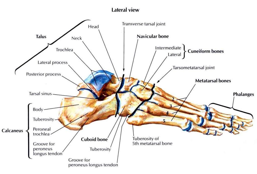
7 The Lower Limb The foot distal part of the ankle the tarsal bones, metatarsal bones, phalanges
8 4 parts: The pelvic girdle The thigh The leg The foot The Lower Limb
9 The pelvic girdle Hip the area from the iliac crest to the thigh the region between the iliac crest and the head of the femur formed by the innominate bones-ossa coxae
10 The hip bone os coxae large and irregular shaped consists of three bones in childhood: ilium ischium pubis fuse at years joined in adult
11 The hip bone 1.The ilium forms the superior 2/3 of the hip bone has ala (wing), is fan-shaped its body representing the handle iliac crest: superior margin of ilium
12 The hip bone the ilium iliac crest internal lip (labium internum) external lips (labium externum)
13 The hip bone the ilium iliac crest end posteriorly posterior superior iliac spine at the level of the fourth lumbar vertebra bilat.* iliac crest end anteriorly anterior superior iliac spine easily felt visible if you are not fatty *: it is important for lumbar puncture
14 The hip bone the ilium Tubercle of the crest is located 5cm posterior to the anterior superior iliac spine ant. inf. iliac spine difficult to identfy by palpation post. inf. iliac spine
15 The hip bone Gluteal face the ilium Pelvic face
16 The hip bone the ilium At the medial side auricular surface for the sacroiliac joint
17 The hip bone 2.The ischium it forms the posteroinferior part of hip L-shaped which passes inferiorly from the acetabulum turns anteriorly to join the pubis body ramus
18 The hip bone the ischium at the inferior end of the body ischial tuberosity» is covered by gluteus muscles when the thigh is extended
19 The hip bone the ischium at the posterior part of the ischium ischial spine (spina ischiadica) separates the» greater sciatic notch (sup.)» lesser sciatic notch (inf.) IS G L
20 greater sciatic notch greater sciatic foramen The hip bone the ilium lesser sciatic notch lesser sciatic foramen
21 The hip bone the ischium the greater sciatic notch is converted greater sciatic foramen by the sacrospinous ligament pass the» the priformis muscle» the vessels and nerves of gluteal region G SSL STL
22 The hip bone the ischium The lesser sciatic notch is converted lesser sciatic foramen by the sacrospinous and sacrotuberous ligament contains:» obtrator internus muscle» pudendal nerve» internal pudendal vessels SSL L STL
23 The hip bone the ischium ramus» extends medially from the body» joins the inf. ramus of the pubis» form ischiopubic ramus which completes the «obturator foramen» inf. ramus of pubis+ramus of ischium=ischiopubic ramus
24 The hip bone the pubis forms anterior part of the hip bone body, lies medially, joins body of the other ones it s called symphysis pubis (cartilaginous joint) ramus (2)» superior ramus passes superiolaterally to the acetabulum» inferior ramus passes posteriorly, inferiorly, laterally to joins ramus of ischium to form ischiopubic ramus
25 The hip bone the pubis the anterior border of the body is thickened pubic crest its lateral ends, pubic tubercule* *: main pubic attachment for the inguinal ligamentbony landmark *
26 The hip bone the obturator foramen oval aperture surrounded by the bodies and rami of the pubis and ischium it lies inferomedial to the acetabulum
27 The hip bone the obturator foramen is nearly closed by the obturator membrane

28 The hip bone the acetabulum cup shape cavity articulates with the head of femur it s names from Roman vinegar cup, it is called acetabulum Until puberty the ilium, ischium and pubis are united by a Y shaped hyaline cartilage At years these bones fuse to form the hip bone (cartilage is replaced by bone)
29 4 parts: The pelvic girdle The thigh The leg The foot The Lower Limb
30 thigh bone is femur longest strongest heaviest bone The thigh Femur articulates with acetabulum and tibia
31 body (shaft) ends (extremities) Proximal end: head neck greater trochanter lesser trochanter articulates with acetabulum The thigh Femur posterior aspect medial aspect
32 The thigh Femur Distal end: broadened articulates with tibia and patella medial aspect anterior aspect
33 Proximal end: head neck greater trochanter lesser trochanter The thigh Femur posterior aspect medial aspect
34 The thigh Femur Proximal end: Head forms about 2/3 of a sphere to fit deeply into the acetabulum sometimes palpable when the thigh is rotated laterally in thin male posterior aspect medial aspect
35 Proximal end: head neck greater trochanter lesser trochanter The thigh Femur posterior aspect
36 The thigh Femur neck between head and body to meet the body neck runs inferolaterally with angle of 125 limited laterally greater trochanter posterior aspect
 anterior aspect")
37 The thigh Femur Intertrochanteric line between greater and lesser trochanter, anteriorly is produced by the attachment of the iliofemoral ligament (massive lig.) anterior aspect
38 The thigh Femur Intertrochanteric crest unites greater and lesser trochanter, posteriorly posterior aspect
39 Proximal end: head neck greater trochanter lesser trochanter The thigh Femur posterior aspect
40 The thigh Femur greater trochanter is large, rectangular projection from the junction of the neck and the body. posterior aspect
41 The thigh greater trochanter is insertion for muscle of gluteal region the most lateral point of the hip region Femur posterior aspect

42 The thigh Femur greater trochanter can be easily palpated on the lateral side of the thigh the most lateral point of the hip region
43 Proximal end: head neck greater trochanter lesser trochanter The thigh Femur posterior aspect

44 The thigh Femur lesser trochanter is located in the posteromedial surface at the inferior end of the intertorachanteric crest in the angle between the neck and body of the femur posterior aspect
45 The thigh Femur Body (shaft) Linea aspera in the middle of its posteriorly has medial and lateral lips Diverge inferiorly to form the supracondylar lines not palpable, covered with large muscle
46 The thigh Femur Body (shaft) Pectineal line runs from the lesser torachanter to the medial lip tendon of the pectineal muscle inserts into it
47 The thigh Femur Distal end:
48 Distal end: Condyle, epicondyle intercondylar notch patellar surface adductor tubercle The thigh Femur


49 The thigh Femur Distal end: broadened for articulation with tibia 2 large condyle project posteriorly are subcutaneous easily palpable Covered by articular surface of condyle separated by a deep U-shaped intercondylar notch
50 The thigh Femur Distal end: at the center of the each condyle is a prominent epicondyle tibial and fibular collateral ligaments are attached to the epicondyles
51 Distal end: articular surfaces of condyle are confluent anteriorly patellar surface The thigh Femur
52 The thigh Femur Patellar surface can be palpated when the leg is flexed. Patella (kneecap) slides during flexion and extension of the leg

53 The adductor tubercle located in the medial side The thigh Femur
54 4 parts: The pelvic girdle The thigh The leg The foot The Lower Limb
55 The Lower Limb The leg (crus) Between knee and ankle tibia fibula are connected by an interosseous membrane» it is composed of strong oblique fibers
56 The Lower Limb Tibia (shine bone) supports most of the weight articulates with the condyle of femur superiorly and the talus inferiorly proximal end of tibia is large superior surface of tibia almost flat Medial-lateral condyles of tibia articulate with the condyles of femur

57 The Lower Limb sup. surface is flat consists of med-lat. tibial plateaus
58 The Lower Limb lat. condyle has facet inferiorly for the head of fibula
59 The Lower Limb Tibial tuberosity is located superior part of anterior surface patellar ligament is attached to the tibial tuberosity
60 The Lower Limb distal end of tibia; is small facet for the fibula and talus project medially and inferiorly medial malleolus
61 The Lower Limb medial malleolus has facet for articulation with talus
62 The Lower Limb body (corpus) Medial surface Lateral surface Posterior surface Medial border Lateral (interosseous border)* border Anterior border anterior aspect

63 The Lower Limb body (corpus) *:lat. border is sharp it gives attachment to the interosseous membrane uniting the tibia and fibula
 runs inferioromedially to the medial border The nutritient foramen is located posterior")
64 The Lower Limb At the posterior surface of tibia Observe a rough diagonal ridge known as the soleal line (soleus muscle is attached) runs inferioromedially to the medial border The nutritient foramen is located posterior aspect
65 The Lower Limb Fibula (calf bone) Pin-like bone lies posterolateral to the tibia little /no function in weight hearing providing support for tibia also provides stability to the ankle joint mainly for the attachment of muscle
66 The Lower Limb head Fibula (calf bone) neck is constricted part interosseous border for attacment to the interosseous memb. nutricient foramen is usually present at the post. side head of fibula is irregular facet for articulation with the lat. tibial condyle of tibia

67 The Lower Limb on the distal end project medially and inferiorly forms lateral malleolus lies more inferior and posterior than does medial malleolus
68 4 parts: The pelvic girdle The thigh The leg The foot The Lower Limb

69 The Lower Limb The foot comprise the tarsus metatarsus phalanges
70 The Lower Limb The foot comprise the tarsus metatarsus phalanges
71 The Lower Limb navicular bone tarsus talus* calcaneus cuboid navicular 3 cuneiforms *:articulates with the tibia
72 The Lower Limb talus body-cuboidal shape on the superior side it has trochlea it is pulley shaped part of talus The inferior surface of talus calcaneus neck the body of talus has an oval area for the articulation with the calcaneus
73 The Lower Limb talus posterior part of body has posterior process has med-lat tubercle 2 tubercle to consist of the groove for the tendon of the flexor hallucis longus muscle
74 The Lower Limb talus head of talus has articular surface for naviculare bone
75 The Lower Limb talus at the medial side of the calcaneus shelf-like projection of calcaneus Sustentaculum tali
76 The Lower Limb talus the neck is slightly constricted inferiorly there is a groove called the sulcus tali for the interosseous lig.
77

78 Calcaneus Largest-strongest 6 surfaces Sup :joins talus Inf :calcaneal tuber Ant :joins cuboid Post :forms heel Lat :fibular trochlea Med :sustentaculum tali
79 Navicular 3 facets Ant cuneiform Post talus Lat cuboid Med tuberosity of navicular
80 Cuboid Most lat. bone distal tarsus Ant base of metatarsals 4-5 Post calcaneus Med lat cuneiform & navicular Inf groove for fibularis longus
81 Cunieform Medial (largest) Lateral Intermedium (smallest) Ant base of metatarsals 1-4 Post Navicular Prof. Dr. H. Selçuk Sürücü
82 Metatarsal 5 bones Base Head Body I shortest & thickest II longest Prof. Dr. H. Selçuk Sürücü
83 Digital 14 bones Base Head Body Proximal, middle & distal phalanges
Copyright 2003 Pearson Education, Inc. publishing as Benjamin Cummings. Dr. Nabil Khouri MD, MSc, Ph.D
 Dr. Nabil Khouri MD, MSc, Ph.D Pelvic Girdle (Hip) Organization of the Lower Limb It is divided into: The Gluteal region The thigh The knee The leg The ankle The foot The thigh and the leg have compartments
Bones of Lower Limb. Dr. Heba Kalbouneh Associate Professor of Anatomy and Histology
 Bones of Lower Limb Dr. Heba Kalbouneh Associate Professor of Anatomy and Histology Bones of the lower limb Hip Bone Made up of 3 bones: 1) Ilium (flat), superior in position 2) Ischium (L), postero-inferior
Bones of the Lower Limb Bone Structure Description Notes. border of the superior ramus. inferolaterally from the pubic symphysis
 Bones of the Lower Limb Bone Structure Description Notes pubis an angulated bone the forms the anterior part of the pelvis one of three bones that form the os coxae: ilium, ischium, pubis; its forms 1/5
Figure 7: Bones of the lower limb
 BONES OF THE APPENDICULAR SKELETON The appendicular skeleton is composed of the 126 bones of the appendages and the pectoral and pelvic girdles, which attach the limbs to the axial skeleton. Although the
It is formed by fusion of 3 bones: I. Ilium (superior bone). II. Pubis (antero-inferior bone). III. Ischium (postero-inferior bone).
. II. Pubis (antero-inferior bone). III. Ischium (postero-inferior bone).") It is formed by fusion of 3 bones: I. Ilium (superior bone). II. Pubis (antero-inferior bone). III. Ischium (postero-inferior bone). Pubis Acetabulum Ana (242 ) The three constituent of bones of the hip
It is formed by fusion of 3 bones: I. Ilium (superior bone). II. Pubis (antero-inferior bone). III. Ischium (postero-inferior bone). Pubis Acetabulum Ana (242 ) The three constituent of bones of the hip
The Appendicular Skeleton
 8 The Appendicular Skeleton PowerPoint Lecture Presentations prepared by Jason LaPres Lone Star College North Harris 8-1 The Pectoral Girdle The Pectoral Girdle Also called shoulder girdle Connects the
Lab Activity 9. Appendicular Skeleton Martini Chapter 8. Portland Community College BI 231
 Lab Activity 9 Appendicular Skeleton Martini Chapter 8 Portland Community College BI 231 Appendicular Skeleton Upper & Lower extremities Shoulder Girdle Pelvic Girdle 2 Humerus 3 Humerus: Proximal End
Chapter 8 Outline. Pectoral Girdle Upper Limb Pelvic Girdle Lower Limb Aging of the Appendicular Skeleton Development of the Appendicular Skeleton
 Chapter 8 Outline Pectoral Girdle Upper Limb Pelvic Girdle Lower Limb Aging of the Appendicular Skeleton Development of the Appendicular Skeleton Figure 8.1 Appendicular Skeleton Pectoral Girdle Clavicle
First practical session. Bones of the gluteal region
 First practical session 2017 Bones of the gluteal region The Hip bone The hip bone is made of: 1 The ilium: superior in position 2 The ischium:postero-inferior in position 3 The pubis: antero-inferior
Pectoral (Shoulder) Girdle
 Girdle") Chapter 8 Skeletal System: Appendicular Skeleton Pectoral girdle Pelvic girdle Upper limbs Lower limbs 8-1 Pectoral (Shoulder) Girdle Consists of scapula and clavicle Clavicle articulates with sternum
Chapter 8 Skeletal System: Appendicular Skeleton Pectoral girdle Pelvic girdle Upper limbs Lower limbs 8-1 Pectoral (Shoulder) Girdle Consists of scapula and clavicle Clavicle articulates with sternum
Amy Warenda Czura, Ph.D. 1 SCCC BIO130 Lab 7 Appendicular Skeleton & Articulations
 The Skeletal System II: Appendicular Skeleton and Articulations Exercises 11, 13 (begins: page 145 in 9 th and 10 th editions) Exercises 10, 11 (begins: page 147 in 11 th edition, page 149 in 12 th edition)
Biology 218 Human Anatomy. Adapted from Martini Human Anatomy 7th ed. Chapter 7 The Skeletal System Appendicular Division
 Adapted from Martini Human Anatomy 7th ed. Chapter 7 The Skeletal System Appendicular Division Introduction The appendicular skeleton includes: Pectoral girdle Shoulder bones Upper limbs Pelvic girdle
Biology 218 Human Anatomy
 Chapter 8 Adapted from Tortora 10 th ed. LECTURE OUTLINE A. Introduction (p. 203) 1. The appendicular skeleton contains 126 bones that form: i. two pectoral (shoulder) girdles two upper limbs i one pelvic
Chapter 8. The Appendicular Skeleton. Lecture Presentation by Lee Ann Frederick University of Texas at Arlington Pearson Education, Inc.
 Chapter 8 The Appendicular Skeleton Lecture Presentation by Lee Ann Frederick University of Texas at Arlington An Introduction to the Appendicular Skeleton The Appendicular Skeleton 126 bones Allows us
Chapter 8B. The Skeletal System: Appendicular Skeleton. The Appendicular Skeleton. Clavicle. Pectoral (Shoulder) Girdle
 Girdle") The Appendicular Skeleton Chapter 8B The Skeletal System: Appendicular Skeleton 126 bones Pectoral (shoulder) girdle Pelvic (hip) girdle Upper limbs Lower limbs Functions primarily to facilitate movement
The Appendicular Skeleton Chapter 8B The Skeletal System: Appendicular Skeleton 126 bones Pectoral (shoulder) girdle Pelvic (hip) girdle Upper limbs Lower limbs Functions primarily to facilitate movement
C. Bones of the Pelvic Girdle
 C. Bones of the Pelvic Girdle 1. 2 coxal bones (a.k.a hip bones): -bony pelvis is made up of hip bones, sacrum, & coccyx -pelvic bones are large & heavy & attach to the axial skeleton via sacrum/coccyx
PRE-LAB EXERCISES. Before we get started, look up the definitions of these common bone marking terms: Canal: Condyle: Facet: Fissure:
 1 PRE-LAB EXERCISES When studying the skeletal system, the bones are often sorted into two broad categories: the axial skeleton and the appendicular skeleton. This lab focuses on the appendicular skeleton,
SKELETAL SYSTEM 206. AXIAL SKELETON 80 APPENDICULAR SKELETON 126 (see Figure 6.1) Clavicle. Clavicle. Pectoral girdles. Scapula. Scapula.
 Clavicle. Clavicle. Pectoral girdles. Scapula. Scapula.") SKELETAL SYSTEM 206 AXIAL SKELETON 80 APPENDICULAR SKELETON 126 (see Figure 6.1) Pectoral girdles 4 Clavicle Scapula 2 2 Clavicle Scapula Humerus 2 Humerus Upper limbs 60 Radius 2 Ulna Carpal bones Metacarpal
SKELETAL SYSTEM 206 AXIAL SKELETON 80 APPENDICULAR SKELETON 126 (see Figure 6.1) Pectoral girdles 4 Clavicle Scapula 2 2 Clavicle Scapula Humerus 2 Humerus Upper limbs 60 Radius 2 Ulna Carpal bones Metacarpal
The thigh. Prof. Oluwadiya KS
 The thigh Prof. Oluwadiya KS www.oluwadiya.com The Thigh: Boundaries The thigh is the region of the lower limb that is approximately between the hip and knee joints Anteriorly, it is separated from the
LAB Notes#1. Ahmad Ar'ar. Eslam
 LAB Notes#1 Ahmad Ar'ar Eslam 1 P a g e Anatomy lab Notes Lower limb bones :- Pelvic girdle: It's the connection between the axial skeleton and the lower limb; it's made up of one bone called the HIP BONE
Copyright 2003 Pearson Education, Inc. publishing as Benjamin Cummings. Dr. Nabil khouri
 Dr. Nabil khouri Appendicular Skeleton The appendicular skeleton is made up of the bones of the upper and lower limbs and their girdles Two girdles: Pectoral girdles attach the upper limbs to the body
Located more distal and anterior together with Trapezoid, anterior to scaphoid Trapezium rarely to be fractured.
 The hand The hand consists of the 3 groups: Proximal part: carpals bones (8) Middle part: metacarpal bones (5) Distal part: fingers or phalanges bones (3 for each finger except for the thumb just 2 bones).
Anatomage Table Instructors Guide- Lower Limb
 The Lower Limb Anatomage Table Instructors Guide- Lower Limb Table of Contents Lower Limb 1- The Skeletal System...3 1: Hip Bone...3 2: Hip Joint and Femur...4 3: Patella and Knee Joint...7 4: Tibia, Fibula,
Biology 152 Appendicular Skeleton Anatomy Objectives
 Biology 152 Appendicular Skeleton Anatomy Objectives We will learn proper bone names, left/right/medial, and the parts of bones in this exercise. Start by learning the names of the bones. As you gain comfort
The Hip (Iliofemoral) Joint. Presented by: Rob, Rachel, Alina and Lisa
 Joint. Presented by: Rob, Rachel, Alina and Lisa") The Hip (Iliofemoral) Joint Presented by: Rob, Rachel, Alina and Lisa Surface Anatomy: Posterior Surface Anatomy: Anterior Bones: Os Coxae Consists of 3 Portions: Ilium Ischium Pubis Bones: Pubis Portion
The Hip (Iliofemoral) Joint Presented by: Rob, Rachel, Alina and Lisa Surface Anatomy: Posterior Surface Anatomy: Anterior Bones: Os Coxae Consists of 3 Portions: Ilium Ischium Pubis Bones: Pubis Portion
Muscles of the lower extremities. Dr. Nabil khouri MD, MSc, Ph.D
 Muscles of the lower extremities Dr. Nabil khouri MD, MSc, Ph.D Posterior leg Popliteal fossa Boundaries Biceps femoris (superior-lateral) Semitendinosis and semimembranosis (superior-medial) Gastrocnemius
Topic 7: Hip and pelvis. Parts of the hip. Parts of the femur
 Topic 7: Hip and pelvis Parts of the hip Parts of the femur Classifying the hip joint Ball and socket Synovial Multiaxial Movements of the hip: Abduction/adduction Flexion/extension Medial/lateral rotation
Lower limb summary. Anterior compartment of the thigh. Done By: Laith Qashou. Doctor_2016
 Lower limb summary Done By: Laith Qashou Doctor_2016 Anterior compartment of the thigh Sartorius Anterior superior iliac spine Upper medial surface of shaft of tibia 1. Flexes, abducts, laterally rotates
Anatomy & Physiology. Muscles of the Lower Limbs.
 Anatomy & Physiology Muscles of the Lower Limbs http://www.ishapeup.com/musclecharts.html Muscles of the Lower Limbs Among the strongest muscles in the body. Because pelvic girdle is composed of heavy,
Anatomy of the lower limb
 Anatomy of the lower limb 1. Bones of the lower limb Pelvis Hip bone/coxal bone Acetabulum o Acetabular margin o Acetabular fossa o Acetabular notch o Lunate surface Ischiopubic ramus Obturator foramen
The Appendicular Skeleton
 8 The Appendicular Skeleton PowerPoint Lecture Presentations prepared by Jason LaPres Lone Star College North Harris An Introduction to the Appendicular Skeleton Learning Outcomes 8-1 Identify the bones
10/12/2010. Upper Extremity. Pectoral (Shoulder) Girdle. Clavicle (collarbone) Skeletal System: Appendicular Skeleton
 Girdle. Clavicle (collarbone) Skeletal System: Appendicular Skeleton") Skeletal System: Appendicular Skeleton Pectoral girdle Pelvic girdle Upper limbs Lower limbs 8-1 Pectoral (Shoulder) Girdle Consists of scapula and clavicle Clavicle articulates with sternum (Sternoclavicular
Skeletal System: Appendicular Skeleton Pectoral girdle Pelvic girdle Upper limbs Lower limbs 8-1 Pectoral (Shoulder) Girdle Consists of scapula and clavicle Clavicle articulates with sternum (Sternoclavicular
Muscles of the Thigh. 6.1 Identify, describe the attachments of and deduce the actions of the muscles of the thigh: Anterior group
 Muscles of the Thigh 6.1 Identify, describe the attachments of and deduce the actions of the muscles of the thigh: Anterior group Sartorius: This is a long strap like muscle with flattened tendons at each
Muscles of Lesson Five. Muscular Nomenclature and Kinesiology - Two. Muscles of Lesson Five, cont. Chapter 16
 Chapter 16 Muscular Nomenclature and Kinesiology - Two Lessons 5-6 Muscles of Lesson Five Iliopsoas (psoas major, iliacus) Hip outward rotators (piriformis, gemellus superior, gemellus inferior, obturator
Appendicular Skeleton. Prof. Abdulameer Al-Nuaimi
 Appendicular Skeleton Prof. Abdulameer Al-Nuaimi a.alnuaimi@sheffield.ac.uk abdulameerh@yahoo.com Hi Prof, It is great to hear from you, I really enjoyed your teaching last year. You taught me the hardest
The Lower Limb II. Anatomy RHS 241 Lecture 3 Dr. Einas Al-Eisa
 The Lower Limb II Anatomy RHS 241 Lecture 3 Dr. Einas Al-Eisa Tibia The larger & medial bone of the leg Functions: Attachment of muscles Transfer of weight from femur to skeleton of the foot Articulations
BONES JOINTS MUSCLES OF THE LOWER LIMB
 BONES JOINTS MUSCLES OF THE LOWER LIMB LOWER LIMB: BONES LOWER LIMB GLUTEAL REGION consists of 6 major segments: FEMORAL REGION (THIGH) KNEE REGION LEG REGION TALOCRURAL REGION (ANKLE) FOOT REGION LOWER
Chapter 8 The Skeletal System: The Appendicular Skeleton. Copyright 2009 John Wiley & Sons, Inc.
 Chapter 8 The Skeletal System: The Appendicular Skeleton Appendicular Skeleton The primary function is movement It includes bones of the upper and lower limbs Girdles attach the limbs to the axial skeleton
Skeletal System Module 13: The Pelvic Girdle and Pelvis
 OpenStax-CNX module: m47993 1 Skeletal System Module 13: The Pelvic Girdle and Pelvis Donna Browne Based on The Pelvic Girdle and Pelvis by OpenStax College This work is produced by OpenStax-CNX and licensed
Human Anatomy Biology 351
 Human Anatomy Biology 351 Lower Limb Please place your name on the back of the last page of this exam. You must answer all questions on this exam. Because statistics demonstrate that, on average, between
The Lower Limb. Anatomy RHS 241 Lecture 2 Dr. Einas Al-Eisa
 The Lower Limb Anatomy RHS 241 Lecture 2 Dr. Einas Al-Eisa The bony pelvis Protective osseofibrous ring for the pelvic viscera Transfer of forces to: acetabulum & head of femur (when standing) ischial
The Dance Hall by Vincent van Gogh,1888
 The Dance Hall by Vincent van Gogh,1888 Articulations of the pelvic girdle Lumbosacral joints, sacroiliac joints & pubic symphysis The remaining joints of the lower limb Hip joint Knee joint Tibiofibular
Pelvic Girdle
 ARTICULATIONS OF LOWER EXTREMITY Pages 429-437 Pelvic Girdle formed by connection of the hip bones and the sacrum Sacroiliac Joints compound joints synovial joint - anterior, between the auricular surfaces
lesser trochanter of femur lesser trochanter of femur iliotibial tract (connective tissue) medial surface of proximal tibia
 medial surface of proximal tibia") LOWER LIMB MUSCLES OF THE APPENDICULAR SKELETON The muscles that act on the lower limb fall into three groups: those that move the thigh, those that move the lower leg, and those that move the ankle, foot,
LOWER LIMB MUSCLES OF THE APPENDICULAR SKELETON The muscles that act on the lower limb fall into three groups: those that move the thigh, those that move the lower leg, and those that move the ankle, foot,
Exercise 11. The Appendicular Skeleton
 Exercise 11 The Appendicular Skeleton The Appendicular Skeleton The appendicular skeleton contains 126 bones. Consists of the upper and lower limbs, the pectoral girdles, and the pelvic girdles. The pectoral
bio4165 lab quiz 1 Posterior View Anterior View Lateral View Anterior View bio fall.quarter lab.quiz.1...page.1 of 6
 B A Posterior View D C E Lateral View bio.4165...fall.quarter.2005...lab.quiz.1...page.1 of 6 F I G 35 Posterior View H bio.4165...fall.quarter.2005...lab.quiz.1...page.2 of 6 J Posterior View L K Inferior
Chapter 7: Skeletal System: Gross Anatomy
 Chapter 7: Skeletal System: Gross Anatomy I. General Considerations A. How many bones in an average adult skeleton? B. Anatomic features of bones are based on II. Axial Skeleton A. Skull 1. Functionally
Practical 1 Worksheet
 Practical 1 Worksheet ANATOMICAL TERMS 1. Use the word bank to fill in the missing words. reference side stand body arms palms anatomical forward All anatomical terms have a(n) point which is called the
Bone Flashcards for 10a
 Bone Flashcards for 0a CLAVICLE (collar bone). Sternal extremity (end) flat end. Acromial extremity (end) rounded end. SCAPULA (shoulder blade). Right or left scapula?. Superior border (superior margin).
Exercise Science Section 2: The Skeletal System
 Exercise Science Section 2: The Skeletal System An Introduction to Health and Physical Education Ted Temertzoglou Paul Challen ISBN 1-55077-132-9 Role of the Skeleton Protection Framework Attachments for
Principles of Anatomy and Physiology
 Principles of Anatomy and Physiology 14 th Edition CHAPTER 8 The Skeletal System: The Appendicular Skeleton The Appendicular Skeleton The 126 bones of the appendicular skeleton are primarily concerned
Contents of the Posterior Fascial Compartment of the Thigh
 Contents of the Posterior Fascial Compartment of the Thigh 1-Muscles: B i c e p s f e m o r i s S e m i t e n d i n o s u s S e m i m e m b r a n o s u s a small part of the adductor magnus (h a m s t
BIOLOGY 113 LABORATORY Skeletal System
 BIOLOGY 113 LABORATORY Skeletal System Objectives Distinguish between the axial and appendicular skeleton. Distinguish between the cranium and facial skeleton. Locate and name the bones of the skull and
Gluteal region DR. GITANJALI KHORWAL
 Gluteal region DR. GITANJALI KHORWAL Gluteal region The transitional area between the trunk and the lower extremity. The gluteal region includes the rounded, posterior buttocks and the laterally placed
Riverside Community College Anatomy & Physiology 2B SPRING 2012 EXAM #1-ABC (Nervous System)
") Riverside Community College Anatomy & Physiology 2B SPRING 2012 EXAM #1-ABC (Nervous System) Name: 1) This vertebra is an example of a(n). 1) A) thoracic B) axis C) atlas D) lumbar E) sacral 1 2) W hich
Riverside Community College Anatomy & Physiology 2B SPRING 2012 EXAM #1-ABC (Nervous System) Name: 1) This vertebra is an example of a(n). 1) A) thoracic B) axis C) atlas D) lumbar E) sacral 1 2) W hich
Chapter 8 The Skeletal System: The Appendicular Skeleton. Copyright 2009 John Wiley & Sons, Inc.
 Chapter 8 The Skeletal System: The Appendicular Skeleton Appendicular Skeleton It includes bones of the upper and lower limbs Girdles attach the limbs to the axial skeleton The pectoral girdle consists
Human Anatomy Biology 351
 Human Anatomy Biology 351 Lower Limb Please place your name on the back of the last page of this exam. You must answer all questions on this exam. Because statistics demonstrate that, on average, between
Bone List Anatomy
 1 Frontal Bone Skull 2 Parietal Bone Skull 3 Occipital Bone Skull 4 Temporal Bone Skull 5 Coronal Suture Skull 6 Sagittal Suture Skull 7 Squamous suture Skull 8 Lambdoid Suture Skull 9 Surpaorbital Ridge
External Acoustic Meatus. Mastoid Process. Zygomatic Process. Temporal Bone
 Bone lab review 1. Frontal Bone 2. Supra-Orbital Foramen 3. Orbit (Orbital Cavity) 4. Superior Orbital Fissure 5. Inferior Orbital Fissure 6. Zygomatic Bone 7. Infra-Orbital Foramen 8. Maxilla 9. Mandible
Chapter 7 Part C The Skeleton
 Chapter 7 Part C The Skeleton Part 2 The Appendicular Skeleton Consists of bones of the limbs and their girdles Pectoral girdle Attaches upper limbs to body trunk Pelvic girdle Attaches lower limbs to
PELVIS & SACRUM Dr. Jamila El-Medany Dr. Essam Eldin Salama
 PELVIS & SACRUM Dr. Jamila El-Medany Dr. Essam Eldin Salama Learning Objectives At the end of the lecture, the students should be able to : Describe the bony structures of the pelvis. Describe in detail
LOWER LIMB. As we know the bony part of the body is divided into Axial and Appendicular (upper and lower Limbs)
") LOWER LIMB As we know the bony part of the body is divided into Axial and Appendicular (upper and lower Limbs) Bones of the Lower limb: 1-Pelvic Girdle: composed of: 1. Right hip bone : is formed by 3
LOWER LIMB As we know the bony part of the body is divided into Axial and Appendicular (upper and lower Limbs) Bones of the Lower limb: 1-Pelvic Girdle: composed of: 1. Right hip bone : is formed by 3
Anatomy images for MSS practical exam- 2019
 Anatomy images for MSS practical exam- 2019 Ilium Ischium Pubis Acetabulaum Iliac crest Iliac tubercle ASIS (muscle and ligament attached) AIIS (muscle attached) PSIS PIIS Ischial spine Ischial tuberosity
Dr.Israa H. Mohsen. Lecture 5. The vertebral column
 Anatomy Lecture 5 Dr.Israa H. Mohsen The vertebral column The vertebral column a flexible structure consisting of 33 vertebrae holds the head and torso upright, serves as an attachment point for the legs,
5.1 Identify, describe the attachments of and deduce the actions of the muscles of the thigh:
 5.1 Identify, describe the attachments of and deduce the actions of the muscles of the thigh: Anterior group Proximal attachment Distal attachment Sartorius ASIS» Upper part of shaft tibia (middle surface)»
The Leg. Prof. Oluwadiya KS
 The Leg Prof. Oluwadiya KS www.oluwadiya.sitesled.com Compartments of the leg 4 Four Compartments: 1. Anterior compartment Deep fibular nerve Dorsiflexes the foot and toes 2. Lateral Compartment Superficial
Lecture 08 THIGH MUSCLES ANTERIOR COMPARTMENT. Dr Farooq Khan Aurakzai. Dated:
 Lecture 08 THIGH MUSCLES ANTERIOR COMPARTMENT BY Dr Farooq Khan Aurakzai Dated: 11.02.2017 INTRODUCTION to the thigh Muscles. The musculature of the thigh can be split into three sections by intermuscular
Hip joint and pelvic girdle. Lower Extremity. Pelvic Girdle 6/5/2017
 Hip joint and pelvic girdle Lower Extremity The relationship between the pelvic girdle and hip is similar to that between the shoulder girdle and shoulder joint. The lower limbs are attached to the axial
The Skeletal System THE APPENDICULAR SKELETON
 The Skeletal System THE APPENDICULAR SKELETON The appendicular skeleton consists of the girdles and the skeleton of the limbs. The upper (anterior) limbs are attached to the pectoral (shoulder) girdle
Important Parts of Bones
 Important Parts of Bones For 2015 Know: Humerus (posterior) Clavical Femur (Anterior) Foot Hand Mandible Os Coxa Scapula Skull (Anterior, Inferior, Lateral) Sternum Humerus (posterior) A. olecranon fossa
Spring Written By: J. E. Sutton. Contents: I. Overview of the Skeleton: II. Appendicular Skeleton III. Axial Skeleton IV.
 Spring 2012 Written By: J. E. Sutton Contents: I. Overview of the Skeleton: II. Appendicular Skeleton III. Axial Skeleton IV. Articulations Overview of the Skeleton: I. Orientation to Human Skeleton: a.
Skeleton of lower extremity Ossa membri inferioris
 Skeleton of lower extremity Ossa membri inferioris Skeleton of lower extremity consists of lower extremity girdle (cingulum membri inferioris) created by pelvis bone (os coxae) and bones of free lower
rotation of the hip Flexion of the knee Iliac fossa of iliac Lesser trochanter Femoral nerve Flexion of the thigh at the hip shaft of tibia
 Anatomy of the lower limb Anterior & medial compartments of the thigh Dr. Hayder The fascia lata encloses the entire thigh like a sleeve/stocking. Three intramuscular fascial septa (lateral, medial, and
Identify the muscles associated with the medial compartment of the thigh. Identify the attachment points of the medial thigh muscles.
 L 8 A B O R A T O R Y Thigh MEDIAL THIGH Identify the muscles associated with the medial compartment of the thigh. Identify the attachment points of the medial thigh muscles. Identify the actions of these
The Hip Joint. Shenequia Howard David Rivera
 The Hip Joint Shenequia Howard David Rivera Topics Of Discussion Movement Bony Anatomy Ligamentous Anatomy Muscular Anatomy Origin/Insertion/Action/Innervation Common Injuries MOVEMENT Flexion Extension
Lectures of Human Anatomy
 Lectures of Human Anatomy Lower Limb Gluteal Region and Hip Joint By DR. ABDEL-MONEM AWAD HEGAZY M.B. with honor 1983, Dipl."Gynecology and Obstetrics "1989, Master "Anatomy and Embryology" 1994, M.D.
Human Anatomy, First Edition McKinley & O'Loughlin
 Human Anatomy, First Edition McKinley & O'Loughlin Chapter 8 : Appendicular Skeleton 8-1 Appendicular Skeleton Includes the bones of the upper and lower limbs. The girdles of bones that attach the upper
Skeleton of the lower limb
 Skeleton of the lower limb (ossa membri inferioris) RNDr. Michaela Račanská, Ph.D. Lecture 3 DENTISTRY Autumn 2016 Skeleton of the lower limb support the weight of the body and transfers that support to
Axial skeleton bones and markings
 Axial skeleton bones and markings Skull Cranial bones Frontal x 1 Supraorbital foramen Occipital x 1 Foramen magnum Occipital condyles Superior nuchal line Inferior nuchal line Anterior cranial fossa External
The University Of Jordan Faculty Of Medicine THE LOWER LIMB. Dr.Ahmed Salman Assistant Prof. of Anatomy. The University Of Jordan
 The University Of Jordan Faculty Of Medicine THE LOWER LIMB Dr.Ahmed Salman Assistant Prof. of Anatomy. The University Of Jordan Gluteal Region Cutaneous nerve supply of (Gluteal region) 1. Lateral cutaneous
Lower Limb Dr. Robin Paudel
 Lower Limb n What is a limb? n Skeleton n Joints n Pelvis or limb girdle n Hip/Hip Muscles n Lumber and sacral plexus getting spinal nerves out onto limb n Muscles anterior and posterior compartments n
Mohammad Ashraf. Abdulrahman Al-Hanbali. Ahmad Salman. 1 P a g e
 - 7 Mohammad Ashraf Abdulrahman Al-Hanbali Ahmad Salman 1 P a g e Structures under the cover of Gluteus Maximus: 1-Bones: Ileum, Femur (Head, greater trochanter and gluteal tuberosity), Ischium (ischial
Anatomy images for MSS practical exam- 2019
 Anatomy images for MSS practical exam- 2019 Ilium Ischium Pubis Acetabulaum Iliac crest Iliac tubercle ASIS (muscle and ligament attached) AIIS (muscle attached) PSIS PIIS Ischial spine Ischial tuberosity
Perpendicular Plate Zygomatic Bone. Mental Foramen Mandible
 Glabella Frontal Middle Nasal Concha Nasal Lacrimal Perpendicular Plate Zygomatic Inferior Nasal Concha Maxilla Mental Mandible Skull (anterior view) Squamosal Suture Coronal Suture Frontal Parietal Nasal
The os coxae or hip bone consists of three flat bones, ilium, ischium and pubis, which fuse together to form the acetabulum.
 The os coxae The os coxae or hip bone consists of three flat bones, ilium, ischium and pubis, which fuse together to form the acetabulum. The ilium extends from the acetabulum upwards forming the lateral
Bony Anatomy. Femur. Femoral Head Femoral Neck Greater Trochanter Lesser Trochanter Intertrochanteric Crest Intertrochanteric Line Gluteal Tuberosity
 Hip Anatomy Bony Anatomy Femur Femoral Head Femoral Neck Greater Trochanter Lesser Trochanter Intertrochanteric Crest Intertrochanteric Line Gluteal Tuberosity Bony Anatomy Pelvic Girdle Acetabulum 3 bones
Main Menu. Joint and Pelvic Girdle click here. The Power is in Your Hands
 1 Hip Joint and Pelvic Girdle click here Main Menu K.6 http://www.handsonlineeducation.com/classes//k6entry.htm[3/23/18, 2:01:12 PM] Hip Joint (acetabular femoral) Relatively stable due to : Bony architecture
~, /' ~::'~ EXTENSOR HALLUCIS LONGUS. Leg-anterolateral :.:~ / ~\,
 TIBIALIS ANTERIOR Lateral condyle of tibia, upper half of lateral surface of tibia, interosseous membrane Medial side and plantar surface of medial cuneiform bone, and base of first metatarsal bone Dorsiflexes
Lesson 24. A & P Hip
 Lesson 24 A & P Hip 1 Aims of the Session This session will allow candidates to have an understanding of the bony prominences and soft tissues of the hip 2 Learning Outcomes By the end of the lesson the
Introduction to Human Osteology Chapter 3: Hands and Feet
 Introduction to Human Osteology Chapter 3: Hands and Feet Roberta Hall Kenneth Beals Holm Neumann Georg Neumann Gwyn Madden Revised in 1978, 1984, and 2008 Bones of the Hand Eight carpal bones, in two
this makes sense, however this is lower order thinking and does not solve the lower leg
 Functional Knee Valgus in a Barbell Squat 1 One of the most common lower leg dysfunction we see in athletes, particularly general population is functional knee valgus, or better referred to as the knees
Acland's DVD Atlas of Human Anatomy. Transcript for Volume Robert D Acland
 Acland's DVD Atlas of Human Anatomy Transcript for Volume 2 2007 Robert D Acland This free downloadable pdf file is to be used for individual study only. It is not to be reproduced in any form without
BLUE SKY SCHOOL OF PROFESSIONAL MASSAGE AND THERAPEUTIC BODYWORK. Musculoskeletal Anatomy & Kinesiology I TERMINOLOGY, STRUCTURES, & SKELETAL OVERVIEW
 BLUE SKY SCHOOL OF PROFESSIONAL MASSAGE AND THERAPEUTIC BODYWORK Musculoskeletal Anatomy & Kinesiology I TERMINOLOGY, STRUCTURES, & SKELETAL OVERVIEW MSAK101-I Session 1 Learning Objectives: 1. Define
The Lower Limb VII: The Ankle & Foot. Anatomy RHS 241 Lecture 7 Dr. Einas Al-Eisa
 The Lower Limb VII: The Ankle & Foot Anatomy RHS 241 Lecture 7 Dr. Einas Al-Eisa Ankle joint Synovial, hinge joint Allow movement of the foot in the sagittal plane only (1 degree of freedom): dorsiflexion:
the Skeletal System provided by Academic Web Services Grand Canyon University
 Anatomy Resource Center Study Guides the Skeletal System HEAD & NECK REGIONAL VIEW SKULL BONES CRANIUM FACE SKULL LANDMARKS ANTERIOR SIDE SUPERIOR/INFERIOR VERTEBRAL COLUMN VERTEBRAL REGIONS CERVICAL C1
Muscle Testing of Knee Extensors. Yasser Moh. Aneis, PhD, MSc., PT. Lecturer of Physical Therapy Basic Sciences Department
 Muscle Testing of Knee Extensors Yasser Moh. Aneis, PhD, MSc., PT. Lecturer of Physical Therapy Basic Sciences Department Muscle Testing of Knee Extensors othe Primary muscle Quadriceps Femoris -Rectus
Leg. Dr. Heba Kalbouneh Associate Professor of Anatomy and Histology
 Leg Dr. Heba Kalbouneh Associate Professor of Anatomy and Histology Skin of the Leg Cutaneous Nerves Medially: The saphenous nerve, a branch of the femoral nerve supplies the skin on the medial surface
Introduction to Anatomy. Dr. Maher Hadidi. Tala Ar ar. Mar/10th/2013
 Sheet Introduction to Anatomy Dr. Maher Hadidi Tala Ar ar 15 Mar/10th/2013 Lower limb The skeleton of the lower limb is the lower appendicular skeleton which consists of 2 parts: 1- Pelvic girdle. 2- Bones
BOGOMOLETS NATIONAL MEDICAL UNIVERSITY. Department of human anatomy GUIDELINES
 BOGOMOLETS NATIONAL MEDICAL UNIVERSITY Department of human anatomy GUIDELINES Academic discipline HUMAN ANATOMY Module 1 Content module 2 The topic of the lesson Bones of the lower limb Course 1 st Number
TABLE OF MUSCLES OF LOWER EXTREMITY 2018zillmusom ANTERIOR THIGH
 TABLE OF MUSCLES OF LOWER EXTREMITY 2018zillmusom ANTERIOR THIGH MUSCLE ORIGIN INSERTION ACTION NERVE Iliopsoas Ilium, vertebra Femur Flex hip joint Femoral nerve (T12-L5) Pectineus Pubis Femur Flex hip