Lab 1: body, skeletal and axial systems
|
|
|
- Baldric Neal
- 5 years ago
- Views:
Transcription

1 Lab 1: body, skeletal and axial systems Types of tissue Epithelia tissue - Sheets of tissue covering other tissues or linin cavities - Protective tissue e.g. skin - Cell types: squamous, cuboid, columnar (can be single layer of cells or stratified) - Single layer epithelium: allows materials to pass, secretion, absorption - Stratified layer epithelium: protects against abrasion, secretes and protects - Pseudostratified: single layer that appears to be stratified, secretes mucus Connective tissue - Connects and supports other tissue - Loose connective tissue: e.g. subcutaneous tissue or superfician fascia - Dense connective tissue: e.g. patella ligament or muscle tendon - Bones - Cartilage: 3 different types. Hyaline (covers joints), elastic (external ear), fibrocartilage (intervertebral disc) - Blood Nervous tissue - Specialised tissue for conduction of nerve impulses to and from the CNS - Brain, spinal chord - Sciatic nerve: (should be able to identify in lab 4) Muscle tissue - Smooth muscle: found in walls of hollow body organs, e.g. gut and blood vessels. Involuntary muscle - Cardiac muscle: heart and bases of great vessels. Involuntary muscle - Skeletal (voluntary muscle): greater mass of muscle in the body. Attach directly or indirectly to bones, cartilages, ligaments, fascia, organs and membranes *voluntary muscles: in arms, legs, hands help you do basic or compex movements **Involuntary muscles: in lungs, intestines help with breathing, propelling food through gut (don t have to think about using these muscles. Types of bone Long bones: found in limbs, length is greater than breadth, have a cylindrical shaft and a swelling at each end. Features: - Diaphysis is a shaft of a long bone (body of the bone) - Epiphyses are swellings at each end - Articular surfaces are the surfaces that participate in a joint - Periosteum is a membrane covering non-articular surfaces. Essential for bone repair and attachment of tendons, ligaments Short bones: found in wrist and ankle, roughly cuboidal shape
2 Sesamoid bones: type of short bone found in certain tendons. E.g. patella Flat bones: found mostly in vault of cranium (skull) Irregular bones: nor long short or flat (e.g. vertebrae) Features of bones Word definition examples foramen A passage through a bone Foramen magnum Meatus A path or passage way to an organ External acoustic meatus Fossa Hollowed or depressed area Middle cranial fossa Process A projection Spinous process of vertebrae Facet A smooth flat articular process Facets on articular processes of vertebra Tuberosity Large flat convexity Iscial tuberosity of pelvis Tubercle A small raised eminence Greater tubercle of humerus Trochanter Large blunt elevation Greater trochanter of femur Malleolus A rounded process Medial malleolus of the tibia Condyle A rounded articular area Medial condyle of femur Head A bony expansion carried on a narrow neck Head of humerus Axial skeleton It is the axis of the body and provides posture and support for the body. It includes the skull, vertebral column, ribs and sternum **paired bones means there are 2 bones on either side of same shape. Cranial exterior Frontal bone Parietal bones (paired) Temporal bones (paired) Occipital bone Sphenoid bone Ethmoid bone Foramen magnum (opening of skull where brain is continuous with spinal cord) Occipital condyles External acoustic meatus Mastoid process (part of temporal bone) Facial skeleton Zygomatic bones (paired) Maxillae (paired) Nasal bones (paired) Mandible Foramina Cribriform plate of ethmoid Optic canal Superior orbital fissure Foramen rotundum Foramen ovale Internal acoustic meatus Jugular foramen
 Posterior cranial fossa (formed by occipital and temporal bones) Hypophyseal")
 NEED TO KNOW: all bones, position, what it articulates with, is it paired or not?")
3 Cranial interior Anterior cranial fossa (formed mainly by frontal bone) Middle cranial fossa (formed by sphenoid and temporal bones) Posterior cranial fossa (formed by occipital and temporal bones) Hypophyseal (pituitary fossa) within the sphenoid bone Petrous part of temporal bone (boundary between middle and cranial fossa) NEED TO KNOW: all bones, position, what it articulates with, is it paired or not? Page 3 of 15


4 Vertebral column The central pillar of the body and provides support for the cranium, shoulder girdle, upper limbs and thoracic cage. It transmits weight to the lower limb. Within the cavity of the vertebral column is the vertebral canal. This is a space for the spinal cord to pass and gives rise to the peripheral nerves. There are 33 vertebrae within the vertebral column. Page 4 of 15
 Sacral vertebrae (5 fused to form sacrum) Coccygeal (lower 3-4")
 *Facet joints")

 Interior articular processes with facets")

5 Vertebral column cervical vertebrae (7, neck) Thoracic vertebrae (12, thorax) Lumbar vertebrae (5, abdomen) Sacral vertebrae (5 fused to form sacrum) Coccygeal (lower 3-4 fused to form coccyx) *Intervertebral discs (fibrocartilaginous joints) Intervertebral foramina (between vertebrae, formed by notes, spinal nerve exit here) *Facet joints (between articular processes) **vertebral column covered in notes of lab 3 Isolated vertebrae Vertebral body Vertebral foramen (vertebral foramen of or vertebrae form the vertebral canal) Pedicle Lamina Spinous process Transverse process (paired) Superior articular processes with facets (paired) Interior articular processes with facets (paired) Superior articular process is above inferior articular process The process is the bone sticking out and the facet is the blue thing Superior vertebra notch Inferior vertebral notch Page 5 of 15
 12 thoracic vertebrae 12 pairs of ribs (numbered 1-12 from top to bottom) - True ribs: ribs 1-7, attach directly to the sternum")
6 Thoracic cage Sternum consists of manubrium, sternal body and xiphoid process. Sits in front of the vertebral column (especially thoracic vertebrae) 12 thoracic vertebrae 12 pairs of ribs (numbered 1-12 from top to bottom) - True ribs: ribs 1-7, attach directly to the sternum via their own costa cartilage - False ribs: ribs 8-10, attach indirectly to the sternum via shared costal cartilage - Floating ribs: ribs 11-12, do not attach to sternum Sternal angle is at the manubrium-sternal joint. 2 nd true rib articulates with sternum here. Lab 2 Appendicular skeleton and articular system Appendicular skeleton Includes parts of the skeleton that connect off the axial skeleton. Includes upper and lower limbs. Be abe to identify and disucss the strcutre, function and features of appendicular skeleton. Shoulder girdle Clavicle articulates with sternum and scapula. Medial/sternal end is rounded. Lateral/acromial end is flattened. Clavicle connects shoulder girdle to axial skeleton. Scapula flat triangular bone that lies on posterior thoracic wall - Glenoid fossa (points laterally and articulates with humeral head) Page 6 of 15
 The prominent spine at the back of the scapula is a good way of telling it which side of scapular we are looking at.")
7 - Anterior surface of scapula (formed by: coracoid process and subscapular fossa) - Posterior surface of scapula (formed by: prominent spine ends laterally as a flat acromion and articulates with clavicle, supraspinous fossae, Infraspinous fossae) The prominent spine at the back of the scapula is a good way of telling it which side of scapular we are looking at. Upper limb Humerus - Proximal bone of upper limb (i.e. arm bone) - Proximal end: Head (articulates with scapula at glenoid fossa), greater tubercle (lateral), lesser tubercle (anterior) (groove in between tubercles for biceps brachii tendon), surgical neck (prone to fractures) - Middle part: shaft and deltoid tuberosity (small protrusion on lateral side of shaft) - Distal end: medial and lateral epicondyles (for muscle attachments), trochlea (medial side, articulates with trochlear notch of ulna), capitulum (lateral side, articulates with head of radius. cap--head) Ulna - Medial bone of the forearm - Proximal end: olecranon process (elbow), trochlear notch (faces anteriorly and articulates with trochlea of humerus), ulna tuberosity (for brachialis attachment, small bump distal to notch). Radial notch (below the trochlear notch and articulates with radial head to form proximal radio-ulnar joint) - Middle part: shaft - Distal end: head (articulates with radius to form distal radio-ulnar joint, head is not proximal for ulna). Page 7 of 15

8 Radius - Lateral bone of forearm - Proximal end: head (articulates to form 2 joints elbow and proximal radio-ulnar joint), neck, radial tuberosity (for biceps brachii muscle) - Middle part: shaft - Distal end: wide carpal articular surface *proximal radio-ulnar joint: radial notch of ulna + head of radius **distal radio-ulnar joint: head of ulna + distal end of radius Wrist or carpus: 8 carpal bones, 2 rows of 4 bones. Metacarpals: bones of the hand (palm). Numbered 1-5 from lateral to medial (note in anatomical position hands facing up so this is from thumb to pinky) Digits: fingers, comprise of bones called phalanges (singular phalanx) - Digit 1: thumb, 2 phalanges (proximal and distal) - Digit 2-5: three phalanges (proximal, middle and distal) Page 8 of 15
 - Illium (superior) - Ischium (inferior to illium) - Pubis - Acetabulum (deep socket for attachment for the head of femur.")
9 Pelvic girdle Pelvic girdle refers to a single pelvic bone. Bony pelvis refers to the ring formed by 2 pelvic bones Hip bone (coxal bone) - Illium (superior) - Ischium (inferior to illium) - Pubis - Acetabulum (deep socket for attachment for the head of femur. The pubis, ischium and illium fuse here) - Iliac crest (structure that you feel when you put hands on hips) - Iliac fossa - Anterior superior iliac spines (ASIS) - Anterior inferior iliac spines (AIIS) - Gluteal surface (posterior surface of ilium, gluteus maximus muscles attach here butt muscles) Page 9 of 15


10 - Ischial tuberosity (rounded part at ischium) - Obturator foramen (hole between pubis and ischium bone) - Greater sciatic notch (above foramen) *note this is LATERAL VIEW. As if looking side on from body Page 10 of 15
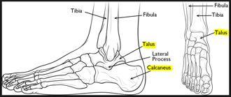
11 Lower limb Femur - Bone of thigh - Proximal end: head, neck, greater trochanter (lateral), lesser trochanter (posterior medial) - Middle part: shaft - Distal end: medial condyle, lateral condyle (condyles articulate with tibia condyles), epicondyles (areas above condyles), patellar surface (articulation with patella) Patella - A sesamoid bone (sesamoid bones are within tendons) - Within tendon of quadriceps femoris muscle (between 2 condyles anteriorly) Tibia - Large weight bearing medial bone of leg (bigger bone of leg Tibia (Tough), fibula (Fragile)) - proximal end: medial condyle, lateral condyle (condyles articulate with femur condyles), tibial tuberosity (attachment for patellar ligament) - middle part: shaft - distal end: medial malleolus (pointy part of ankle that we can feel articulate with talus at ankle joint), inferior articular surface for angle joint fibular - slender bone of leg - not part of knee joint but forms part of ankle joint - distal end: lateral malleolus (articulate with talus at ankle joint) ankle/tarsus - 7 tarsal bone, only need to identify the 2 below - Talus (other big bone other than calcaneus) - calcaneus (heel) metatarsals - bones of the foot - 5 metatarsals numbered 1-5 from medial to lateral (opposite to hand, big toe to little toe) Digits - Toes, comprised of phalanges - Digit 1: big toe, 2 phalanges (proximal and distal) - Digit 2-5: 3 phalanges (proximal, middle, distal) *lateral malleolus of fibula and medial malleolus of tibia form and arch to articulate with talus Page 11 of 15

12 Page 12 of 15
 Gomphosis (teeth) Syndesmosis (tibiofibular and radiulnar joints) Cartilaginous joints -")
 - Cavity contains synovial fluid - Examples: Non axial (plane) Uni axial (hinge pivot) Bi axial (condylar, saddle) Multiaxial (ball & socket) Shoulder girdle Gleno-humeral Articulating")

13 Articular system The type and range of joints are codependent. Fibrous joints - Immovable joints - Fibres connect articulating bones - Examples: Synathosis (sutures) Gomphosis (teeth) Syndesmosis (tibiofibular and radiulnar joints) Cartilaginous joints - Slightly moveable joints - Cartilage connect articulating bones - Examples: Synchondrosis (plate in humerus) Symphysis (pubic symphysis between 2 hip bones) Synovial joints - Freely moveable joints (most common) - Cavity contains synovial fluid - Examples: Non axial (plane) Uni axial (hinge pivot) Bi axial (condylar, saddle) Multiaxial (ball & socket) Shoulder girdle Gleno-humeral Articulating bones: glenoid fossa head of humerus. Both articulating surfaces covered in hyaline cartilage Joint type: synovia ball and socket Movements: flexion/extension; abduction/adduction; medial/lateral rotation; circumduction Articular capsule surrounds the joint Glenoid labrum fibrocartilage attached at the edges of glenoid cavity Coracoacromial arch coracoacromial ligament connects acromion and coracoid process. This forms a bridge and prevents superior displacement of humerus Subdeltoid bursa provides cushion around joint Tendon of Long head of biceps brachii inserts at top of glenoid fossa. Elongated bursa covers this tendon Page 13 of 15

 - glenoid labrum (increases depth of glenoid cavity) - tendon sheath and bursae (reduce friction) Knee joint Includes femur, tibia and patella (NOT fibula) Articulating")
14 Rotator cuff muscles originate at scapula and insert at head of humerus. They rotate and support the head of humerus at the shoulder joint. There is a shallow articular surface area of glenoid fossa and large articular surface of humeral head. This allows greater range of movement Stability of shoulder joint is due to: - coracormial arch (prevents superior displacement) - rotator cuff muscles (supports shoulder joint, tendons merge with joint capsule to reinforce it) - glenoid labrum (increases depth of glenoid cavity) - tendon sheath and bursae (reduce friction) Knee joint Includes femur, tibia and patella (NOT fibula) Articulating bones: medial and lateral condyles of femur, tibia, patella surface Type of joint: gliding joint, modified hinge joint Movements: extension/flexion; medial/lateral rotation Articular capsule Synovial membrane Hyaline cartilage medial collateral ligament (tibial collateral ligament) lateral collateral ligament (fibular collateral ligament) Collateral ligaments are extracapsular ligaments and prevent medial and lateral displacement. Page 14 of 15


15 anterior cruciate ligaments posterior cruciate ligaments Cruciate ligaments cross over Anterior cruciate ligament Originates from anterior part of tibia Prevents anterior displacement of tibia Attaches to lateral femoral condyle Posterior cruciate ligament Originates from posterior part of tibia Prevents posterior displacement of tibia Attaches to medial femoral condyle medial and lateral menisci attaches to tibia condyle. Similar to gleno labrum, it deepens surface Suprapatellar bursa above patella, deep to quadriceps tendon Patella protects the patella tendon, and holds it away from femur and tibia for greater angle and greater movement Other joints of limbs Elbow joint - Trochlear notch of ulna trochlea of humerus - head of radius humeral capitulum - flexion/extension proximal and distal radio-ulnar - radius pivots around ulna during pronation and supination wrist joint - distal radius carpal bones (Not ulna) hip joint - head of femur acetabulum of hip bone - much more stable that shoulder joint ankle joint - medial malleolus, lateral malleolus talus - dorsiflexion, plantarflexion, inversion, eversion Page 15 of 15
Chapter 7: Skeletal System: Gross Anatomy
 Chapter 7: Skeletal System: Gross Anatomy I. General Considerations A. How many bones in an average adult skeleton? B. Anatomic features of bones are based on II. Axial Skeleton A. Skull 1. Functionally
Biology 2401 The Skeletal System
 Biology 2401 The Skeletal System Purpose: The lab will describe the microscopic and gross anatomy of bone, identify bones of the body, and identify important bone markings. I. Overview of the Skeleton
Bone Flashcards for 10a
 Bone Flashcards for 0a CLAVICLE (collar bone). Sternal extremity (end) flat end. Acromial extremity (end) rounded end. SCAPULA (shoulder blade). Right or left scapula?. Superior border (superior margin).
The Appendicular Skeleton
 8 The Appendicular Skeleton PowerPoint Lecture Presentations prepared by Jason LaPres Lone Star College North Harris 8-1 The Pectoral Girdle The Pectoral Girdle Also called shoulder girdle Connects the
Bone List Anatomy
 1 Frontal Bone Skull 2 Parietal Bone Skull 3 Occipital Bone Skull 4 Temporal Bone Skull 5 Coronal Suture Skull 6 Sagittal Suture Skull 7 Squamous suture Skull 8 Lambdoid Suture Skull 9 Surpaorbital Ridge
Spring Written By: J. E. Sutton. Contents: I. Overview of the Skeleton: II. Appendicular Skeleton III. Axial Skeleton IV.
 Spring 2012 Written By: J. E. Sutton Contents: I. Overview of the Skeleton: II. Appendicular Skeleton III. Axial Skeleton IV. Articulations Overview of the Skeleton: I. Orientation to Human Skeleton: a.
11/25/2012. Chapter 7 Part 2: Bones! Skeletal Organization. The Skull. Skull Bones to Know Cranium
 Chapter 7 Part 2: Bones! 5) Distinguish between the axial and appendicular skeletons and name the major parts of each 6) Locate and identify the bones and the major features of the bones that compose the
Important Parts of Bones
 Important Parts of Bones For 2015 Know: Humerus (posterior) Clavical Femur (Anterior) Foot Hand Mandible Os Coxa Scapula Skull (Anterior, Inferior, Lateral) Sternum Humerus (posterior) A. olecranon fossa
BIOLOGY 113 LABORATORY Skeletal System
 BIOLOGY 113 LABORATORY Skeletal System Objectives Distinguish between the axial and appendicular skeleton. Distinguish between the cranium and facial skeleton. Locate and name the bones of the skull and
Perpendicular Plate Zygomatic Bone. Mental Foramen Mandible
 Glabella Frontal Middle Nasal Concha Nasal Lacrimal Perpendicular Plate Zygomatic Inferior Nasal Concha Maxilla Mental Mandible Skull (anterior view) Squamosal Suture Coronal Suture Frontal Parietal Nasal
Axial skeleton bones and markings
 Axial skeleton bones and markings Skull Cranial bones Frontal x 1 Supraorbital foramen Occipital x 1 Foramen magnum Occipital condyles Superior nuchal line Inferior nuchal line Anterior cranial fossa External
Pectoral (Shoulder) Girdle
 Girdle") Chapter 8 Skeletal System: Appendicular Skeleton Pectoral girdle Pelvic girdle Upper limbs Lower limbs 8-1 Pectoral (Shoulder) Girdle Consists of scapula and clavicle Clavicle articulates with sternum
Chapter 8 Skeletal System: Appendicular Skeleton Pectoral girdle Pelvic girdle Upper limbs Lower limbs 8-1 Pectoral (Shoulder) Girdle Consists of scapula and clavicle Clavicle articulates with sternum
bio4165 lab quiz 1 Posterior View Anterior View Lateral View Anterior View bio fall.quarter lab.quiz.1...page.1 of 6
 B A Posterior View D C E Lateral View bio.4165...fall.quarter.2005...lab.quiz.1...page.1 of 6 F I G 35 Posterior View H bio.4165...fall.quarter.2005...lab.quiz.1...page.2 of 6 J Posterior View L K Inferior
Exercise Science Section 2: The Skeletal System
 Exercise Science Section 2: The Skeletal System An Introduction to Health and Physical Education Ted Temertzoglou Paul Challen ISBN 1-55077-132-9 Role of the Skeleton Protection Framework Attachments for
CHAPTER 7, PART II (BONES)
") Anatomy Name: CHAPTER 7, PART II (BONES) Entry #: INSTRUCTIONS: 1) READ Chapter 7, pg. 140-161. 2) Using the outline, make a note card for each underlined bone name or phrase. 3) On each note card, put
Anatomy Name: CHAPTER 7, PART II (BONES) Entry #: INSTRUCTIONS: 1) READ Chapter 7, pg. 140-161. 2) Using the outline, make a note card for each underlined bone name or phrase. 3) On each note card, put
8.2: Fibrous Joints. There are three (3) types of fibrous joints (synarthroses): Syndesmosis Suture Gomphosis. Interosseus membrane of leg.
 types of fibrous joints (synarthroses): Syndesmosis Suture Gomphosis. Interosseus membrane of leg.") 8.1: Introduction Are known as articulations Functional junctions between bones Bind parts of skeletal system together Make bone growth possible Permit parts of the skeleton to change shape during childbirth
8.1: Introduction Are known as articulations Functional junctions between bones Bind parts of skeletal system together Make bone growth possible Permit parts of the skeleton to change shape during childbirth
Anatomy & Physiology Skeletal System Worksheet
 1. Name the five functions of the skeleton. c) d) e) Anatomy & Physiology Skeletal System Worksheet 2. The term for the shaft of a bone is:. 3. The bony struts found in spongy bone are called. 4. In ossification,
Chapter 8. The Appendicular Skeleton. Lecture Presentation by Lee Ann Frederick University of Texas at Arlington Pearson Education, Inc.
 Chapter 8 The Appendicular Skeleton Lecture Presentation by Lee Ann Frederick University of Texas at Arlington An Introduction to the Appendicular Skeleton The Appendicular Skeleton 126 bones Allows us
10/12/2010. Upper Extremity. Pectoral (Shoulder) Girdle. Clavicle (collarbone) Skeletal System: Appendicular Skeleton
 Girdle. Clavicle (collarbone) Skeletal System: Appendicular Skeleton") Skeletal System: Appendicular Skeleton Pectoral girdle Pelvic girdle Upper limbs Lower limbs 8-1 Pectoral (Shoulder) Girdle Consists of scapula and clavicle Clavicle articulates with sternum (Sternoclavicular
Skeletal System: Appendicular Skeleton Pectoral girdle Pelvic girdle Upper limbs Lower limbs 8-1 Pectoral (Shoulder) Girdle Consists of scapula and clavicle Clavicle articulates with sternum (Sternoclavicular
the Skeletal System provided by Academic Web Services Grand Canyon University
 Anatomy Resource Center Study Guides the Skeletal System HEAD & NECK REGIONAL VIEW SKULL BONES CRANIUM FACE SKULL LANDMARKS ANTERIOR SIDE SUPERIOR/INFERIOR VERTEBRAL COLUMN VERTEBRAL REGIONS CERVICAL C1
Dr.Israa H. Mohsen. Lecture 5. The vertebral column
 Anatomy Lecture 5 Dr.Israa H. Mohsen The vertebral column The vertebral column a flexible structure consisting of 33 vertebrae holds the head and torso upright, serves as an attachment point for the legs,
External Acoustic Meatus. Mastoid Process. Zygomatic Process. Temporal Bone
 Bone lab review 1. Frontal Bone 2. Supra-Orbital Foramen 3. Orbit (Orbital Cavity) 4. Superior Orbital Fissure 5. Inferior Orbital Fissure 6. Zygomatic Bone 7. Infra-Orbital Foramen 8. Maxilla 9. Mandible
Ch. 5 - Skeletal System
 Ch. 5 - Skeletal System Bones are living, ever-changing structures. This allows them grow and adapt to new situations that the body encounters. The functions of the skeletal system: 1) support bones are
Chapter 8B. The Skeletal System: Appendicular Skeleton. The Appendicular Skeleton. Clavicle. Pectoral (Shoulder) Girdle
 Girdle") The Appendicular Skeleton Chapter 8B The Skeletal System: Appendicular Skeleton 126 bones Pectoral (shoulder) girdle Pelvic (hip) girdle Upper limbs Lower limbs Functions primarily to facilitate movement
The Appendicular Skeleton Chapter 8B The Skeletal System: Appendicular Skeleton 126 bones Pectoral (shoulder) girdle Pelvic (hip) girdle Upper limbs Lower limbs Functions primarily to facilitate movement
Lab Exercise #04 The Skeletal System Student Performance Objectives
 Lab Exercise #04 The Skeletal System Student Performance Objectives The material that you are required to learn in this exercise can be found in either the lecture text or the supplemental materials provided
PRE-LAB EXERCISES. Before we get started, look up the definitions of these common bone marking terms: Canal: Condyle: Facet: Fissure:
 1 PRE-LAB EXERCISES When studying the skeletal system, the bones are often sorted into two broad categories: the axial skeleton and the appendicular skeleton. This lab focuses on the appendicular skeleton,
Copyright 2003 Pearson Education, Inc. publishing as Benjamin Cummings. Dr. Nabil khouri
 Dr. Nabil khouri Appendicular Skeleton The appendicular skeleton is made up of the bones of the upper and lower limbs and their girdles Two girdles: Pectoral girdles attach the upper limbs to the body
Biology 218 Human Anatomy
 Chapter 8 Adapted from Tortora 10 th ed. LECTURE OUTLINE A. Introduction (p. 203) 1. The appendicular skeleton contains 126 bones that form: i. two pectoral (shoulder) girdles two upper limbs i one pelvic
Biology 218 Human Anatomy. Adapted from Martini Human Anatomy 7th ed. Chapter 7 The Skeletal System Appendicular Division
 Adapted from Martini Human Anatomy 7th ed. Chapter 7 The Skeletal System Appendicular Division Introduction The appendicular skeleton includes: Pectoral girdle Shoulder bones Upper limbs Pelvic girdle
Riverside Community College Anatomy & Physiology 2B SPRING 2012 EXAM #1-ABC (Nervous System)
") Riverside Community College Anatomy & Physiology 2B SPRING 2012 EXAM #1-ABC (Nervous System) Name: 1) This vertebra is an example of a(n). 1) A) thoracic B) axis C) atlas D) lumbar E) sacral 1 2) W hich
Riverside Community College Anatomy & Physiology 2B SPRING 2012 EXAM #1-ABC (Nervous System) Name: 1) This vertebra is an example of a(n). 1) A) thoracic B) axis C) atlas D) lumbar E) sacral 1 2) W hich
Amy Warenda Czura, Ph.D. 1 SCCC BIO130 Lab 7 Appendicular Skeleton & Articulations
 The Skeletal System II: Appendicular Skeleton and Articulations Exercises 11, 13 (begins: page 145 in 9 th and 10 th editions) Exercises 10, 11 (begins: page 147 in 11 th edition, page 149 in 12 th edition)
Lab Activity 9. Appendicular Skeleton Martini Chapter 8. Portland Community College BI 231
 Lab Activity 9 Appendicular Skeleton Martini Chapter 8 Portland Community College BI 231 Appendicular Skeleton Upper & Lower extremities Shoulder Girdle Pelvic Girdle 2 Humerus 3 Humerus: Proximal End
Anatomy. Anatomy deals with the structure of the human body, and includes a precise language on body positions and relationships between body parts.
 Anatomy deals with the structure of the human body, and includes a precise language on body positions and relationships between body parts. Proper instruction on safe and efficient exercise technique requires
Principles of Anatomy and Physiology
 Principles of Anatomy and Physiology 14 th Edition CHAPTER 8 The Skeletal System: The Appendicular Skeleton The Appendicular Skeleton The 126 bones of the appendicular skeleton are primarily concerned
Chapter 7: Skeletal System
 Chapter 7: Skeletal System The Skeletal System Introduction P. 182 Bone is an organ made up of tissues: It is made up of the following components. Cartilage Blood Nerves Bone Connective Bone Classification
Bio 5/6 5 The Skeletal System Study Guide
 Name: THE SKELETAL SYSTEM: 5 The Skeletal System Study Guide Period: The skeleton is constructed of two of the most supportive tissues found in the human body - cartilage and bone. Besides supporting and
Skeletal System - Prelab 1
 Skeletal System - Prelab 1 1. Which bones contain the paranasal sinuses? What function do the sinuses serve? 2. What two areas are separated from each other by the hard palate? Name the two bones that
Skeletal System. Supplementary Information
 Skeletal System Supplementary Information COMMON ANATOMICAL TERMS Planes run through the body side to side and front to back eg. median plane Surfaces of the body are also named eg. anterior surface This
Overview of the Skeleton: Bone Markings
 Name Overview of the Skeleton: Bone Markings Match the terms in column B with the appropriate description in column A. Column A 1. sharp, slender process* 2. small rounded projection* 3. narrow ridge of
TEST YOURSELF- Chapter 7
 TEST YOURSELF- Chapter 7 Cranial Bones 1. Give the name of the bone for each of the following markings. Some of the markings are found on more than one bone. List all that apply. Cranium a. Frontal squama:
Lab 6, 7, 8: Skeletal System
 107 Lab 6, 7, 8: Skeletal System Adult Skull Bony orbit (FLEZMS) Frontal bone supraorbital foramen frontal sinus Lacrimal bone Ethmoid bone perpendicular plate of ethmoid middle nasal conchae cribriform
SKELETON FUNCTIONS OF BONE:
 SKELETON FUNCTIONS OF BONE: SKELETON: 1. Performs a mechanical function in forming the skeletal support of the body and in forming a leverage system whereby work and movement are possible. 2. Serves as
Bones of Thorax (Rib Cage)
") Musculoskeletal System (Part A-2) Module 7 -Chapter 10 Overview Muscles Attachments Bones Bone types Surface features of bones Divisions of the skeletal system Joints or Articulations Susie Turner, M.D.
Musculoskeletal System (Part A-2) Module 7 -Chapter 10 Overview Muscles Attachments Bones Bone types Surface features of bones Divisions of the skeletal system Joints or Articulations Susie Turner, M.D.
The Skeletal System THE APPENDICULAR SKELETON
 The Skeletal System THE APPENDICULAR SKELETON The appendicular skeleton consists of the girdles and the skeleton of the limbs. The upper (anterior) limbs are attached to the pectoral (shoulder) girdle
BLUE SKY SCHOOL OF PROFESSIONAL MASSAGE AND THERAPEUTIC BODYWORK. Musculoskeletal Anatomy & Kinesiology I TERMINOLOGY, STRUCTURES, & SKELETAL OVERVIEW
 BLUE SKY SCHOOL OF PROFESSIONAL MASSAGE AND THERAPEUTIC BODYWORK Musculoskeletal Anatomy & Kinesiology I TERMINOLOGY, STRUCTURES, & SKELETAL OVERVIEW MSAK101-I Session 1 Learning Objectives: 1. Define
Bone Composition. Bone is very strong for its relatively light weight The major components of bone are:
 Human Bones Bone Composition Bone is very strong for its relatively light weight The major components of bone are: Calcium carbonate Calcium phosphate Collagen Water Cortical Bone Spongy Bone Medullary
The Appendicular Skeleton
 8 The Appendicular Skeleton PowerPoint Lecture Presentations prepared by Jason LaPres Lone Star College North Harris An Introduction to the Appendicular Skeleton Learning Outcomes 8-1 Identify the bones
BIO 137 AXIAL SKELETON BONE STUDY THE HUMAN SKELETON
 BIO 137 THE AXIAL SKELETON MARY CATHERINE FLATH, Ph.D. THE HUMAN SKELETON AXIAL SKULL HYOID THORACIC CAGE VERTEBRAL COLUMN APPENDICULAR PECTORAL GIRDLE UPPER LIMBS PELVIC GIRDLE LOWER LIMBS AXIAL SKELETON
Biology 152 Appendicular Skeleton Anatomy Objectives
 Biology 152 Appendicular Skeleton Anatomy Objectives We will learn proper bone names, left/right/medial, and the parts of bones in this exercise. Start by learning the names of the bones. As you gain comfort
BONE CHALLENGE DANIL HAMMOUDI.MD
 BONE CHALLENGE DANIL HAMMOUDI.MD Bone Basic functions? A. support B. protection C. movement assistance in D. RBC formation-hemopoiesis E. mineral homeostasis +importance of calcium F. energy supply -yellow
Axial Skeleton BONE TERMINOLOGY FEATURES
 Axial Skeleton BONE TERMINOLOGY FEATURES Tuberosity Rounded area on bone often roughened for muscle attachment. Tubercle Rounded projection on bone. This is called a tuberosity on the femur. Crest Ridgeline
Chapter 8 The Skeletal System: The Appendicular Skeleton. Copyright 2009 John Wiley & Sons, Inc.
 Chapter 8 The Skeletal System: The Appendicular Skeleton Appendicular Skeleton The primary function is movement It includes bones of the upper and lower limbs Girdles attach the limbs to the axial skeleton
Chapter 8 The Skeletal System: The Appendicular Skeleton. Copyright 2009 John Wiley & Sons, Inc.
 Chapter 8 The Skeletal System: The Appendicular Skeleton Appendicular Skeleton It includes bones of the upper and lower limbs Girdles attach the limbs to the axial skeleton The pectoral girdle consists
Skeletal System A&P Week 11
 Skeletal System A&P Week 11 Bones 206 bones in the body Smallest are in the middle ear cavity Malleus, incus, stapes Functions of Bone Framework and support Protection Contains/protects red bone marrow
SKELETAL SYSTEM 206. AXIAL SKELETON 80 APPENDICULAR SKELETON 126 (see Figure 6.1) Clavicle. Clavicle. Pectoral girdles. Scapula. Scapula.
 Clavicle. Clavicle. Pectoral girdles. Scapula. Scapula.") SKELETAL SYSTEM 206 AXIAL SKELETON 80 APPENDICULAR SKELETON 126 (see Figure 6.1) Pectoral girdles 4 Clavicle Scapula 2 2 Clavicle Scapula Humerus 2 Humerus Upper limbs 60 Radius 2 Ulna Carpal bones Metacarpal
SKELETAL SYSTEM 206 AXIAL SKELETON 80 APPENDICULAR SKELETON 126 (see Figure 6.1) Pectoral girdles 4 Clavicle Scapula 2 2 Clavicle Scapula Humerus 2 Humerus Upper limbs 60 Radius 2 Ulna Carpal bones Metacarpal
Joints Dr. Ali Ebneshahidi
 Joints Dr. Ali Ebneshahidi Function of Joints 1. Serve as functional junctions between bones. 2. Bind bones, strokes, and other related tissues together. 3. Allow bone growth to occur. 4. Permit certain
Human Anatomy - Problem Drill 06: The Skeletal System Axial Skeleton & Articualtions
 Human Anatomy - Problem Drill 06: The Skeletal System Axial Skeleton & Articualtions Question No. 1 of 10 Instructions: (1) Read the problem and answer choices carefully, (2) Work the problems on paper
The Skeletal System: Axial Skeleton
 The Skeletal System: Axial Skeleton The Big Idea The Axial Skeleton & Homeostasis The bones of the axial skeleton contribute to homeostasis by protecting many of the body s organs such as the brain, spinal
CLASSIFICATION OF JOINTS STRUCTURAL VS FUNCTIONAL
 CHAPTER 8 JOINTS CLASSIFICATION OF JOINTS STRUCTURAL VS FUNCTIONAL The most moveable type of joint is a 1) Synarthrosis 2) Amphiarthrosis 3) Diarthrosis FIBROUS JOINTS Figure 8.1 Fibrous joints. (a) Suture
NOTES SKELETAL SYSTEM
 NOTES for the SKELETAL SYSTEM Anatomy & Physiology 2016 Johnson The Skeletal System I. System includes 4 basic parts: A. Bones (206 of em) B. Joints C. Cartilages D. Ligaments II. Bones have 5 basic functions:
TEST BANK FOR THE HUMAN BODY IN HEALTH AND ILLNESS 5TH EDITION BY BARBARA HERLIHY Chapter 8: Skeletal System
 Link download Full : http://testbankair.com/download/test-bank-for-thehuman-body-in-health-and-illness-5th-edition-by-barbara-herlihy/ TEST BANK FOR THE HUMAN BODY IN HEALTH AND ILLNESS 5TH EDITION BY
Chapter 7 Skeletal System. Skeletal System: Bone Functions: Describe the role the skeletal system plays in each of the following functions.
 Chapter 7 Skeletal System Skeletal System: Bone Functions: Describe the role the skeletal system plays in each of the following functions. support protection muscle attachment - movement blood production
UNIT 4 - SKELETAL SYSTEM LECTURE NOTES
 UNIT 4 - SKELETAL SYSTEM LECTURE NOTES 4.01 FUNCTIONS OF THE SKELETAL SYSTEM A. Support 1. Provides a framework for the body 2. Supports soft tissue 3. Serves as a point of attachment for ligaments, tendons,
Appendicular Skeleton. Prof. Abdulameer Al-Nuaimi
 Appendicular Skeleton Prof. Abdulameer Al-Nuaimi a.alnuaimi@sheffield.ac.uk abdulameerh@yahoo.com Hi Prof, It is great to hear from you, I really enjoyed your teaching last year. You taught me the hardest
The skeletal system is the framework for the muscular system to attach to so we can move.
 Skeletal System The skeletal system is the framework for the muscular system to attach to so we can move. BONE: A rigid connective tissue Helps to move & support the body Protect the organs (skull, ribs)
Lab Unit One Flashcards
 CLAVICLE (collar bone). Sternal extremity (end) flat end. Acromial extremity (end) rounded end.. Conoid tubercle near round end SCAPULA (shoulder blade). Right or left scapula?. Superior border (superior
Skeletal system. Prof. Abdulameer Al-Nuaimi. E. mail:
 Skeletal system Prof. Abdulameer Al-Nuaimi E-mail: a.al-nuaimi@sheffield.ac.uk E. mail: abdulameerh@yahoo.com Functions of Bone and The Skeletal System Support: The skeleton serves as the structural framework
Figure ) The area that causes the lengthwise growth of a long bone is indicated by letter. Diff: 2 Page Ref:
 The area that causes the lengthwise growth of a long bone is indicated by letter. Diff: 2 Page Ref:") Essentials of Anatomy and Physiology, 9e (Marieb) Chapter 5 The Skeletal System Short Answer Figure 5.1 Using Figure 5.1, identify the following: 1) Spongy bone is indicated by letter. Diff: 1 Page Ref:
Essentials of Anatomy and Physiology, 9e (Marieb) Chapter 5 The Skeletal System Short Answer Figure 5.1 Using Figure 5.1, identify the following: 1) Spongy bone is indicated by letter. Diff: 1 Page Ref:
Labs 6, 7, 8: Skeletal System
 153 Labs 6, 7, 8: Skeletal System Unit 6: Skeletal System: Bone tissue, Bones and Joints (p. 105-152) Ex. 6-1: Histology of Osseous Tissue, p. 113 Model: Osteon Tiss Lamella Osteocyte Lacunae Canaliculi
Nervous & Skeletal Systems. Virtual Science University
 Nervous & Skeletal Systems Virtual Science University 1 Nervous & Skeletal Systems Texas TEK B.10(A) The student will interpret the function of systems in organisms (humans) including the nervous and skeletal
The Skeletal System. Mosby items and derived items 2010, 2006, 2002, 1997, 1992 by Mosby, Inc., an affiliate of Elsevier Inc.
 The Skeletal System Functions of Skeletal System Provides internal framework that supports the body Protects internal organs Helps fight disease by producing white blood cells 2 Functions of Skeletal System
Cornell Notes Name: Date: Topic: CH 5. Subject: The Skeletal System
 Cornell Notes Name: Date: Topic: CH 5 Questions/Main Ideas: Record Notes: We are revisiting Ch 3B on Connective Tissue prior to our study of Ch 5 Skeletal start on p.91-95 I. Types of Connective A. Bone
Connects arm to thorax 3 joints. Glenohumeral joint Acromioclavicular joint Sternoclavicular joint
 Connects arm to thorax 3 joints Glenohumeral joint Acromioclavicular joint Sternoclavicular joint Scapula Elevation Depression Protraction (abduction) Retraction (adduction) Downward Rotation Upward Rotation
Chapter 5 The Skeletal System
 Chapter 5 The Skeletal System The Skeletal System Parts of the skeletal system Bones (skeleton) Joints Cartilages Ligaments (bone to bone)(tendon=bone to muscle) Divided into two divisions Axial skeleton:
Introduction. Skeletal Nomenclature. Lesson One: Bones of the Upper Extremity. Shoulder Girdle: Clavicle. Scapula: Bony Markings
 Chapter 14 Skeletal Nomenclature Susan G. Salvo Bones help locate muscles Can also be areas to avoid Chapter divided into 5 lessons covering bones, bony markings, and joints Introduction 1 2 Lesson One:
Bio 103 Skeletal System 45
 45 Lecture Outline: SKELETAL SYSTEM [Chapters 7, 8] Introduction A. Components B. Functions 1. 2. 3. 4. Classification and Parts A. Bone Shapes 1. Long: 2. Short: 3. Flat: 4. Irregular: 5. Sesamoid: B.
Sports Medicine Part I : ANATOMY OF THE SPINE, ABDOMEN AND SHOULDER COMPLEX
 Sports Medicine 25 1.1 Part I : ANATOMY OF THE SPINE, ABDOMEN AND SHOULDER COMPLEX c.w.p. Wagner High School, Sports Medicine, A. Morgan, T. Morgan 2008 Anatomy of the Upper Body In this section of the
Human Skeletal System Glossary
 Acromegaly Apatite Acromegaly - is a condition which involves excessive growth of the jaw, hands, and feet. It results from overproduction of somatotropin in adults (after fusion of the ossification centres
Skeletal System. Skeleton. Support. Function of Bones. Movement. Protection 10/15/12
 Skeleton Skeletal System 1 Axial Skeleton-Bones that form the longitudinal axis of the body (skull and spinal column). Appendicular Skeleton-Bones of the limbs and girdles. Also include joints, ligaments
Classification of bones
 Classification of bones compact intramembranous axial histology development regional spongy Intra cartilaginous appendicular flat Irregular shape Sesamoid Long Short Wormian pneumatic Classification
Skeletal system overview. Classification of Bones
 Skeletal system overview BIOL241 Lab #9 Classification of Bones Bone are identified by: shape internal tissues bone markings 1 1. Flat bones 2. Long bones 3. Short bones 4. Irregular bones 5. Sutural bones
I. Introduction. Unit Two. of the Skeletal System. II. Classification of Joints. URLs for this chapter:
 8 URLs for this chapter: http://www.vh.org/adult/provider/radiology/joint Fluoro/JointFluoroHP.html of the Skeletal System Karen Webb Smith Unit Two http://www.science.ubc.ca/~biomania/tutorial/bonejt/
Labs 9 and 10. Classification of Bones. Bone Shapes 1/05/13. Skeletal system overview. Bone are identified by:
 Labs 9 and 10 Skeletal system overview Classification of Bones Bone are identified by: shape internal tissues bone markings 1. Flat bones 2. Long bones 3. Short bones 4. Irregular bones 5. Sutural bones
Copyright 2010 Pearson Education, Inc.
 E. VERTEBRAL COLUMN 1. The vertebral column extends from the skull to the pelvis and forms the vertical axis of the skeleton. 2. The vertebral column is composed of vertebrae that are separated by intervertebral
APPENDICULAR SKELETON 126 AXIAL SKELETON SKELETAL SYSTEM. Cranium. Skull. Face. Skull and associated bones. Auditory ossicles. Associated bones.
 SKELETAL SYSTEM 206 AXIAL SKELETON 80 APPENDICULAR SKELETON 26 Skull Skull and associated s 29 Cranium Face Auditory ossicles 8 4 6 Associated s Hyoid Thoracic cage 25 Sternum Ribs 24 Vertebrae 24 column
UNIT 2 - CHAPTER 8: JOINTS OF THE SKELETAL SYSTEM LEARNING OUTCOMES:
 LEARNING OUTCOMES: 8.1 Types of Joints 1. Explain how joints can be classified according to the type of tissue that binds the bones together and the degree of movement possible at the joint. (p. 268) 2.
Hole s Human Anatomy and Physiology
 Hole s Human Anatomy and Physiology 1 Chapter 7 Skeletal System Bone Classification Long Bones Short Bones Flat Bones Irregular Bones Sesamoid (Round) Bones 2 Parts of a Long Bone epiphysis distal proximal
Exercise 13. Articulations and Body Movements
 Exercise 13 Articulations and Body Movements Articulations Articulations, or joints, are points where a bone is connected to one or more other bones. Articulations hold the skeleton together. Articulations
Anatomy and Physiology 2016
 Anatomy and Physiology 2016 O = Temporal line I = coronoid process (Mandible) A = elevates mandible (chewing) O = galea aponeurotica (layer of dense fibrous tissue which covers the upper part of the cranium)
UNIT 2 - CHAPTER 8: JOINTS OF THE SKELETAL SYSTEM LEARNING OUTCOMES:
 LEARNING OUTCOMES: 8.1 Introduction 1. List the functions of joints. 2. Explain how joints can be classified according to the type of tissue that binds the bones together and the degree of movement possible
Exercise 11. The Appendicular Skeleton
 Exercise 11 The Appendicular Skeleton The Appendicular Skeleton The appendicular skeleton contains 126 bones. Consists of the upper and lower limbs, the pectoral girdles, and the pelvic girdles. The pectoral
Skeletal Considerations for Movement. Kinesiology RHS 341 Lecture 2 Dr. Einas Al-Eisa
 Skeletal Considerations for Movement Kinesiology RHS 341 Lecture 2 Dr. Einas Al-Eisa The Skeletal System Bones, cartilage, ligaments, & joints Consists of approximately 20% of total body weight Bone constitutes
Skeletal System. Chapter 7.1. Objective- Read 7.1 and understand that bones are alive and multifunctional. Introduction:
 Chapter 7.1 Skeletal System Objective- Read 7.1 and understand that bones are alive and multifunctional. Introduction: A. Bones are very active tissues B. Each bone is made up of several types of tissues
In-Depth Foundations: Anatomy Terms to Know
 Be familiar with / able to identify and define all the following parts. The Spine Cranium Vertebrae Cervical, Thoracic, Lumbar Sacrum Coccyx Bones of Upper Body Cranium Mastoid process; Occipital condyle,
Introduction. Fibrous Joints. 8.1: Types of Joints. Cartilaginous Joints. Fibrous Joints 12/14/2016. Chapter 08 Lecture Outline
 Introduction Chapter 08 Lecture Outline See separate PowerPoint slides for all figures and tables preinserted into PowerPoint without notes. Joints (Articulations): Functional junctions between bones Bind
The skeleton consists of: Bones: special connective tissue, hard. Cartilage: special connective tissue, less hard than bones. Joints: joint is the
 The skeleton consists of: Bones: special connective tissue, hard. Cartilage: special connective tissue, less hard than bones. Joints: joint is the location at witch two bones make contact, whereas ligaments
PowerPoint Lecture Slides prepared by Janice Meeking, Mount Royal College C H A P T E R. Joints: Part A. Copyright 2010 Pearson Education, Inc.
 PowerPoint Lecture Slides prepared by Janice Meeking, Mount Royal College C H A P T E R 8 Joints: Part A Warm Up 11/28/16 Happy Thanksgiving welcome back! J (be ready to share something fun you did over
2 skull, vertebral column, thoracic cage
 CHAPTER 7-SKELTON FILL-IN NOTES 2 skull, vertebral column, thoracic cage 3 Fig. 7.1 pg. 199 4 I. Skull: A. : Encloses and the brain - 8 bones B. : 14 bones Cranium A. Forehead (brain) Anterior part of
Copyright 2004 Lippincott Williams & Wilkins. 2. Bone Structure. Copyright 2004 Lippincott Williams & Wilkins
 Chapter 7 The Skeleton: Bones and Joints The Skeleton Skeletal system is made up of bones and joints and supporting connective tissue. 1. Bone Functions 1. To store calcium salts 2. To protect delicate