Cervical Spine Disorder in Children
|
|
|
- Jessie Harvey
- 5 years ago
- Views:
Transcription
1 Cervical Spine Disorder in Children

2 Embryology of Vertebra Para axial Mesoderm 42~44, 42~44 somite 4 occipital somite 8 cervical somite 12 thoracic somite 5 lumbar somite 10 sacral somite 5 coccyx somite Ventral part sclerotome Dorsomedial myotome Dorsolateral dermatome
3 Atlas, odontoid, axis 4 occiput somite 8 cervical somite 1 st 2 nd 3 rd 4 th st occiput somite nd occiput somite rd occiput somite th occiput somite Occipital bone & Foramen magnum 4 occiput somite proatlas 4 th occiput somite atlas lat mass, ring odontoid tip Odontoid from C1 Axis body from C2
4 Atlas, odontoid, axis C1 (atlas) Lateral mass ring from 4 th occiput somite Atlas ring from C1 somite C2 (axis) Odontoid tip from 4 th occiput somite Odontoid body from C1 somite C2 body from C2 somite

5 Atlas, odontoid, axis 4 th occiput somite 4 th occiput somite C2 somite C1 somite C1 somite C2 somite C2
6 Odontoid Anomalies
7 Odontoid anomaly / Lig instability Spondyloepiphyseal dysplasia Morquio disease(mucopolysaccharidosis) Down syndrome Osteogenesis imperfecta Neurofibromatosis


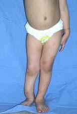
8 Spondyloepiphyseal dysplasia Extension Flexion


9 Neurofibromatosis
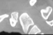
10 Os Odontoideum : Separation of dens from C2 body Frequency : rare, but exact frequency unknown Common in Morquio, MED, Down synd Symptoms Local mechanical neck pain Neurologic symptoms Neurovascular symptoms vertebral a compression cervical & brainstem ischemia gait ataxia, syncope, vertigo, and visual disturbances, brainstem infarcts & seizures
11 Os Odontoideum
12 Etiology: Controversy, vs - Cong failure of fusion of dens & body persistent ossiculum terminale Ossicle much smaller, at the level of C1 Associated with Klippel Feil syndrome, occipitalized atlas, or basilar invagination
13 persistent ossiculum terminale 4 th occiput somite 4~6 Fusion at age 12yrs C2 somite C1 somite C2
14 - Fracture or excessive movement of odontoid synchondrosis before closure age of 5-6 years pulling of fragment by alar lig, Non-union prox part of fragment- circulation(+) by alar lig, distal part- AVN(+) resorbed &rounded 2 types of os odontoideum orthotopic os odontoideum : anatomic position dystopic os odontoideum : non-anatomic position
 Ischemic & resoption Fusion 3~6yrs C2 somite Fx at")
15 Etiology - Os Odontoideum 4 th occiput somite retraction by apical lig C2 somite C1 somite Circulation(+) Ischemic & resoption Fusion 3~6yrs C2 somite Fx at synchondrosis


16 orthotopic os odontoideum dystopic os odontoideum

17 Odontoid ossification Grows & ossifies until age 18 Tip of dens reach upper margin of arch of C1 at age 9 (easily mistaken for hypoplasia)
18 Indications for surgery Significant Instability Neurologic or neurovascular involvement Persistent and disabling pain Progressive instability, SAC < 13mm


19 Instability index (a-b/a) : 53% Extension A a b Flexion B


20 Cervical decompression and fusion
21 Atlanto-Occipital fusion


22 Atlanto-occipital fusion CVJ 0.14 ~ 2.76%. 38% Chiari malformation Basilar impression : common
23 Atlanto-occipital fusion (Occipitalization of Atlas) Clinical findings : short neck, low hairline, Instability(+) in 50% Sxs: : neck pain, neurology limited neck motion Tx : Myelopathy(-), instability(-) no Tx Myelopathy(+),instability(+) fusion
24 Klippel - Feil syndrome

25 Klippel-Feil syndrome : Cong fusion of cervical vertebra Failure of segmentation of somite Clinical Triad : Short, webbed neck Low hair line Restricted neck motion In practice,, any failure of segmentation in C-spine C is called Klippel-Feil syndrome
26 Associated Anomalies Facial abnomaly, webbing, torticollis (26%) Sprengel s deformity (50%) Scoliosis (60%) Renal abnormalities (35%) cause of Death Deafness (15~35%) Congenital heart disease (14~29%) Synkinesia, mirror movement (20%)
27 Klippel-Feil syndrome Renal anomalies Spinal cord neurology MRI Hypermobility degeneration canal stenosis Basilar impression from occiput-cervical anomay Instability & Neurology
28 Klippel-Feil synd Head tilt or rotation Halo immobilzation Surgical fusion progression of deformity rigid head tilt or rotation severe neck pain neurologic deficit occipitocervical instability with neurology
29 Cervical Spine in Down syndrome
30 Cervical anomalies in Down synd (Trisomy 21) Atlantoaxial instability due to lig laxity Occipitalization of atlas Hypoplasia of post arch of C1 Os Odontoideum
31 Incidence of instability : 10~25% But, most children are asymptomatic Symptom(+) among instabilities : variable (0~18%),. :.
32 High failure rates of fusion & resorption (d/t collagen defect & T-cell immune deficiency) Less likelihood of improvement of neurology Low rates of neurology with variable Sxs (tingling sense ~ Sudden death in normal pts) Development of neurology with increasing AGE Preventive fusion need? All needs fusion? When to fuse?
33 Treatment guidelines Myelopathy(+) needs fusion Marked instability(+), myelopathy(-) fusion Instability(+), neurology(-) no surgery (segal,1991)
34 Guidelines for Down s syndrome American Academy of Pediatrics, Competitive high risk sports instability. 2.
35 Atlantoaxial rotatory subluxation
36 Instability Developmental anomaly Trauma Inflammation of adjacent neck tissues URI, Grisel synd, Adenitis, Ear infection X-ray evaluation AP, Lat view Open mouth view Flexion & Extension lat view (!)

37 Fielding & Hawkins classification
38 Tx of atlantoaxial rotatory sublux (Philips, Hensinger 1989) -, < 1 wk, : soft collar & rest < 1~4wks : Halo traction, firm color 6wks > 4 wks : Halo traction, Halo vest 6wks Failed reduction CR under G/A or Arthrodesis C1-C2 dislocation on visit Arthrodesis


39


40 Atlantoaxial rotary subluxation (type II M / 17 C.C. : Torticollis (for 3mo.) Trauma(-), URI(+) Type II
 for 3 months Fielding")

41 Atlantoaxial rotary subluxation (type III) Atlantoaxial rotary subluxation for 3 months Fielding & Hawkins : Type III Traction & fusion
42
43 (, X-ray) (SCIWORA- spinal cord injury without radiologic abnormalities) Occiput-C2 (8~10 ) 70% of cervical injuries Ligament laxity, hypermobility, more horizontal facets Large head size fulcrum at C3 Breech delivery, Child abuse, shaking Motor vehicle, fall down, athetic injury
44 SCIWORA (spinal cord injury without radiologic abnormalities) Unique to children under 8yrs 7-66% of pts. with c-spine inj. Sp. column elastic limit : 2 inches Sp. cord elastic limit : 1/4 inches
45 Facial abrasions, head trauma, clavicle fx, High speed motor vehicle injury fall from height X-ray Cervical AP, Lat, both oblique, open mouth view flexion-extension lat view
46 (x) (o) (o).
47 X-ray evaluation - cervical alignment Spinolaminar line of Swischuk C mm. Ant vertebral body line pseudosubluxation 8, 4mm Prevertebral soft tissue swelling 5~6mm at C2 level Post interspinous process distance Cervical lordosis


48 Pseudosubluxation Ligamentous laxity Luscha jt underdevelopment Shallow, horizontal facet jt 4mm
 -4mm SAC(Space available for cord) -13mm Power s ratio- 1.")
49 X-ray evaluation Atlantoaxial instability C1/C2 10 ADI (Atlanto-dens interval) -4mm SAC(Space available for cord) -13mm Power s ratio- 1.0 BC / AO=1.0
50 Atlantoccipital Instability Down syndrome Juvenile rheumatoid arthritis Larsen s synd Spondyloepiphyseal dysplasia Os odontoideum Hypochondroplasia Achondroplasia
51 X-ray evaluation Basilar Impression Primary Basilar impression Klippel-Feil synd Atlas occipitalization Odontoid or atlas anomaly Secondary Basilar impression Skull base softening dz RA, OI, Paget dz, Morquio Renal dystrophy
52 33 mm 28 mm Best screening line with high reproducerbility
53 Acknowledgement
54 .
55

56

57
58
59

60
61 Cervical spine problems in Down s syndrome Due to generalized lig. Laxity C0-C1 instability : 15/33 C1-C2 instability : 22/33 Most children are asymptomatic Symptomatic in 2.6% (UMN signs)
62 CVJ problems in Down Lig. Laxity bony anomalies CVJ. C0-C1 & C1-C2 instabilities, rotary luxations, Os odontoideum(10/33) C1-C2 up to 40% < 1%, C0-C1 instability 66%. High rates of nonunion & infection with long fusion (d/t collagen defect & deficient T-cell immune system)


63 Basilar impression Most comm. anomaly of upper c-spine Sx. onset : 3rd to 5th decade Neck pain, Myelopathy Platybasia : basal angle > 140 Midsagittal diameter of F. Magnum < 19 mm
64 Cervical spine affections Several anomalies coexist Coexisting other organ anomalies Upper(C0-C2) & lower C-lesions Craniovertebral junction anomalies

65


66

67 Clinical manifestations Pain Torticollis Weakness Sensory disturbance Gait difficulties Incoordination
68 D.Dx of Torticollis Nonosseus - congenital muscular - Sandifer s syndrome - neurogenic - inflammatory Osseus - basilar impression - atlanto-occipital anomalies - unilateral absence of C1 - familial cervical dysplasia - atlantoaxial rotary displacement - neoplasms

69
70 Clinical manifestations Mechanical (direct compression, traction) Vascular (ischemia, infarction) Alterations in CSF (hydroceph., syrinx) Instability, deformity, myelopathy
71 Odontoid anomalies Neurol. involvement (even transient) Flex-Ext view instability > 10 mm SAC < 13 mm Progressive instability Persistent neck complaints
72

73 Os odontoideum Neurocentral sychondrosis failure Nonunion d/t unrecognized Fx. Retraction by alar, apical ligament Orthotopic or dystopic position Pain, torticollis, N. compromise
74 Klippel - Feil syndrome C-spine fusion abnormality : Tip of Iceberg Prognostic factors : Associated anomalies Instability of adjacent segments
75 Klippel - Feil syndrome Require the skills of a detective to unravel the full syndrome Hidden features may jeopardize the pt s health more than the anomalous fusion itself
 1 : 2 : 3 : 2 sets of block vertebrae with open intervening disc space Craniocervical")
76 Nagib s at-risk groups (J. Neurosurg., 1984) 1 : 2 : 3 : 2 sets of block vertebrae with open intervening disc space Craniocervical anomalies with fusion below C2 Associated with canal stenosis
77 occiput C1 C2 C3 C4 C5 C6 C7 T1 T2
78 Atlantoaxial instability C1-C2 is the most mobile joint in Cx. spine (50% of rotation occurs) > 10 > 4.5mm ADI > 4 mm SAC < 13 mm
79 Apex erosion Routine screening Abnormal : 33 mm, 28 mm
80 Chamberlain s line McGregor s line Wackenheim s line : Easiest method

81

82
 F : Digastric line (mid-oa joint")
83 E : Bimastoid line (within 3mm ~ +10 mm) F : Digastric line (mid-oa joint 10mm )


84


85


86


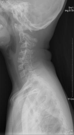
87 M/5, L/E paraparesis, minor fall Os odontoideum Neutral Flexion Extension


88 Atlanto-occipital instability More than 1mm translation on flexion-extension x-ray


89
90 Odontoid anomalies Mechanical & neurological Sx Down s syndrome Spondyloepiphyseal dysplasia Mucopolysaccharidoses

")
91 Os odotoideum in 3/11patients -- atlantoaxial instability during cervical motion -- Instability index 36%, 46%, 53%(cervical myelopathy) Extension Flexion
Cranial Vertebral Junction Anatomy and Pathology
 Cranial Vertebral Junction Anatomy and Pathology October 2016 Mary Scanlon MD FACR Goals and Objective Goal-Understand CVJ anatomy & pathology Objective-After attending this lecture you will be able to
PEDIATRIC CERVICAL SPINE DEFORMITY.
 PEDIATRIC CERVICAL SPINE DEFORMITY www.fisiokinesiterapia.biz DEVELOPMENT EACH VERTEBRAE DEVELOP FROM THE CAUDAL AND CRANIAL ½ OF 2 SCLEROTOMES C1 and C2 primitive centrum fuse to form odontoid process
Musculoskeletal Development and Sports Injuries in Pediatric Patients
 Dynamic Chiropractic October 21, 2010, Vol. 28, Issue 22 Musculoskeletal Development and Sports Injuries in Pediatric Patients By Deborah Pate, DC, DACBR Physical activity is extremely important for everyone,
May have excessive movement in the unfused segment to compensate. Flexion extension better preserved than lateral bend or rotation
 IV CONGENITAL SPINE KLIPPEL FLAIL SYNDROME Prevalence 0.60% Mainly around upper 3 vertebrae [75%] Commonest: C2 3 Lower Cervical spine fusion may be associated with syndromes: Fetal alcohol syndrome Goldenhar
Timing of Surgery for Cervical Spine Anomalies
 Timing of Surgery for Cervical Spine Anomalies Ilkka Helenius, MD, PhD Professor and Chairman Department of Pediatric Orthopedic Surgery University of Turku and Turku University Hospital, Finland Disclosures
Ligaments of the vertebral column:
 In the last lecture we started talking about the joints in the vertebral column, and we said that there are two types of joints between adjacent vertebrae: 1. Between the bodies of the vertebrae; which
Spinal Cord Injuries: The Basics. Kadre Sneddon POS Rounds October 1, 2003
 Spinal Cord Injuries: The Basics Kadre Sneddon POS Rounds October 1, 2003 Anatomy Dorsal columntouch, vibration Corticospinal tract- UMN Anterior horn-lmn Spinothalamic tractpain, temperature (contralateral)
Common fracture & dislocation of the cervical spine. Theerachai Apivatthakakul Department of Orthopaedic Chiangmai University
 Common fracture & dislocation of the cervical spine Theerachai Apivatthakakul Department of Orthopaedic Chiangmai University Objective Anatomy Mechanism and type of injury PE.and radiographic evaluation
Involvement of the spine is common in rheumatoid. Incidence been reported to be 85% radiologically but only 30% have neurological signs and symptoms.
 RHEUMATOID SPINE Involvement of the spine is common in rheumatoid. Incidence been reported to be 85% radiologically but only 30% have neurological signs and symptoms. When neurology is present it may manifest
Malformations of the cranio-cervical junction: basilar impression Gonzalo Bertullo 1, Viviana Cabrera 2
 Malformations of the cranio-cervical junction: basilar impression Gonzalo Bertullo 1, Viviana Cabrera 2 Abstract Cranio-cervical junction abnormalities are a rare combination of congenital or acquired
The craniocervical junction
 Anver Jameel, MD The craniocervical junction A biomechanical and anatomical unit that extends from the skull base to C2 Includes the clivus, foramen magnum and contiguous occipital bone, the occipital
Radiology of Cervical Spine Trauma. Cervical Spine Trauma. Imaging Standards. Canadian C. Spine Rule 11/28/2016
 Radiology of Cervical Spine Trauma Dr. Steven J. Gould, D.C. Board Certified Chiropractic Radiologist Cleveland Chiropractic College, KC. MO. Radiology Residency at CCC, KC Cervical Spine Trauma Vertebral
Paediatric cervical spine injuries: A pictorial review
 Paediatric cervical spine injuries: A pictorial review Poster No.: C-2863 Congress: ECR 2010 Type: Educational Exhibit Topic: Pediatric Authors: L. L. Wang, W. Thomas, K. Ng, C. C. Hiew ; Randwick/AU,
Comprehension of the common spine disorder.
 Objectives Comprehension of the common spine disorder. Disc degeneration/hernia. Spinal stenosis. Common spinal deformity (Spondylolisthesis, Scoliosis). Osteoporotic fracture. Anatomy Anatomy Anatomy
Case report. Open Access. Abstract
 Open Access Case report Orthotopic ossiculum terminale persistens and atlantoaxial instability in a child less than 12 years of age: a case report and review of the literature Ashwin Viswanathan 1, William
Spine Trauma- Part B
 Spine Trauma- Part B Cervical Spine Injuries Atlanto- Occipital Dislocation Hyperextension and distraction mechanism Down s syndrome, RA more susceptible Asymmetric lateral masses on odontoid view Widened
Spinal Trauma. Dr T G Kruger
 Spinal Trauma Dr T G Kruger Epidemiology Spine injury in 6% of trauma patients Multiple levels involved in 20% of cases 80% of spinal cord injury patients have concurrent other system injuries 41% have
2. The vertebral arch is composed of pedicles (projecting from the body) and laminae (uniting arch posteriorly).
 and laminae (uniting arch posteriorly).") VERTEBRAL COLUMN 2018zillmusom I. VERTEBRAL COLUMN - functions to support weight of body and protect spinal cord while permitting movements of trunk and providing for muscle attachments. A. Typical vertebra
VERTEBRAL COLUMN 2018zillmusom I. VERTEBRAL COLUMN - functions to support weight of body and protect spinal cord while permitting movements of trunk and providing for muscle attachments. A. Typical vertebra
SARI Therapeutic Riding GUIDELINES FOR PHYSICIANS / THERAPISTS
 SARI Therapeutic Riding GUIDELINES FOR PHYSICIANS / THERAPISTS CONTRAINDICATIONS AND PRECAUTIONS FOR THERAPEUTIC RIDING The following conditions may represent precautions or contraindications to therapeutic
CASE OF THE WEEK PROFESSOR YASSER METWALLY
 CLINICAL PICTURE CLINICAL PICTURE A 13 years old male patient presented clinically with atrophy of the small muscles of the hands, bulbar cranial nerve manifestations and cerebellar manifestations. Physical
Clinical Guidelines for Spinal Manipulation in Naturopathic Practice
 QUALITY ASSURANCE REPORT ON NATUROPATHIC MANIPULATION Clinical Guidelines for Spinal Manipulation in Naturopathic Practice Executive Summary April 19, 2005 Clinical guidelines are designed to assist clinicians
Torticollis in children - differential diagnosis approach
 Torticollis in children - differential diagnosis approach Poster No.: C-2101 Congress: ECR 2015 Type: Educational Exhibit Authors: A. Eran, A. Ilivitzki; Haifa/IL Keywords: Neoplasia, Infection, Education,
Digital Motion X-ray Cervical Spine
 NAME OF PATIENT: CASE STUDY 4 DATE OF REPORT: DATE OF EXAMINATION: REFERRING PHYSICIAN: TESTING FACILITY: Digital Motion X-ray Cervical Spine 1. In the neutral lateral projection: Shows reversal of the
Spinal canal stenosis Degenerative diseases F 06
 What is spinal canal stenosis? The condition known as spinal canal stenosis is a narrowing (stenosis) of the spinal canal that in most cases develops due to the degenerative (wear-induced) deformation
New Riders Application
 3885B 96 St, Delta BC V4K 3N3 Tel/Fax : 604-590-0097 www.ponypals.org email ponypalstra@yahoo.ca New Riders Application Date of application: Parent or Care Worker Rider Name: Telephone: Home Address: Problem/Diagnosis:
Imaging of Cervical Spine Trauma Tudor H Hughes, M.D.
 Imaging of Cervical Spine Trauma Tudor H Hughes, M.D. General Considerations Most spinal fractures are due to a single episode of major trauma. Fatigue fractures of the spine are unusual except in the
Dear Doctor: Included in this packet are: 1. A list of contraindication and precautions. 2. A physician referral form.
 Dear Doctor: Thank you for completing the referral form for your patient to ride at Rivers Edge Equestrian Centre Therapeutic Riding Program. Your comments will greatly help our therapists and instructors
Rheumatoid Arthritis and the Cervical Spine. Radiology Rounds November 21, 2006 Derek Haaland
 Rheumatoid Arthritis and the Cervical Spine Radiology Rounds November 21, 2006 Derek Haaland Laiho et al. Semin Arthritis Rheum. 2004:34;267. Laiho et al. Semin Arthritis Rheum. 2004:34;267. *Shen et al.
Cervical spine trauma accounts for approximately 1.5% of
 Cervical Spine Injuries in Children: Attention to Radiographic Differences and Stability Compared to Those in the Adult Patient Pankaj A. Gore, MD, Steve Chang, MD, and Nicholas Theodore, MD The relative
Dr Ajit Singh Moderator Dr P S Chandra Dr Rajender Kumar
 BIOMECHANICS OF SPINE Dr Ajit Singh Moderator Dr P S Chandra Dr Rajender Kumar What is biomechanics? Biomechanics is the study of the consequences of application of external force on the spine Primary
SUBAXIAL CERVICAL SPINE TRAUMA- DIAGNOSIS AND MANAGEMENT
 SUBAXIAL CERVICAL SPINE TRAUMA- DIAGNOSIS AND MANAGEMENT 1 Anatomy 3 columns- Anterior, middle and Posterior Anterior- ALL, Anterior 2/3 rd body & disc. Middle- Posterior 1/3 rd of body & disc, PLL Posterior-
A Review of the Diagnosis and Treatment of Atlantoaxial Dislocations
 Global Spine Journal Review Article 197 A Review of the Diagnosis and Treatment of Atlantoaxial Dislocations Sun Y. Yang 1 Anthony J. Boniello 1 Caroline E. Poorman 1 Andy L. Chang 1 Shenglin Wang 2 Peter
Spinal Trauma at the Pediatric Age
 Spinal Trauma at the Pediatric Age Burçak B LG NER Nejat AKALAN ABSTRACT Spinal trauma is relatively rare in pediatric patients. The anatomy and biomechanics of the growing spine produce failure patterns
Inferior view of the skull showing foramina (Atlas of Human Anatomy, 5th edition, Plate 12)
") Section 1 Head and Neck Skull, Basal View Incisive foramen Choanae Foramen ovale Foramen lacerum Foramen spinosum Carotid canal Jugular fossa Mastoid process Inferior view of the skull showing foramina
Section 1 Head and Neck Skull, Basal View Incisive foramen Choanae Foramen ovale Foramen lacerum Foramen spinosum Carotid canal Jugular fossa Mastoid process Inferior view of the skull showing foramina
EVALUATION AND MANAGEMENT OF CERVICAL SPINE DISORDERS
 CERVICAL SPINE EVALUATION AND MANAGEMENT OF CERVICAL SPINE DISORDERS Gregory M Yoshida MD Supports the skull Allows movement of the head Houses the spinal cord CERVICAL SPINE Unique anatomy Upper C spine
Objectives. Comprehension of the common spine disorder
 Objectives Comprehension of the common spine disorder Disc degeneration/hernia Spinal stenosis Common spinal deformity (Spondylolisthesis, Scoliosis) Osteoporotic fracture Destructive spinal lesions Anatomy
Cervical Spine Injuries in the Athlete: The pain in the neck. BrianBraaksma, MD Orthopedic Spine Surgeon
 Cervical Spine Injuries in the Athlete: The pain in the neck BrianBraaksma, MD Orthop Surgeon Outline Incidence Pathophysiology Diving injuries Football Sprain Stingers and burners Transient Quad Fractures
Outline. Epidemiology Indications for C-spine imaging Modalities Interpretation Types of fractures
 C-Spine Plain Films Outline Epidemiology Indications for C-spine imaging Modalities Interpretation Types of fractures Epidemiology 7000-10000 c-spine injuries treated each year Additional 5000 die at the
Ultimate Spinal Analysis PA USA-XRAY ( )
") Page: 1 Spine Atlas Angle 7.24 S Atlas Angle 21.67 S The Atlas Angle is a measurement of the stability of the Atlas. The Atlas Plane Line is compared to true horizontal. Any increase or decrease of this
Page: 1 Spine Atlas Angle 7.24 S Atlas Angle 21.67 S The Atlas Angle is a measurement of the stability of the Atlas. The Atlas Plane Line is compared to true horizontal. Any increase or decrease of this
Subaxial Cervical Spine Trauma Dr Hesarikia BUMS
 Subaxial Cervical Spine Trauma Dr. Hesarikia BUMS Subaxial Cervical Spine From C3-C7 ROM Majority of cervical flexion Lateral bending Approximately 50% rotation Ligamentous Anatomy Anterior ALL, PLL, intervertebral
Craniovertebral Junction Embryology and Anatomy. Presented by: Amandeep Moderators: S.S.Kale G.D.Satyarthi. CVJ-Embryology & Anatomy
 Craniovertebral Junction Embryology and Anatomy Presented by: Amandeep Moderators: S.S.Kale G.D.Satyarthi CVJ-Embryology & Anatomy CVJ-Embryology SOMITE-The building block of vertebrae, skeletal muscle
Treatment of Down syndrome associated craniovertebral junction abnormalities
 J Neurosurg (Spine 2) 93:205 213, 2000 Treatment of Down syndrome associated craniovertebral junction abnormalities DEREK A. TAGGARD, M.D., ARNOLD H. MENEZES, M.D., AND TIMOTHY C. RYKEN, M.D. Division
Human Anatomy and Physiology - Problem Drill 07: The Skeletal System Axial Skeleton
 Human Anatomy and Physiology - Problem Drill 07: The Skeletal System Axial Skeleton Question No. 1 of 10 Which of the following statements about the axial skeleton is correct? Question #01 A. The axial
Fractures of the thoracic and lumbar spine and thoracolumbar transition
 Most spinal column injuries occur in the thoracolumbar transition, the area between the lower thoracic spine and the upper lumbar spine; over half of all vertebral fractures involve the 12 th thoracic
T32. Page 1. Transorbital endoscopic amygdalohippocampectomy. Financial Disclosures None TEA. Lucas Lab Funding sources
 Transorbital endoscopic amygdalohippocampectomy Financial Disclosures None Lucas Lab Funding sources Tim Lucas, MD, PhD Neurosurgery Timothy.Lucas@uphs.upenn.edu 26 April, 2015 T32 2 TEA Range of globe
LUMBAR SPINAL STENOSIS
 LUMBAR SPINAL STENOSIS Always occurs in the mobile segment. Factors play role in Stenosis Pre existing congenital or developmental narrowing of the lumbar spinal canal Translation of one anatomic segment
VERTEBRAL COLUMN VERTEBRAL COLUMN
 VERTEBRAL COLUMN FUNCTIONS: 1) Support weight - transmits weight to pelvis and lower limbs 2) Houses and protects spinal cord - spinal nerves leave cord between vertebrae 3) Permits movements - *clinical
A study of indications and assessment of fusion rates for atlantoaxial subluxation
 International Surgery Journal Reddy AM et al. Int Surg J. 2016 Feb;3(1):211-216 http://www.ijsurgery.com pissn 2349-3305 eissn 2349-2902 Research Article DOI: http://dx.doi.org/10.18203/2349-2902.isj20160228
8/31/2018 IMPORTANT CONSIDERATIONS. Signalment History Symmetry Progression of signs Painful vs non-painful SURGICAL CONSIDERATIONS
 IMPORTANT CONSIDERATIONS Signalment History Symmetry Progression of signs Painful vs non-painful SURGICAL CONSIDERATIONS Specific region of TL spine Differences in size and shape of articular processes
CERVICAL SPINE EVALUATION MARK FIGUEROA PHYSICAL THERAPIST
 CERVICAL SPINE EVALUATION MARK FIGUEROA PHYSICAL THERAPIST OVERVIEW OF CLINICAL REASONING Stage of disorder Pathoanatomical diagnosis Signs and symptoms Consideration of the evidence gathered Common sense
Chapter 7 Craniocervical Developmental Anatomy and Its Implications
 Chapter 7 Craniocervical Developmental Anatomy and Its Implications Arnold H. Menezes, M.D. INTRODUCTION Congenital and developmental osseous anomalies and abnormalities that affect the craniovertebral
Subaxial Cervical Spine Trauma
 Subaxial Cervical Spine Trauma Pooria Salari, MD Assistant Professor Of Orthopaedics Department of Orthopaedic Surgery St. Louis University School of Medicine St. Louis, Missouri, USA Initial Evaluation
Imaging of Cervical Spine Trauma
 Imaging of Cervical Spine Trauma C Craig Blackmore, MD, MPH Professor of Radiology and Adjunct Professor of Health Services University of Washington, Harborview Medical Center Salary support: AHRQ grant
Spine and Spinal Cord Injury in Children
 Spine and Spinal Cord Injury in Children S. Danielle Brown, MS, RN, CNRN, SCRN Director, Research Coordination and Education Barrow Neurological Institute at Phoenix Children s Hospital Introduction Trauma
Instability of the upper cervical spine
 Archives of Disease in Childhood, 1989, 64, 283-288 Special report Instability of the upper cervical spine SKELETAL DYSPLASIA GROUP* There are probably about 100 different types of skeletal dysplasia (osteochondrodystrophy),
Key Primary CPT Codes: Refer to pages: 7-9 Last Review Date: October 2016 Medical Coverage Guideline Number:
 National Imaging Associates, Inc. Clinical guidelines CERVICAL SPINE SURGERY: ANTERI CERVICAL DECOMPRESSION WITH FUSION CERVICAL POSTERI DECOMPRESSION WITH FUSION CERVICAL ARTIFICIAL DISC CERVICAL POSTERI
Surgical management of combined fracture of atlas associated with fracture of axis vertebrae (CAAF): Case Series
: Case Series") Romanian Neurosurgery (2015) XXIX 3: 335-341 335 Surgical management of combined fracture of atlas associated with fracture of axis vertebrae (CAAF): Case Series Guru Dutta Satyarthee, Gaurang Vaghani,
Romanian Neurosurgery (2015) XXIX 3: 335-341 335 Surgical management of combined fracture of atlas associated with fracture of axis vertebrae (CAAF): Case Series Guru Dutta Satyarthee, Gaurang Vaghani,
102 Results RESULTS. Age Mean=S.D Range 42= years -84 years Number % <30 years years >50 years
 102 Results RESULTS A total of 50 cases were studied 39 males and 11females.Their age ranged between 16 years and 84 years (mean 42years). T1 and T2WI were acquired for all cases in sagittal and axial
MDCT and MRI evaluation of cervical spine trauma
 Insights Imaging (2014) 5:67 75 DOI 10.1007/s13244-013-0304-2 PICTORIAL REVIEW MDCT and MRI evaluation of cervical spine trauma Michael Utz & Shadab Khan & Daniel O Connor & Stephen Meyers Received: 10
Trisomy 21, or Down s syndrome, is the most common
 Review Article Nepal Journal of Neuroscience 2:52-58, 2005 Down s and Craniovertebral Instability: Topic Review and Treatment Recommendations Douglas Brockmeyer, MD Division of Pediatric Neurosurgery Primary
1/15/2012. Cervical Spine Trauma. Who to Image. Who to Image. Who to Image. Who to Image. Trauma Cx Spine Protocols NEXUS. CCR and Nexus CCR CCR
 Trauma Cx Spine Protocols Cervical Spine Trauma Issues The clinically negative Cx-spine Does everyone need a CT Dr. Tudor H. Hughes M.D., FRCR Department of Radiology University of California School of
PREPARED FOR. Marsha Eichhorn DATE OF INJURY : N/A DATE OF ANALYSIS : 12/14/2016 DATE OF IMAGES : 12/8/2016. REFERRING DOCTOR : Dr.
 Accent on Health Chiropractic 405 Firemans Ave PREPARED FOR Marsha Eichhorn DATE OF INJURY : N/A DATE OF ANALYSIS : 12/14/2016 DATE OF IMAGES : 12/8/2016 REFERRING DOCTOR : Dr. David Bohn This report contains
Cervical Degenerative Disease - Surgical Approaches to CSM 가톨릭의대인천성모병원척추센터 김종태
 KNS Main Topic Session Spine Surgery : Case-Based Lecture of Spinal Disease Cervical Degenerative Disease - Surgical Approaches to CSM 가톨릭의대인천성모병원척추센터 김종태 Cervical Spondylotic Myelopathy ( CSM ) (1984,
Cervical Spine Trauma 2016 Nordic Trauma Society
 Cervical Spine Trauma 2016 Nordic Trauma Society Stuart E. Mirvis. M.D., FACR Department of Radiology and Maryland Shock-Trauma Center University of Maryland School of Medicine Topics to Review Definition
Os odontoideum is an anatomical abnormality in
 Neurosurg Focus 31 (6):E10, 2011 Incidental os odontoideum: current management strategies Paul Klimo Jr., M.D., M.P.H., 1 Valerie Coon, M.D., 2 and Douglas Brockmeyer, M.D. 2 1 Semmes-Murphey Neurologic
Imaging of Trauma to the Spine. Orthopedic Diplomate Program University of Bridgeport College of Chiropractic
 Imaging of Trauma to the Spine Orthopedic Diplomate Program University of Bridgeport College of Chiropractic Jefferson Fracture Yee, LL: The Jefferson Fracture, Radiology Cases in Pediatric Emergency Medicine.
eck and Low ack pain: ddressing he Surgical valuation
 eck and Low ack pain: ddressing he Surgical valuation KI FOX, DO T WORTH BRAIN & SPINE Goals Review anatomy Identify sources of pain Imaging: the good, the bad, and the ugly PE: findings to determine source
SCIWORA Rozlyn McTeer BSN, RN, CEN Pediatric Trauma Coordinator Trauma Services OBJECTIVES DEFINITION 11/8/2017. Identify SCIWORA.
 SCIWORA Rozlyn McTeer BSN, RN, CEN Pediatric Trauma Coordinator Trauma Services Identify SCIWORA. OBJECTIVES Identify the population at risk. To identify anatomic and physiologic reasons for SCIWORA. To
International Journal of Pharma and Bio Sciences
 Original Research Article Anatomy and Allied sciences International Journal of Pharma and Bio Sciences ISSN 0975-6299 SCREENING FOR ANOMALIES IN OCCIPITO-CERVICAL JUNCTION USING CRANIOMETRY IN COMPUTED
Case Report A Case of Delayed Myelopathy Caused by Atlantoaxial Subluxation without Fracture
 Case Reports in Orthopedics Volume 2013, Article ID 421087, 4 pages http://dx.doi.org/10.1155/2013/421087 Case Report A Case of Delayed Myelopathy Caused by Atlantoaxial Subluxation without Fracture Ryo
Anterior atlantoaxial subluxation with Down syndrome and arthritis: case report
 Case Report nterior atlantoaxial subluxation with Down syndrome and arthritis: case report Carlos ndres Ferreira Prada 1, Maria Gabriela Sanchez Paez 1, ndreina Martinez mado 2 1 Neurological Surgery Service
Structure and Function of the Vertebral Column
 Structure and Function of the Vertebral Column Posture Vertebral Alignment Does it really matter? Yes it does! Postural Curves The vertebral column has a series of counterbalancing curves posterior anterior
Atlantoaxial joint distraction as a treatment for basilar invagination: A report of an experience with 11 cases
 Original Article Atlantoaxial joint distraction as a treatment for basilar invagination: A report of an experience with 11 cases Atul Goel, Abhidha Shah Department of Neurosurgery, King Edward 7 th Memorial
Human Anatomy - Problem Drill 06: The Skeletal System Axial Skeleton & Articualtions
 Human Anatomy - Problem Drill 06: The Skeletal System Axial Skeleton & Articualtions Question No. 1 of 10 Instructions: (1) Read the problem and answer choices carefully, (2) Work the problems on paper
Clarification of Terms
 Clarification of Terms The Spine, Spinal Column, and Vertebral Column are synonymous terms referring to the bony components housing the spinal cord Spinal Cord = made of nervous tissue Facet = a small,
Differential Diagnosis. Anamnesis. Examination: EWMM Nederland - cursus Kiss
 Differential Diagnosis Anamnesis Pregnancy and birth Neurophysiological development Vegetative development Slow c.q. disturbed development Extra information other disciplins, Growing book Examination:
Clarification of Terms
 Clarification of Terms The Spine, Spinal Column, and Vertebral Column are synonymous terms referring to the bony components housing the spinal cord Spinal Cord = made of nervous tissue Facet = a small,
Raymond Wiegand, D.C. Spine Rehabilitation Institute of Missouri
 2D Pattern matching of frontal plane radiograph to 3D model identifies structural and functional deficiencies of the spinal pelvic system in consideration of mechanical spine pain (AKA Spine distortion
Why does sideflexion increase ipsilateral vertebral artery occlusion with contralateral atlanto-axial rotation? Thomas Langer
 Why does sideflexion increase ipsilateral vertebral artery occlusion with contralateral atlanto-axial rotation? Thomas Langer 1 Introduction When the head and neck are placed in the premanipulative position
Craniovertebral Junction Realignment for the Treatment of Basilar Invagination With Syringomyelia: Preliminary Report of 12 Cases
 Neurol Med Chir (Tokyo) 45, 512 518, 2005 Craniovertebral Junction Realignment for the Treatment of Basilar Invagination With Syringomyelia: Preliminary Report of 12 Cases Atul GOEL and Praveen SHARMA
ESSENTIALS OF PLAIN FILM INTERPRETATION: SPINE DR ASIF SAIFUDDIN
 ESSENTIALS OF PLAIN FILM INTERPRETATION: SPINE DR ASIF SAIFUDDIN Consultant Musculoskeletal Radiologist Royal National Orthopaedic Hospital Stanmore,UK. INTRODUCTION 2 INTRODUCTION 3 INTRODUCTION Spinal
Subaxial Cervical Spine Trauma. Introduction. Anatomic Considerations 7/23/2018
 Subaxial Cervical Spine Trauma Sheyan J. Armaghani, MD Florida Orthopedic Institute Assistant Professor USF Dept of Orthopedics Introduction Trauma to the cervical spine accounts for 5 of all spine injuries
Upper Cervical Spine - Occult Injury and Trigger for CT Exam
 Upper Cervical Spine - Occult Injury and Trigger for CT Exam Main Menu Introduction Clinical clearance of C-SpineC Radiographic evaluation Norms for C-spineC Triggers for CT exam: Odontoid Lateral view
Clarification of Terms
 Clarification of Terms The Spine, Spinal Column, and Vertebral Column are synonymous terms referring to the bony components housing the spinal cord Spinal Cord = made of nervous tissue Facet = a small,
the cervical spine in early rheumatoid disease
 Annals of the Rheumatic Diseases, 1981, 40, 109-114 A prospective study of the radiological changes in the cervical spine in early rheumatoid disease J. WINFIELD, D. COOKE,' A. S. BROOK,2 AND MARY CORBETT
Chapter 2 Diagnostic Algorithms. 4 Traumatic Neck Pain Algorithm
 Chapter 2 Diagnostic Algorithms 4 Traumatic Neck Pain Algorithm Patient presents with a traumatic onset of neck pain. In general, radiographs should be ordered with a history of recent, significant trauma.
RETROLISTHESIS. Retrolisthesis. is found mainly in the cervical spine and lumbar region but can also be often seen in the thoracic spine
 RETROLISTHESIS A retrolisthesis is a posterior displacement of one vertebral body with respect to adjacent vertebrae Typically a vertebra is to be in retrolisthesis position when it translates backward
River North Pain Management Consultants, S.C., Axel Vargas, M.D., Regional Anesthesiology and Interventional Pain Management.
 River North Pain Management Consultants, S.C., Axel Vargas, M.D., Regional Anesthesiology and Interventional Pain Management. Chicago, Illinois, 60611 Phone: (888) 951-6471 Fax: (888) 961-6471 Clinical
Spinal Dynamics I: The Axio-atlanto-occipital Assemblage
 Spinal Dynamics I: The Axio-atlanto-occipital Assemblage Bones interact through joints. The relative placements of bones across joints determine how they move in space. In this section we will consider
(iv) Cervical spine problems in children
 Cervical spine problems in children") Current Orthopaedics (2006) 20, 274 285 Available at www.sciencedirect.com journal homepage: www.elsevier.com/locate/cuor MINI-SYMPOSIUM: CHILDREN S ORTHOPAEDIC SURGERY (iv) Cervical spine problems in
Current Orthopaedics (2006) 20, 274 285 Available at www.sciencedirect.com journal homepage: www.elsevier.com/locate/cuor MINI-SYMPOSIUM: CHILDREN S ORTHOPAEDIC SURGERY (iv) Cervical spine problems in
Spine. Neuroradiology. Spine. Spine Pathology. Distribution of fractures. Radiological algorithm. Role of radiology 18/11/2015
 Spine Neuroradiology Spine Prof.Dr.Nail Bulakbaşı X Ray: AP/L/Oblique Vertebra & disc spaces CT & CTA Vertebra, discs, vessels MRI & MRA Vertebra, disc, vessels, meninges Spinal cord & nerves Myelography
Spinal Column. Anatomy Of The Spine
 Anatomy Of The Spine The spine is a flexible column, composed of a stack of individual bones. Each bone is called a vertebra. There are seven vertebrae in the neck (cervical vertebrae) twelve in the thoracic
The paediatric plain cervical-spine film - got it cracked?
 The paediatric plain cervical-spine film - got it cracked? Poster No.: C-1690 Congress: ECR 2014 Type: Educational Exhibit Authors: J. B. Davies, K. Khanna, S. G. Cross; London/UK Keywords: Anatomy, Emergency,
Copyright 2010 Pearson Education, Inc. Copyright 2010 Pearson Education, Inc. Figure Sectioned spinous process. Interspinous.
 PowerPoint Lecture Slides prepared by Janice Meeking, Mount Royal College C H A P T E R 7 The Skeleton: Part B Vertebral Column Transmits weight of trunk to lower limbs Surrounds and protects spinal cord
Thoracic Spine Applied Anatomy. Jason Zafereo, PT, OCS, FAAOMPT
 Thoracic Spine Applied Anatomy Jason Zafereo, PT, OCS, FAAOMPT Clinical i l Orthopedic Rehabilitation ti Education Objectives Discuss concepts relevant to thoracic pain of red flag origin Discuss concepts
Orthopedic Issues in Children with Special Healthcare Needs
 Orthopedic Issues in Children with Special Healthcare Needs Kathryn A Keeler, MD Assistant Professor University of Missouri-Kansas City School of Medicine, Department of Orthopaedic Surgery and Department
Sir William Asher ANATOMY
 SPINAL CORD INJURY BASICS RELATED TO LIFE CARE PLANNING Lesson 1 Sir William Asher Picture the pathetic patient lying long abed, the urine leaking from his distended bladder, the lime draining from his
Cervical Spine in Baseball
 Cervical Spine in Baseball Robert G Watkins, IV, MD Co-Director, Marina Spine Center Marina del Rey, CA Vice Chief of Staff Cedars-Marina del Rey Hospital Disclosures n Pioneer / RTI Consulting, Royalties
CTQ 6/24/2018. Age-Related Disc Degeneration. Age-Related Disc Degeneration. Acknowledgement. Cervical Spondylosis. Key Points.
 Acknowledgement Thank you to A. Jay Khanna, MD for providing many of the images and slides Francis H. Shen, MD Warren G. Stamp Endowed Professor Division Head, Spine Division Co-Director, Spine Center
DEGENERATIVE SPINAL DISEASE PRABIN SHRESTHA ANISH M SINGH B&B HOSPITAL
 SPINAL CHAPTER, NESON DEGENERATIVE SPINAL DISEASE PRABIN SHRESTHA ANISH M SINGH B&B HOSPITAL INTRODUCTION DEGENERATIVE SPINAL DISEASE Gradual loss of normal structure and function of spine with time Also