SPLIT NOTOCHORD SYNDROME ASSOCIATION. DR. Hasan Nugud Consultant Paediatric Surgeon
|
|
|
- William Potter
- 6 years ago
- Views:
Transcription
1 SPLIT NOTOCHORD SYNDROME ASSOCIATION DR. Hasan Nugud Consultant Paediatric Surgeon
2 CASE PRESENTATION :- New born baby, boy, referred to the paediatric surgical team at the age of 14 hours. Birth History :- Born in Al Wasl Hospital at term by NVD with B.W. of kg. Apgar score was 8 & 9 at 1 & 5 min respectively.
3 Maternal History :- Mother is 29 years old. P2+1. Antenatal USS was normal. No history of polyhydramnios. The first baby died at the age of 6 months because of cardiac abnormality.
4 Split Notochord Syndrome Association ON EXAMINATION :- After birth, the baby became tachypneic, grunting, cyanosed for which he was transferred to the SBCU, intubated and connected to a mechanical ventilator. The initial blood gases showed ph:7.324, PO 2 :40.2, PCO 2 :46.7, HCO 3 :23.6.
5 GENERAL EXAMINATION :- The baby was on ventilator with high parameters, pink in color, no pallor, no dysmorphic features, no skin lesions. Head & neck examination was normal. Chest examination revealed good bilateral air entry but it was more on the left side. Cardiovascular system was normal.
6 Abdomen was soft, scaphoid, no palpable masses, no organomegaly. Locomotor system was normal. External genitalia Male with bilateral impalpable testicles. Anus & perineum was normal. Chest x-ray revealed bowel gas shadow at the right hemithorax and Bifid Dorsal Spine.
7 Bowel shadows In the left hemithorax Bifid upper Thoracic spine
8 Split Notochord Syndrome Association INVESTIGATIONS :- Initial blood tests was normal and new blood gases showed ph:7.5, PO 2 :36, PCO 2 :30.3, HCO 3 :24.9. Upper GI contrast study showed the contrast passing through the small bowel and filling the right hemithorax with diagnosis of right congenital diaphragmatic hernia.

9 Upper GI contrast Study showing Bowel loops in The left hemithorax
10 Split Notochord Syndrome Association Treatment :- The baby underwent surgery on the same day through right sub-costal incision. The liver was mobilized and a mid-ileal loop was seen herniating through a defect in the root of the mesentery. It was difficult to reduce all the bowel loops.
11 Small bowel was resected and primary end to end ileal anastomosis was done. The herniating intestine was delivered in pieces and the defect was closed. No defect in the right diaphragm was seen. The wound closed in layers.

12 Post operative x-ray Showing all bowel loops Reduced to the abdominal Cavity. Normal right dome Of the diaphragm
13 Split Notochord Syndrome Association Post operative period :- Uneventful apart from one attack of convulsion on the 9 th post operative day. Then the baby discharged home in good general condition on 19 th Jan 02. He attended clinic appointment after one week and was doing well.
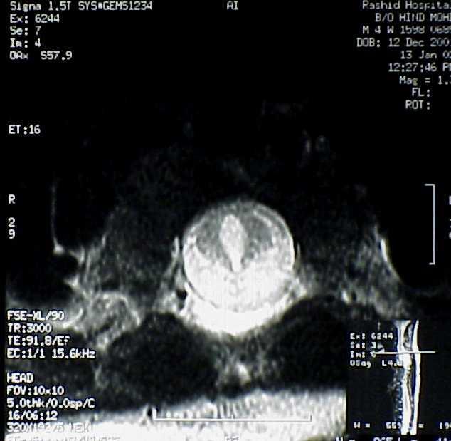
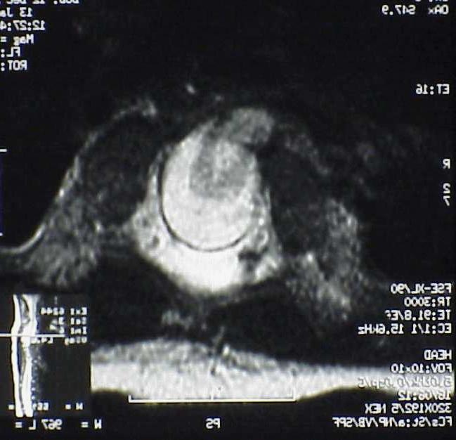
14 Split Notochord Syndrome Association The first x-ray impression was right diaphragmatic hernia with completely split upper thoracic spine and this abnormality could be described as thoracic spine split notochord with anterior thoracic meningocele or diastematomelia for which CT or MRI of the thoracic spine was suggested.
15 Split Notochord Syndrome Association

16 Split Notochord Syndrome Association

17 Split Notochord Syndrome Association

18 Split Notocohrd Syndrome Association
19 Split Notochord Syndrome Association On (MRI) Spinal dysraphism in the form of ; Upper dorsal scoliosis, Spina bifida occulta of C7 down to T6, Butterfly T3, T4 and T5 vertebrae. Diastematomelia of the upper dorsal cord with No MRI evidence of bony or connective tissue, Finally, anterior thoracic meningomyelocele.
20 DIASTEMATOMYELIA (DM) DM is a rare form of spinal dysraphism characterized by a sagittal cleft of varying extent in the spinal cord, conus medullaris and filum terminale with splaying of the posterior vertebral elements. This condition is the result of the presence of an osseous, cartilaginous or fibrous septum producing a complete or incomplete sagittal division of the spinal cord into two hemicords.
21 DIASTEMATOMYELIA (DM) In about 50% of patients, each hemicord contained within it s own dural tube and separated by a dura-sheathed rigid median septum, while in the remaining part they lie within a single dural tube separated by a non rigid, fibrous median septum as seen in our patient. Associated intra-medullary tumours with a DM have been rarely described.
22 DIASTEMATOMYELIA (DM) It may be isolated or associated with other segmental anomalies of the vertebral bodies. DM usually occurs between T9 and S1 levels. Cervical DM is a very rare entity. There is a female predominance in these patients and it was more remarkable in Type I DM than in Type II DM.
23 DIASTEMATOMYELIA (DM) Pang and his colleagues have suggested an alternative classification to deal with all double spinal cord malformations as a split spinal cord malformations with Type I and Type II split spinal cord malformation (SSCMs).
24 DIASTEMATOMYELIA (DM) Initially, the embryo has two layers endoderm & ectoderm. Mesoderm forms between the two, but for a short while, at the primitive pit, the two layers remain in contact. A transient opening, the neurenteric canal, appears connecting the neural ectoderm with the gastro-intestinal endoderm.
25 DIASTEMATOMYELIA (DM) The notochord forms in the mesoderm just caudal to the neurenteric canal. As it migrates, the notochord is split by the persistent neurenteric canal, resulting in the development of spina bifida or other vertebral anomalies and anterior or posterior myelomeningocele. The term split notochord syndrome has been applied to the defect.
26 POSSIBLE ANOMALIES RELATED TO FAILURE OF REGRESSION OF THE NEURENTERIC CANAL ARE :- Complete dorsal enteric fistula Fibrous cord passing through the spinal cord as in DM. Intra-spinal enteric cyst. Dorsal enteric sinus Neurenteric cyst and Enteric duplications.
27 SYMPTOMS - Skin stigmata Orthopedic deformities of foot. Spina bifida aperta Weakness in lower extremities Scoliosis Bladder and bowel disturbance Short &/thin leg Back pain
28 DIASTEMATOMYELIA (DM) Skin findings Orthopedic deformities Neurological findings Hypertrichosis Scoliosis Paraparesis Capillary haemangioma Kyphosis Unilateral leg paresis Hyperpigmentation Subcutaneous lipoma Unilateral leg atrophy Pes cavus/valgus Bladder and bowel dysfunction. Dimple Trophic ulcers
29 RADIOLOGICAL FINDINGS Plain x-ray Bifid lamina. Scoliosis. Hemi vertebra. Accessory lamina. Fused ribs. Widened interpediculate distance Bony median septum. Bifid vertebra. Kypho-scoliosis. Sacral agenesis. Blocked vertebra.
30 MRI FINDINGS Splitting of the spinal cord and other dysraphic lesions such as myelomeningocele, meningocele and lipoma. Low conus medullaris, thick filum terminale, hydromelia, split vertebrae and rigid median septum.
31 DIASTEMATOMYELIA (DM) CT Myelography was superior to other radiological tools in defining the type of DM. Prenatal and post natal ultrasound can be of diagnostic value.
32 ASSOCIATED LESIONS 85% of the patients had more than one spinal lesion as Thick filum terminale. Myelomeningocele. Meningocele. lipo-myelomeningocele. intra-dural arachnoid cyst. dermal sinus tract. dermoid cyst. teratoma. dorsal lipoma.
33 DIASTEMATOMYELIA (DM) Treatment :- When diagnosed, all cases of DM should be surgically treated even if the patient was neurologically asymptomatic.
34 OUTCOME & COMPLICATIONS OF TREATMENT A good outcome is expected aiming at improvement or stabilization of the deficit. COMPLICATIONS Transient. Permanent.
35 PATHOLOGICAL FINDINGS Pathological examination of the specimen from the median septa revealed :- * Fetal renal tissue * Lymphoid tissue * Dermoid cyst * Muscle tissue * Ganglion and blood vessels. * Bone and cartilage.
36 DIASTEMATOMYELIA (DM) Endodermal, ectodermal and mesanchymal structure detected within the median septa support the theory of endomesanchymal tract and ectoendodermal adhesion.
37 THANK YOU
Pediatric Spinal Anomalies
 Department of Radiology University of California San Diego Pediatric Spinal Anomalies John R. Hesselink, M.D. Spine Embryogenesis 1. Primitive streak 2. Proliferation of cells at primitive pit (Hensen's
Long segment composite split cord malformation with double bony spur
 Long segment composite split cord malformation with double bony spur Anand Sharma, Achal Sharma, R.S. Mittal SMS Medical College, Jaipur, India Abstract: A composite type of SCM is very rare and only a
Persistent Terminal Ventricle
 Persistent Terminal Ventricle Ventriculus Terminalis Incomplete regression of TV of 2 neurulation, continuity with central canal small cavity PTV vs terminal myelocystocele (?severe manifestation from
University Journal of Surgery and Surgical Specialties
 University Journal of Surgery and Surgical Specialties ISSN 2455-2860 Volume 2 Issue 1 2016 TWO RARE CASES OF DIASTEMATOMYELIA MUTHURAMAN P Department of Neuro Surgery, THANJAVUR MEDICAL COLLEGE Abstract
Role of helical CT and MRI in the evaluation of spinal dysraphism
 International Journal of Advances in Medicine Kumaran SK et al. Int J Adv Med. 2017 Feb;4(1):124-132 http://www.ijmedicine.com pissn 2349-3925 eissn 2349-3933 Original Research Article DOI: http://dx.doi.org/10.18203/2349-3933.ijam20170095
Wound healing in trophic ulcers in spina bifida patients
 J Neurosurg 82:000 000, 1995 Wound healing in trophic ulcers in spina bifida patients VINOD KUMAR SRIVASTAVA, M.B.B.S, M.CH. Neurosurgical Unit, J. N. Medical College, Aligarh Muslim University, Aligarh,
Prenatal ultrasound evaluation of fetal diastematomyelia: two cases of type I split cord malformation
 Ultrasound Obstet Gynecol 2000; 15: 78 82. Prenatal ultrasound evaluation of fetal diastematomyelia: two cases of type I split cord malformation L.M. ALLEN and R.K. SILVERMAN Perinatal Center, SUNY Health
ISUOG Basic Training. Examining Fetal Anatomy from Longitudinal Sections Titia Cohen-Overbeek, The Netherlands
 ISUOG Basic Training Examining Fetal Anatomy from Longitudinal Sections Titia Cohen-Overbeek, The Netherlands Learning objectives 2 & 3 At the end of the lecture you will be able to: describe how to obtain
Diastematomyelia: A Case with Familial Aggregation of Neural Tube Defects
 Case Study TheScientificWorldJOURNAL (2004) 4, 847 852 ISSN 1537-744X; DOI 10.1100/tsw.2004.140 Diastematomyelia: A Case with Familial Aggregation of Neural Tube Defects Nuray Öksüz Kanbur 1, *, Pınar
A Retrospective Analysis of Clinical Profile and Surgical Outcome in Patients with Spinal Dysraphism at Tertiary Care Center
 Original Research Article A Retrospective Analysis of Clinical Profile and Surgical Outcome in Patients with Spinal Dysraphism at Tertiary Care Center Premlal KV * Assistant Professor, Department of Neurosurgery,
Ligaments of the vertebral column:
 In the last lecture we started talking about the joints in the vertebral column, and we said that there are two types of joints between adjacent vertebrae: 1. Between the bodies of the vertebrae; which
Neonatal Spinal Ultrasound Imaging - A Pictorial Review from The Royal Liverpool Children Hospital, Alder Hey, Liverpool
 Neonatal Spinal Ultrasound Imaging - A Pictorial Review from The Royal Liverpool Children Hospital, Alder Hey, Liverpool Poster No.: C-0081 Congress: ECR 2012 Type: Educational Exhibit Authors: K. Chetcuti,
Spinal dysraphism: genetic relation to
 Journal of Medical Genetics (1976). 13, 343-350. Spinal dysraphism: genetic relation to neural tube malformations C. 0. CARTER, K. A. EVANS, and K. TILL* From MRC Clinical Genetics Unit, Institute of Child
University Journal of Surgery and Surgical Specialties
 University Journal of Surgery and Surgical Specialties ISSN 2455-2860 Volume 2 Issue 1 2016 Profile of paediatric patients with split cord malformation MANORANJITHAKUMARI M Department of Neuro Surgery,
Skeletal System. Prof. Dr. Malak A. Al-yawer Department of Anatomy/Embryology Section
 Skeletal System Prof. Dr. Malak A. Al-yawer Department of Anatomy/Embryology Section Learning objectives At the end of this lecture, the medical student will be able to: State the embryonic origin of skeletal
Development of Spinal Cord & Vertebral Column. Dr. Sanaa Alshaarawi & Prof. Ahmed Fathalla
 Development of Spinal Cord & Vertebral Column Dr. Sanaa Alshaarawi & Prof. Ahmed Fathalla OBJECTIVES At the end of the lecture, students should be able to: q Describe the development of the spinal cord
Chapter 8. Pediatric Surgery
 Chapter 8 Pediatric Surgery 8.1 Hydrocephalus Hydrocephalus is a congenital disorder. There may be difficulties during normal vaginal delivery due large size of the head. In 1970s, when these pictures
Dorsal dermal sinus in children
 Dorsal dermal sinus in children Poster No.: C-2581 Congress: ECR 2015 Type: Educational Exhibit Authors: J. Marjanovic, A. Paterson, P. C. McSherry, A. Nixon, A. 1 1 2 1 2 1 1 2 TRIPALO BATOS, T. Grmoja
Central Nervous System Congenital Abnormalities
 Central Nervous System Congenital Abnormalities Eva Brichtova, M.D., Ph.D., Department of Pediatric Sugery, Orthopaedics and Traumatology, University Hospital Brno Neural tube defects Dysraphism uncomplete
Sonography of the Neonatal Spine: Part 2, Spinal Disorders
 Neonatal Spine Sonography Pediatric Imaging Pictorial Essay Downloaded from www.ajronline.org by 148.251.232.83 on 04/11/18 from IP address 148.251.232.83. Copyright RRS. For personal use only; all rights
Congenital Spine and Spinal Cord Malformations Pictorial Review
 JR Integrative Imaging LIFELONG LERNING FOR RDIOLOGY ongenital Spine and Spinal ord Malformations Pictorial Review Stephanie L. Rufener 1,2, Mohannad Ibrahim 2, harles. Raybaud 3, Hemant. Parmar 2 Downloaded
Sonography of the Neonatal Spine: Part 1, Normal Anatomy, Imaging Pitfalls, and Variations That May Simulate Disorders
 Sonography of Neonatal Spine Pediatric Imaging Pictorial Essay Downloaded from www.ajronline.org by 46.3.195.60 on 02/04/18 from IP address 46.3.195.60. Copyright RRS. For personal use only; all rights
Spine and spinal cord
 NEURORADIOLOGY Spine and spinal cord Erika Vörös University of Szeged Department of Radiology SZEGED DISEASES OF SPINE AND SPINAL CORD I. Non-tumourous diseases developmental anomalies vascular disorders
Formation defects Scoliosis Deformities I 07 1
 What is congenital scoliosis? Congenital scoliosis is a spinal deformity with lateral deviation and rotation of the spinal column, where congenital dysfunctions in embryonal vertebra development cause
Anatomy of the Nervous System. Brain Components
 Anatomy of the Nervous System Brain Components NERVOUS SYSTEM INTRODUCTION Is the master system of human body, controlling the functions of rest of the body systems Nervous System CLASSIFICATION A. Anatomical
Case Report Ipsilateral Hip Dysplasia in Patients with Sacral Hemiagenesis: A Report of Two Cases
 Case Reports in Orthopedics Volume 2015, Article ID 854151, 4 pages http://dx.doi.org/10.1155/2015/854151 Case Report Ipsilateral Hip Dysplasia in Patients with Sacral Hemiagenesis: A Report of Two Cases
Development of the Digestive System. W.S. O The University of Hong Kong
 Development of the Digestive System W.S. O The University of Hong Kong Plan for the GI system Then GI system in the abdomen first develops as a tube suspended by dorsal and ventral mesenteries. Blood
What Every Spine Surgeon Should Know About Neurosurgical Issues
 What Every Spine Surgeon Should Know About Neurosurgical Issues Amer Samdani, MD Chief of Surgery Shriners Hospitals for Children Philadelphia, PA Objectives Main intraspinal lesions Chiari malformation
The spinal dermal-sinus-like stalk
 Childs Nerv Syst (2009) 25:191 197 DOI 10.1007/s00381-008-0669-6 ORIGINAL PAPER The spinal dermal-sinus-like stalk J. van Aalst & E. A. M. Beuls & E. M. J. Cornips & H. W. M. van Straaten & A. F. M. Boselie
ISCHIOPUBIC HYPOPLASIA : A RARE CONSTITUENT OF CONGENITAL SYNDROMES
 ISCHIOPUBIC HYPOPLASIA : A RARE CONSTITUENT OF CONGENITAL SYNDROMES N. K. SFEROPOULOS, I. TSITOURIDIS Hypoplasia of the ischiopubic region is described in four patients. An adolescent was referred for
Radiologic and pathologic features of spinal dysraphism. A pictorial review.
 Radiologic and pathologic features of spinal dysraphism. A pictorial review. Poster No.: C-0586 Congress: ECR 2011 Type: Educational Exhibit Authors: N. Arcalis, J. L. Ribó, J. Muchart, L. Riaza, J. Blanch
Prospective Evaluation of Role of MRI in Suspected Spinal Dysraphism and Its Management
 IOSR Journal of Dental and Medical Sciences (IOSR-JDMS) e-issn: 2279-0853, p-issn: 2279-0861.Volume 17, Issue 5 Ver. 1 (May. 2018), PP 21-28 www.iosrjournals.org Prospective Evaluation of Role of MRI in
May have excessive movement in the unfused segment to compensate. Flexion extension better preserved than lateral bend or rotation
 IV CONGENITAL SPINE KLIPPEL FLAIL SYNDROME Prevalence 0.60% Mainly around upper 3 vertebrae [75%] Commonest: C2 3 Lower Cervical spine fusion may be associated with syndromes: Fetal alcohol syndrome Goldenhar
LUMPS, TUFTS AND DIMPLES IT S THE PITS!! Session Information. Faculty Disclosure Information
 Session Information Session Title: Lumps, Tufts and Dimples Session Number: F3056, F2130 Faculty Name: Mark S. Dias, MD, FAAP Faculty Institution: Penn State Children s Hospital, Penn State University
Development of the Digestive System. W.S. O School of Biomedical Sciences, University of Hong Kong.
 Development of the Digestive System W.S. O School of Biomedical Sciences, University of Hong Kong. Organization of the GI tract: Foregut (abdominal part) supplied by coeliac trunk; derivatives include
Endoscopic Assisted resection for congenital Midline Nasal Mass
 Endoscopic Assisted resection for congenital Midline Nasal Mass Ahmed Aly Ibrahim A.prof ORL Department Alexandria University Emad. A Magdy prof ORL Department Alexandria University Haytham Morsi,MD Mohammad
Diastematomyelia in Children: Metrizamide and CT
 403 Diastematomyelia in Children: Metrizamide and CT Metrizamide Myelography Giuseppe Scotti' Mark. Musgrave Derek C. Harwood-Nash Charles R. Fitz Sylvester H. Chuang Diastematomyelia is an uncommon dysraphic
Ligamentous Integrity in Spinal Cord Injury without Radiographic Abnormality. Dr Anria Horn Dr Stewart Dix-Peek
 Ligamentous Integrity in Spinal Cord Injury without Radiographic Abnormality Dr Anria Horn Dr Stewart Dix-Peek Introduction Spinal Cord Injury Without Radiographic Abnormality SCIWORA Pang, Wilberger 1982
Symptomatic Multiple Level Lateral Meningoceles with Intraspinal Meningocele: A Case Study and Its Surgical Management
 THIEME Original Article 15 Symptomatic Multiple Level Lateral Meningoceles with Intraspinal Meningocele: A Case Study and Its Surgical Management Vernon Velho 1 Sachin Guthe 1 Pravin Survashe 1 Poonam
Midgut. Over its entire length the midgut is supplied by the superior mesenteric artery
 Gi Embryology 3 Midgut the midgut is suspended from the dorsal abdominal wall by a short mesentery and communicates with the yolk sac by way of the vitelline duct or yolk stalk Over its entire length the
Case report: Imaging findings in a butterfly vertebra
 Acta Neurol. Belg., 2011, 111, 344-348 Case report: Imaging findings in a butterfly vertebra Cedric BOULET, Ann SCHIETTECATTE, Johan DE MEY and Michel DE MAESENEER Department of Radiology, UZ Brussel,
Split cord malformation in children undergoing neurosurgical intervetion in India: a descriptive study
 ORIGINAL ARTICLE Journal of Pediatric Neurology 2004; 2(1): 21-27 www.jpneurology.org in children undergoing neurosurgical intervetion in India: a descriptive study Raj Kumar 1, Vinita Singh 2, Satya Narayan
Disclosures None. Spinal Dysraphism
 Spinal Dysraphism Andrew Jea MD MHA FAAP Professor and Chief Section of Pediatric Neurosurgery Riley Hospital for Children Department of Neurosurgery Indiana University School of Medicine Goodman Campbell
AXIAL SKELETON FORM THE VERTICAL AXIS OF THE BODY CONSISTS OF 80 BONES INCLUDES BONES OF HEAD, VERTEBRAL COLUMN, RIBS,STERNUM
 AXIAL SKELETON FORM THE VERTICAL AXIS OF THE BODY CONSISTS OF 80 BONES INCLUDES BONES OF HEAD, VERTEBRAL COLUMN, RIBS,STERNUM APPENDICULAR SKELETON BONES OF THE FREE APPENDAGES & THEIR POINTS OF ATTACHMENTS
Asymptomatic posterior cervical myelomeningocele with tethered cord in an adolescent: a rare form of spinal dysraphism with rare presentation
 Romanian Neurosurgery (2016) XXX 1: 113-117 113 Asymptomatic posterior cervical myelomeningocele with tethered cord in an adolescent: a rare form of spinal dysraphism with rare presentation Gangesh Gunjan,
Case Report Surgical Treatment of a Patient with Human Tail and Multiple Abnormalities of the Spinal Cord and Column
 SAGE-Hindawi Access to Research Advances in Orthopedics Volume 2011, Article ID 153797, 4 pages doi:10.4061/2011/153797 Case Report Surgical Treatment of a Patient with Human Tail and Multiple Abnormalities
A Very Unusual Case of a Dorsal Heteropagus Twin
 PRG A Very Unusual Case of a Dorsal Heteropagus Twin Nathan David P. Concepcion, MD 1, Bernard F. Laya, DO 1, Eduardo P. Manrique, MD 2 and Faith Caroline D. Bayabos, MD 1 1 Section of Pediatric Radiology,
Spinal congenital dermal sinus with dual ostia
 J Neurosurg Pediatrics 3:000 000, 3:407 411, 2009 Spinal congenital dermal sinus with dual ostia Clinical article Ch a n g Su b Le e, M.D., 1 Ji Ho o n Ph i, M.D., 2 Se u n g -Ki Kim, M.D., Ph.D., 2 By
The Fetal Care Center at NewYork-Presbyterian/ Weill Cornell Medicine
 The Fetal Care Center at NewYork-Presbyterian/ Weill Cornell Medicine Prompt and Personalized Care for Women with Complex Pregnancies A Team of Experts additional training in maternal and fetal complications
Vertebral Column. Backbone consists of 26 vertebrae. Five vertebral regions. Cervical
 Vertebral Column Backbone consists of 26 vertebrae. Five vertebral regions Cervical vertebrae (7) in the neck. Thoracic vertebrae (12) in the thorax. Lumbar vertebrae (5) in the lower back. Sacrum (5,
Lumbosacral lipomas: critical survey of 26 cases submitted to laminectomy
 J. Neurol. Neuirosurg. Psychiat.. 1967, 30, 174 Lumbosacral lipomas: critical survey of 26 cases submitted to laminectomy L. P. LASSMAN AND C. C. MICHAEL JAMES From the Regional Neurological Centre, Newcastle
Tethered spinal cord syndrome: a developmental overview
 International Journal of Sciences & Applied Research www.ijsar.in Tethered spinal cord syndrome: a developmental overview Anushi Singh 1 *, Rekha Kumari 2 1 CHN Department, School of Nursing Science and
Ultrasound Anomaly Details
 Appendix 2. Association of Copy Number Variants With Specific Ultrasonographically Detected Fetal Anomalies Ultrasound Anomaly Details Abdominal wall Bladder exstrophy Body-stalk anomaly Cloacal exstrophy
Note: Please refer to handout Spinal Plexuses and Representative Spinal Nerves for
 Chapter 13 Outline Note: Please refer to handout Spinal Plexuses and Representative Spinal Nerves for what you need to know from Exhibits 13.1 13.4 I. INTRODUCTION A. The spinal cord and spinal nerves
8/31/2018 IMPORTANT CONSIDERATIONS. Signalment History Symmetry Progression of signs Painful vs non-painful SURGICAL CONSIDERATIONS
 IMPORTANT CONSIDERATIONS Signalment History Symmetry Progression of signs Painful vs non-painful SURGICAL CONSIDERATIONS Specific region of TL spine Differences in size and shape of articular processes
A Congenital Defect in the Spinal Cord of the Manx Cat
 Vet, Path. 8: 232-238 (1971) A Congenital Defect in the Spinal Cord of the Manx Cat A. H. MARTIN Department of Anatomy, University of Wisconsin, Madison Wisc. Abstract. The lumbar part of the spinal cords
Axial Skeleton: Vertebrae and Thorax
 Axial Skeleton: Vertebrae and Thorax Function of the vertebral column (spine or backbone): 1) 2) 3) Composition of Vertebral column The vertebral column is formed by 33 individual vertebrae (some of which
Management of Bone and Spinal Cord in Spinal Surgery.
 Management of Bone and Spinal Cord in Spinal Surgery. G. Saló, PhD, MD. Senior Consultant Spine Unit. Hospital del Mar. Barcelona. Ass. Prof. Universitat Autònoma de Barcelona. Introduction The management
Remember from the first year embryology Trilaminar disc has 3 layers: ectoderm, mesoderm, and endoderm
 Development of face Remember from the first year embryology Trilaminar disc has 3 layers: ectoderm, mesoderm, and endoderm The ectoderm forms the neural groove, then tube The neural tube lies in the mesoderm
Bronchioles. Alveoli. Type I alveolar cells are very thin simple squamous epithelial cells and form most of the lining of an alveolus.
 276 Bronchioles Bronchioles continue on to form bronchi. The primary identifying feature is the loss of hyaline cartilage. The epithelium has become simple ciliated columnar, and there is a complete ring
1 Normal Anatomy and Variants
 1 Normal Anatomy and Variants 1.1 Normal Anatomy MR Technique. e standard MR protocol for a routine evaluation of the spine always comprises imaging in sagittal and axial planes, while coronal images are
Development of pancreas and Small Intestine. ANATOMY DEPARTMENT DR.SANAA AL-AlSHAARAWY DR.ESSAM Eldin Salama
 Development of pancreas and Small Intestine ANATOMY DEPARTMENT DR.SANAA AL-AlSHAARAWY DR.ESSAM Eldin Salama OBJECTIVES At the end of the lecture, the students should be able to : Describe the development
NEURORADIOLOGY. Part III. Angela Csomor University of Szeged Department of Radiology
 NEURORADIOLOGY Part III Angela Csomor University of Szeged Department of Radiology DISEASES OF SPINE AND SPINAL CORD I. Non-tumourous diseases developmental anomalies vascular disorders inflammatory processes
Introduction to Neurosurgical Subspecialties:
 Introduction to Neurosurgical Subspecialties: Pediatric Neurosurgery Brian L. Hoh, MD 1 and Gregory J. Zipfel, MD 2 1 University of Florida, 2 Washington University Pediatric Neurosurgery Pediatric neurosurgeons
A Model of Care for Patients with Spina Bifida. Dr C West AM Spina Bifida Service The Children s Hospital at Westmead 2012
 A Model of Care for Patients with Spina Bifida Dr C West AM Spina Bifida Service The Children s Hospital at Westmead 2012 Explanation of Model of Care To provide a framework for holistic care of patient
Development of the urinary system
 Development of the urinary system WSO School of Biomedical Sciences, University of Hong Kong. 3 sets of kidneys developing in succession (temporally and spatially) : Pronephros ] Mesonephros ]- Intermediate
CAUDAL REGRESSION SYNDROME
 CAUDAL REGRESSION SYNDROME *Prateek Gehlot 1 and Jagdish Mandliya 2 1 Department of Radio-Diagnosis, R.D.Gardi Medical College,, Ujjain (MP). 2 Department of Pediatrics, R.D.Gardi Medical College, Ujjain
Human Anatomy. Spinal Cord and Spinal Nerves
 Human Anatomy Spinal Cord and Spinal Nerves 1 The Spinal Cord Link between the brain and the body. Exhibits some functional independence from the brain. The spinal cord and spinal nerves serve two functions:
Case Report Occult Spinal Dysraphism in the Presence of Rare Cutaneous Stigma in a Neonate: Importance of Ultrasound and Magnetic Resonance Imaging
 Case Reports in Medicine Volume 2013, Article ID 468376, 4 pages http://dx.doi.org/10.1155/2013/468376 Case Report Occult Spinal Dysraphism in the Presence of Rare Cutaneous Stigma in a Neonate: Importance
Metastatic Spinal Disease
 Metastatic Spinal Disease Mr Neil Chiverton Consultant Spinal Surgeon, Sheffield Objectives The scale and nature of the problem NICE recommendations Surgical decision making Case illustrations Incidence
Spinal Column. Anatomy Of The Spine
 Anatomy Of The Spine The spine is a flexible column, composed of a stack of individual bones. Each bone is called a vertebra. There are seven vertebrae in the neck (cervical vertebrae) twelve in the thoracic
Scoliosis: Orthopaedic Perspectives
 Scoliosis: Orthopaedic Perspectives Scott B. Rosenfeld, MD Division of Pediatric Orthopaedic Surgery Texas Children s Hospital Page 0 xxx00.#####.ppt 9/23/2012 8:26:24 AM I have no disclosures Disclosures
Welcome to ANAT 10A! What is Anatomy? Different levels of Anatomy The Language of Anatomy Pearson Education, Inc.
 Welcome to ANAT 10A! What is Anatomy? Different levels of Anatomy The Language of Anatomy Introduction Anatomy means to dissect: (ANAT 10A) The study of internal & external body structures The study of
Congenital Tethered Spinal Cord Syndrome in Adults
 Page 1 of 7 http://www.medscape.com/ To Print: Click your browser's PRINT button. NOTE: To view the article with Web enhancements, go to: http://www.medscape.com/viewarticle/405679 Congenital Tethered
Basic Training. ISUOG Basic Training The 20 Planes Approach to the Routine Mid Trimester Scan
 ISUOG The 20 Planes Approach to the Routine Mid Trimester Scan Learning objective At the end of the lecture you will be able to: Explain how to perform a structured routine examination, including measurements,
Congenital Spinal Lipoma: analyzing the perplexed nomenclature and our management
 Congenital Spinal Lipoma: analyzing the perplexed nomenclature and our management Nidal Khasawneh MD *, Rami Alqroom MD *, Firas Sha'ban MD *, Rafeed Al Drous MD *, Rima Nserat **, Amer Al Shurbaji MD
The vault bones Frontal Parietals Occiput Temporals Sphenoid Ethmoid
 The Vertebral Column Head, Neck and Spine Bones of the head Some consider the bones of the head in terms of the vault bones and the facial bones hanging off the front of them The vault bones Frontal Parietals
THE VERTEBRAL COLUMN. Average adult length: In male: about 70 cms. In female: about 65 cms.
 THE VERTEBRAL COLUMN Average adult length: In male: about 70 cms. In female: about 65 cms. 1 Vertebral Column (Regions and Curvatures) Curvatures of the vertebral column: A. Primary curvature: C-shaped;
Development of the Urinary System
 Development of the Urinary System Lecture Objectives Understand the development of the kidney and related organs of the urinary system. Define the pronephrons, mesonephrons and metanephrons. Understand
Fetal Medicine. Case Presentations. Dr Ermos Nicolaou Fetal Medicine Unit Chris Hani Baragwanath Hospital. October 2003
 Case Presentations Dr Ermos Nicolaou Fetal Medicine Unit Chris Hani Baragwanath Hospital October 2003 Case 1 Ms A M 22year old P0 G1 Referred from Sebokeng Hospital at 36w for polyhydramnios On Ultrasound:
Johnson Rogers and colleagues- used Term LMM
 LIPOMENINGOMYELOCELE: CLASSIFICATION, MANAGEMENT AND CONTROVERSIES Definition : Lipomyelomeningocele is a form of OSD in which a subcutaneous fibrofatty mass traverses the lumbodorsal fascia, causes a
We are IntechOpen, the world s leading publisher of Open Access books Built by scientists, for scientists. International authors and editors
 We are IntechOpen, the world s leading publisher of Open Access books Built by scientists, for scientists 3,500 108,500 1.7 M Open access books available International authors and editors Downloads Our
W IDENING of an intervertebral foramen
 Agenesis of a Pedicle in the Cervical Spine LESLIE i. ZATZ, M.D., PETER W. BURGESS, M.D. AND John W. HANBERY, M.D. Departments of Radiology and of Surgery, Neurosurgical Division, Stanford University School
Chapter 13. The Spinal Cord & Spinal Nerves. Spinal Cord. Spinal Cord Protection. Meninges. Together with brain forms the CNS Functions
 Spinal Cord Chapter 13 The Spinal Cord & Spinal Nerves Together with brain forms the CNS Functions spinal cord reflexes integration (summation of inhibitory and excitatory) nerve impulses highway for upward
Congenital tethered spinal cord syndrome in adults
 Neurosurg Focus 10 (1):Article 7, 2001, Click here to return to Table of Contents Congenital tethered spinal cord syndrome in adults BERMANS J. ISKANDAR, M.D., BENJAMIN B. FULMER, M.D., MARK N. HADLEY,
Early Development of Neural Tube Development of Medulla Spinalis and Peripheral Nervous System. Assoc.Prof. E.Elif Güzel, M.D.
 Early Development of Neural Tube Development of Medulla Spinalis and Peripheral Nervous System Assoc.Prof. E.Elif Güzel, M.D. Third week of Embryogenesis Primitive streak/pit appears on the epiblast (day
Purpose: To discuss the fatty filum terminale which is incidentally demonstrated on MRI concerning the causes of TCS
 ISPUB.COM The Internet Journal of Spine Surgery Volume 3 Number 1 T Iizuka Citation T Iizuka.. The Internet Journal of Spine Surgery. 2006 Volume 3 Number 1. Abstract Background: Fatty filum terminale
Lecture 14: The Spinal Cord
 Lecture 14: The Spinal Cord M/O Chapters 16 69. Describe the relationship(s) between the following structures: root, nerve, ramus, plexus, tract, nucleus, and ganglion. 70. Trace the path of information
Bony framework of the vertebral column Structure of the vertebral column
 5.1: Vertebral column & back. Overview. Bones o vertebral column. o typical vertebra. o vertebral canal. o spinal nerves. Joints o Intervertebral disc. o Zygapophyseal (facet) joint. Muscles o 2 compartments:
Neuropathic bladder and spinal dysraphism
 Archives of Disease in Childhood, 1981, 56, 176-180 Neuropathic bladder and spinal dysraphism MALGORZATA BORZYSKOWSKI AND B G R NEVILLE Evelina Children's Department, Guy's Hospital, London SUMMARY The
Management of Tethered Cord Syndrome in Adults: Experience of 23 Cases
 DOI: 10.5137/1019-5149.JTN.15892-15.1 Received: 31.08.2015 / Accepted: 02.10.2015 Published Online: 02.06.2016 Original Investigation Management of Tethered Cord Syndrome in Adults: Experience of 23 Cases
(i) Family 1. The male proband (1.III-1) from European descent was referred at
 Family 1. The male proband (1.III-1) from European descent was referred at") 1 Supplementary Note Clinical descriptions of families (i) Family 1. The male proband (1.III-1) from European descent was referred at age 14 because of scoliosis. He had normal development. Physical evaluation
1 Supplementary Note Clinical descriptions of families (i) Family 1. The male proband (1.III-1) from European descent was referred at age 14 because of scoliosis. He had normal development. Physical evaluation
Basic Training. ISUOG Basic Training Examining the Upper Lip, Face & Profile
 ISUOG Examining the Upper Lip, Face & Profile Learning objectives At the end of the lecture you will be able to: Describe how to obtain the 3 planes required to assess the anatomy of the fetal face Recognise
SWISS SOCIETY OF NEONATOLOGY. Cantrell s pentalogy: an unusual midline defect
 SWISS SOCIETY OF NEONATOLOGY Cantrell s pentalogy: an unusual midline defect October 2004 2 Cevey-Macherel MN, Meijboom EJ, Di Bernardo S, Truttmann AC, Division of Neonatology and Division of Pediatric
VERTEBRAL COLUMN ANATOMY IN CNS COURSE
 VERTEBRAL COLUMN ANATOMY IN CNS COURSE Vertebral body Sections of the spine Atlas (C1) Axis (C2) What type of joint is formed between atlas and axis? Pivot joint What name is given to a fracture of both
Congenital Spinal Dermal Sinuses Long-Term Analysis at King Hussein Medical Center.
 Congenital Spinal Dermal Sinuses Long-Term Analysis at King Hussein Medical Center. Nidal Khasawneh MD*, Rami Al Qroom MD*, Luma Fayyad MD*, Sura AlRawabdeh MD*, Hayat Al-khasawneh MD*. ABSTRACT Objectives:
Case Based Urology Learning Program
 Case Based Urology Learning Program Resident s Corner: UROLOGY Case Number 23 CBULP 2011 077 Case Based Urology Learning Program Editor: Associate Editors: Manager: Case Contributors: Steven C. Campbell,
Human Anatomy - Problem Drill 11: The Spinal Cord and Spinal Nerves
 Human Anatomy - Problem Drill 11: The Spinal Cord and Spinal Nerves Question No. 1 of 10 Instructions: (1) Read the problem statement and answer choices carefully, (2) Work the problems on paper as needed,
Week 14. Development of the Musculoskeletal System
 Week 14 Development of the Musculoskeletal System Skeletal System Derived from: paraxial mesoderm somites and somitomeres sclerotome sclerotome differentiation induced by SHH from notochord and floor plate