Persistent Terminal Ventricle
|
|
|
- Gabriel Shannon Lane
- 5 years ago
- Views:
Transcription

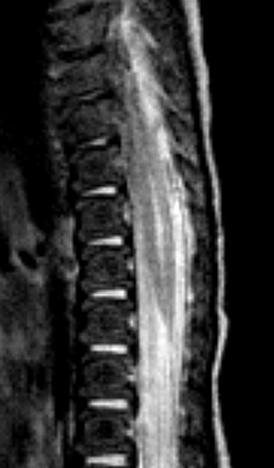
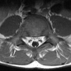
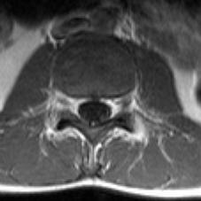
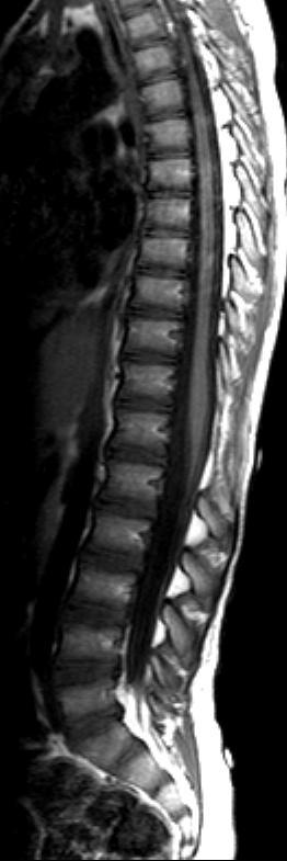
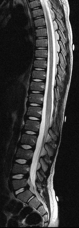
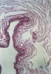
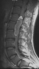
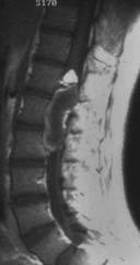
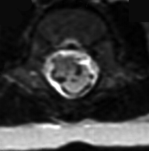
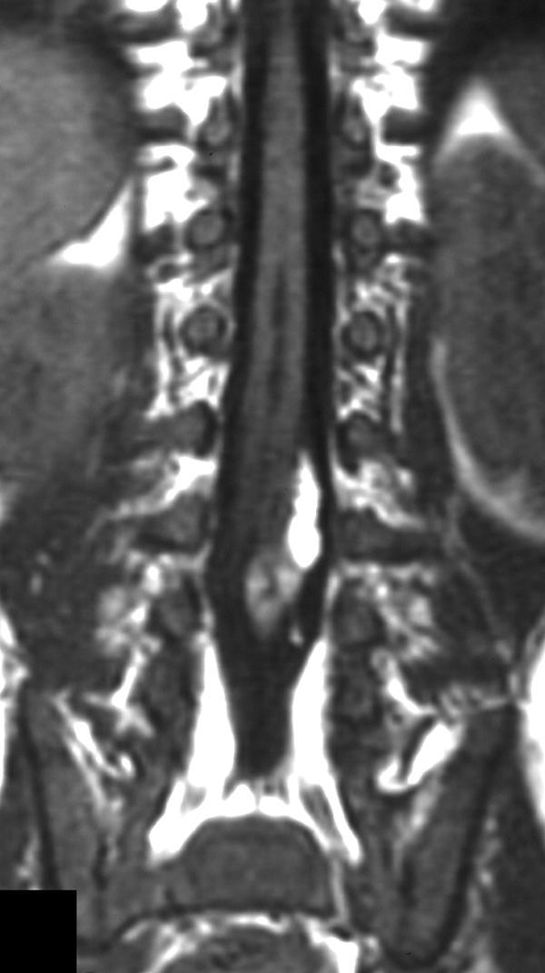
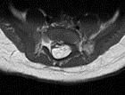
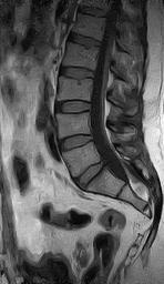
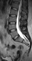
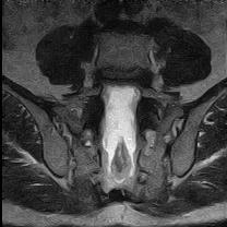
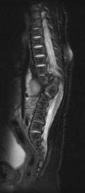
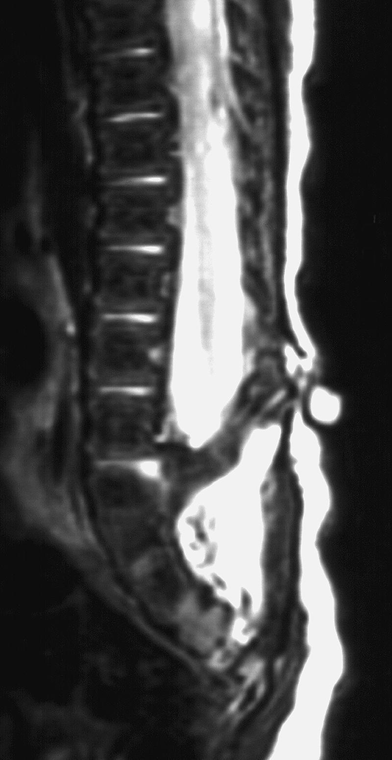
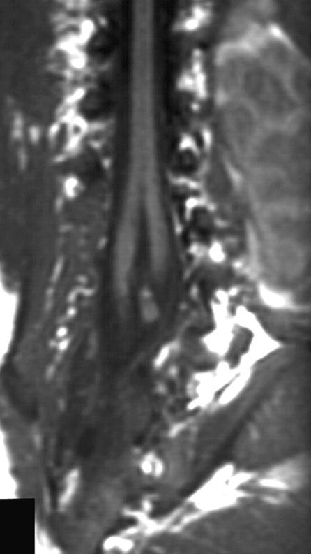
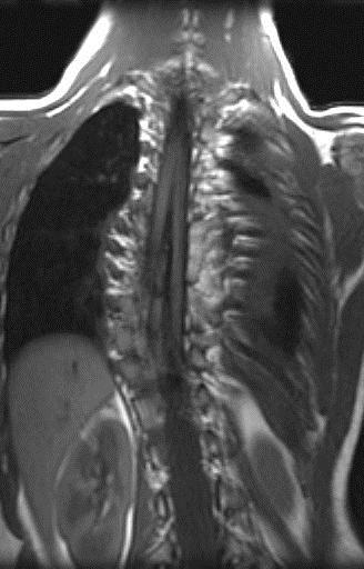
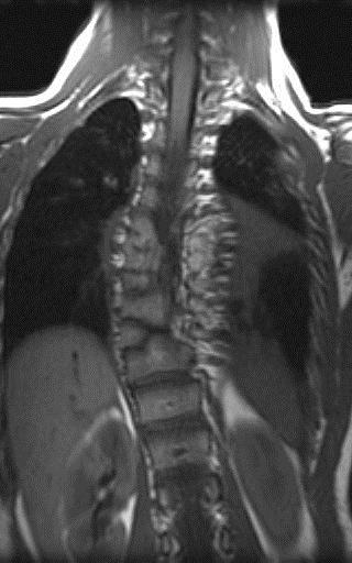
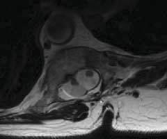
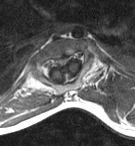
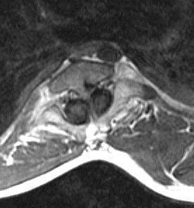
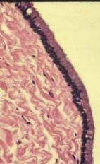
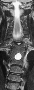
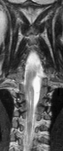
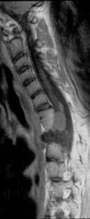
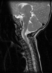
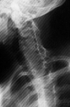
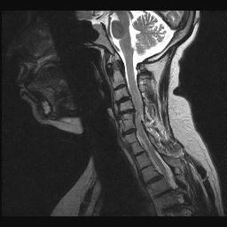
1 Persistent Terminal Ventricle Ventriculus Terminalis Incomplete regression of TV of 2 neurulation, continuity with central canal small cavity PTV vs terminal myelocystocele (?severe manifestation from inability of CSF to escape)


2 Persistent Terminal Ventricle Common, transient finding until 5 yrs Identical to CSF, in conus or conus-filum transition Anatomic variant, usually incidental but > 4-5mm can be associated with pain and neurologic symptoms

3 Terminal Ventricle

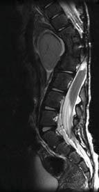
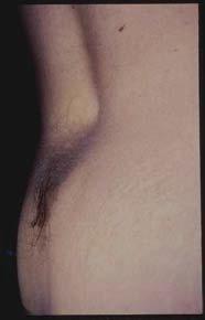
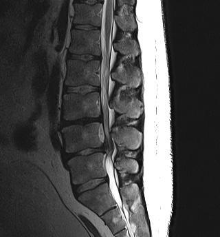
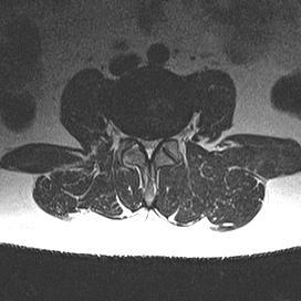
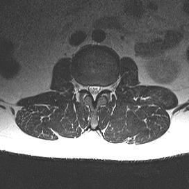
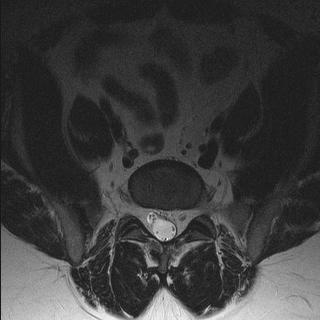
4 Fibrolipomatous Infiltration of Filum Anomaly of 2 neurulation, from totipotential caudal cell mass Axial T1 most sensitive?small amount of fat anatomical variant vs any fat or thickening (>2mm) = tether


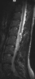
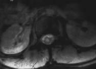
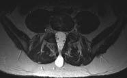
5 Fatty Filum and Ventriculis Terminalis
6 Incidental Fatty Filum


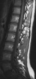
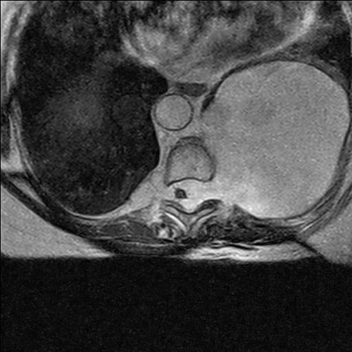
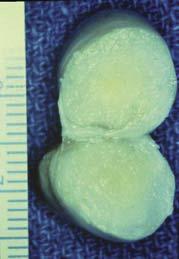
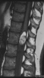
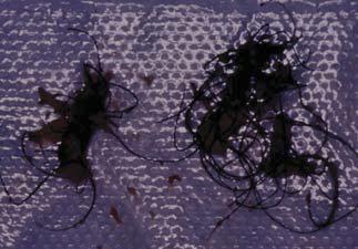
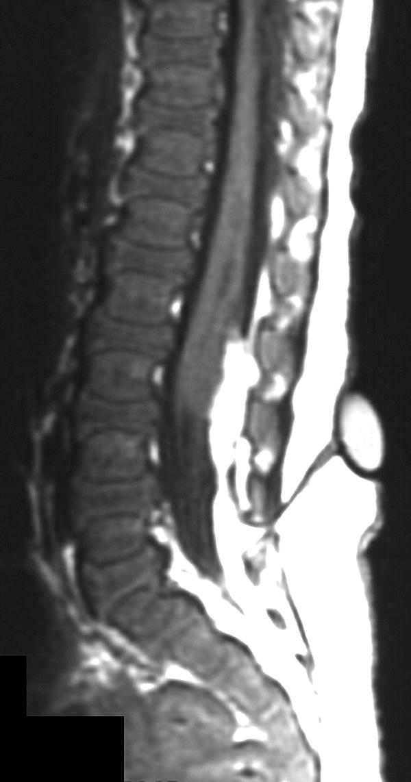
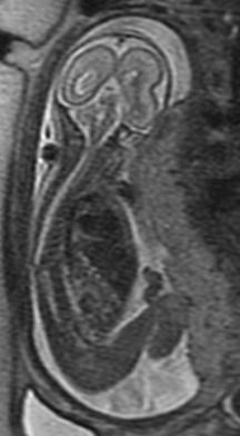
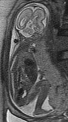
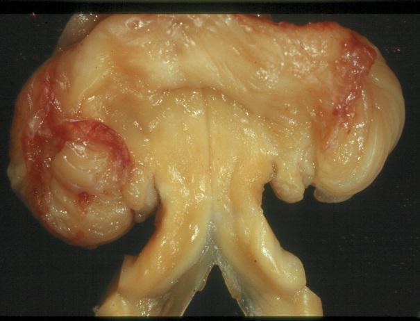
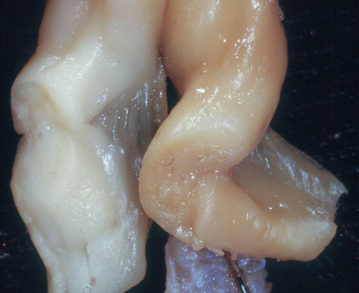
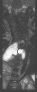
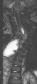
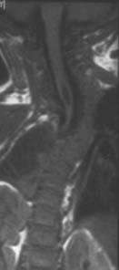
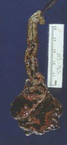
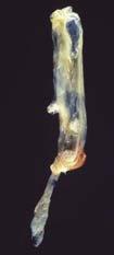
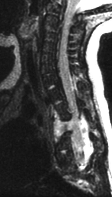
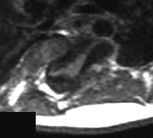
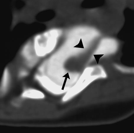
7 Terminal Myelocystocele Large, skin covered lumbosacral mass Dilatation of terminal ventricle High assoc with caudal cell mass anomalies (GU, lower GI, abd wall) Cerebellar herniation late due to lesser CSF sump effect? Incontinent, extremely poor LE function Neural elements attenuated
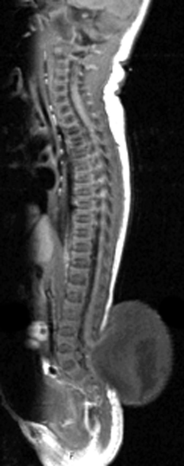
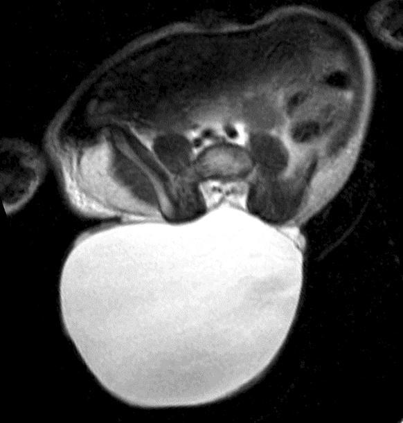
8 Terminal Myelocystocele

9 Myelocele (Myeloschisis) Placode of OSD in plane with back Less common, embryologically similar Clinical signs & function similar


10 Lateral Thoracic and Lumbar Myeloceles

11 Meningocele Meningeal-lined CSF sac protruding through defect Cord does not enter sac, may be assoc with hypertrophy of filum or cord tethering? From CSF pulsations, Weakened or abnormal dura? Lateral assoc with NF I, also post-trauma, connective tissue disorders
12 Dorsal meningocele


13 Thoracic Meningocele

14 Dural Ectasia, Meningocele assoc with NF-1

15 Spinal Lipoma Premature disjunction of cutaneous ectoderm from neuroectoderm allows mesenchyme to contact inner portion of neural tube. Mesenchyme intended for other cell types regresses into fat when exposed to CSF As tube tries to close, mesenchyme fat, may interferes with neurulation Lipomas contain ectodermal, mesodermal, endodermal elements = teratomas Grows in proportion to overall adipose

16 Intraspinal Lipoma Mesodermal cells contact primitive ependyma LS, any level Usually intradural, rarely entirely intramedullary


17 Spinal Lipomas
18 Epidermoid / Dermoid Trapped epithelial mesenchymal tissue Developmental Post procedure May grow over time Related to teratomas sometimes
19 Epidermoid dermoid

20 Dermal Sinus Proteus Infxn of Dermoid, EDA

21 Spinal Teratoma
22 Lipomyelocele (with dermal sinus) Placode within canal
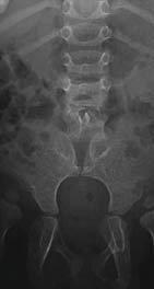
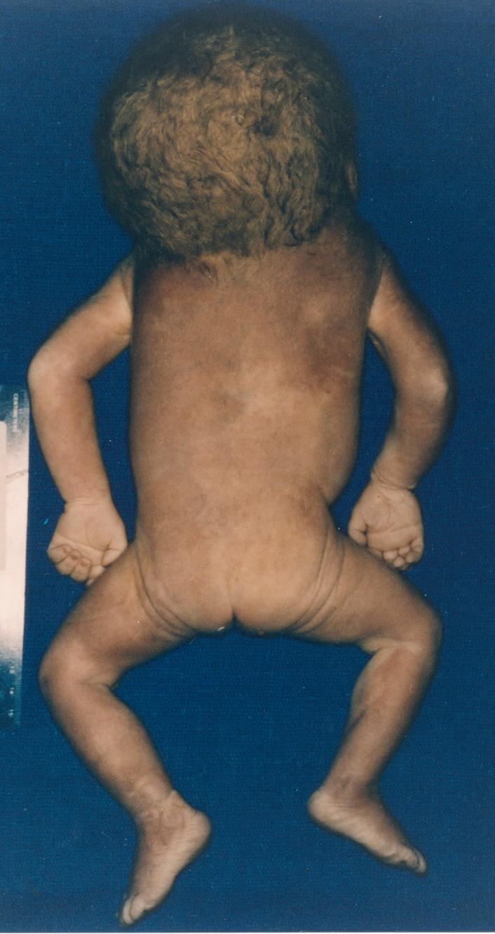
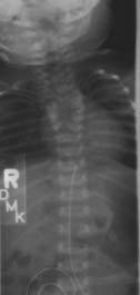
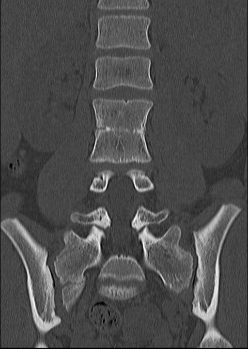
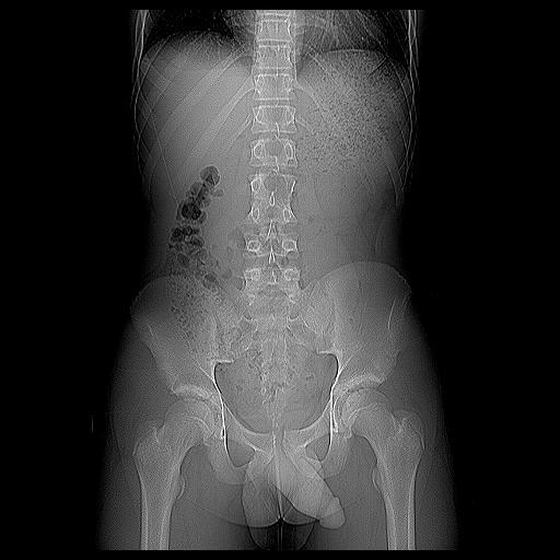
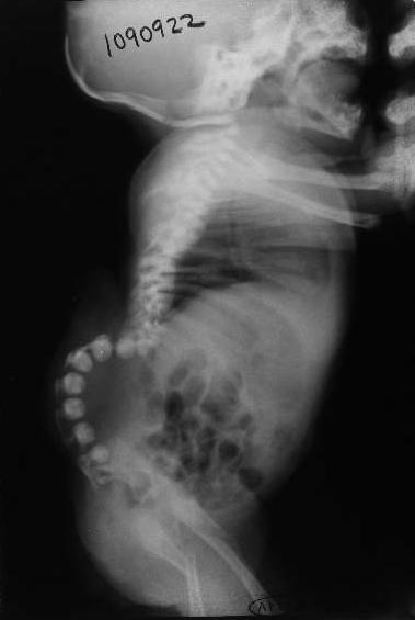
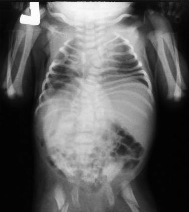
23 Caudal Regression Syndrome Spectrum from coccygeal/ls hypogenesis sirenomelia Infants of diabetic mothers, 1 in 7500 live births Assoc with: OEIS (omphalocele, exstrophy, imperforate anus, spinal anomalies) VACTERL (vertebral, renal, cardiac, limb anomalies with anorectal atresia & TEF) Currarino triad (sacral hypogenesis, anorectal malformations, presacral teratoma or meningocele)

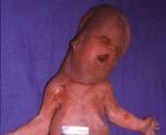
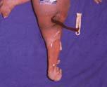
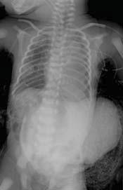
24 Caudal Regression: Sirenomelia
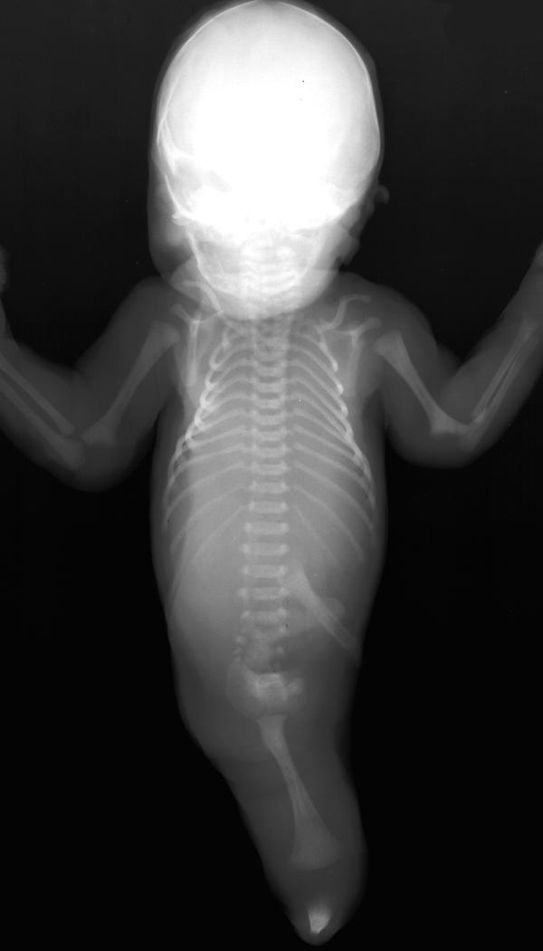
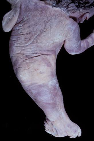
25 Sirenomelia

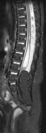
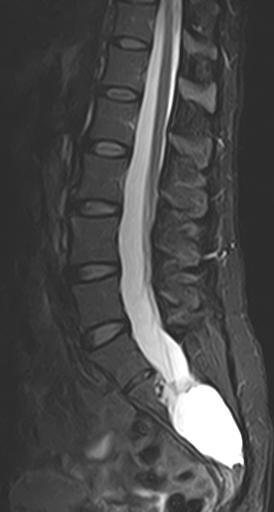
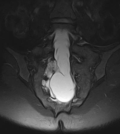
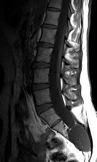
26 Caudal Regression Syndrome Level determines type & severity Type I: above S1, even mid-thoracic, cord terminates high, blunted tip & deformation of cauda common, ant & post separation of roots Type II: Below S2, less severe, distal-most conus absent, tethered by tight filum, lipoma, or CSD with subcutaneous mass Mild CRS: only tip of conus absent, cord not tethered, may be missed Type II

27 Type I CRS
28 CRS with fibrolipoma
29 Fibrolipoma, Tether, Sacral Hypoplasia CRS Type I
30 Terminal Myelocystocele and Sacral Hypoplasia CRS Type I
31 Segmental Spinal Dysgenesis Focal segment of lumbar or thoracic spine ageneticmarkedly hypogenetic, cord segmentally disrupted, distal canal unaffected Distal cord large, focal kyphosis early presentation Anomalous lower extremities, incontinence Frequency of CRS (11:1), indicates higher degree of susceptibility of the caudal cell mass to derangement
32 Segmental dysgenesis
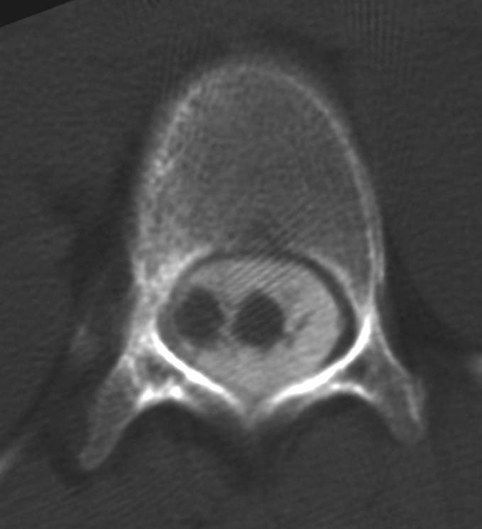
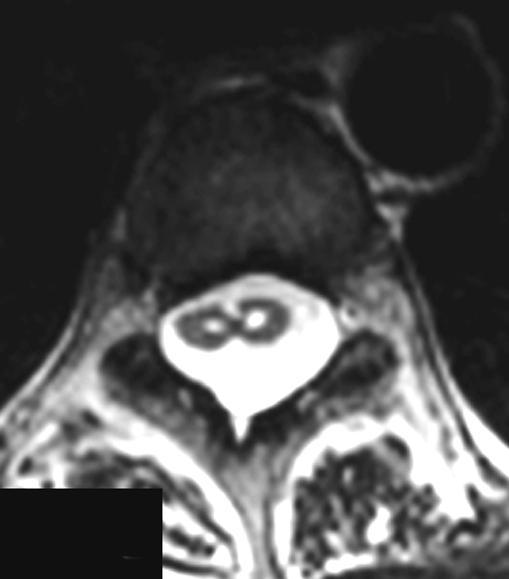
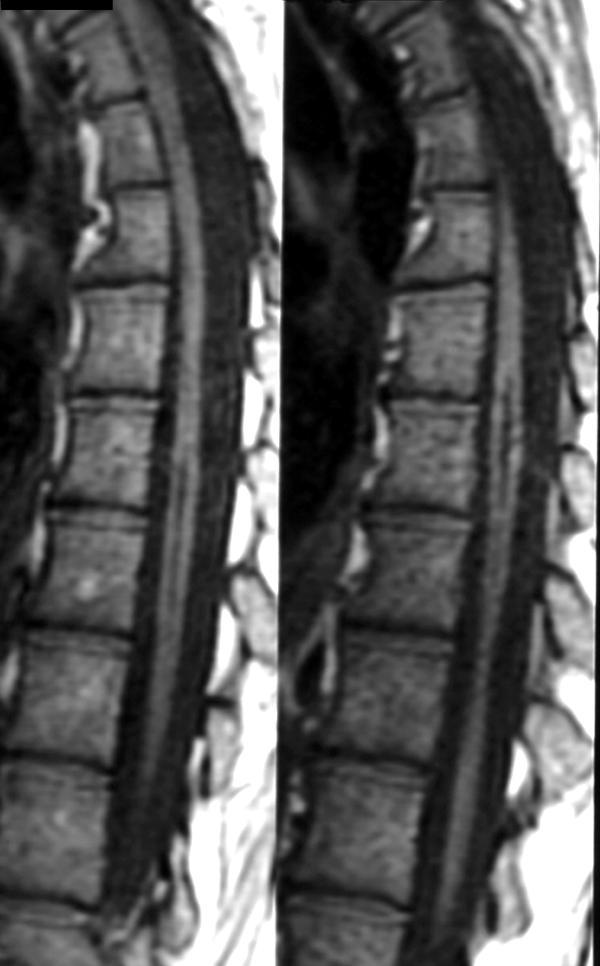
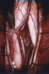
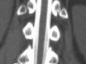
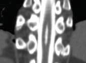
33 Duplication Anomalies Diastematomyelia Diplomyelia Trimyelia
34 Split Cord Malformation Diastematomyelia (splitting) & diplomyelia (duplication), not radiologically distinguishable Type I: less common, osseous septum dividing two dural tubes & hemicords, assoc vertebral anomalies Type II: more common, hemicords in single dural tube ± fibrous septum Each hemicord contains a central canal, dorsal horn & ventral horn, each 1 nerve root Can be asymmetrical, smaller easily missed Diplomyelia two complete separate cords
35 Split Cord Malformations Septum causes tethering,?scm around CT junction under-recognized Despite lack of signs and sx, ~ 75% abnormal voiding on formal testing Cutaneous stigmata (Type I), F > M Presentation (any age): scoliosis, tethering, unilateral lower extremity weakness, wasting
36 Type I Diastematomyelia
37 Type I Diastematomyelia
38 Diastematomyelia Type 1
39 Type I Diastematomyelia


40 Diastem Type 1 with bony spur
41 Type II Diastematomyelia
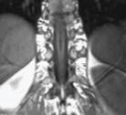
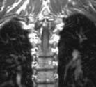

42 Diastematomyelia Type 2

43 Trimyelia fatal anomaly

44 Neurenteric Cyst Partial regression of neurenteric canal (of Kovalesky) Lined by secretory epithelium, contents similar to CSF or proteinaceous Intraspinal, ventral to thoracic cord, anywhere Associated with dysplastic vertebrae more than GI, respiratory anomalies Presents late teens, compressive signs & symptoms
45 Neurenteric Cyst and Canal
46 Neuroenteric Cyst
47 Neuroenteric Cyst
48 Neuroenteric Cyst
49 Dorsal Enteric Fistula Complex anomaly split notochord Persistence of neurenteric Canal of Kovalesky connects neural tube and archenteron. Fistula from bowel to dorsal skin Extends through spine and canal, usually lumbar High assoc with other CNS & non-cns anomalies (renal, GI, CDH, pulmonary)
50 Confused Sclerotome Syndrome Transitional anatomy - Hox Rib and Vertebral Fusion anomalies, Klippel-Feil Sprengels Deformity Congenital Short Pedicles

51 Sclerotomes Split!
52 Sprengels Deformity Congenital High Scapula, Otto Sprengel % female, Left > Right Assoc with other adjacent bony anomalies, Klippel Feil Omovertebral bone may connect scapula and lower cervical vertebra
53 CSS Sprengels, Klippel Feil
54 Congenital Short Pedicles Very common, not well defined Greater than 2/1 AP vertebral body to pedicle? Less than 1 cm pedicle Most Neurorads use the eyeball test
55 Congenital Short Pedicles


56 CSS Multiple fatal anomalies
57
58 CSS with craniorachischisis

59 Klippel Feil Syndrome

60 C1 Anomaly
61 Conjoined Nerve Root Two nerve sets; same root sleeve Most common nerve root anomaly Uncommonly associated with bony anomalies Usually asymptomatic but may induce diagnostic confusion and subsequent therapeutic misadventure. 6-8% incidence in some studies, if so underdiagnosed

62 Conjoined Nerve Root
63 Conclusion Spinal anomalies arise from diverse and complex etiologies and exhibit complex and overlapping appearances. There will come a time when our understanding of the underlying mechanisms of both normal and abnormal spinal cord development will make these differences much more coherent than they are now.
64
65
")
66 16mm (~8week) human embryo
Pediatric Spinal Anomalies
 Department of Radiology University of California San Diego Pediatric Spinal Anomalies John R. Hesselink, M.D. Spine Embryogenesis 1. Primitive streak 2. Proliferation of cells at primitive pit (Hensen's
SPLIT NOTOCHORD SYNDROME ASSOCIATION. DR. Hasan Nugud Consultant Paediatric Surgeon
 SPLIT NOTOCHORD SYNDROME ASSOCIATION DR. Hasan Nugud Consultant Paediatric Surgeon CASE PRESENTATION :- New born baby, boy, referred to the paediatric surgical team at the age of 14 hours. Birth History
Sonography of the Neonatal Spine: Part 2, Spinal Disorders
 Neonatal Spine Sonography Pediatric Imaging Pictorial Essay Downloaded from www.ajronline.org by 148.251.232.83 on 04/11/18 from IP address 148.251.232.83. Copyright RRS. For personal use only; all rights
Long segment composite split cord malformation with double bony spur
 Long segment composite split cord malformation with double bony spur Anand Sharma, Achal Sharma, R.S. Mittal SMS Medical College, Jaipur, India Abstract: A composite type of SCM is very rare and only a
CAUDAL REGRESSION SYNDROME
 CAUDAL REGRESSION SYNDROME *Prateek Gehlot 1 and Jagdish Mandliya 2 1 Department of Radio-Diagnosis, R.D.Gardi Medical College,, Ujjain (MP). 2 Department of Pediatrics, R.D.Gardi Medical College, Ujjain
Congenital Spine and Spinal Cord Malformations Pictorial Review
 JR Integrative Imaging LIFELONG LERNING FOR RDIOLOGY ongenital Spine and Spinal ord Malformations Pictorial Review Stephanie L. Rufener 1,2, Mohannad Ibrahim 2, harles. Raybaud 3, Hemant. Parmar 2 Downloaded
1 Normal Anatomy and Variants
 1 Normal Anatomy and Variants 1.1 Normal Anatomy MR Technique. e standard MR protocol for a routine evaluation of the spine always comprises imaging in sagittal and axial planes, while coronal images are
Neonatal Spinal Ultrasound Imaging - A Pictorial Review from The Royal Liverpool Children Hospital, Alder Hey, Liverpool
 Neonatal Spinal Ultrasound Imaging - A Pictorial Review from The Royal Liverpool Children Hospital, Alder Hey, Liverpool Poster No.: C-0081 Congress: ECR 2012 Type: Educational Exhibit Authors: K. Chetcuti,
Radiologic and pathologic features of spinal dysraphism. A pictorial review.
 Radiologic and pathologic features of spinal dysraphism. A pictorial review. Poster No.: C-0586 Congress: ECR 2011 Type: Educational Exhibit Authors: N. Arcalis, J. L. Ribó, J. Muchart, L. Riaza, J. Blanch
Development of Spinal Cord & Vertebral Column. Dr. Sanaa Alshaarawi & Prof. Ahmed Fathalla
 Development of Spinal Cord & Vertebral Column Dr. Sanaa Alshaarawi & Prof. Ahmed Fathalla OBJECTIVES At the end of the lecture, students should be able to: q Describe the development of the spinal cord
University Journal of Surgery and Surgical Specialties
 University Journal of Surgery and Surgical Specialties ISSN 2455-2860 Volume 2 Issue 1 2016 Profile of paediatric patients with split cord malformation MANORANJITHAKUMARI M Department of Neuro Surgery,
Diastematomyelia: A Case with Familial Aggregation of Neural Tube Defects
 Case Study TheScientificWorldJOURNAL (2004) 4, 847 852 ISSN 1537-744X; DOI 10.1100/tsw.2004.140 Diastematomyelia: A Case with Familial Aggregation of Neural Tube Defects Nuray Öksüz Kanbur 1, *, Pınar
Dorsal dermal sinus in children
 Dorsal dermal sinus in children Poster No.: C-2581 Congress: ECR 2015 Type: Educational Exhibit Authors: J. Marjanovic, A. Paterson, P. C. McSherry, A. Nixon, A. 1 1 2 1 2 1 1 2 TRIPALO BATOS, T. Grmoja
Development of the Digestive System. W.S. O The University of Hong Kong
 Development of the Digestive System W.S. O The University of Hong Kong Plan for the GI system Then GI system in the abdomen first develops as a tube suspended by dorsal and ventral mesenteries. Blood
Spinal congenital dermal sinus with dual ostia
 J Neurosurg Pediatrics 3:000 000, 3:407 411, 2009 Spinal congenital dermal sinus with dual ostia Clinical article Ch a n g Su b Le e, M.D., 1 Ji Ho o n Ph i, M.D., 2 Se u n g -Ki Kim, M.D., Ph.D., 2 By
Anatomy of the Nervous System. Brain Components
 Anatomy of the Nervous System Brain Components NERVOUS SYSTEM INTRODUCTION Is the master system of human body, controlling the functions of rest of the body systems Nervous System CLASSIFICATION A. Anatomical
Development of the Digestive System. W.S. O School of Biomedical Sciences, University of Hong Kong.
 Development of the Digestive System W.S. O School of Biomedical Sciences, University of Hong Kong. Organization of the GI tract: Foregut (abdominal part) supplied by coeliac trunk; derivatives include
Essentials of Clinical MR, 2 nd edition. 51. Primary Neoplasms
 51. Primary Neoplasms As with spinal central canal neoplasms in other regions, those of the lumbar spine may be classified as extradural, intradural extramedullary, and medullary. If an extradural lesion
Formation defects Scoliosis Deformities I 07 1
 What is congenital scoliosis? Congenital scoliosis is a spinal deformity with lateral deviation and rotation of the spinal column, where congenital dysfunctions in embryonal vertebra development cause
Ligaments of the vertebral column:
 In the last lecture we started talking about the joints in the vertebral column, and we said that there are two types of joints between adjacent vertebrae: 1. Between the bodies of the vertebrae; which
Role of helical CT and MRI in the evaluation of spinal dysraphism
 International Journal of Advances in Medicine Kumaran SK et al. Int J Adv Med. 2017 Feb;4(1):124-132 http://www.ijmedicine.com pissn 2349-3925 eissn 2349-3933 Original Research Article DOI: http://dx.doi.org/10.18203/2349-3933.ijam20170095
Scoliosis: Orthopaedic Perspectives
 Scoliosis: Orthopaedic Perspectives Scott B. Rosenfeld, MD Division of Pediatric Orthopaedic Surgery Texas Children s Hospital Page 0 xxx00.#####.ppt 9/23/2012 8:26:24 AM I have no disclosures Disclosures
The spinal dermal-sinus-like stalk
 Childs Nerv Syst (2009) 25:191 197 DOI 10.1007/s00381-008-0669-6 ORIGINAL PAPER The spinal dermal-sinus-like stalk J. van Aalst & E. A. M. Beuls & E. M. J. Cornips & H. W. M. van Straaten & A. F. M. Boselie
Moderators Dr A Suri Dr Deepak Gupta. Presented by Dr A Bisht
 Moderators Dr A Suri Dr Deepak Gupta Presented by Dr A Bisht A distinct group of congenital anomalies characterized by a failure of midline structures of ecto- and mesodermal origin to fuse Nicolas Tulp
A Retrospective Analysis of Clinical Profile and Surgical Outcome in Patients with Spinal Dysraphism at Tertiary Care Center
 Original Research Article A Retrospective Analysis of Clinical Profile and Surgical Outcome in Patients with Spinal Dysraphism at Tertiary Care Center Premlal KV * Assistant Professor, Department of Neurosurgery,
Midgut. Over its entire length the midgut is supplied by the superior mesenteric artery
 Gi Embryology 3 Midgut the midgut is suspended from the dorsal abdominal wall by a short mesentery and communicates with the yolk sac by way of the vitelline duct or yolk stalk Over its entire length the
Case Report Fatal Meningitis in a 14-Month-Old with Currarino Triad
 Case Reports in Radiology Volume 2016, Article ID 1346895, 5 pages http://dx.doi.org/10.1155/2016/1346895 Case Report Fatal Meningitis in a 14-Month-Old with Currarino Triad Hanan Mohammed Al Qahtani,
Prenatal ultrasound evaluation of fetal diastematomyelia: two cases of type I split cord malformation
 Ultrasound Obstet Gynecol 2000; 15: 78 82. Prenatal ultrasound evaluation of fetal diastematomyelia: two cases of type I split cord malformation L.M. ALLEN and R.K. SILVERMAN Perinatal Center, SUNY Health
disclosure Pediatric Tethered cord Syndrome Learning Objectives overview definiton Hoffman 1976 Pediatrics Grand Rounds 26 June 2015
 disclosure Pediatric Tethered cord Syndrome None Izabela, Tarasiewicz, MD,FRCS(C), has no relationships with commercial companies to disclose. Izabela Tarasiewicz MD. FRCS(C) Pediatric Neurosurgery overview
Johnson Rogers and colleagues- used Term LMM
 LIPOMENINGOMYELOCELE: CLASSIFICATION, MANAGEMENT AND CONTROVERSIES Definition : Lipomyelomeningocele is a form of OSD in which a subcutaneous fibrofatty mass traverses the lumbodorsal fascia, causes a
University Journal of Surgery and Surgical Specialties
 University Journal of Surgery and Surgical Specialties ISSN 2455-2860 Volume 2 Issue 1 2016 TWO RARE CASES OF DIASTEMATOMYELIA MUTHURAMAN P Department of Neuro Surgery, THANJAVUR MEDICAL COLLEGE Abstract
Neuroanatomy. Assistant Professor of Anatomy Faculty of Medicine The University of Jordan Dr Maha ELBeltagy
 Neuroanatomy Dr. Maha ELBeltagy Assistant Professor of Anatomy Faculty of Medicine The University of Jordan 2018 Development of the Central Nervous System Development of the nervous system Development
Skeletal System. Prof. Dr. Malak A. Al-yawer Department of Anatomy/Embryology Section
 Skeletal System Prof. Dr. Malak A. Al-yawer Department of Anatomy/Embryology Section Learning objectives At the end of this lecture, the medical student will be able to: State the embryonic origin of skeletal
Sonography of the Neonatal Spine: Part 1, Normal Anatomy, Imaging Pitfalls, and Variations That May Simulate Disorders
 Sonography of Neonatal Spine Pediatric Imaging Pictorial Essay Downloaded from www.ajronline.org by 46.3.195.60 on 02/04/18 from IP address 46.3.195.60. Copyright RRS. For personal use only; all rights
Disclosures None. Spinal Dysraphism
 Spinal Dysraphism Andrew Jea MD MHA FAAP Professor and Chief Section of Pediatric Neurosurgery Riley Hospital for Children Department of Neurosurgery Indiana University School of Medicine Goodman Campbell
A Very Unusual Case of a Dorsal Heteropagus Twin
 PRG A Very Unusual Case of a Dorsal Heteropagus Twin Nathan David P. Concepcion, MD 1, Bernard F. Laya, DO 1, Eduardo P. Manrique, MD 2 and Faith Caroline D. Bayabos, MD 1 1 Section of Pediatric Radiology,
Asymptomatic posterior cervical myelomeningocele with tethered cord in an adolescent: a rare form of spinal dysraphism with rare presentation
 Romanian Neurosurgery (2016) XXX 1: 113-117 113 Asymptomatic posterior cervical myelomeningocele with tethered cord in an adolescent: a rare form of spinal dysraphism with rare presentation Gangesh Gunjan,
Spine. Neuroradiology. Spine. Spine Pathology. Distribution of fractures. Radiological algorithm. Role of radiology 18/11/2015
 Spine Neuroradiology Spine Prof.Dr.Nail Bulakbaşı X Ray: AP/L/Oblique Vertebra & disc spaces CT & CTA Vertebra, discs, vessels MRI & MRA Vertebra, disc, vessels, meninges Spinal cord & nerves Myelography
Symptomatic Multiple Level Lateral Meningoceles with Intraspinal Meningocele: A Case Study and Its Surgical Management
 THIEME Original Article 15 Symptomatic Multiple Level Lateral Meningoceles with Intraspinal Meningocele: A Case Study and Its Surgical Management Vernon Velho 1 Sachin Guthe 1 Pravin Survashe 1 Poonam
May have excessive movement in the unfused segment to compensate. Flexion extension better preserved than lateral bend or rotation
 IV CONGENITAL SPINE KLIPPEL FLAIL SYNDROME Prevalence 0.60% Mainly around upper 3 vertebrae [75%] Commonest: C2 3 Lower Cervical spine fusion may be associated with syndromes: Fetal alcohol syndrome Goldenhar
Split cord malformation in children undergoing neurosurgical intervetion in India: a descriptive study
 ORIGINAL ARTICLE Journal of Pediatric Neurology 2004; 2(1): 21-27 www.jpneurology.org in children undergoing neurosurgical intervetion in India: a descriptive study Raj Kumar 1, Vinita Singh 2, Satya Narayan
Development of the urinary system
 Development of the urinary system WSO School of Biomedical Sciences, University of Hong Kong. 3 sets of kidneys developing in succession (temporally and spatially) : Pronephros ] Mesonephros ]- Intermediate
Prospective Evaluation of Role of MRI in Suspected Spinal Dysraphism and Its Management
 IOSR Journal of Dental and Medical Sciences (IOSR-JDMS) e-issn: 2279-0853, p-issn: 2279-0861.Volume 17, Issue 5 Ver. 1 (May. 2018), PP 21-28 www.iosrjournals.org Prospective Evaluation of Role of MRI in
Wound healing in trophic ulcers in spina bifida patients
 J Neurosurg 82:000 000, 1995 Wound healing in trophic ulcers in spina bifida patients VINOD KUMAR SRIVASTAVA, M.B.B.S, M.CH. Neurosurgical Unit, J. N. Medical College, Aligarh Muslim University, Aligarh,
Lecture 21Development of respiratory system Dr. Rehan Asad At the end of session students should able to Describe formation of lung buds Describe
 Lecture 21Development of respiratory system Dr. Rehan Asad At the end of session students should able to Describe formation of lung buds Describe development of larynx, trachea and bronchi. Describe the
MR Imaging in the Tethered Spinal Cord Syndrome
 27 MR Imaging in the Tethered Spinal Cord Syndrome Narasimhachari Raghavan 1 A. James Barkovich 1 Michael Edwards 2 David Norman 1 MR examinations of the spine were reviewed in 25 patients with a clinical
A Journey Down The Canal
 A Journey Down The Canal Radiological Assessment of Spinal Cord Masses John Berry-Candelario HMS III Gillian Lieberman, MD BIDMC Objectives Patient review Anatomy of the spine Imaging techniques Classification
LUMPS, TUFTS AND DIMPLES IT S THE PITS!! Session Information. Faculty Disclosure Information
 Session Information Session Title: Lumps, Tufts and Dimples Session Number: F3056, F2130 Faculty Name: Mark S. Dias, MD, FAAP Faculty Institution: Penn State Children s Hospital, Penn State University
Intraoperative Sonography in Spinal Dysraphism and Syringohydromyelia
 329 Intraoperative Sonography in Spinal Dysraphism and Syringohydromyelia Robert M. Quencerl erta M. Montalvo 2 Thomas P. Naidich 3 M. Judith Donovan Post 1 arth. Green 4 Larry K. Page 4 The use of intraoperative
Terminal and nonterminal myelocystoceles
 See the corresponding editorial in this issue, pp 85 86. J Neurosurg (2 Suppl Pediatrics) 107:87 97, 2007 Terminal and nonterminal myelocystoceles NATARAJAN MUTHUKUMAR, M.CH. Department of Neurosurgery,
Magnetic resonance imaging in the prenatal diagnosis of neural tube defects
 Insights Imaging (2013) 4:225 237 DOI 10.1007/s13244-013-0223-2 PICTORIAL REVIEW Magnetic resonance imaging in the prenatal diagnosis of neural tube defects A. Zugazaga Cortazar & C. Martín Martinez &
What Every Spine Surgeon Should Know About Neurosurgical Issues
 What Every Spine Surgeon Should Know About Neurosurgical Issues Amer Samdani, MD Chief of Surgery Shriners Hospitals for Children Philadelphia, PA Objectives Main intraspinal lesions Chiari malformation
Spinal canal stenosis Degenerative diseases F 06
 What is spinal canal stenosis? The condition known as spinal canal stenosis is a narrowing (stenosis) of the spinal canal that in most cases develops due to the degenerative (wear-induced) deformation
NEURORADIOLOGY. Part III. Angela Csomor University of Szeged Department of Radiology
 NEURORADIOLOGY Part III Angela Csomor University of Szeged Department of Radiology DISEASES OF SPINE AND SPINAL CORD I. Non-tumourous diseases developmental anomalies vascular disorders inflammatory processes
Congenital Brain and Spinal Cord Malformations and Their Associated Cutaneous Markers
 CLINICAL REPORT Guidance for the Clinician in Rendering Pediatric Care Congenital Brain and Spinal Cord Malformations and Their Associated Cutaneous Markers Mark Dias, MD, FAANS, FAAP, Michael Partington,
Endoscopic Assisted resection for congenital Midline Nasal Mass
 Endoscopic Assisted resection for congenital Midline Nasal Mass Ahmed Aly Ibrahim A.prof ORL Department Alexandria University Emad. A Magdy prof ORL Department Alexandria University Haytham Morsi,MD Mohammad
Spinal Cord and Properties of Cerebrospinal Fluid: Options for Drug Delivery. SMA Foundation New York
 Spinal Cord and Properties of Cerebrospinal Fluid: Options for Drug Delivery New York Why Do We Need to Know about the Spinal Cord Anatomy and Properties of Cerebrospinal Fluid? SMA therapeutics need to
SPINAL CORD AND PROPERTIES OF CEREBROSPINAL FLUID: OPTIONS FOR DRUG DELIVERY
 SPINAL CORD AND PROPERTIES OF CEREBROSPINAL FLUID: OPTIONS FOR DRUG DELIVERY WHY DO WE NEED TO KNOW ABOUT THE SPINAL CORD ANATOMY AND PROPERTIES OF CEREBROSPINAL FLUID? SMA therapeutics need to reach cells
Early Development of Neural Tube Development of Medulla Spinalis and Peripheral Nervous System. Assoc.Prof. E.Elif Güzel, M.D.
 Early Development of Neural Tube Development of Medulla Spinalis and Peripheral Nervous System Assoc.Prof. E.Elif Güzel, M.D. Third week of Embryogenesis Primitive streak/pit appears on the epiblast (day
Chapter 8. Pediatric Surgery
 Chapter 8 Pediatric Surgery 8.1 Hydrocephalus Hydrocephalus is a congenital disorder. There may be difficulties during normal vaginal delivery due large size of the head. In 1970s, when these pictures
Role of MRI in the Evaluation of Spinal Dysraphism
 Role of MRI in the Evaluation of Spinal Dysraphism 1 Dr Ravi N, 2 Dr Ramesh V, 3 Dr Naveen K, G, 4 Dr R Nagaraj, 2 Dr darsh K M, 2 Dr Manjappa H, 2 Dr Lakshmeesha M T 1 ssociate Professor, 2 Resident,
Development of Respiratory System. Dr. Sanaa Alshaarawy& Dr. Saeed Vohra
 Development of Respiratory System Dr. Sanaa Alshaarawy& Dr. Saeed Vohra OBJECTIVES At the end of the lecture the students should be able to: Identify the development of the laryngeotracheal (respiratory)
Case Report Surgical Treatment of a Patient with Human Tail and Multiple Abnormalities of the Spinal Cord and Column
 SAGE-Hindawi Access to Research Advances in Orthopedics Volume 2011, Article ID 153797, 4 pages doi:10.4061/2011/153797 Case Report Surgical Treatment of a Patient with Human Tail and Multiple Abnormalities
Spectrum of Magnetic Resonance Imaging findings in infective intra spinal complications of dermal sinus and associated inclusion cysts
 Spectrum of Magnetic Resonance Imaging findings in infective intra spinal complications of dermal sinus and associated inclusion cysts Poster No.: C-1443 Congress: ECR 2015 Type: Educational Exhibit Authors:
Imaging in neurofibromatosis type 1: An original research article with focus on spinal lesions
 Original Research Article Imaging in neurofibromatosis type 1: An original research article with focus on spinal lesions Kalpesh Patel 1*, Siddharth Zala 2, C. Raychaudhuri 3 1 Assistant Professor, 2 1
Introduction to Neuroimaging spine. John J. McCormick MD
 Introduction to Neuroimaging spine John J. McCormick MD Neuroanatomy Netter drawings Radiographic Anatomy Cervical Spine Cervical Spine Oblique View Cervical Spine Dens View Thoracic Spine Lumbar Spine
Week 14. Development of the Musculoskeletal System
 Week 14 Development of the Musculoskeletal System Skeletal System Derived from: paraxial mesoderm somites and somitomeres sclerotome sclerotome differentiation induced by SHH from notochord and floor plate
Synovial cyst of spinal facet
 Case report CHUN C. KAO, M.D., STEFAN S. WINKLER, M.D., AND J. H. TURNER, M.D. Sections of Neurosurgery, Radiology, and Pathology, Madison Veterans Administration Hospital, and University of Wisconsin,
B. Normal variants and pitfalls
 B. Normal variants and pitfalls Poster No.: A-371 Congress: ECR 2011 Type: Invited Speaker Authors: B. Tins; Oswestry/UK Keywords: Musculoskeletal spine, Anatomy DOI: 10.1594/ecr2011/A-371 Any information
A Congenital Defect in the Spinal Cord of the Manx Cat
 Vet, Path. 8: 232-238 (1971) A Congenital Defect in the Spinal Cord of the Manx Cat A. H. MARTIN Department of Anatomy, University of Wisconsin, Madison Wisc. Abstract. The lumbar part of the spinal cords
Introduction to Neurosurgical Subspecialties:
 Introduction to Neurosurgical Subspecialties: Pediatric Neurosurgery Brian L. Hoh, MD 1 and Gregory J. Zipfel, MD 2 1 University of Florida, 2 Washington University Pediatric Neurosurgery Pediatric neurosurgeons
Neurologic improvement after thoracic, thoracolumbar, and lumbar spinal cord (conus medullaris) injuries
 injuries") Thomas Jefferson University Jefferson Digital Commons Department of Orthopaedic Surgery Faculty Papers Department of Orthopaedic Surgery 1-2011 Neurologic improvement after thoracic, thoracolumbar, and
Thomas Jefferson University Jefferson Digital Commons Department of Orthopaedic Surgery Faculty Papers Department of Orthopaedic Surgery 1-2011 Neurologic improvement after thoracic, thoracolumbar, and
Tethered spinal cord syndrome: a developmental overview
 International Journal of Sciences & Applied Research www.ijsar.in Tethered spinal cord syndrome: a developmental overview Anushi Singh 1 *, Rekha Kumari 2 1 CHN Department, School of Nursing Science and
MRI of chronic spinal cord injury
 The British Journal of Radiology, 76 (2003), 347 352 DOI: 10.1259/bjr/11881183 E 2003 The British Institute of Radiology Pictorial review MRI of chronic spinal cord injury 1 K POTTER, FRCR and 1 A SAIFUDDIN,
Spinal Cord Injuries: The Basics. Kadre Sneddon POS Rounds October 1, 2003
 Spinal Cord Injuries: The Basics Kadre Sneddon POS Rounds October 1, 2003 Anatomy Dorsal columntouch, vibration Corticospinal tract- UMN Anterior horn-lmn Spinothalamic tractpain, temperature (contralateral)
AXIAL SKELETON FORM THE VERTICAL AXIS OF THE BODY CONSISTS OF 80 BONES INCLUDES BONES OF HEAD, VERTEBRAL COLUMN, RIBS,STERNUM
 AXIAL SKELETON FORM THE VERTICAL AXIS OF THE BODY CONSISTS OF 80 BONES INCLUDES BONES OF HEAD, VERTEBRAL COLUMN, RIBS,STERNUM APPENDICULAR SKELETON BONES OF THE FREE APPENDAGES & THEIR POINTS OF ATTACHMENTS
Development of the Axial Skeleton and Limbs. Professor Alfred Cuschieri Department of Anatomy University of Malta
 Development of the Axial Skeleton and Limbs Professor Alfred Cuschieri Department of Anatomy University of Malta During the Fourth Week the Embryo Is Segmented. Each segment consists of: a segment of neural
Human Anatomy. Spinal Cord and Spinal Nerves
 Human Anatomy Spinal Cord and Spinal Nerves 1 The Spinal Cord Link between the brain and the body. Exhibits some functional independence from the brain. The spinal cord and spinal nerves serve two functions:
Giant thoracolumbar extradural arachnoid cyst : A case report
 Acta Orthop. Belg., 2008, 74, 709-713 CASE REPORT Giant thoracolumbar extradural arachnoid cyst : A case report Muddassir RASHID, Anjum SYED, Ibne AHMAD, EKRAMULLAH From the Jawaharlal Nehru Medical College
Central Nervous System Congenital Abnormalities
 Central Nervous System Congenital Abnormalities Eva Brichtova, M.D., Ph.D., Department of Pediatric Sugery, Orthopaedics and Traumatology, University Hospital Brno Neural tube defects Dysraphism uncomplete
ANATOMY OF SPINAL CORD. Khaleel Alyahya, PhD, MEd King Saud University School of
 ANATOMY OF SPINAL CORD Khaleel Alyahya, PhD, MEd King Saud University School of Medicine @khaleelya OBJECTIVES At the end of the lecture, students should be able to: Describe the external anatomy of the
LUMBAR SPINAL STENOSIS
 LUMBAR SPINAL STENOSIS Always occurs in the mobile segment. Factors play role in Stenosis Pre existing congenital or developmental narrowing of the lumbar spinal canal Translation of one anatomic segment
Radiographic characteristics in congenital scoliosis associated with split cord malformation: a retrospective study of 266 surgical cases
 Feng et al. BMC Musculoskeletal Disorders (2017) 18:420 DOI 10.1186/s12891-017-1782-z RESEARCH ARTICLE Open Access Radiographic characteristics in congenital scoliosis associated with split cord malformation:
Lipomyelomeningocele for the Urologist: should we view it the same as myelomeningocele?
 Lipomyelomeningocele for the Urologist: should we view it the same as myelomeningocele? Grace Yoshiba BS Chris Halline, BA Earl Y. Cheng MD Theresa A. Meyer RN Ilina Rosoklija MPH Robin Bowman MD Elizabeth
Spine and spinal cord
 NEURORADIOLOGY Spine and spinal cord Erika Vörös University of Szeged Department of Radiology SZEGED DISEASES OF SPINE AND SPINAL CORD I. Non-tumourous diseases developmental anomalies vascular disorders
Series of vertebral synostosis-clinically implied
 Original article: Series of vertebral synostosis-clinically implied Dr. Somanath Deepa, Dr. Rajasekar SS Department of Anatomy, Sri Manakula Vinayagar Medical College and Hospital, Puducherry (U.T.), India.
CNS Embryology 5th Menstrual Week (Dorsal View)
") Imaging of the Fetal Brain; Normal & Abnormal Alfred Abuhamad, M.D. Eastern Virginia Medical School CNS Embryology 5th Menstrual Week (Dorsal View) Day 20 from fertilization Neural plate formed in ectoderm
Imaging of the Fetal Brain; Normal & Abnormal Alfred Abuhamad, M.D. Eastern Virginia Medical School CNS Embryology 5th Menstrual Week (Dorsal View) Day 20 from fertilization Neural plate formed in ectoderm
I have no disclosures
 I have no disclosures Provide an overview of the spectrum of congenital upper extremity anomalies Describe the key imaging findings of these abnormalities Discuss the important clinical features of these
Congenital Spinal Lipoma: analyzing the perplexed nomenclature and our management
 Congenital Spinal Lipoma: analyzing the perplexed nomenclature and our management Nidal Khasawneh MD *, Rami Alqroom MD *, Firas Sha'ban MD *, Rafeed Al Drous MD *, Rima Nserat **, Amer Al Shurbaji MD
CT Myelography and MR Imaging of Extramedullary Cysts of the Spinal Canal in Adult and Pediatric Patients
 Downloaded from www.ajronline.org by 37.44.199.154 on 12/23/17 from IP address 37.44.199.154. opyright RRS. For personal use only; all rights reserved T Myelography and MR Imaging of Extramedullary ysts
W IDENING of an intervertebral foramen
 Agenesis of a Pedicle in the Cervical Spine LESLIE i. ZATZ, M.D., PETER W. BURGESS, M.D. AND John W. HANBERY, M.D. Departments of Radiology and of Surgery, Neurosurgical Division, Stanford University School
Central Nervous System: Part 2
 Central Nervous System: Part 2 1. Meninges 2. CSF 3. Spinal Cord and Spinal Nerves Explain spinal cord anatomy, including gray and white matter and meninges (give the general functions of this organ).
Human Anatomy - Problem Drill 11: The Spinal Cord and Spinal Nerves
 Human Anatomy - Problem Drill 11: The Spinal Cord and Spinal Nerves Question No. 1 of 10 Instructions: (1) Read the problem statement and answer choices carefully, (2) Work the problems on paper as needed,
Purpose: To discuss the fatty filum terminale which is incidentally demonstrated on MRI concerning the causes of TCS
 ISPUB.COM The Internet Journal of Spine Surgery Volume 3 Number 1 T Iizuka Citation T Iizuka.. The Internet Journal of Spine Surgery. 2006 Volume 3 Number 1. Abstract Background: Fatty filum terminale
Radiography of the Spine
 Radiography of the Spine Radiography of the Spine Attila ARANY-TóTH, DVM Complex anatomy Vertebrae: 7 cervical, 13 thoracal, 7 lumbal, 3 sacral, n caudal Thorough neurological examination - localization!!!
A QUANTITATIVE STUDY OF THE PRENATAL CHANGES IN ANGULATION OF THE SPINAL NERVES
 A QUANTITATIVE STUDY OF THE PRENATAL CHANGES IN ANGULATION OF THE SPINAL NERVES ALEXANDER BARRY Department of Anatomy, Wniversity of Michigan, Ann Arbor SEVEN FIGURES The University of Michigan Embryological
Comprehension of the common spine disorder.
 Objectives Comprehension of the common spine disorder. Disc degeneration/hernia. Spinal stenosis. Common spinal deformity (Spondylolisthesis, Scoliosis). Osteoporotic fracture. Anatomy Anatomy Anatomy
Chiari III Joseph Junewick, MD FACR
 Chiari III Joseph Junewick, MD FACR 07/02/2010 History Newborn with suboccipital mass. Diagnosis Chiari III Additional Clinical Surgery-Skin covered suboccipital cystic mass confined by the dura. Pathology-Leptomeningeal
Visualization of Sacral Nerve Roots via Percutaneous Intraspinal Navigation (PIN) V
 V") AJNR Am J Neuroradiol 26:2420 2424, October 2005 Technical Note Visualization of Sacral Nerve Roots via Percutaneous Intraspinal Navigation (PIN) V Takuya Fujimoto, Brian P. Giles, Robert E. Replogle,
AJNR Am J Neuroradiol 26:2420 2424, October 2005 Technical Note Visualization of Sacral Nerve Roots via Percutaneous Intraspinal Navigation (PIN) V Takuya Fujimoto, Brian P. Giles, Robert E. Replogle,