LUMPS, TUFTS AND DIMPLES IT S THE PITS!! Session Information. Faculty Disclosure Information
|
|
|
- Madeline Andrews
- 6 years ago
- Views:
Transcription

1 Session Information Session Title: Lumps, Tufts and Dimples Session Number: F3056, F2130 Faculty Name: Mark S. Dias, MD, FAAP Faculty Institution: Penn State Children s Hospital, Penn State University Hershey, PA Faculty Disclosure Information In the past 12 months, I have no relevant financial relationships with the manufacturer(s) of any commercial product(s) and/or provider(s) of commercial services discussed in this CME activity. I do (or) do not intend to discuss an unapproved/investigative use of a commercial product/device in my presentation. LUMPS, TUFTS AND DIMPLES IT S THE PITS!! Mark S. Dias, MD, FAAP Departments of Neurosurgery and Pediatrics Penn State Children s Hospital Penn State University College of Medicine Hershey, PA 1


2 Embryology of the Nervous System: Gastrulation Primitive streak elongates from caudal to cranial end (POD 13-16) Prospective mesoderm ingresses through primitive groove Remaining epiblast cells spread out, replace ingressing mesoderm cells Embryology of the Nervous System: Gastrulation Hensen s node at cranial end of the primitive streak Prospective notochordal cells ingress through node and become notochord Primitive streak caudally, becomes caudal cell mass 2


3 Embryology of the Nervous System: Primary Neurulation POD Neural groove forms as median hingepoint Neural folds develop Neural folds fuse Separation of neuro- and cutaneous ectoderm (dysjunction) Embryology of the Nervous System: Primary Neurulation Cranial neuropores last points to close Caudal neuropore at level of S2 Cranial neuropore at lamina terminalis, just above optic chiasm Additional occipital neuropore Neuropores most common sites of dermal sinus tracts 3


4 Embryology of Nervous System: Secondary Neurulation Begins on POD 27 Involves spinal cord caudal to S2 and filum terminale Formed from caudal cell mass Occurs in the presence of an intact cutaneous ectoderm Species specific Embryology of the Nervous System: Ascent of Conus Medullaris POD 42-54: Retrogressive differentiation Caudal neural tube thinner No mantle zone Rudimentary marginal zone Looks less well developed POD 54 and beyond: Differential growth of spinal cord and vertebral column Embryology of the Nervous System: Ascent of Conus Medullaris Most ascent pre-natally Conus at or above L2-3 disc space at birth Conus at or above L1-2 disc space by 2 months post-natal 4
: Middle third L2 Below inferior third of L2 considered radiographically tethered Kesler, Dias, Kalapos: Neurosurg Focus 2007 Innocent Coccygeal Dimples Coccygeal")
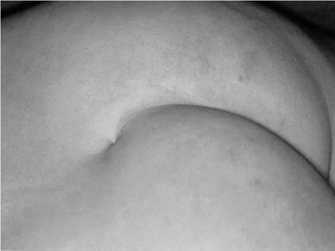
5 L S1 Level of Normal Conus Medullaris 100 children with brain tumors having screening whole spine imaging Level of conus medullaris measured Mode: L1-2 disc space Mean: Inferior third of L1 Lower border (95% confidence limits): Middle third L2 Below inferior third of L2 considered radiographically tethered Kesler, Dias, Kalapos: Neurosurg Focus 2007 Innocent Coccygeal Dimples Coccygeal - within the gluteal cleft Tip of coccyx palpable within few millimeters Normal gluteal cleft Shallow, non-complex dimple No tufts of hair, hemangiomata, skin appendages or skin tags No neurological, urological, or orthopedic abnormalities Remnant of tail bud/hensen s node 5

 Benign coccygeal dimple Benign")
6 Innocent Coccygeal Dimples Prevalence up to 4% of population Not related to pilonidal cysts in adults No convincing evidence for relationship with congenital spinal cord malformations Only 7 cases with SC abnormalities in literature 5/7 cases had other cutaneous markers 2 not clear whether dimple was within gluteal cleft Powell: 2000 cases, 1 with tract to outer dura Herman 0/53, Gibson 0/75 had abnormalities Weprin and Oakes: 1000 cases, no evidence of tethering or deterioration (imaging not done) Benign coccygeal dimple Benign coccygeal dimple 6
 Lumbosacral (above gluteal crease) Abnormal gluteal cleft Deep or complex Tufts of hair,")
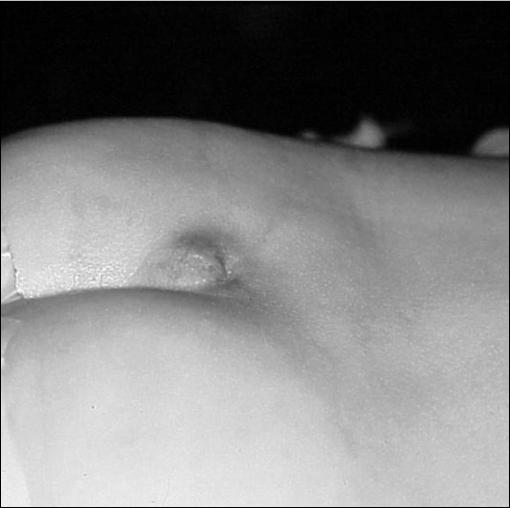
7 Spinal Dermal Sinus Tracts (DST) Incidence 1 in 2500 births 1% cervical 10% thoracic 41% lumbar 35% lumbosacral May be associated with other congenital spinal cord malformations Split cord malformations, lipoma, thickened filum terminale, endodermal or dermoid cyst Spinal Dermal Sinus Tracts (DST) Lumbosacral (above gluteal crease) Abnormal gluteal cleft Deep or complex Tufts of hair, hemangiomata, appendages or skin tags Neurological, urological, or orthopedic abnormalities 7


8 8

9 9


10 Embryology of DST Failure of neuroectoderm to separate from cutaneous ectoderm during dysjunction Tongue of cutaneous ectoderm remains attached to neural tube Persistence of tract variable Some tracts involve subcutaneous tissues only, most remain attached to spinal cord Most common at posterior neuropore Tract therefore ends along dorsum of spinal cord at level of S2, separate from filum terminale 10
")
 Mass effect from expanding dermoid inclusion")
11 Pathophysiology of Deterioration in Spinal DST Spinal cord tethering Bacterial meningitis (portal of entry) Aseptic meningitis (desquamation of epithelial debris within CSF) Mass effect from expanding dermoid inclusion cyst 11

12 Imaging of Spinal DST Spinal Ultrasound Good screening tool, but not sensitive Probably not worthwhile after 6 months of age Magnetic resonance Imaging Definitive neuroimaging study More sensitive than sonography Missed 2/3 of tracts in one study Conus may not be abnormally low Need for operation depends upon clinical appearance and location of dimple!!!! Dermal Sinus Tract with Fascial Penetration Clinical Presentation of Spinal DST Focal neurological deficits in 2/3 at initial neurosurgical evaluation Motor weakness 39% Sensory changes 25% Gait changes 18% Sphincter disturbances 21% Bowel and bladder changes 14% Age < 1 yr: 50% with neurological deficits Age > 1 yr: 92% with neurological deficits Infection in 3/28 (1 with meningitis) Ackerman, Menezes: Pediatrics,


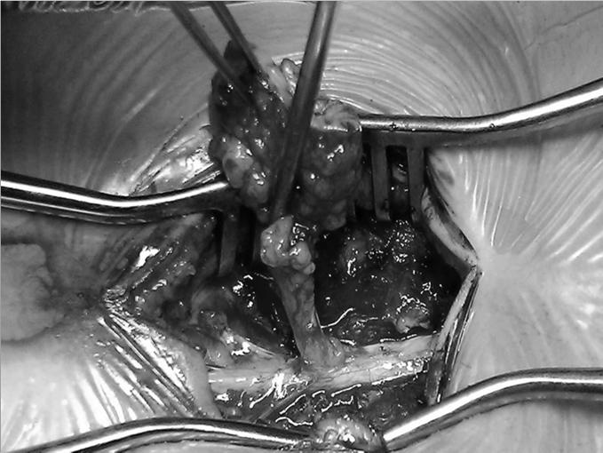
13 Management of Spinal DST Linear vertical incision, ellipse around tract Follow tract to defect in lumbodorsal fascia Laminectomy adjacent to tract Identify penetration of dura Open dura cranial and caudal to track, ellips dural opening around tract Follow tract to conus medullaris Look for separate filum terminale 13


14 Meningocele Manque Likely related embryologically to DST Scarified, cigarette paper, cigarette burn skin lesion May be tender to touch No CSF leakage Fibrous tract or atretic peripheral nerves, dorsal root ganglion cells 50% associated with split cord malformations 14

15 15

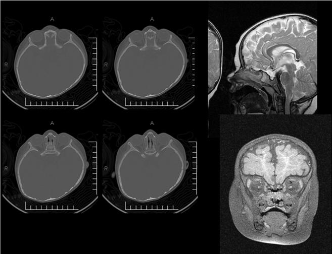
16 Cranial Dermal Sinus Tracts Less common than spinal DST Occipital, retro-auricular, nasofrontal location Modes of deterioratoin Intracranial suppurative infection Epidural abscess, subdural empyema, recurrent bouts of meningitis, brain abscess Growing intracranial dermoid or epidermoid masses Occipital DST Cutaneous pit or tract usually located near occipital external protuberance Skull X-rays and CT/or scans demonstrate bone defect at site of cutaneous lesion MRI better demonstrates soft tissue tract Invariable intracranial extension Associated hairy nevus (2/8 cases) or subcutaneous mass (4 of 8 cases) 16


17 Frontonasal DST Dimple or dermoid extending anywhere from nasal tip to glabella Innocuous looking Travel between nasal bone and nasal cartilage toward anterior skull base 90% end extra-cranially 10% extend through foramen cecum, anterior to crista galli, and end intracranially Extradural, intra-falx, subarachnoid space, lamina terminalis 17


18 18


19 Embryology of Cranial DST Failure of dysjunction at cranial neuropores Occipital midline: occipital lobes and/or cerebellum Lamina terminalis 19


20 Surgical Management of Frontonasal DST If no intracranial extension on neuroimaging, excise superficial tract, follow toward skull base If tract ends extracranially, no need for further exploration If intracranial extension found, plan intracranial exposure at same or later operation If obvious intracranial extension, operation planned jointly with plastic and neurosurgery 20

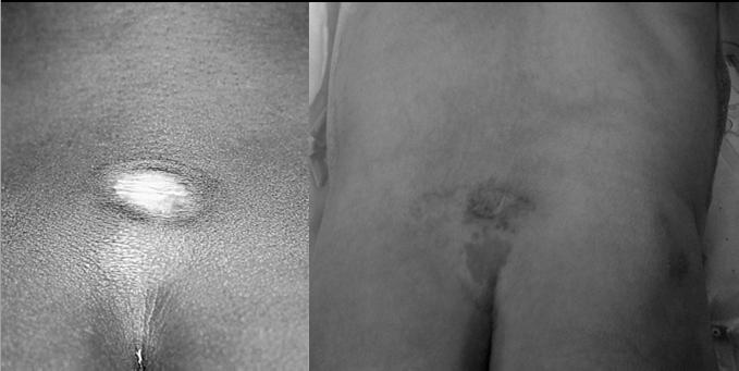
21 Flammeus Nevus and Spinal Lipomas Flat, not raised, port wine in color Similar in appearance to stork bite Irregular outline, blanches with pressure May be associated with other cutaneous markers of spinal dysraphism Dermal sinus tract, subcutaneous masses Meningocele manque Fawn s tail Hairy nevus Tubbs, JNS:Pediatrics, 2004 Management of Flammeus Nevus Incidence of underlying spinal cord malformations is unknown 21 (17%) of 120 patients with occult spinal dysraphism in one series had isolated flammeus nevus as the only manifestation (N=21) Lipoma 3 Fatty filum terminale 5 Dermal sinus tract with lipoma 2 Syringomyelia 8 Meningocele manque 1 Split cord malformation 2 No specific malformation associated with flammeus nevus Tubbs, JNS:Pediatrics,
 Diastematomyelia, diplomyelia Present in 11/14")
 Dysplastic skin in 2/14 (14%) Subcutaneous mass in 2 (14%)")
22 Hypertrichosis (Fawn s Tail) Most commonly associated with, and specific for, split cord malformations (SCM) Diastematomyelia, diplomyelia Present in 11/14 (79%) of patients with SCM Associated with other cutaneous markers Flammeus nevus in 3/14 (21%) Dysplastic skin in 2/14 (14%) Subcutaneous mass in 2 (14%) 22
 Differentiation arbitrary and confusing, not helpful since BOTH")

23 Human Tails and Pseudotails True tail: contains both cutaneous and bony structures (stiff), remnant of human tail Pseudotail: lacks bony structures (soft) Differentiation arbitrary and confusing, not helpful since BOTH are associated with congenital dysraphic malformations Congenital spinal anomalies in 50% Spinal cord tethering in 25% MRI for further evaluation Surgical exploration, excision and spinal cord untethering Lu, Pediatr Neurol


24 Atretic Parietal Encephaloceles Small, usually elevated lesions with dysplastic skin over dome Whorling pattern of surrounding hair No CSF leakage Bisects sagittal sinus Underlying MRI abnormalities common, MRV demonstrates persistent embryonic prosencephalic vein Neurological/cognitive abnormalities in 40% 24

25 Cutis Aplasia Congenita Rare, most often involves midline scalp May involve scalp, scalp and bone or scalp, bone and dura Overlies superior sagittal sinus Significant hemorrhage possible Sometimes associated with other malformations Cerebral malformations, TEF, frontonasal dysplasia, congenital heart defects, facial palsy, mental retardation, cleft-lip/palate Adams Oliver syndrome (CAC + limb defects) Cutis Aplasia Congenita: Management Early identification of lesion Do NOT allow to dry out. Keep moist!!!! Cover with wet gauze and plastic wrap Wet to wet dressings Cover with petroleum based antibiotic ointment Two surgical management strategies Non-operative: wet to wet dressing changes Operative: primary closure or scalp rotation flaps 25
26 Conclusions Innocent coccygeal dimples are located within the gluteal cleft No associated cutaneous lesions Depth of pit not important No need for further workup, imaging, or surgery Pathological lumbosacral DST are more cranially located, outside of gluteal cleft Imaging important for surgical planning but of no importance for determining need for surgery Might as well go directly to MRI rather than US Prophylactic surgical exploration necessary Conclusions Cranial DST Recognize importance of seemingly innocent midline dimple or bump anywhere on nose Suspicion in cases of intracranial suppurative infections in expected locations, or recurrent bouts of unexplained meningitis Neuroimaging may include both CT and MRI Do not inject dye into any tract Combined surgical approach with experienced pediatric neurosurgical and plastic/craniofacial surgical expertise Conclusions Flammeus nevus is associated with various forms of spinal dysraphism (incidence unknown) Hypertrichosis (fawn s tail) most often associated with, and specific for, SCM Human tails associated with spinal cord malformations in 50%, tethering in 25% 26


27 Conclusions Atretic encephaloceles look benign, associated with underlying brain malformations Whorls of hair, skin covered Cutis aplasia congenita similar but without whorls of hair, not skin covered, sometimes has underlying skull and dural defect Hemorrhage from SSS, keep it moist! Surgical and non-surgical management proposed Conclusions Midline cutaneous anomalies other than coccygeal dimples should be considered to have associated underlying CNS malformations and should be referred to a specialist for evaluation and treatment 27
Pediatric Spinal Anomalies
 Department of Radiology University of California San Diego Pediatric Spinal Anomalies John R. Hesselink, M.D. Spine Embryogenesis 1. Primitive streak 2. Proliferation of cells at primitive pit (Hensen's
SPLIT NOTOCHORD SYNDROME ASSOCIATION. DR. Hasan Nugud Consultant Paediatric Surgeon
 SPLIT NOTOCHORD SYNDROME ASSOCIATION DR. Hasan Nugud Consultant Paediatric Surgeon CASE PRESENTATION :- New born baby, boy, referred to the paediatric surgical team at the age of 14 hours. Birth History
Congenital Brain and Spinal Cord Malformations and Their Associated Cutaneous Markers
 CLINICAL REPORT Guidance for the Clinician in Rendering Pediatric Care Congenital Brain and Spinal Cord Malformations and Their Associated Cutaneous Markers Mark Dias, MD, FAANS, FAAP, Michael Partington,
Dorsal dermal sinus in children
 Dorsal dermal sinus in children Poster No.: C-2581 Congress: ECR 2015 Type: Educational Exhibit Authors: J. Marjanovic, A. Paterson, P. C. McSherry, A. Nixon, A. 1 1 2 1 2 1 1 2 TRIPALO BATOS, T. Grmoja
University Journal of Surgery and Surgical Specialties
 University Journal of Surgery and Surgical Specialties ISSN 2455-2860 Volume 2 Issue 1 2016 TWO RARE CASES OF DIASTEMATOMYELIA MUTHURAMAN P Department of Neuro Surgery, THANJAVUR MEDICAL COLLEGE Abstract
Endoscopic Assisted resection for congenital Midline Nasal Mass
 Endoscopic Assisted resection for congenital Midline Nasal Mass Ahmed Aly Ibrahim A.prof ORL Department Alexandria University Emad. A Magdy prof ORL Department Alexandria University Haytham Morsi,MD Mohammad
Persistent Terminal Ventricle
 Persistent Terminal Ventricle Ventriculus Terminalis Incomplete regression of TV of 2 neurulation, continuity with central canal small cavity PTV vs terminal myelocystocele (?severe manifestation from
Anatomy of the Nervous System. Brain Components
 Anatomy of the Nervous System Brain Components NERVOUS SYSTEM INTRODUCTION Is the master system of human body, controlling the functions of rest of the body systems Nervous System CLASSIFICATION A. Anatomical
The spinal dermal-sinus-like stalk
 Childs Nerv Syst (2009) 25:191 197 DOI 10.1007/s00381-008-0669-6 ORIGINAL PAPER The spinal dermal-sinus-like stalk J. van Aalst & E. A. M. Beuls & E. M. J. Cornips & H. W. M. van Straaten & A. F. M. Boselie
Long segment composite split cord malformation with double bony spur
 Long segment composite split cord malformation with double bony spur Anand Sharma, Achal Sharma, R.S. Mittal SMS Medical College, Jaipur, India Abstract: A composite type of SCM is very rare and only a
Early Development of Neural Tube Development of Medulla Spinalis and Peripheral Nervous System. Assoc.Prof. E.Elif Güzel, M.D.
 Early Development of Neural Tube Development of Medulla Spinalis and Peripheral Nervous System Assoc.Prof. E.Elif Güzel, M.D. Third week of Embryogenesis Primitive streak/pit appears on the epiblast (day
Ligaments of the vertebral column:
 In the last lecture we started talking about the joints in the vertebral column, and we said that there are two types of joints between adjacent vertebrae: 1. Between the bodies of the vertebrae; which
Tethered spinal cord syndrome: a developmental overview
 International Journal of Sciences & Applied Research www.ijsar.in Tethered spinal cord syndrome: a developmental overview Anushi Singh 1 *, Rekha Kumari 2 1 CHN Department, School of Nursing Science and
Sonography of the Neonatal Spine: Part 2, Spinal Disorders
 Neonatal Spine Sonography Pediatric Imaging Pictorial Essay Downloaded from www.ajronline.org by 148.251.232.83 on 04/11/18 from IP address 148.251.232.83. Copyright RRS. For personal use only; all rights
Cutaneous abnormalities of the back may represent
 Coccygeal Pits Bradley E. Weprin, MD*, and W. Jerry Oakes, MD ABSTRACT. Background. Congenital dermal sinuses represent cutaneous depressions or tracts that are lined by stratified squamous epithelium.
Guidelines in the management of neural tube defects and hydrocephalus
 Guidelines in the management of neural tube defects and hydrocephalus Dominic Venne, MD, MSc, FRCSC, Division of Neurosurgery Sheikh Khalifa Medical City Abu Dhabi, UAE 1. Introduction: Neural tube defects
Remember from the first year embryology Trilaminar disc has 3 layers: ectoderm, mesoderm, and endoderm
 Development of face Remember from the first year embryology Trilaminar disc has 3 layers: ectoderm, mesoderm, and endoderm The ectoderm forms the neural groove, then tube The neural tube lies in the mesoderm
Wound healing in trophic ulcers in spina bifida patients
 J Neurosurg 82:000 000, 1995 Wound healing in trophic ulcers in spina bifida patients VINOD KUMAR SRIVASTAVA, M.B.B.S, M.CH. Neurosurgical Unit, J. N. Medical College, Aligarh Muslim University, Aligarh,
Sonography of the Neonatal Spine: Part 1, Normal Anatomy, Imaging Pitfalls, and Variations That May Simulate Disorders
 Sonography of Neonatal Spine Pediatric Imaging Pictorial Essay Downloaded from www.ajronline.org by 46.3.195.60 on 02/04/18 from IP address 46.3.195.60. Copyright RRS. For personal use only; all rights
Neuroanatomy. Assistant Professor of Anatomy Faculty of Medicine The University of Jordan Dr Maha ELBeltagy
 Neuroanatomy Dr. Maha ELBeltagy Assistant Professor of Anatomy Faculty of Medicine The University of Jordan 2018 Development of the Central Nervous System Development of the nervous system Development
University Journal of Surgery and Surgical Specialties
 University Journal of Surgery and Surgical Specialties ISSN 2455-2860 Volume 2 Issue 1 2016 Profile of paediatric patients with split cord malformation MANORANJITHAKUMARI M Department of Neuro Surgery,
Human Anatomy. Spinal Cord and Spinal Nerves
 Human Anatomy Spinal Cord and Spinal Nerves 1 The Spinal Cord Link between the brain and the body. Exhibits some functional independence from the brain. The spinal cord and spinal nerves serve two functions:
Spinal congenital dermal sinus with dual ostia
 J Neurosurg Pediatrics 3:000 000, 3:407 411, 2009 Spinal congenital dermal sinus with dual ostia Clinical article Ch a n g Su b Le e, M.D., 1 Ji Ho o n Ph i, M.D., 2 Se u n g -Ki Kim, M.D., Ph.D., 2 By
Diastematomyelia: A Case with Familial Aggregation of Neural Tube Defects
 Case Study TheScientificWorldJOURNAL (2004) 4, 847 852 ISSN 1537-744X; DOI 10.1100/tsw.2004.140 Diastematomyelia: A Case with Familial Aggregation of Neural Tube Defects Nuray Öksüz Kanbur 1, *, Pınar
A Retrospective Analysis of Clinical Profile and Surgical Outcome in Patients with Spinal Dysraphism at Tertiary Care Center
 Original Research Article A Retrospective Analysis of Clinical Profile and Surgical Outcome in Patients with Spinal Dysraphism at Tertiary Care Center Premlal KV * Assistant Professor, Department of Neurosurgery,
Prenatal ultrasound evaluation of fetal diastematomyelia: two cases of type I split cord malformation
 Ultrasound Obstet Gynecol 2000; 15: 78 82. Prenatal ultrasound evaluation of fetal diastematomyelia: two cases of type I split cord malformation L.M. ALLEN and R.K. SILVERMAN Perinatal Center, SUNY Health
Central Nervous System Congenital Abnormalities
 Central Nervous System Congenital Abnormalities Eva Brichtova, M.D., Ph.D., Department of Pediatric Sugery, Orthopaedics and Traumatology, University Hospital Brno Neural tube defects Dysraphism uncomplete
disclosure Pediatric Tethered cord Syndrome Learning Objectives overview definiton Hoffman 1976 Pediatrics Grand Rounds 26 June 2015
 disclosure Pediatric Tethered cord Syndrome None Izabela, Tarasiewicz, MD,FRCS(C), has no relationships with commercial companies to disclose. Izabela Tarasiewicz MD. FRCS(C) Pediatric Neurosurgery overview
Development of Spinal Cord & Vertebral Column. Dr. Sanaa Alshaarawi & Prof. Ahmed Fathalla
 Development of Spinal Cord & Vertebral Column Dr. Sanaa Alshaarawi & Prof. Ahmed Fathalla OBJECTIVES At the end of the lecture, students should be able to: q Describe the development of the spinal cord
Neonatal Spinal Ultrasound Imaging - A Pictorial Review from The Royal Liverpool Children Hospital, Alder Hey, Liverpool
 Neonatal Spinal Ultrasound Imaging - A Pictorial Review from The Royal Liverpool Children Hospital, Alder Hey, Liverpool Poster No.: C-0081 Congress: ECR 2012 Type: Educational Exhibit Authors: K. Chetcuti,
CNS Embryology 5th Menstrual Week (Dorsal View)
") Imaging of the Fetal Brain; Normal & Abnormal Alfred Abuhamad, M.D. Eastern Virginia Medical School CNS Embryology 5th Menstrual Week (Dorsal View) Day 20 from fertilization Neural plate formed in ectoderm
Imaging of the Fetal Brain; Normal & Abnormal Alfred Abuhamad, M.D. Eastern Virginia Medical School CNS Embryology 5th Menstrual Week (Dorsal View) Day 20 from fertilization Neural plate formed in ectoderm
Neurosurgery. Neurosurgery
 Neurosurgery Neurosurgery Neurosurgery Telephone Numbers: Appointment: 202-476-3020 Fax: 202-476-3091 Administration: 202-476-3020 Evenings and Weekends: 202-476-5000 Robert Keating, MD, Chief The Division
Introduction and Basic structural organization of the nervous system
 Introduction and Basic structural organization of the nervous system **the slides are in bold and the book is in red Done by : razan krishan & marah marahleh INTRODUCTION The nervous system, along with
Human Anatomy - Problem Drill 11: The Spinal Cord and Spinal Nerves
 Human Anatomy - Problem Drill 11: The Spinal Cord and Spinal Nerves Question No. 1 of 10 Instructions: (1) Read the problem statement and answer choices carefully, (2) Work the problems on paper as needed,
SKULL AS A WHOLE + ANTERIOR CRANIAL FOSSA
 SKULL AS A WHOLE + ANTERIOR CRANIAL FOSSA LEARNING OBJECTIVES At the end of this lecture, the student should be able to know: Parts of skeleton (axial and appendicular) Parts of skull Sutures of skull
Chapter 12b. Overview
 Chapter 12b Spinal Cord Overview Spinal cord gross anatomy Spinal meninges Sectional anatomy Sensory pathways Motor pathways Spinal cord pathologies 1 The Adult Spinal Cord About 18 inches (45 cm) long
Case Report Surgical Treatment of a Patient with Human Tail and Multiple Abnormalities of the Spinal Cord and Column
 SAGE-Hindawi Access to Research Advances in Orthopedics Volume 2011, Article ID 153797, 4 pages doi:10.4061/2011/153797 Case Report Surgical Treatment of a Patient with Human Tail and Multiple Abnormalities
Role of helical CT and MRI in the evaluation of spinal dysraphism
 International Journal of Advances in Medicine Kumaran SK et al. Int J Adv Med. 2017 Feb;4(1):124-132 http://www.ijmedicine.com pissn 2349-3925 eissn 2349-3933 Original Research Article DOI: http://dx.doi.org/10.18203/2349-3933.ijam20170095
Disclosures None. Common Neurosurgical Problems Seen in Office Encounters. Macrocephaly Low Back Pain Sacral Dimple Concussion Chiari Malformation
 Common Neurosurgical Problems Seen in Office Encounters When to Manage, When to Refer Andrew Jea MD FAAP Professor and Chief of Pediatric Neurosurgery Riley Hospital for Children Indiana University School
Superior View of the Skull (Norma Verticalis) Anteriorly the frontal bone articulates with the two parietal bones AT THE CORONAL SUTURE
 Anteriorly the frontal bone articulates with the two parietal bones AT THE CORONAL SUTURE") Superior View of the Skull (Norma Verticalis) Anteriorly the frontal bone articulates with the two parietal bones AT THE CORONAL SUTURE 1 The two parietal bones articulate in the midline AT THE SAGITTAL
Superior View of the Skull (Norma Verticalis) Anteriorly the frontal bone articulates with the two parietal bones AT THE CORONAL SUTURE 1 The two parietal bones articulate in the midline AT THE SAGITTAL
Asymptomatic posterior cervical myelomeningocele with tethered cord in an adolescent: a rare form of spinal dysraphism with rare presentation
 Romanian Neurosurgery (2016) XXX 1: 113-117 113 Asymptomatic posterior cervical myelomeningocele with tethered cord in an adolescent: a rare form of spinal dysraphism with rare presentation Gangesh Gunjan,
What Every Spine Surgeon Should Know About Neurosurgical Issues
 What Every Spine Surgeon Should Know About Neurosurgical Issues Amer Samdani, MD Chief of Surgery Shriners Hospitals for Children Philadelphia, PA Objectives Main intraspinal lesions Chiari malformation
Gross Morphology of Spinal Cord
 Gross Morphology of Spinal Cord Done By : Rahmeh Alsukkar ** I did my best and sorry for any mistake ** the sheet does not contain pictures, tables and some slides so please be careful and go back to slides
Gross Morphology of Spinal Cord
 Gross Morphology of Spinal Cord Lecture Objectives Describe the gross anatomical features of the spinal cord. Describe the level of the different spinal segments compared to the level of their respective
Essentials of Clinical MR, 2 nd edition. 51. Primary Neoplasms
 51. Primary Neoplasms As with spinal central canal neoplasms in other regions, those of the lumbar spine may be classified as extradural, intradural extramedullary, and medullary. If an extradural lesion
ANATOMY OF SPINAL CORD. Khaleel Alyahya, PhD, MEd King Saud University School of
 ANATOMY OF SPINAL CORD Khaleel Alyahya, PhD, MEd King Saud University School of Medicine @khaleelya OBJECTIVES At the end of the lecture, students should be able to: Describe the external anatomy of the
REVIEW OF CLINICAL EMBRYOLOGY OF HEAD AND NECK
 REVIEW OF CLINICAL EMBRYOLOGY OF HEAD AND NECK OUTLINE - EMBRYOLOGY UNDERLYING CLINICAL CONDITIONS I. EARLY DEVELOPMENT OF FACE: CLEFT LIP, CLEFT PALATE, OBSTRUCTED NASOLACRIMAL DUCT II. BRANCHIAL ARCHES
The CNS Part II pg
 The CNS Part II pg. 455-474 Protection of the Brain Objectives Describe how the meninges, cerebrospinal fluid, and the blood brain barrier protect the CNS. Explain how Cerebrospinal fluid is formed, and
Chapter 13. The Spinal Cord & Spinal Nerves. Spinal Cord. Spinal Cord Protection. Meninges. Together with brain forms the CNS Functions
 Spinal Cord Chapter 13 The Spinal Cord & Spinal Nerves Together with brain forms the CNS Functions spinal cord reflexes integration (summation of inhibitory and excitatory) nerve impulses highway for upward
Objectives. 1. Recognizing benign skin lesions. 2.Know which patients will likely need surgical intervention.
 The Joy of Pediatric Skin Dr. Claire Sanger University of Kentucky Plastic & Reconstructive Surgery Objectives 1. Recognizing benign skin lesions 2.Know which patients will likely need surgical intervention.
Embryology of the Nervous System. Steven McLoon Department of Neuroscience University of Minnesota
 Embryology of the Nervous System Steven McLoon Department of Neuroscience University of Minnesota In the blastula stage embryo, the embryonic disk has two layers. During gastrulation, epiblast cells migrate
Skeletal System. Prof. Dr. Malak A. Al-yawer Department of Anatomy/Embryology Section
 Skeletal System Prof. Dr. Malak A. Al-yawer Department of Anatomy/Embryology Section Learning objectives At the end of this lecture, the medical student will be able to: State the embryonic origin of skeletal
Brain and spinal nerve. By: shirin Kashfi
 Brain and spinal nerve By: shirin Kashfi Nervous system: central nervous system (CNS) peripheral nervous system (PNS) Brain (cranial) nerves Spinal nerves Ganglions (dorsal root ganglions, sympathetic
SYLLABUS BDS I PROFESSIONAL GENERAL HUMAN ANATOMY INCLUDING EMBRYOLOGY AND HISTOLOGY
 GENERAL HUMAN ANATOMY INCLUDING EMBRYOLOGY AND HISTOLOGY I. General Anatomy 1. Anatomical terms 2. Skin, superficial fascia & deep fascia 3. Cardiovascular system, portal system, collateral circulation
The dura is sensitive to stretching, which produces the sensation of headache.
 Dural Nerve Supply Branches of the trigeminal, vagus, and first three cervical nerves and branches from the sympathetic system pass to the dura. Numerous sensory endings are in the dura. The dura is sensitive
It is well known that tethered cord syndrome (TCS) is associated with dorsal midline skin stigmata (MSS).
 is associated with dorsal midline skin stigmata (MSS).") Low-Risk Lumbar Skin Stigmata in Infants: The Role of Ultrasound Screening Ben-Sira Liat, MD, Ponger Penina, MD, Miller Elka, MD, Beni-Adani Liana, MD, and Constantini Shlomi, MD, MSc Objective To reassess
Low-Risk Lumbar Skin Stigmata in Infants: The Role of Ultrasound Screening Ben-Sira Liat, MD, Ponger Penina, MD, Miller Elka, MD, Beni-Adani Liana, MD, and Constantini Shlomi, MD, MSc Objective To reassess
Chiari Malformations. Google. Objectives Seventh Annual NKY TBI Conference 3/22/13. Kerry R. Crone, M.D.
 Chiari Malformations Kerry R. Crone, M.D. Professor of Neurosurgery and Pediatrics University of Cincinnati College of Medicine University of Cincinnati Medical Center Cincinnati Children s Hospital Medical
Spectrum of Magnetic Resonance Imaging findings in infective intra spinal complications of dermal sinus and associated inclusion cysts
 Spectrum of Magnetic Resonance Imaging findings in infective intra spinal complications of dermal sinus and associated inclusion cysts Poster No.: C-1443 Congress: ECR 2015 Type: Educational Exhibit Authors:
Congenital Spinal Dermal Sinuses Long-Term Analysis at King Hussein Medical Center.
 Congenital Spinal Dermal Sinuses Long-Term Analysis at King Hussein Medical Center. Nidal Khasawneh MD*, Rami Al Qroom MD*, Luma Fayyad MD*, Sura AlRawabdeh MD*, Hayat Al-khasawneh MD*. ABSTRACT Objectives:
THE ESSENTIAL BRAIN INJURY GUIDE
 THE ESSENTIAL BRAIN INJURY GUIDE Neuroanatomy & Neuroplasticity Section 2 Contributors Erin D. Bigler, PhD Michael R. Hoane, PhD Stephanie Kolakowsky-Hayner, PhD, CBIST, FACRM Dorothy A. Kozlowski, PhD
Case Report Occult Spinal Dysraphism in the Presence of Rare Cutaneous Stigma in a Neonate: Importance of Ultrasound and Magnetic Resonance Imaging
 Case Reports in Medicine Volume 2013, Article ID 468376, 4 pages http://dx.doi.org/10.1155/2013/468376 Case Report Occult Spinal Dysraphism in the Presence of Rare Cutaneous Stigma in a Neonate: Importance
MID LINE ANOMALIES: WHEN TO WORRY?
 MID LINE ANOMALIES: WHEN TO WORRY? F019 Lumps & Bumps in Children Kristen Hook, MD Associate Professor, Pediatrics & Dermatology University of Minnesota, Masonic Children s Hospital Division Director DISCLOSURE
Sacral Dimples Holly A. Zywicke and Curtis J. Rozzelle. DOI: /pir
 Sacral Dimples Holly A. Zywicke and Curtis J. Rozzelle Pediatrics in Review 2011;32;109 DOI: 10.1542/pir.32-3-109 The online version of this article, along with updated information and services, is located
Biological Bases of Behavior. 3: Structure of the Nervous System
 Biological Bases of Behavior 3: Structure of the Nervous System Neuroanatomy Terms The neuraxis is an imaginary line drawn through the spinal cord up to the front of the brain Anatomical directions are
The Nervous System. PowerPoint Lecture Slides C H A P T E R 7. Prepared by Patty Bostwick-Taylor, Florence-Darlington Technical College
 PowerPoint Lecture Slides Prepared by Patty Bostwick-Taylor, Florence-Darlington Technical College C H A P T E R 7 The Nervous System NERVOUS SYSTEM OVERVIEW Essential Question: What are the primary functions
Congenital Spine and Spinal Cord Malformations Pictorial Review
 JR Integrative Imaging LIFELONG LERNING FOR RDIOLOGY ongenital Spine and Spinal ord Malformations Pictorial Review Stephanie L. Rufener 1,2, Mohannad Ibrahim 2, harles. Raybaud 3, Hemant. Parmar 2 Downloaded
Brain Meninges, Ventricles and CSF
 Brain Meninges, Ventricles and CSF Lecture Objectives Describe the arrangement of the meninges and their relationship to brain and spinal cord. Explain the occurrence of epidural, subdural and subarachnoid
Central Nervous System: Part 2
 Central Nervous System: Part 2 1. Meninges 2. CSF 3. Spinal Cord and Spinal Nerves Explain spinal cord anatomy, including gray and white matter and meninges (give the general functions of this organ).
1 Normal Anatomy and Variants
 1 Normal Anatomy and Variants 1.1 Normal Anatomy MR Technique. e standard MR protocol for a routine evaluation of the spine always comprises imaging in sagittal and axial planes, while coronal images are
HEAD AND NECK IMAGING. James Chen (MS IV)
") HEAD AND NECK IMAGING James Chen (MS IV) Anatomy Course Johns Hopkins School of Medicine Sept. 27, 2011 OBJECTIVES Introduce cross sectional imaging of head and neck Computed tomography (CT) Review head
HEAD AND NECK IMAGING James Chen (MS IV) Anatomy Course Johns Hopkins School of Medicine Sept. 27, 2011 OBJECTIVES Introduce cross sectional imaging of head and neck Computed tomography (CT) Review head
NEUROCRANIUM VISCEROCRANIUM VISCEROCRANIUM VISCEROCRANIUM
 LECTURE 4 SKULL NEUROCRANIUM VISCEROCRANIUM VISCEROCRANIUM VISCEROCRANIUM CRANIUM NEUROCRANIUM (protective case around brain) VISCEROCRANIUM (skeleton of face) NASOMAXILLARY COMPLEX MANDIBLE (DESMOCRANIUM)
The Spinal Cord. The Nervous System. The Spinal Cord. The Spinal Cord 1/2/2016. Continuation of CNS inferior to foramen magnum.
 The Nervous System Spinal Cord Continuation of CNS inferior to foramen magnum Simpler than the brain Conducts impulses to and from brain Two way conduction pathway Reflex actions Passes through vertebral
Imaging in neurofibromatosis type 1: An original research article with focus on spinal lesions
 Original Research Article Imaging in neurofibromatosis type 1: An original research article with focus on spinal lesions Kalpesh Patel 1*, Siddharth Zala 2, C. Raychaudhuri 3 1 Assistant Professor, 2 1
Organization of The Nervous System PROF. SAEED ABUEL MAKAREM
 Organization of The Nervous System PROF. SAEED ABUEL MAKAREM Objectives By the end of the lecture, you should be able to: List the parts of the nervous system. List the function of the nervous system.
Chapter 7 The Skeletal System:The Axial Skeleton
 Chapter 7 The Skeletal System:The Axial Skeleton Axial Skeleton 80 bones lie along longitudinal axis skull, hyoid, vertebrae, ribs, sternum, ear ossicles Appendicular Skeleton 126 bones upper & lower limbs
THIEME. Scalp and Superficial Temporal Region
 CHAPTER 2 Scalp and Superficial Temporal Region Scalp Learning Objectives At the end of the dissection of the scalp, you should be able to identify, understand and correlate the clinical aspects: Layers
Radiologic and pathologic features of spinal dysraphism. A pictorial review.
 Radiologic and pathologic features of spinal dysraphism. A pictorial review. Poster No.: C-0586 Congress: ECR 2011 Type: Educational Exhibit Authors: N. Arcalis, J. L. Ribó, J. Muchart, L. Riaza, J. Blanch
UNIVERSITY OF NAIROBI
 UNIVERSITY OF NAIROBI UNIVERSITY EXAMINATIONS 2013/2014 LEVEL I MID-SEMESTER II EXAMINATION FOR THE DEGREE OF BACHELOR OF SCIENCE IN NURSING (BScN) AND BACHELOR OF PHARMACY (B.PHARM) MARKING SCHEME HNS101/UPC106:
Biological Early Detection of Nasal Dermoid Cyst Using CT/ MRI Scan - A Review
 Biological Early Detection of Nasal Dermoid Cyst Using CT/ MRI Scan - A Review Neeraj Singla 1, Sugandha Sharma 2 M.Tech Student, Dept. of CSE, CGC, Gharuan, Punjab, India 1 Assistant Professor, Dept.
Biology 218 Human Anatomy. Adapted from Martini Human Anatomy 7th ed. Chapter 6 The Skeletal System: Axial Division
 Adapted from Martini Human Anatomy 7th ed. Chapter 6 The Skeletal System: Axial Division Introduction The axial skeleton: Composed of bones along the central axis of the body Divided into three regions:
A Case of Naso-Ethmoidal Meningoencephalocele
 A Case of Naso-Ethmoidal Meningoencephalocele Divyanshu Dubey, Sonjjay Pande, Pradeep Dubey, Anshudha Sawhney Vol. 3 No. 8 (August 2011) International Journal of Collaborative Research on Internal Medicine
Chapter 14. The Nervous System. The Spinal Cord and Spinal Nerves. Lecture Presentation by Steven Bassett Southeast Community College
 Chapter 14 The Nervous System The Spinal Cord and Spinal Nerves Lecture Presentation by Steven Bassett Southeast Community College Introduction The Central Nervous System (CNS) consists of: The spinal
Spinal Cord and Properties of Cerebrospinal Fluid: Options for Drug Delivery. SMA Foundation New York
 Spinal Cord and Properties of Cerebrospinal Fluid: Options for Drug Delivery New York Why Do We Need to Know about the Spinal Cord Anatomy and Properties of Cerebrospinal Fluid? SMA therapeutics need to
Surgery for Spinal Cord Lipomas
 39 Original Article Surgery for Spinal Cord Lipomas Manish K. Kasliwal and Ashok K. Mahapatra Department of Neurosurgery, Neurosciences Centre, All India Institute of Medical Sciences, New Delhi, India
Split cord malformation in children undergoing neurosurgical intervetion in India: a descriptive study
 ORIGINAL ARTICLE Journal of Pediatric Neurology 2004; 2(1): 21-27 www.jpneurology.org in children undergoing neurosurgical intervetion in India: a descriptive study Raj Kumar 1, Vinita Singh 2, Satya Narayan
Surgical Privileges Form: "Neurosurgery" Clinical Privileges Request. Requested (To be completed by the applicant) Not Recommended (For committee use)
 Not Recommended (For committee use)") Surgical Form: Clinical Request "Neurosurgery" Applicant s Name:. License No. (If Any):... Date:... Scope of Practice:. Facility:.. Place of Work:. the applicant) CATEGORY I: Core : 1. Interpretation of
Surgical Form: Clinical Request "Neurosurgery" Applicant s Name:. License No. (If Any):... Date:... Scope of Practice:. Facility:.. Place of Work:. the applicant) CATEGORY I: Core : 1. Interpretation of
SPINAL CORD AND PROPERTIES OF CEREBROSPINAL FLUID: OPTIONS FOR DRUG DELIVERY
 SPINAL CORD AND PROPERTIES OF CEREBROSPINAL FLUID: OPTIONS FOR DRUG DELIVERY WHY DO WE NEED TO KNOW ABOUT THE SPINAL CORD ANATOMY AND PROPERTIES OF CEREBROSPINAL FLUID? SMA therapeutics need to reach cells
Nsci 2100: Human Neuroanatomy Examination 1
 Name KEY Lab Section Nsci 2100: Human Neuroanatomy Examination 1 On this page, write your name and lab section. On your scantron answer sheet, enter your name (last name, space, first name), internet ID
Spinal cord. We have extension of the pia mater below L1-L2 called filum terminale
 Spinal cord Part of the CNS extend from foramen magnum to the level of L1-L2 (it is shorter than the vertebral column) it is covered by spinal meninges. It is cylindrical in shape. It s lower end become
Management of Nasal Dermoid Sinus Cyst by Modified
 AIJCR CASE REPORT Management of Nasal Dermoid Sinus Cyst by Modified Bipedicle 10.5005/jp-journals-10013-1247 Advancement Flap Technique Management of Nasal Dermoid Sinus Cyst by Modified Bipedicle Advancement
River North Pain Management Consultants, S.C., Axel Vargas, M.D., Regional Anesthesiology and Interventional Pain Management.
 River North Pain Management Consultants, S.C., Axel Vargas, M.D., Regional Anesthesiology and Interventional Pain Management. Chicago, Illinois, 60611 Phone: (888) 951-6471 Fax: (888) 961-6471 Clinical
THE BACK THE SPINAL CORD
 THE BACK THE SPINAL CORD The structures in the vertebral canal: the spinal cord spinal nerve roots spinal meninges the neurovascular structures THE SPINAL CORD The spinal cord occupies the superior 2/3
Moderators Dr A Suri Dr Deepak Gupta. Presented by Dr A Bisht
 Moderators Dr A Suri Dr Deepak Gupta Presented by Dr A Bisht A distinct group of congenital anomalies characterized by a failure of midline structures of ecto- and mesodermal origin to fuse Nicolas Tulp
Neck lumps in children
 Neck lumps in children Midline Lateral Midline neck lumps Thyroglossal cyst - 80% Dermoid cyst Submental lymph node Ectopic thyroid Some rare lesions Thyroglossal cyst Diagnosis: midline, usually overlying
Case Report Ipsilateral Hip Dysplasia in Patients with Sacral Hemiagenesis: A Report of Two Cases
 Case Reports in Orthopedics Volume 2015, Article ID 854151, 4 pages http://dx.doi.org/10.1155/2015/854151 Case Report Ipsilateral Hip Dysplasia in Patients with Sacral Hemiagenesis: A Report of Two Cases
Cerebral hemisphere. Parietal Frontal Occipital Temporal
 Cerebral hemisphere Sulcus / Fissure Central Precental gyrus Postcentral gyrus Lateral (cerebral) Parieto-occipital Cerebral cortex Frontal lobe Parietal lobe Temporal lobe Insula Amygdala Hippocampus
Ventriculus Terminalis of the Conus Medullaris: MR Imaging in Four Patients with Congenital Dilatation
 733 Ventriculus Terminalis of the Conus Medullaris: MR Imaging in Four Patients with Congenital Dilatation Robert Sigal 1 2 Alban Denys 2 Philippe Halimi 2 Lorraine Shapeero 1 3 Dominique Doyon 2 Frank
The Spinal Cord & Spinal Nerves
 The Spinal Cord & Spinal Nerves Together with brain forms the CNS Functions spinal cord reflexes integration (summation of inhibitory and excitatory) nerve impulses highway for upward and downward travel
CAUDAL REGRESSION SYNDROME
 CAUDAL REGRESSION SYNDROME *Prateek Gehlot 1 and Jagdish Mandliya 2 1 Department of Radio-Diagnosis, R.D.Gardi Medical College,, Ujjain (MP). 2 Department of Pediatrics, R.D.Gardi Medical College, Ujjain