Central Nervous System Congenital Abnormalities
|
|
|
- Lily Stevens
- 5 years ago
- Views:
Transcription
1 Central Nervous System Congenital Abnormalities Eva Brichtova, M.D., Ph.D., Department of Pediatric Sugery, Orthopaedics and Traumatology, University Hospital Brno
2 Neural tube defects Dysraphism uncomplete neural tube closure with possible herniation 1. Cranial dysraphism 2. Spinal dysraphism Total dysraphism craniorachischisis non developed calvarium with complete spinal canal splitting (mostly abortus)
3 1. Cranial dysraphism uncomplete neural tube closure with cranial bifidum (middleline calvaria defect) with possible cephalocele Cephalocele a) cranial meningocele: dura mater and CSF herniation b) encephalocele: cerebral tissue herniation c) anencephaly: open dysraphism, without calvaria bones Localisation middle line, frontal, parietal or occipital Diagnosis X-rays skull and spine skeleton defects Ultrasound hernia content CT or MRI detailed information

4 Cranial dysraphism Cranial meningocele mostly has a good prognosis Encephalocele is mostly accompanied by hydrocephalus, mikrocephaly, mental retardation, epileptic seizures
5 Cranial dysraphism a) Microcephalia b) Hydranencephalia a loss of almost all cerebral tissue c) Holoprosencephalia hemispheral development disturbance d) Lissencephalia severe disturbance of neural tissue migration agyria: completely smooth cerebral surface pachygyria: few flat gyruses polymicrogyria: small gyruses, shallow sulci (similar to pachygyria) e) Porencephalia f) Agenesis of corpus callosum g) Dandy-Walker syndrom (cerebellar hypoplasia) h) Macroencephaly megalencephaly i) Schizencefaly

6 Cranial dysraphism Schizencefalia
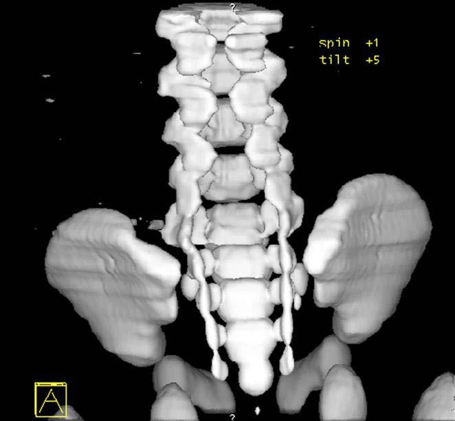
7 2. Spinal dysraphism - spina bifida a) Spina bifida occulta congenital absence of processus spinosi and vertebral arches changes cutaneous changes in middle lumbosacral region: hypertrichosis lipoma dyscoloration dermal sinus (cave infectious complications) Serious conditions associated Syringomyelia Diastematomyelia Tethred cord syndrom X-rays diagnosis L5-S1 level dorsal part of spinal canal - closure defect

8 Spinal dysraphism Diastematomyelia in CT 3D
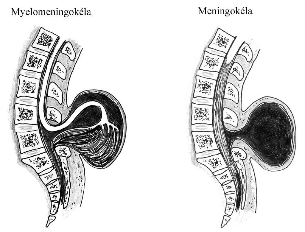
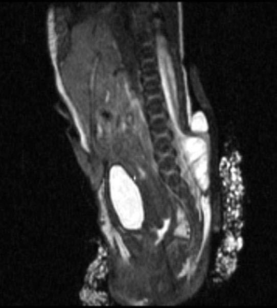
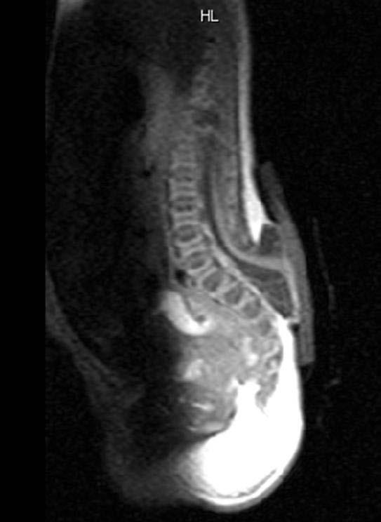
9 Spinal dysraphism a) Spina bifida aperta seu spina bifida cystica eningocele vertebral arches defect, meningeal cyst, in 1/3 neurological deficit myelomeningocele - vertebral arches defect, meningeal cyst, structural and functional nervous tissue abnormalities Myelomeningocele epidemiology: 1 from 1000 newborns Clinical features lower extremities paresis proprioceptive reflexes disturbance incontinence Associated conditions hydrocephalus (in %) Chiari malformation (in 80 %)
10 Spinal dysraphism

11 Spinal dysraphism

12 Spinal dysraphism

13 Spinal dysraphism

14 Spinal dysraphism
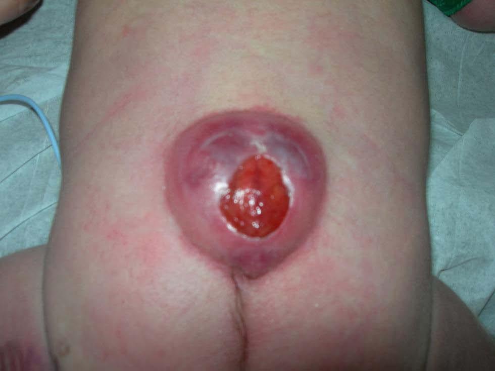
15 Spinal dysraphism
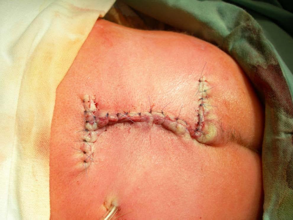
16 Spinal dysraphism
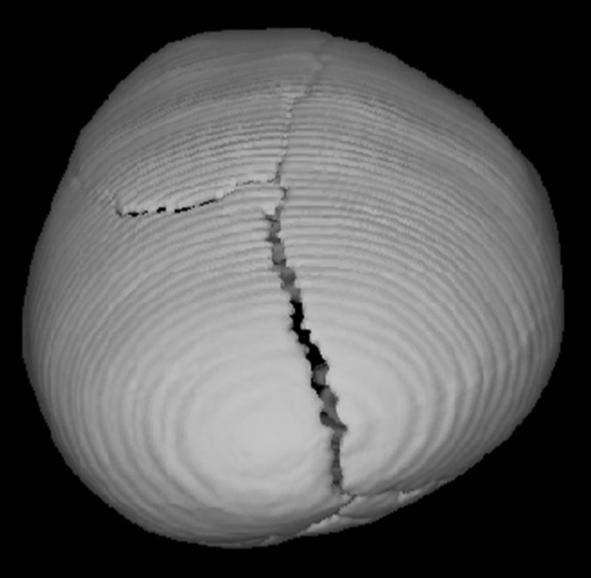
17 Craniostenosis (caniosynostosis) Premature skull sutures synostosis 1852 Rudolf Virchow 1 from 2100 children

18 Sutures of the skull
19 1. Skaphocephalia - dolichocephalia Premature sagital suture synostosis Výskyt %
20 Skaphocephalia - dolichocephalia

21 Skaphocephalia - dolichocephalia

22 Skaphocephalia - dolichocephalia
23 2. Brachycephalia Premature coronar suture synostosis % a) Frontal plagiocephalia one side coronar suture b) Occipital plagiocephalia one side lambdoid suture

24 Brachycephalia

25 Brachycephalia
26 3. Trigonocephalia Premature sutura metopica synostosis 10%

27 Trigonocephalia

28 Trigonocephalia

29 Trigonocephalia

30 Trigonocephalia

31 Trigonocephalia

32 Trigonocephalia
33 4. Morbus Crouzon dysostosis craniofacialis (1912) Turicephaly Shallow orbits Exophalmos Hypertelorism Hypoplasia of middle facial skelet 1 from children

34 Morbus Crouzon

35 Morbus Crouzon

36 Morbus Crouzon

37 Morbus Crouzon

38 Morbus Crouzon

39 Morbus Crouzon

40 Post surgery care Preventive protective helmets
Fetal Medicine. Case Presentations. Dr Ermos Nicolaou Fetal Medicine Unit Chris Hani Baragwanath Hospital. October 2003
 Case Presentations Dr Ermos Nicolaou Fetal Medicine Unit Chris Hani Baragwanath Hospital October 2003 Case 1 Ms A M 22year old P0 G1 Referred from Sebokeng Hospital at 36w for polyhydramnios On Ultrasound:
Malformations of the Nervous System November 10, Dr. Peter Ostrow
 Malformations of the Nervous System November 10, 2016 Dr. Peter Ostrow Malformations of the Nervous System 1. Abnormal closure of the neural tube 1. Disorders of forebrain formation 1. Cortical anomalies
Introduction to Neurosurgical Subspecialties:
 Introduction to Neurosurgical Subspecialties: Pediatric Neurosurgery Brian L. Hoh, MD 1 and Gregory J. Zipfel, MD 2 1 University of Florida, 2 Washington University Pediatric Neurosurgery Pediatric neurosurgeons
CNS Embryology 5th Menstrual Week (Dorsal View)
") Imaging of the Fetal Brain; Normal & Abnormal Alfred Abuhamad, M.D. Eastern Virginia Medical School CNS Embryology 5th Menstrual Week (Dorsal View) Day 20 from fertilization Neural plate formed in ectoderm
Imaging of the Fetal Brain; Normal & Abnormal Alfred Abuhamad, M.D. Eastern Virginia Medical School CNS Embryology 5th Menstrual Week (Dorsal View) Day 20 from fertilization Neural plate formed in ectoderm
Spine and spinal cord
 NEURORADIOLOGY Spine and spinal cord Erika Vörös University of Szeged Department of Radiology SZEGED DISEASES OF SPINE AND SPINAL CORD I. Non-tumourous diseases developmental anomalies vascular disorders
SPLIT NOTOCHORD SYNDROME ASSOCIATION. DR. Hasan Nugud Consultant Paediatric Surgeon
 SPLIT NOTOCHORD SYNDROME ASSOCIATION DR. Hasan Nugud Consultant Paediatric Surgeon CASE PRESENTATION :- New born baby, boy, referred to the paediatric surgical team at the age of 14 hours. Birth History
Central nervous system. Obstetrics Content Outline Obstetrics - Fetal Abnormalities
 Obstetrics Content Outline Obstetrics - Fetal Abnormalities Many congenital malformations of the CNS result from incomplete closure of the neural tube Effective February 2007 10 16% the most common neural
Neuroembryology II. Dr. Newton COPH G210
 Neuroembryology II Dr. Newton COPH G210 Anterior and posterior neuropore closure at E25 & E27, respectively, is essential for normal nervous system development. NTDs occur 1/1K births. Incidence can be
Pediatric Spinal Anomalies
 Department of Radiology University of California San Diego Pediatric Spinal Anomalies John R. Hesselink, M.D. Spine Embryogenesis 1. Primitive streak 2. Proliferation of cells at primitive pit (Hensen's
Spinal dysraphism: genetic relation to
 Journal of Medical Genetics (1976). 13, 343-350. Spinal dysraphism: genetic relation to neural tube malformations C. 0. CARTER, K. A. EVANS, and K. TILL* From MRC Clinical Genetics Unit, Institute of Child
A Retrospective Analysis of Clinical Profile and Surgical Outcome in Patients with Spinal Dysraphism at Tertiary Care Center
 Original Research Article A Retrospective Analysis of Clinical Profile and Surgical Outcome in Patients with Spinal Dysraphism at Tertiary Care Center Premlal KV * Assistant Professor, Department of Neurosurgery,
A Case of Naso-Ethmoidal Meningoencephalocele
 A Case of Naso-Ethmoidal Meningoencephalocele Divyanshu Dubey, Sonjjay Pande, Pradeep Dubey, Anshudha Sawhney Vol. 3 No. 8 (August 2011) International Journal of Collaborative Research on Internal Medicine
Supplementary Online Content
 Supplementary Online Content Honein MA, Dawson AL, Petersen E, et al; US Zika Pregnancy Registry Collaboration. Birth Defects Among Fetuses and Infants of US Women With Laboratory Evidence of Possible
Neuroanatomy. Assistant Professor of Anatomy Faculty of Medicine The University of Jordan Dr Maha ELBeltagy
 Neuroanatomy Dr. Maha ELBeltagy Assistant Professor of Anatomy Faculty of Medicine The University of Jordan 2018 Development of the Central Nervous System Development of the nervous system Development
NEURORADIOLOGY Part I
 NEURORADIOLOGY Part I Vörös Erika University of Szeged Department of Radiology SZEGED DISEASES OF CNS BRAIN Developmental anomalies Cerebrovascular disorders Tumours Inflammatory diseases Trauma DISEASES
Developmental Neuropathology
 Developmental Neuropathology Pathology, Radiology, and Clinical Correlations Reid Heffner MD Distinguished Teaching Professor Department of Pathology and Anatomy I HAVE NO CONFLICTS OF INTEREST OR DISCLOSURES
Role of helical CT and MRI in the evaluation of spinal dysraphism
 International Journal of Advances in Medicine Kumaran SK et al. Int J Adv Med. 2017 Feb;4(1):124-132 http://www.ijmedicine.com pissn 2349-3925 eissn 2349-3933 Original Research Article DOI: http://dx.doi.org/10.18203/2349-3933.ijam20170095
Brain Imaging. Bearbeitet von Klaus Sartor, Stefan Hähnel, Bodo Kress
 Brain Imaging Bearbeitet von Klaus Sartor, Stefan Hähnel, Bodo Kress 1. Auflage 2007. Taschenbuch. 312 S. Paperback ISBN 978 3 13 143961 1 Format (B x L): 12,5 x 19 cm Weitere Fachgebiete > Medizin > Sonstige
Congenital malformation & hydrocephalus
 Congenital malformation & hydrocephalus Objectives: 1- Know the common types of congenital malformations of the CNS and have a basic knowledge of their pathological features. 2- Correlate CNS normal development
Neuro. Development. Judy Philbrook, NNP-BC. ! Primary neurulation! Prosencepahlic! Neuronal proliferation. ! 3-4 weeks! 2-3 months!
 Neuro Judy Philbrook, NNP-BC Microsoft clip art Development! Primary neurulation! Prosencepahlic! Neuronal proliferation! Neuronal migration! Organization! Myelination! 3-4 weeks! 2-3 months! 3-4 months!
Skeletal System. Prof. Dr. Malak A. Al-yawer Department of Anatomy/Embryology Section
 Skeletal System Prof. Dr. Malak A. Al-yawer Department of Anatomy/Embryology Section Learning objectives At the end of this lecture, the medical student will be able to: State the embryonic origin of skeletal
Neurosurgery. Neurosurgery
 Neurosurgery Neurosurgery Neurosurgery Telephone Numbers: Appointment: 202-476-3020 Fax: 202-476-3091 Administration: 202-476-3020 Evenings and Weekends: 202-476-5000 Robert Keating, MD, Chief The Division
University Journal of Surgery and Surgical Specialties
 University Journal of Surgery and Surgical Specialties ISSN 2455-2860 Volume 2 Issue 1 2016 TWO RARE CASES OF DIASTEMATOMYELIA MUTHURAMAN P Department of Neuro Surgery, THANJAVUR MEDICAL COLLEGE Abstract
Appendix 3.5 Case Inclusion Guidance for Potentially Zika-related Birth Defects
 Appendix 3.5 Case Inclusion Guidance for Potentially Zika-related Birth Defects Appendix 3.5 A3.5-1 Case Definition Appendix 3.5 Case Inclusion Guidance for Potentially Zika-related Birth Defects Contents
Symposium: OB/GY US (Room B) CNS Anomalies
 CNS Anomalies") 82 Symposium: OB/GY US (Room B) 11 : 50 1 2 : 10 CNS Anomalies Brain area Midline structure S u p r a t e n t o r i a l ventricular system Cerebral hemisphere Posterior fossa Head size and shape Image
82 Symposium: OB/GY US (Room B) 11 : 50 1 2 : 10 CNS Anomalies Brain area Midline structure S u p r a t e n t o r i a l ventricular system Cerebral hemisphere Posterior fossa Head size and shape Image
Week 14. Development of the Musculoskeletal System
 Week 14 Development of the Musculoskeletal System Skeletal System Derived from: paraxial mesoderm somites and somitomeres sclerotome sclerotome differentiation induced by SHH from notochord and floor plate
Chapter 8. Pediatric Surgery
 Chapter 8 Pediatric Surgery 8.1 Hydrocephalus Hydrocephalus is a congenital disorder. There may be difficulties during normal vaginal delivery due large size of the head. In 1970s, when these pictures
Guidelines in the management of neural tube defects and hydrocephalus
 Guidelines in the management of neural tube defects and hydrocephalus Dominic Venne, MD, MSc, FRCSC, Division of Neurosurgery Sheikh Khalifa Medical City Abu Dhabi, UAE 1. Introduction: Neural tube defects
Chiari III Joseph Junewick, MD FACR
 Chiari III Joseph Junewick, MD FACR 07/02/2010 History Newborn with suboccipital mass. Diagnosis Chiari III Additional Clinical Surgery-Skin covered suboccipital cystic mass confined by the dura. Pathology-Leptomeningeal
Sonography of the Neonatal Spine: Part 2, Spinal Disorders
 Neonatal Spine Sonography Pediatric Imaging Pictorial Essay Downloaded from www.ajronline.org by 148.251.232.83 on 04/11/18 from IP address 148.251.232.83. Copyright RRS. For personal use only; all rights
Romanian Neurosurgery Volume XXXI Number April-June. Article
 Romanian Neurosurgery Volume XXXI Number 2 2017 April-June Article Association of abnormal metopic suture causing hypertelorism, interfrontal encephalocele with craniofacial cosmetic deformity associated
HYDROCEPHALUS OF THE INFANT (ABOUT 86 CASES)
") HYDROCEPHALUS OF THE INFANT (ABOUT 86 CASES) K.EL KHOU;R.ANDALOUSSI;L.OUZIDANE Pediatric radiology department-chu Ibn Rochd Casablanca-Morroco Morroco. Introduction Hydrocephalus of infant is a progressive
HYDROCEPHALUS OF THE INFANT (ABOUT 86 CASES) K.EL KHOU;R.ANDALOUSSI;L.OUZIDANE Pediatric radiology department-chu Ibn Rochd Casablanca-Morroco Morroco. Introduction Hydrocephalus of infant is a progressive
NYEIS Version 4.3 (ICD) ICD - 10 Codes Available in NYEIS at time of version launch (9/23/2015)
 ICD - 10 Codes Available in NYEIS at time of version launch (9/23/2015)") D82.1 Di George's syndrome E63.9 Nutritional deficiency, unspecified E70.21 Tyrosinemia E70.29 Other disorders of tyrosine metabolism E70.30 Albinism, unspecified E70.5 Disorders of tryptophan metabolism
D82.1 Di George's syndrome E63.9 Nutritional deficiency, unspecified E70.21 Tyrosinemia E70.29 Other disorders of tyrosine metabolism E70.30 Albinism, unspecified E70.5 Disorders of tryptophan metabolism
NEURORADIOLOGY. Part III. Angela Csomor University of Szeged Department of Radiology
 NEURORADIOLOGY Part III Angela Csomor University of Szeged Department of Radiology DISEASES OF SPINE AND SPINAL CORD I. Non-tumourous diseases developmental anomalies vascular disorders inflammatory processes
Central nervous system
 Chapter 2 Central nervous system NORMAL SONOGRAPHIC ANATOMY The fetal brain undergoes major developmental changes throughout pregnancy. At 7 weeks of gestation, a sonolucent area is seen in the cephalic
Prenatal Diagnosis of Central Nervous System (CNS) Pathologies: does Fetal MRI help in their management?
 Pathologies: does Fetal MRI help in their management?") Prenatal Diagnosis of Central Nervous System (CNS) Pathologies: does Fetal MRI help in their management? Daniela Prayer, Division of Neuroradiology and Musculoskeletal Radiology Medical University Vienna/Austria
Prenatal Diagnosis of Central Nervous System (CNS) Pathologies: does Fetal MRI help in their management? Daniela Prayer, Division of Neuroradiology and Musculoskeletal Radiology Medical University Vienna/Austria
Wound healing in trophic ulcers in spina bifida patients
 J Neurosurg 82:000 000, 1995 Wound healing in trophic ulcers in spina bifida patients VINOD KUMAR SRIVASTAVA, M.B.B.S, M.CH. Neurosurgical Unit, J. N. Medical College, Aligarh Muslim University, Aligarh,
The Brain: Prenatal and Postnatal Effects of Congenital Heart Disease. Dianna M. E. Bardo, M D Swedish Cherry Hill Radia, Inc.
 The Brain: Prenatal and Postnatal Effects of Congenital Heart Disease Dianna M. E. Bardo, M D Swedish Cherry Hill Radia, Inc. Seattle, WA embryology We recognize the VACTERL association and frequency of
Persistent Terminal Ventricle
 Persistent Terminal Ventricle Ventriculus Terminalis Incomplete regression of TV of 2 neurulation, continuity with central canal small cavity PTV vs terminal myelocystocele (?severe manifestation from
Arnold Chiari Malformation - A hospital based autopsy study
 Rapotra Megha et al / International Journal of Biomedical Research 2017; 8(05): 250-254. 250 International Journal of Biomedical Research ISSN: 0976-9633 (Online); 2455-0566 (Print) Journal DOI: https://dx.doi.org/10.7439/ijbr
Chiari Malformations. Google. Objectives Seventh Annual NKY TBI Conference 3/22/13. Kerry R. Crone, M.D.
 Chiari Malformations Kerry R. Crone, M.D. Professor of Neurosurgery and Pediatrics University of Cincinnati College of Medicine University of Cincinnati Medical Center Cincinnati Children s Hospital Medical
Dorsal dermal sinus in children
 Dorsal dermal sinus in children Poster No.: C-2581 Congress: ECR 2015 Type: Educational Exhibit Authors: J. Marjanovic, A. Paterson, P. C. McSherry, A. Nixon, A. 1 1 2 1 2 1 1 2 TRIPALO BATOS, T. Grmoja
Ultrasound Anomaly Details
 Appendix 2. Association of Copy Number Variants With Specific Ultrasonographically Detected Fetal Anomalies Ultrasound Anomaly Details Abdominal wall Bladder exstrophy Body-stalk anomaly Cloacal exstrophy
Anatomy, Terminology and Treatment in Pediatric Neurosurgery Part I
 Anatomy, Terminology and Treatment in Pediatric Neurosurgery Part I John Ragheb, MD, FACS, FAAP Professor of Neurosurgery and Pediatrics, Affiliated Faculty of University of Miami, Miller School of Medicine
Original Article SURGICAL MANAGEMENT & CLINICAL OUTCOME OF OCCIPITAL ENCEPHALOCOELE
 Original Article OF OCCIPITAL ENCEPHALOCOELE Najm us saqib *, Sharif Alqadhi *, Usman Ahmed Khan * * Department Neurosurgey Khoula hospital, Ministry of Health, Muscat, Oman. ABSTRACT: INTRODUCTION: Encephalocele
Spectrum of Cranio-facial anomalies during 2 Ultrasound. trimester on
 Spectrum of Cranio-facial anomalies during 2 Ultrasound nd trimester on Poster No.: C-0378 Congress: ECR 2015 Type: Scientific Exhibit Authors: K. Dave, S. Solanki; Ahmedabad/IN Keywords: Obstetrics (Pregnancy
University Journal of Surgery and Surgical Specialties
 University Journal of Surgery and Surgical Specialties ISSN 2455-2860 Volume 2 Issue 1 2016 Profile of paediatric patients with split cord malformation MANORANJITHAKUMARI M Department of Neuro Surgery,
Ligaments of the vertebral column:
 In the last lecture we started talking about the joints in the vertebral column, and we said that there are two types of joints between adjacent vertebrae: 1. Between the bodies of the vertebrae; which
In-Training Examination for Diagnostic Radiology Residents Rationales
 28th Annual In-Training Examination for Diagnostic Radiology Residents Rationales Sponsored by: Commission on Education Committee on Residency Training in Diagnostic Radiology February 3, 2005 The American
Spina bifida in Sudan
 imedpub Journals http://journals.imedpub.com JOURNAL OF NEUROLOGY AND NEUROSCIENCE Spina bifida in Sudan Darrag Salim, Abubakr 1, Awad Elzain, Mohammed 1, Adil Mohamed, Alla 1 Abstract Introduction: Neural
Central Nervous System Practical Exam. Chapter 12 Nervous System Cells. 1. Please identify the flagged structure.
 Central Nervous System Practical Exam Chapter 12 Nervous System Cells 1. Please identify the flagged structure. 2. Please identify the flagged structure. 3. Please identify the flagged structure. 4. A
Prospective Evaluation of Role of MRI in Suspected Spinal Dysraphism and Its Management
 IOSR Journal of Dental and Medical Sciences (IOSR-JDMS) e-issn: 2279-0853, p-issn: 2279-0861.Volume 17, Issue 5 Ver. 1 (May. 2018), PP 21-28 www.iosrjournals.org Prospective Evaluation of Role of MRI in
meninges Outermost layer of the meninge dura mater arachnoid mater pia mater membranes located between bone and soft tissue of the nervous system
 membranes located between bone and soft tissue of the nervous system meninges Outermost layer of the meninge dura mater middle layer of the meninges, contains no blood vessels arachnoid mater Innermost
Diastematomyelia: A Case with Familial Aggregation of Neural Tube Defects
 Case Study TheScientificWorldJOURNAL (2004) 4, 847 852 ISSN 1537-744X; DOI 10.1100/tsw.2004.140 Diastematomyelia: A Case with Familial Aggregation of Neural Tube Defects Nuray Öksüz Kanbur 1, *, Pınar
Complex Hydrocephalus
 2012 Hydrocephalus Association Conference Washington, DC - June 27-July1, 2012 Complex Hydrocephalus Marion L. Walker, MD Professor of Neurosurgery & Pediatrics Primary Children s Medical Center University
SKULL AS A WHOLE + ANTERIOR CRANIAL FOSSA
 SKULL AS A WHOLE + ANTERIOR CRANIAL FOSSA LEARNING OBJECTIVES At the end of this lecture, the student should be able to know: Parts of skeleton (axial and appendicular) Parts of skull Sutures of skull
Central Nervous System (CNS) -> brain and spinal cord. Major Divisions of the nervous system:
 -> brain and spinal cord. Major Divisions of the nervous system:") Central Nervous System (CNS) -> brain and spinal cord Major Divisions of the nervous system: Afferent (sensory input) -> cell bodies outside of the central nervous system (CNS), carry info into the CNS
Central Nervous System (CNS) -> brain and spinal cord Major Divisions of the nervous system: Afferent (sensory input) -> cell bodies outside of the central nervous system (CNS), carry info into the CNS
BIOL Dissection of the Sheep and Human Brain
 BIOL 2401 Dissection of the Sheep and Human Brain Laboratory Objectives After completing this lab, you should be able to: Identify the main structures in the sheep brain and to compare them with those
Department of Cognitive Science UCSD
 Department of Cognitive Science UCSD Verse 1: Neocortex, frontal lobe, Brain stem, brain stem, Hippocampus, neural node, Right hemisphere, Pons and cortex visual, Brain stem, brain stem, Sylvian fissure,
DEPARTMENT OF NEUROSURGERY. Records, Archival Collection - AC 13. Boston Children s Hospital Archives
 DEPARTMENT OF NEUROSURGERY Records, 1929-1999 Archival Collection - AC 13 Boston Children s Hospital Archives Contact Information: 300 Longwood Avenue Hospital Library Boston, MA 02135 Phone: 617-355-5286
Hydrocephalus in children. Eva Brichtova, M.D., Ph.D., Department of Pediatric Sugery, Orthopaedics and Traumatology, University Hospital Brno
 Hydrocephalus in children Eva Brichtova, M.D., Ph.D., Department of Pediatric Sugery, Orthopaedics and Traumatology, University Hospital Brno Ventricle system Ventricle system, cerebral cisterns Hydrocephalus
PARA210 SUMMARY Hyperglycaemia (DKA & HHS) Brain & Nervous System Anatomy & Physiology Degenerative Neurological Disorders
 Brain & Nervous System Anatomy & Physiology Degenerative Neurological Disorders") PARA210 SUMMARY Page Topic 01-03 Diabetes Mellitus 04-05 Hyperglycaemia (DKA & HHS) 06-13 Toxicology 14-18 12 Lead ECG 19-21 Brain & Nervous System Anatomy & Physiology 22-24 Degenerative Neurological
PARA210 SUMMARY Page Topic 01-03 Diabetes Mellitus 04-05 Hyperglycaemia (DKA & HHS) 06-13 Toxicology 14-18 12 Lead ECG 19-21 Brain & Nervous System Anatomy & Physiology 22-24 Degenerative Neurological
intracranial anomalies
 Chapter 5: Fetal Central Nervous System 84 intracranial anomalies Hydrocephaly Dilatation of ventricular system secondary to an increase in the amount of CSF. Effects of hydrocephalus include flattening
Chiari malformations. A fact sheet for patients and carers
 A fact sheet for patients and carers Chiari malformations This fact sheet provides information on Chiari malformations. It focuses on Chiari malformations in adults. Our fact sheets are designed as general
Basic Training. ISUOG Basic Training Distinguishing Between Normal & Abnormal Appearances of the Skull & Brain
 ISUOG Distinguishing Between Normal & Abnormal Appearances of the Skull & Brain Learning objectives At the end of the lecture you will be able to: Describe how to obtain the 3 planes required to assess,
Neuropathic bladder and spinal dysraphism
 Archives of Disease in Childhood, 1981, 56, 176-180 Neuropathic bladder and spinal dysraphism MALGORZATA BORZYSKOWSKI AND B G R NEVILLE Evelina Children's Department, Guy's Hospital, London SUMMARY The
Cerebellar tonsil herniation: Its diverse pathogenesis
 Cerebellar tonsil herniation: Its diverse pathogenesis Poster No.: C-0959 Congress: ECR 2014 Type: Educational Exhibit Authors: H. Mukai, H. Yokota, T. Horikoshi, K. Motoori, T. Uno; Chiba/JP Keywords:
Prenatal Prediction of The Neurologically Impaired Neonate By Ultrasound
 Prenatal Prediction of The Neurologically Impaired Neonate By Ultrasound Robert H. Debbs, D.O.,F.A.C.O.O.G. Professor of OB-GYN Perelman School of Medicine, University of Pennsylvania Director, Pennsylvania
Case Based Urology Learning Program
 Case Based Urology Learning Program Resident s Corner: UROLOGY Case Number 23 CBULP 2011 077 Case Based Urology Learning Program Editor: Associate Editors: Manager: Case Contributors: Steven C. Campbell,
8.3 The Central Nervous System. SBI4U Ms. Ho-Lau
 8.3 The Central Nervous System SBI4U Ms. Ho-Lau The Central Nervous System the structural and functional centre for the entire nervous system the site of neural integration and processing The Central
Asymptomatic posterior cervical myelomeningocele with tethered cord in an adolescent: a rare form of spinal dysraphism with rare presentation
 Romanian Neurosurgery (2016) XXX 1: 113-117 113 Asymptomatic posterior cervical myelomeningocele with tethered cord in an adolescent: a rare form of spinal dysraphism with rare presentation Gangesh Gunjan,
Central nervous system
 Central nervous system By Dr. Mohsen Dashti Clinical Medicine & Pathology 316 7 th Lecture Lecture outline Review of structure & function. Symptoms, signs & tests. Specific diseases. Review of structure
A Congenital Defect in the Spinal Cord of the Manx Cat
 Vet, Path. 8: 232-238 (1971) A Congenital Defect in the Spinal Cord of the Manx Cat A. H. MARTIN Department of Anatomy, University of Wisconsin, Madison Wisc. Abstract. The lumbar part of the spinal cords
Dissection of the Sheep Brain
 Dissection of the Sheep Brain Laboratory Objectives After completing this lab, you should be able to: 1. Identify the main structures in the sheep brain and to compare them with those of the human brain.
Chiari FAQ's. 1. What is a Chiari Malformation?
 Chiari FAQ's These FAQ's are for informational purposes only and in no way represent an attempt to provide medical advice. This information may or may not apply to your case and anyone with a question
ISUOG Basic Training. Distinguishing Between Normal & Abnormal Appearances of the Skull & Brain. Seshadri Suresh, India
 ISUOG Basic Training Distinguishing Between Normal & Abnormal Appearances of the Skull & Brain Seshadri Suresh, India Learning objectives 4 & 5 At the end of the lecture you will be able to: Describe how
Fetal CNS MRI. Daniela Prayer. Division of Neuroradiology And Musculoskeletal Radiology. Medical University of Vienna, AUSTRIA
 Fetal CNS MRI Daniela Prayer Division of Neuroradiology And Musculoskeletal Radiology Medical University of Vienna, AUSTRIA Methods Normal development Malformations Acquired pathology MR- methods for assessment
Cleidocranial Dysplasia
 BRIEF REPORTS Cleidocranial Dysplasia Anita Sharma Rohtash Yadav Kuldip Ahlawat Cleidocranial dysplasia (CCD), is characterized by short stature, typical facial features and variable degree of pan-skeletal
ISUOG Basic Training. Examining Fetal Anatomy from Longitudinal Sections Titia Cohen-Overbeek, The Netherlands
 ISUOG Basic Training Examining Fetal Anatomy from Longitudinal Sections Titia Cohen-Overbeek, The Netherlands Learning objectives 2 & 3 At the end of the lecture you will be able to: describe how to obtain
Slide 1. Slide 2. Slide 3. Tomography vs Topography. Computed Tomography (CT): A simplified Topographical review of the Brain. Learning Objective
: A simplified Topographical review of the Brain. Learning Objective") Slide 1 Computed Tomography (CT): A simplified Topographical review of the Brain Jon Wheiler, ACNP-BC Slide 2 Tomography vs Topography Tomography: A technique for displaying a representation of a cross
Slide 1 Computed Tomography (CT): A simplified Topographical review of the Brain Jon Wheiler, ACNP-BC Slide 2 Tomography vs Topography Tomography: A technique for displaying a representation of a cross
The Nervous System PART B
 7 The Nervous System PART B PowerPoint Lecture Slide Presentation by Jerry L. Cook, Sam Houston University ESSENTIALS OF HUMAN ANATOMY & PHYSIOLOGY EIGHTH EDITION ELAINE N. MARIEB Central Nervous System
Giant high occipital encephalocele
 122 Agrawal et al Giant high occipital encephalocele Giant high occipital encephalocele Amit Agrawal 1, Umamaheshwara Reddy V. 2, Kishor V. Hegde 2, Suneetha P. 2, Divya Siddharth Kolikipudi 2 Narayana
Case Report Occult Spinal Dysraphism in the Presence of Rare Cutaneous Stigma in a Neonate: Importance of Ultrasound and Magnetic Resonance Imaging
 Case Reports in Medicine Volume 2013, Article ID 468376, 4 pages http://dx.doi.org/10.1155/2013/468376 Case Report Occult Spinal Dysraphism in the Presence of Rare Cutaneous Stigma in a Neonate: Importance
PATIENTS AND METHODS:
 ORIGINAL ARTICLE Pattern of congenital brain malformations at a referral hospital in Saudi Arabia: An MRI study Ibrahim A. Alorainy BACKGROUND: More than 2000 different congenital cerebral malformations
Djamila Kafoufi Al Galaa Military Hospital Cairo
 Djamila Kafoufi Al Galaa Military Hospital Cairo Herniation cerebellar tonsils below the foramen magnum, Hans Chiari 4 types Chiari I less than 5mm,HDC rare,syringomyelia often present. Chiari II,protrusion
The Central Nervous System
 The Central Nervous System Bởi: OpenStaxCollege The central nervous system (CNS) is made up of the brain, a part of which is shown in [link] and spinal cord and is covered with three layers of protective
Treatment of cervicodorsalepidermiod cyst
 Treatment of cervicodorsalepidermiod cyst Ayied Motteb Turkey Department of Medicine, College of medicine, University of Tikret, Tekrit, Iraq Received 22 / 9/2011 Accepted 22/11/2011 Abstract Intramedullary
Chapter 5: Fetal Central Nervous System 71
 71 Chapter 5 Fetal Central Nervous System Embryology NEURULATION begins with the formation of the neural plate, the neural folds and their ultimate fusion and closure as the NEURAL TUBE. NEURAL PLATE -
Congenital Anomalies
 Congenital Anomalies Down Syndrome 7580 7580 DOWN''S SYNDROME Q900 Q90.0 : Trisomy 21, meiotic nondisjunction 7580 7580 DOWN''S SYNDROME Q901 Q90.1 : Trisomy 21, mosaicism (mitotic nondisjunction) 7580
LUMPS, TUFTS AND DIMPLES IT S THE PITS!! Session Information. Faculty Disclosure Information
 Session Information Session Title: Lumps, Tufts and Dimples Session Number: F3056, F2130 Faculty Name: Mark S. Dias, MD, FAAP Faculty Institution: Penn State Children s Hospital, Penn State University
BRAIN PART I (A & B): VENTRICLES & MENINGES
: VENTRICLES & MENINGES") BRAIN PART I (A & B): VENTRICLES & MENINGES Cranial Meninges Cranial meninges are continuous with spinal meninges Dura mater: inner layer (meningeal layer) outer layer (endosteal layer) fused to periosteum
BRAIN PART I (A & B): VENTRICLES & MENINGES Cranial Meninges Cranial meninges are continuous with spinal meninges Dura mater: inner layer (meningeal layer) outer layer (endosteal layer) fused to periosteum
Somatic Nervous Systems. III. Autonomic Nervous System. Parasympathetic Nervous System. Sympathetic Nervous Systems
 7/21/2014 Outline Nervous System - PNS and CNS I. II. Two Parts of the Nervous System Central Nervous System vs Peripheral Nervous System Peripheral Nervous System A. B. Brain and Spinal Cord III. Autonomic
Does not intend to discuss commercial products or services. Does not intend to discuss non-fda approved uses of products/providers of services.
 Date: 3/17/2017 Lecture title: Rate of shunt revision as a function of age in patients with shunted hydrocephalus due to myelomeningocele Anastasia Arynchyna, MPH, CCRP Clinical Research Manager, Department
Long segment composite split cord malformation with double bony spur
 Long segment composite split cord malformation with double bony spur Anand Sharma, Achal Sharma, R.S. Mittal SMS Medical College, Jaipur, India Abstract: A composite type of SCM is very rare and only a
Case report: Imaging findings in a butterfly vertebra
 Acta Neurol. Belg., 2011, 111, 344-348 Case report: Imaging findings in a butterfly vertebra Cedric BOULET, Ann SCHIETTECATTE, Johan DE MEY and Michel DE MAESENEER Department of Radiology, UZ Brussel,
Brain Meninges, Ventricles and CSF
 Brain Meninges, Ventricles and CSF Lecture Objectives Describe the arrangement of the meninges and their relationship to brain and spinal cord. Explain the occurrence of epidural, subdural and subarachnoid
Ch 13: Central Nervous System Part 1: The Brain p 374
 Ch 13: Central Nervous System Part 1: The Brain p 374 Discuss the organization of the brain, including the major structures and how they relate to one another! Review the meninges of the spinal cord and
Ventricles, CSF & Meninges. Steven McLoon Department of Neuroscience University of Minnesota
 Ventricles, CSF & Meninges Steven McLoon Department of Neuroscience University of Minnesota 1 Coffee Hour Thursday (Sept 14) 8:30-9:30am Surdyk s Café in Northrop Auditorium Stop by for a minute or an
CODE TABLES ICD-9-CM to ICD-10-CM code conversions are presented in the tables where appropriate
 CODE TABLES ICD-9-CM to ICD-10-CM code conversions are presented in the tables where appropriate Table 1: Codes to Identify Neuroimaging in Administrative Claims with ICD-10-CM Code Conversion Imaging
Johnson Rogers and colleagues- used Term LMM
 LIPOMENINGOMYELOCELE: CLASSIFICATION, MANAGEMENT AND CONTROVERSIES Definition : Lipomyelomeningocele is a form of OSD in which a subcutaneous fibrofatty mass traverses the lumbodorsal fascia, causes a