Biological Early Detection of Nasal Dermoid Cyst Using CT/ MRI Scan - A Review
|
|
|
- Henry Reeves
- 5 years ago
- Views:
Transcription
1 Biological Early Detection of Nasal Dermoid Cyst Using CT/ MRI Scan - A Review Neeraj Singla 1, Sugandha Sharma 2 M.Tech Student, Dept. of CSE, CGC, Gharuan, Punjab, India 1 Assistant Professor, Dept. of CSE, CGC, Gharuan, Punjab, India 2 ABSTRACT- Dermoid cyst occuere in brain,nasal,ovarian under which the nasal dermoid cyst surgery is very complicated when it occure under which dermoid cyst early detection is possible by CT or MRI scan during pregnancy of lady so it will not occurred and no need of surgery. Dermoid cysts are closed sacs that form within the body. A cyst may contain fluid, pus, some other tissue from the body, or foreign matter.dermoid cysts form in or on the skin, usually on the face near the eyebrow, on the scalp, or on the chest over the collarbone. Rarely, a dermoid cyst near the surface of the body extends below the skin and connects to deeper structures. Very rarely, dermoid cysts develop in other areas of the body instead of the skin. Dermoid cysts are common in children. Most of the time they are painless and benign, which means they don't threaten your child's health. But they may become infected. Surgeons remove dermoid cysts to prevent infection. Keywords: Dermoid Cyst, Radiological Imaging, Indonasal Surgery, Dermoid Sinus Cyst I. INTRODUCTION Dermoid Cyst: Dermoid cysts grow slowly and are not tender unless ruptured. They usually occur on the face, inside the skull, on the lower back and in the ovaries. Superficial dermoid cysts (ones near the surface of the skin) on the face can usually be removed without complications. Removal of other, rarer dermoid cysts requires special techniques and training. These rarer dermoid cysts occur in four major areas: DERMOID CYSTS IN THE BRAIN: Dermoid cysts occur very rarely here. A neurosurgeon may need to remove them if they cause problems. DERMOID CYSTS IN THE NASAL SINUSES: These are also very rare. Only a handful of cases involving dermoid cysts located here are reported each year. Removal of these cysts is extremely complicated. OVARIAN DERMOID CYSTS: These growths can develop in a woman during her reproductive years. They can cause torsion (twisting), infection, rupture and cancer of the ovary. These dermoid cysts can be removed with either conventional surgery or laparoscopy (surgery that uses small incisions and specially designed instruments to enter the abdomen or pelvis). DERMOID CYSTS OF THE SPINAL CORD: A sinus tract, which is a narrow connection from a deep pit in the skin, usually connects these very rare cysts to the skin surface. This type of dermoid cyst can become infected. Removal is often incomplete, but the outcome is usually excellent. Symptoms: The symptoms of epidermoid and dermoid tumors vary depending on their location. Cysts in the scalp are usually painless, moveable, rubbery masses that may slowly increase in size over time. The skin over the cyst is usually normal. Cysts in the bone may feel somewhat firmer and are usually less mobile. Although they usually only cause cosmetic problems, cysts in the skull may penetrate into the brain. Diagnosis: Most scalp lesions can be accurately diagnosed by physical exam and x-rays or other imaging may not be needed. Lesions that appear to involve the skull usually require skull x-rays or less commonly, a CT or MRI scan to make sure that there is no penetration into the brain. Treatment: Because of the potential to slowly enlarge and possibly penetrate through the skull, surgical removal is usually recommended. Most simple cysts can easily be removed with a short surgical procedure lasting less than an hour. Most children can go home the same day of surgery. Most children can resume regular activity, including bathing, in 2 to 3 days after surgery. Copyright to IJIRCCE
2 II. COMPARATIVE STUDY Cysts located in the brain are not truly brain tumors because they do not arise from the brain tissue itself. Although they tend to be (benign noncancerous), they are sometimes found in parts of the brain that control vital functions. There are four main types of cysts found in the brain: Arachnoid Cyst (also called Leptomeningeal Cyst): An enlarged, fluid-filled area of the subarachnoid space that occurs in both adults and children. Colloid Cyst: Although scientists are not sure of the definitive cause of colloid cysts, most agree that these cysts begin during embryonic development of the central nervous system. Malignant forms are unknown. Dermoid Cyst: These cysts most likely form during the early weeks of fetal development even though symptoms may not be noticed until years later. They are usually benign. Epidermoid Cyst (also called Epidermoid Tumor): Often referred to as epidermoid tumors. Likely form during the early weeks of fetal development even though the symptoms may not be noticed until decades later. A. Location Cysts can appear in a variety of locations within the brain. Arachnoid Cysts appear in the subarachnoid space (between the arachnoid and pia mater layers of the meninges). Colloid Cysts are typically attached to the roof of the third ventricle and the choroid plexus. Dermoid Cysts, though rarely found in the brain, are usually located at the lower back portion of the brain (the posterior fossa) in older adults and in the lower end of the spine in older children and young adults. Epidermoid Cysts tend to be located in the area where the top part of the brain meets the brain stem. FIGURE -1[3] B. Description Just like a cyst anywhere else in your body, a cyst in the brain is a tumor-like sphere filled with fluid much like a balloon filled with water. They may contain fluid, blood, tissue, or tumor cells. Arachnoid Cysts are enlarged, fluid-filled areas between layers of the covering of the brain. Colloid Cysts tend to contain a thick, gel-like substance called colloid. Dermoid and Epidermoid Cysts are tumor-like spheres. Symptoms Symptoms depend on the size and location of the cyst. C. Incidence Arachnoid Cysts can be found in both adults and children. Colloid Cysts almost always occur in adults. Dermoid Cysts in the brain tend to occur in children under 10 years old. Epidermoid Cysts are most commonly found in middle-aged adults. Cause Copyright to IJIRCCE

3 The exact cause of cysts is unknown, but it is suspected that certain types form in the early weeks of fetal growth. Nothing specific can be done to prevent the development of cysts. D. Treatment The type, size, and location of the cyst determine how it will be addressed. Treatment methods for the various types of cysts are briefly explained below: Arachnoid Cyst: Treatment may be watchful waiting, or the cyst may require surgery. Colloid Cyst: Surgery is typically recommended. However, removal can be challenging because of its location. Dermoid and Epidermoid Cysts: Surgery is typically recommended. If complete removal is not possible, the remaining portion of the cyst may regrow. Fortunately, this growth may be very slow and it could be years before symptoms return. Figure 2 : Dermoid cyst presenting as midline nasal mass The nose is formed from the frontonasal process and two nasal placodes which de-velop dorsal to the stomodaeum (primitive mouth) during the fourth week of embryological life. The nasal placodes consist of medial and lateral processes and become more prominent. The medial pro-cesses approach one another and eventual-ly fuse in the midline. The lateral processes become less prominent as the maxillary process fuses with them. A deep groove in this region, called the nasal-maxillary groove becomes the nasolacrimal duct. As the external nose is develops, other neural crest cells migrate through the frontonasal process to form the posterior septum, ethmoid bone, and sphenoid. The nasal septum develops around week five from the frontonasal process, growing in an anterior-posterior direction. During formation of the skull base and nose, mesenchymal structures are formed from several centers that eventually fuse and begin to ossify. Before they fuse there are recognised spaces between them that are important in the development of congenital midline nasal masses. These include the fonticulus nasofrontalis, the prenasal space, and the foramen caecum (Figures 3a-c). The fonticulus frontalis is the space between the frontal and nasal bones. The prenasal space is located between the nasal bones and the nasal capsule (precursor of nasal septum and nasal cartilages). During foetal develop-ment these spaces are close by fusing and ossifying. Abnormal development of these structures is thought to be involved in the formation of nasal dermoids. A widely accepted theory of dermoid sinus cyst development is the pre nasal space theory[7] E. Pre-operative evaluation Clinical evaluation Encephalocoeles are pulsatile, compressi-ble masses that expand on crying and on bilateral compression of the internal jugu-lar veins (Furstenberg test); neither glio-mas nor dermoids expand with crying or the Furstenberg test. However a negative Furstenberg test does not exclude intra-cranial extension; hence imaging is essential. Radiological Imaging If a dermoid is suspected, imaging is man-datory to determine the extent of the cyst or tract, to exclude an intracranial con-nection and to plan surgery. A CT scan delineates the bony anatomy and may indicate an intracranial connection (Figure 4). Copyright to IJIRCCE

. Figure 5: MRI scan of nasal dermoid cyst II.")
4 Figure 4: CT scan of nasal dermoid cyst However, in order to exclude this, MRI scanning is required and is becoming the imaging method of choice, as both false-positives and false-negatives for intracranial involvement are found with CT. It is prudent in many cases (especially when a child requires general anaesthaesia or sedation for imaging) to either obtain both images at a single sitting or to proceed directly to MRI scan (Figure 5). Figure 5: MRI scan of nasal dermoid cyst II. CASE REPORT-SURGERY OF CHILD Early surgical intervention is recommend-ded to avoid further distortion of the nose or bony atrophy caused by growth of the mass or recurrent infection. [5]Biopsy is con-traindicated due to risk of CSF leakage in cases with intracranial connections. The surgical objective is complete surgical ex-cision at the first operation. Two factors determine the surgical approach Several surgical approaches have been described; occasionally more than one incision is required especially in the presence of a nasal pit or skin breakdown A. Transverse rhinotomy Septorhinoplasty approach Vertical rhinotomy B. Horizontal nasofrontal incision with eyebrow extensions C. Endoscopic approaches A. Transverse rhinotomy: This can be used for small to moderate-sized lesions without intracranial extension. The sinus punctum is excised within a transversely oriented ellipse of skin and the tract is cannulated with a lacrimal probe and dissect-ed. Medial or lateral osteotomies may be performed if necessary. If placed in a natural nasal skin crease it Copyright to IJIRCCE


5 leaves a very favourable scar. B. Open septorhinoplasty approach: This provides wide exposure, but with a con-cealed, aesthetically pleasing scar, for larger lesions and for patients with dam-aged bone and cartilage from prior surgery or recurrent infection or with intracranial extension. A separate excision of a sinus opening may be required; with intracranial extension a combined intracranial ap-proach may be required. C. Endoscopic approach: An endonasal en-doscopic approach is recommended when a dermoid is located within the nasal cavity with little or no cutaneous involvement. It can be combined with a small external midline excision of the cutaneous punc-tum.[6] Although there are reports of ade-quate visualisation of the skull base through intercartilagenous incisions to allow passage of an endoscope and instru-ments, intracranial extension is a relative contraindication to endoscopic approach. IV. FLOWCHART DERMOID CYST TYPES NASAL DERMOID BRAIN DERMOID SPINAL DERMOID DERMOID OVARIES DERMOID COMPLICATED RARE CYST RARE CYST LESS COMPLICATED FIGURE -6- TYPES OF DERMOID CYST V. NASAL DERMOID SURGERY IS VERY COMPLICATED SO ITS EARLY DETECTION IS NECESSAY Copyright to IJIRCCE


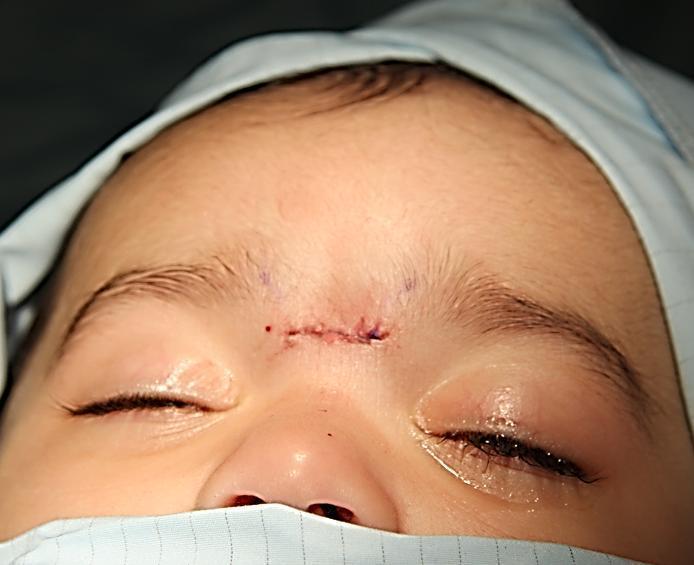
6 FIGURE 7 SURGERY OF NASAL DERMOID Copyright to IJIRCCE

7 V.CONCLUSION The dermoid sinus with or without cyst is an extensive lesion extending into the nasal cartilage and bone, usually passing deeply at the osteocartilaginous junction on the dorsum of the nose.the simplest cleft malformation in the frontobasal region is the dermal sinus which may be accompanied by dermoid cyst. This group includes a large proportion of the congenital nasal fistulas and dermoid cysts of the nose and frontal area, which are infrequently seen by the ENT surgeon. These fistulae may extend from the skin to the frontal bone leading to pressure atrophy and narrowing of the frontal sinus, if it is developed. They may extend through the skull base to the cranial cavity ending as an extradural cyst. Complete excision of dermoid cyst is treatment of choice. Surgical approaches for the removal of nasal dermoid depend upon extension of lesion. Different approaches have being advocated for removal of nasal dermoid which ranges from external approaches, such as midline vertical incision, transverse incision, lateral rhinotomy, external rhinotomy, inverted U incision, degloving procedure and craniotomy for intracranial extension of nasal dermoid.dermoid cyst is rare and often presents from birth appearing on the nose as a midline mass anywhere from the glabella to the columella. They rarely exceed 4 cm in size and possibly originate from inclusion of epidermal tissues in aberrant sites during embryonic development. Nasal dermoid cyst surgery is very complicated so its early detection is necessary. REFERENCES 1. Rohrich RJ, Lwe JB, Schwart,. The role of open rhinoplasty in management of nasal dermoid cysts, Pp , Kelly JH, Strome M, Hall B. Surgical update on nasal dermoid. Arch Otolaryngology pp: , Hughes, et al. The management of the congenital midline mass-a review. Otolaryngology-Head and Neck Surgery pp:222-23, Jaffe BF., Classification and management of anomalies of the nose. Otolaryngologic Clinic of North America pp: , Wardinsky, et al. Nasal dermoid sinus cysts: Association with intracranial extension and multiple malformations. pp:87-95, Reza Rahbar, et al. The presentation and management of nasal dermoid. Arch Otolaryngology Head Neck Surg Vol 129., Pratt LW. Midline cy-sts of the nasal dorsum: Embryologic origin and treatment. Laryngoscope pp , Michelle Wyatt. Nasal obstruction in children. Scott Brown s Otolaryngology , BIOGRAPHY ER. NEERAJ SINGLA, pursuing M.Tech -CSE from CGC, Gharuan and done B.Tech. degree from Punjab technical university. He is author of one international journals. ER. SUGANDHA SHARMA is working as Assistant professor in CGC Gharuan in CSE department.. She completed her M.E in Computer Science and Engineering from University Institute of Engineering and Technology, Panjab University in She is Author of EIGHT International Journals. Fields of Specialization is Image Processing. Copyright to IJIRCCE
Endoscopic Assisted resection for congenital Midline Nasal Mass
 Endoscopic Assisted resection for congenital Midline Nasal Mass Ahmed Aly Ibrahim A.prof ORL Department Alexandria University Emad. A Magdy prof ORL Department Alexandria University Haytham Morsi,MD Mohammad
Remember from the first year embryology Trilaminar disc has 3 layers: ectoderm, mesoderm, and endoderm
 Development of face Remember from the first year embryology Trilaminar disc has 3 layers: ectoderm, mesoderm, and endoderm The ectoderm forms the neural groove, then tube The neural tube lies in the mesoderm
Cysts Arachnoid Cyst (also called Leptomeningeal Cyst)
") Cysts This article was provided to us by David Schiff, MD, Co-Director of the Neuro-Oncology Center and Professor of Neurology, Neurosurgery, and Medicine at the University of Virginia, Charlottesville.
Cysts This article was provided to us by David Schiff, MD, Co-Director of the Neuro-Oncology Center and Professor of Neurology, Neurosurgery, and Medicine at the University of Virginia, Charlottesville.
NEUROCRANIUM VISCEROCRANIUM VISCEROCRANIUM VISCEROCRANIUM
 LECTURE 4 SKULL NEUROCRANIUM VISCEROCRANIUM VISCEROCRANIUM VISCEROCRANIUM CRANIUM NEUROCRANIUM (protective case around brain) VISCEROCRANIUM (skeleton of face) NASOMAXILLARY COMPLEX MANDIBLE (DESMOCRANIUM)
The dura is sensitive to stretching, which produces the sensation of headache.
 Dural Nerve Supply Branches of the trigeminal, vagus, and first three cervical nerves and branches from the sympathetic system pass to the dura. Numerous sensory endings are in the dura. The dura is sensitive
Essentials in Head and Neck Embryology. Part 3 Development of the head, face, and oral cavity
 Essentials in Head and Neck Embryology Part 3 Development of the head, face, and oral cavity Outline General overview of prenatal development Embryonic period phase 1 Formation of bilaminar disk Formation
Meninges and Ventricles
 Meninges and Ventricles Irene Yu, class of 2019 LEARNING OBJECTIVES Describe the meningeal layers, the dural infolds, and the spaces they create. Name the contents of the subarachnoid space. Describe the
Meningioma tumor. Meningiomas are named according to their location (Fig. 1) and cause various symptoms: > 1
 and cause various symptoms: > 1") Meningioma tumor Overview A meningioma is a type of tumor that grows from the protective membranes, called meninges, which surround the brain and spinal cord. Most meningiomas are benign (not cancer) and
Meningioma tumor Overview A meningioma is a type of tumor that grows from the protective membranes, called meninges, which surround the brain and spinal cord. Most meningiomas are benign (not cancer) and
Brain Meninges, Ventricles and CSF
 Brain Meninges, Ventricles and CSF Lecture Objectives Describe the arrangement of the meninges and their relationship to brain and spinal cord. Explain the occurrence of epidural, subdural and subarachnoid
The management of midline transcranial nasal dermoid sinus cysts *
 British Journal of Plastic Surgery (2005) 58, 1043 1050 The management of midline transcranial nasal dermoid sinus cysts * M. Hanikeri*, N. Waterhouse, N. Kirkpatrick, D. Peterson, I. Macleod Department
Nasal region. cartilages: septal cartilage (l); lateral nasal cartilage (2); greater alar cartilages (2); lesser alar cartilages (?
; lateral nasal cartilage (2); greater alar cartilages (2); lesser alar cartilages (?") Nasal region skull bones: nasal and frontal processes of maxilla cartilages: septal cartilage (l); lateral nasal cartilage (2); greater alar cartilages (2); lesser alar cartilages (?) 1 Nasal cavity Roof
Nasal region skull bones: nasal and frontal processes of maxilla cartilages: septal cartilage (l); lateral nasal cartilage (2); greater alar cartilages (2); lesser alar cartilages (?) 1 Nasal cavity Roof
Management of Nasal Dermoid Sinus Cyst by Modified
 AIJCR CASE REPORT Management of Nasal Dermoid Sinus Cyst by Modified Bipedicle 10.5005/jp-journals-10013-1247 Advancement Flap Technique Management of Nasal Dermoid Sinus Cyst by Modified Bipedicle Advancement
04 Development of the Face and Neck. Development of the Face Development of the neck
 04 Development of the Face and Neck Development of the Face Development of the neck Development of the face Overview of facial development The fourth week ~ the twelfth week of prenatal development Between
Neuroanatomy. Assistant Professor of Anatomy Faculty of Medicine The University of Jordan Dr Maha ELBeltagy
 Neuroanatomy Dr. Maha ELBeltagy Assistant Professor of Anatomy Faculty of Medicine The University of Jordan 2018 Development of the Central Nervous System Development of the nervous system Development
PEDIATRICS WK 3 HEAD AND NECK ALISON WALLACE MD, PHD
 PEDIATRICS WK 3 HEAD AND NECK ALISON WALLACE MD, PHD Topics 1. Cervical lymphadenopathy 2. Lymphatic malformation 3. Thyroglossal duct cysts 4. Branchial cleft cysts 5. Thyroid masses CASE 1 Case 1 A 2
HEAD AND NECK IMAGING. James Chen (MS IV)
") HEAD AND NECK IMAGING James Chen (MS IV) Anatomy Course Johns Hopkins School of Medicine Sept. 27, 2011 OBJECTIVES Introduce cross sectional imaging of head and neck Computed tomography (CT) Review head
HEAD AND NECK IMAGING James Chen (MS IV) Anatomy Course Johns Hopkins School of Medicine Sept. 27, 2011 OBJECTIVES Introduce cross sectional imaging of head and neck Computed tomography (CT) Review head
Ventricles, CSF & Meninges. Steven McLoon Department of Neuroscience University of Minnesota
 Ventricles, CSF & Meninges Steven McLoon Department of Neuroscience University of Minnesota 1 Coffee Hour Thursday (Sept 14) 8:30-9:30am Surdyk s Café in Northrop Auditorium Stop by for a minute or an
Intranasal location of lacrimal sac in Thai cadavers
 Asian Biomedicine Vol. 4 No. 2 April 2010; 323-327 Clinical report Napas Tanamai a, Teeraporn Ratanaanekchai a, Sanguansak Thanaviratananich a, Kowit Chaisiwamongkol b, Thanarat Chantaumpalee b a Department
General Anatomy p. 1 Organization of the Human Body p. 1 Skeleton of the Human Body p. 4 Ossification of the Bones p. 6 Bone Structure p. 8 Joints p.
 General Anatomy p. 1 Organization of the Human Body p. 1 Skeleton of the Human Body p. 4 Ossification of the Bones p. 6 Bone Structure p. 8 Joints p. 10 Principal Joints (Immovable) p. 12 Synovial Joints
Superior View of the Skull (Norma Verticalis) Anteriorly the frontal bone articulates with the two parietal bones AT THE CORONAL SUTURE
 Anteriorly the frontal bone articulates with the two parietal bones AT THE CORONAL SUTURE") Superior View of the Skull (Norma Verticalis) Anteriorly the frontal bone articulates with the two parietal bones AT THE CORONAL SUTURE 1 The two parietal bones articulate in the midline AT THE SAGITTAL
Superior View of the Skull (Norma Verticalis) Anteriorly the frontal bone articulates with the two parietal bones AT THE CORONAL SUTURE 1 The two parietal bones articulate in the midline AT THE SAGITTAL
Radiological anatomy of frontal sinus By drtbalu
 2009 Radiological anatomy of frontal sinus By drtbalu Anatomy of frontal sinus is highly variable. Precise understanding of these variables will help a surgeon to avoid unnecessary complications during
Department of Cognitive Science UCSD
 Department of Cognitive Science UCSD Verse 1: Neocortex, frontal lobe, Brain stem, brain stem, Hippocampus, neural node, Right hemisphere, Pons and cortex visual, Brain stem, brain stem, Sylvian fissure,
Congenital Neck Masses C. Stefan Kénel-Pierre, MD
 Congenital Neck Masses C. Stefan Kénel-Pierre, MD SUNY-LICH Medical Center Department of Surgery Case Presentation xx year old male presents with sudden onset left lower neck swelling x 1 week Denies pain,
REVIEW OF CLINICAL EMBRYOLOGY OF HEAD AND NECK
 REVIEW OF CLINICAL EMBRYOLOGY OF HEAD AND NECK OUTLINE - EMBRYOLOGY UNDERLYING CLINICAL CONDITIONS I. EARLY DEVELOPMENT OF FACE: CLEFT LIP, CLEFT PALATE, OBSTRUCTED NASOLACRIMAL DUCT II. BRANCHIAL ARCHES
Structure Location Function
 Frontal Bone Cranium forms the forehead and roof of the orbits Occipital Bone Cranium forms posterior and inferior portions of the cranium Temporal Bone Cranium inferior to the parietal bone forms the
Pediatric Spinal Anomalies
 Department of Radiology University of California San Diego Pediatric Spinal Anomalies John R. Hesselink, M.D. Spine Embryogenesis 1. Primitive streak 2. Proliferation of cells at primitive pit (Hensen's
Neck lumps in children
 Neck lumps in children Midline Lateral Midline neck lumps Thyroglossal cyst - 80% Dermoid cyst Submental lymph node Ectopic thyroid Some rare lesions Thyroglossal cyst Diagnosis: midline, usually overlying
Congenital Nasal Neuroglial Heterotopia and Encephaloceles: An Update on Current Evaluation and Management
 The Laryngoscope VC 2016 The American Laryngological, Rhinological and Otological Society, Inc. Congenital Nasal Neuroglial Heterotopia and Encephaloceles: An Update on Current Evaluation and Management
Chapter 7: Head & Neck
 Chapter 7: Head & Neck Osteology I. Overview A. Skull The cranium is composed of irregularly shaped bones that are fused together at unique joints called sutures The skull provides durable protection from
Anatomy of the Nervous System. Brain Components
 Anatomy of the Nervous System Brain Components NERVOUS SYSTEM INTRODUCTION Is the master system of human body, controlling the functions of rest of the body systems Nervous System CLASSIFICATION A. Anatomical
intracranial anomalies
 Chapter 5: Fetal Central Nervous System 84 intracranial anomalies Hydrocephaly Dilatation of ventricular system secondary to an increase in the amount of CSF. Effects of hydrocephalus include flattening
Thyroglossal cyst our experience
 Volume 3 Issue 1 2013 ISSN: 2250-0359 Thyroglossal cyst our experience Balasubramanian Thiagarajan 1 Ulaganathan Venkatesan 2 Geetha Ramamoorthy 1 1 Stanley Medical College 2 Meenakshi Medical College
THIEME. Scalp and Superficial Temporal Region
 CHAPTER 2 Scalp and Superficial Temporal Region Scalp Learning Objectives At the end of the dissection of the scalp, you should be able to identify, understand and correlate the clinical aspects: Layers
Trigeminal Nerve (V)
") Trigeminal Nerve (V) Lecture Objectives Discuss briefly how the face is developed. Follow up the course of trigeminal nerve from its point of central connections, exit and down to its target areas. Describe
Trigeminal Nerve (V) Lecture Objectives Discuss briefly how the face is developed. Follow up the course of trigeminal nerve from its point of central connections, exit and down to its target areas. Describe
Chapter 7 Part A The Skeleton
 Chapter 7 Part A The Skeleton Why This Matters Understanding the anatomy of the skeleton enables you to anticipate problems such as pelvic dimensions that may affect labor and delivery The Skeleton The
NASOLACRIMAL DUCT OBSTRUCTION (BLOCKED TEAR DUCT) AND TEARY EYE - PATIENT INFORMATION
 AND TEARY EYE - PATIENT INFORMATION") NASOLACRIMAL DUCT OBSTRUCTION (BLOCKED TEAR DUCT) AND TEARY EYE - PATIENT INFORMATION What is lacrimal sac and nasolacrimal duct? The lacrimal apparatus is composed of a lacrimal gland (tear producing
NASOLACRIMAL DUCT OBSTRUCTION (BLOCKED TEAR DUCT) AND TEARY EYE - PATIENT INFORMATION What is lacrimal sac and nasolacrimal duct? The lacrimal apparatus is composed of a lacrimal gland (tear producing
Bony orbit Roof The orbital plate of the frontal bone Lateral wall: the zygomatic bone and the greater wing of the sphenoid
 Bony orbit Roof: Formed by: The orbital plate of the frontal bone, which separates the orbital cavity from the anterior cranial fossa and the frontal lobe of the cerebral hemisphere Lateral wall: Formed
ANATOMY & PHYSIOLOGY I Laboratory Version B Name Section. REVIEW SHEET Exercise 10 Axial Skeleton
 ANATOMY & PHYSIOLOGY I Laboratory Version B Name Section REVIEW SHEET Exercise 10 Axial Skeleton 1 POINT EACH. THE SKULL MULTIPLE CHOICE 1. The major components of the axial skeleton include the 7. The
Citation Hong Kong Practitioner, 1996, v. 18 n. 12, p
 Title Radiological conference. Frontal sinus osteoma complicated by pneumocephalus Author(s) Fung, WT; Peh, WCG Citation Hong Kong Practitioner, 1996, v. 18 n. 12, p. 658-662 Issued Date 1996 URL http://hdl.handle.net/10722/44655
PTERYGOPALATINE FOSSA
 PTERYGOPALATINE FOSSA Outline Anatomical Structure and Boundaries Foramina and Communications with other spaces and cavities Contents Pterygopalatine Ganglion Especial emphasis on certain arteries and
SPLIT NOTOCHORD SYNDROME ASSOCIATION. DR. Hasan Nugud Consultant Paediatric Surgeon
 SPLIT NOTOCHORD SYNDROME ASSOCIATION DR. Hasan Nugud Consultant Paediatric Surgeon CASE PRESENTATION :- New born baby, boy, referred to the paediatric surgical team at the age of 14 hours. Birth History
The sebaceous glands (glands of Zeis) open directly into the eyelash follicles, ciliary glands (glands of Moll) are modified sweat glands that open
 open directly into the eyelash follicles, ciliary glands (glands of Moll) are modified sweat glands that open") The Orbital Region The orbits are a pair of bony cavities that contain the eyeballs; their associated muscles, nerves, vessels, and fat; and most of the lacrimal apparatus upper eyelid is larger and more
The Orbital Region The orbits are a pair of bony cavities that contain the eyeballs; their associated muscles, nerves, vessels, and fat; and most of the lacrimal apparatus upper eyelid is larger and more
Dr. Sami Zaqout Faculty of Medicine IUG
 The Nose External Nose Nasal Cavity External Nose Blood and Nerve Supplies of the External Nose Blood Supply of the External Nose The skin of the external nose Branches of the ophthalmic and the maxillary
Consumer summary. Endoscopic modified Lothrop procedure for the. treatment of chronic frontal sinusitis
 ASERNIP S Australian Safety and Efficacy Register of New Interventional Procedures Surgical Consumer summary Endoscopic modified Lothrop procedure for the treatment of chronic frontal sinusitis (The report
Embryo#1. Mohammad Hisham Al-Mohtaseb باشق جهاد. 0 P a g e
 Embryo#1 Mohammad Hisham Al-Mohtaseb باشق جهاد 0 P a g e Before you start, it is important to link what you learn in gross anatomy with developmental stages discussed in embryology. Cells that form organs
Basic Training. ISUOG Basic Training Examining the Upper Lip, Face & Profile
 ISUOG Examining the Upper Lip, Face & Profile Learning objectives At the end of the lecture you will be able to: Describe how to obtain the 3 planes required to assess the anatomy of the fetal face Recognise
 Drawings illustrating the human pharyngeal apparatus. Drawings illustrating the human pharyngeal apparatus. Drawings illustrating the human pharyngeal apparatus. Drawings illustrating the human pharyngeal
Core Curriculum Syllabus Emergencies in Otolaryngology-Head and Neck Surgery FACIAL FRACTURES
 Core Curriculum Syllabus Emergencies in Otolaryngology-Head and Neck Surgery A. General Considerations FACIAL FRACTURES Look for other fractures like skull and/or cervical spine fractures Test function
Pharyngeal Apparatus. Pouches Endoderm Grooves Ectoderm Arch Neural Crest Somitomeres Aortic Arch - Vessel
 Pharyngeal Apparatus Pouches Endoderm Grooves Ectoderm Arch Neural Crest Somitomeres Aortic Arch - Vessel Segmental Organization Humans: Arch 1-4 prominent Arch 5 absent Arch 6 - transient First Arch Face
An important indication for imaging the anterior skull base
 ORIGINAL RESEARCH D.C. Hughes M.J. Kaduthodil D.J.A. Connolly P.D. Griffiths Dimensions and Ossification of the Normal Anterior Cranial Fossa in Children BACKGROUND AND PURPOSE: Interpretation of CT of
Omran Saeed. Luma Taweel. Mohammad Almohtaseb. 1 P a g e
 2 Omran Saeed Luma Taweel Mohammad Almohtaseb 1 P a g e I didn t include all the photos in this sheet in order to keep it as small as possible so if you need more clarification please refer to slides In
Biology 218 Human Anatomy. Adapted from Martini Human Anatomy 7th ed. Chapter 6 The Skeletal System: Axial Division
 Adapted from Martini Human Anatomy 7th ed. Chapter 6 The Skeletal System: Axial Division Introduction The axial skeleton: Composed of bones along the central axis of the body Divided into three regions:
Development of the Pharyngeal Arches
 Development of the Pharyngeal Arches Thomas A. Marino, Ph.D. Temple University School of Medicine Competencies: Upon completion of this section of the course, the student must be able to: 1. Recall the
in compact bone, large vertical canals carrying blood vessels and nerves. in compact bone, large horizontal canals carrying blood vessels and nerves.
 Carl Christensen, PhD Skeletal System (Bones`) Bio. 2304 Human Anatomy 1. Identify a term for each of the following: shaft of a long bone ends of a long bone ossified remnant of the "growth plate" connective
TRANSVERSE SECTION PLANE Scalp 2. Cranium. 13. Superior sagittal sinus
 TRANSVERSE SECTION PLANE 1 1. Scalp 2. Cranium 3. Superior sagittal sinus 4. Dura mater 5. Falx cerebri 6. Frontal lobes of the cerebrum 7. Middle meningeal artery 8. Cortex, grey matter 9. Cerebral vessels
Head & Neck Clinical Sub Group. Network Agreed Imaging Guidelines for UAT and Thyroid Cancer. Measure Nos: 11-1C-105i & 11-1C-106i
 Greater Manchester, Lancashire & South Cumbria Strategic Clinical Network & Senate Head & Neck Clinical Sub Group Network Agreed Imaging Guidelines for UAT and Thyroid Cancer Measure Nos: 11-1C-105i &
Regional Human Anatomy (HBA 461/561/540): Course Objectives
: Course Objectives") Regional Human Anatomy (HBA 461/561/540): Course Objectives This is a 5-credit course that consists of 1-hour lectures followed by 3-hour labs. It is organized into three modules (see syllabus): Module
Regional Human Anatomy (HBA 461/561/540): Course Objectives This is a 5-credit course that consists of 1-hour lectures followed by 3-hour labs. It is organized into three modules (see syllabus): Module
Boundaries Septum Turbinates & Meati Lamellae Drainage Pathways Variants
 The Fastest 20 Minutes in Michelle A. Michel, MD Professor of Radiology and Otolaryngology Medical College of Wisconsin, Milwaukee Overview Nasal cavity Anterior skull base Ostiomeatal complex Frontal
Anatomy and Physiology. Bones, Sutures, Teeth, Processes and Foramina of the Human Skull
 Anatomy and Physiology Chapter 6 DRO Bones, Sutures, Teeth, Processes and Foramina of the Human Skull Name: Period: Bones of the Human Skull Bones of the Cranium: Frontal bone: forms the forehead and the
LESSON ASSIGNMENT. Positioning for Exams of the Cranium, Sinuses, and Mandible. After completing this lesson, you should be able to:
 LESSON ASSIGNMENT LESSON 5 Positioning for Exams of the Cranium, Sinuses, and Mandible. LESSON ASSIGNMENT Paragraphs 5-1 through 5-9. LESSON OBJECTIVES After completing this lesson, you should be able
Brain ميهاربا لض اف دمح ا د The Meninges 1- Dura Mater of the Brain endosteal layer does not extend meningeal layer falx cerebri tentorium cerebelli
 .احمد د فاضل ابراهيم Lecture 15 Brain The Meninges Three protective membranes or meninges surround the brain in the skull: the dura mater, the arachnoid mater, and the pia mater 1- Dura Mater of the Brain
Chapter 13: Mass in the Neck. Raymond P. Wood II:
 Chapter 13: Mass in the Neck Raymond P. Wood II: In approaching the problem of a mass in the neck, one immediately encounters the fact that there are normally palpable masses in the neck (eg, almost all
Infratemporal fossa: Tikrit University college of Dentistry Dr.Ban I.S. head & neck Anatomy 2 nd y.
 Infratemporal fossa: This is a space lying beneath the base of the skull between the lateral wall of the pharynx and the ramus of the mandible. It is also referred to as the parapharyngeal or lateral pharyngeal
MANAGEMENT OF CSF. Steven D. Schaefer, MD, FACS. Department of Otolaryngology New York Eye and Ear Infirmary
 MANAGEMENT OF CSF RHINORRHEA, MENIGIOCELES, Steven D. Schaefer, MD, FACS Professor and Chair Department of Otolaryngology New York Eye and Ear Infirmary New York Medical College Anatomy and Physiology
CT - Brain Examination
 CT - Brain Examination Submitted by: Felemban 1 CT - Brain Examination The clinical indication of CT brain are: a) Chronic cases (e.g. headache - tumor - abscess) b) ER cases (e.g. trauma - RTA - child
FRONTAL SINUPLASTY P R E P A R E D A N D P R E S E N T E D B Y : D R. Y A H Y A F A G E E H R 4 16/ 12/ 2013
 FRONTAL SINUPLASTY P R E P A R E D A N D P R E S E N T E D B Y : D R. Y A H Y A F A G E E H R 4 16/ 12/ 2013 ANATOMY: FRONTAL SINUS Not present at birth Starts developing at 4 years Radiographically visualized
Exercise 10. The Axial Skeleton
 Exercise 10 The Axial Skeleton The Axial Skeleton Consists of the skeletal structures found along the midline of the body. Includes the skull, hyoid, vertebrae, ribs, sternum, and sacrum. The cartilages
Complex Hydrocephalus
 2012 Hydrocephalus Association Conference Washington, DC - June 27-July1, 2012 Complex Hydrocephalus Marion L. Walker, MD Professor of Neurosurgery & Pediatrics Primary Children s Medical Center University
LUMPS, TUFTS AND DIMPLES IT S THE PITS!! Session Information. Faculty Disclosure Information
 Session Information Session Title: Lumps, Tufts and Dimples Session Number: F3056, F2130 Faculty Name: Mark S. Dias, MD, FAAP Faculty Institution: Penn State Children s Hospital, Penn State University
THE THORACIC WALL. Boundaries Posteriorly by the thoracic part of the vertebral column. Anteriorly by the sternum and costal cartilages
 THE THORACIC WALL Boundaries Posteriorly by the thoracic part of the vertebral column Anteriorly by the sternum and costal cartilages Laterally by the ribs and intercostal spaces Superiorly by the suprapleural
Fracture frontal bone and its management
 From the SelectedWorks of Balasubramanian Thiagarajan March 1, 2013 Fracture frontal bone and its management Balasubramanian Thiagarajan Available at: https://works.bepress.com/drtbalu/14/ ISSN: 2250-0359
Mucocele of paranasal sinuses
 From the SelectedWorks of Balasubramanian Thiagarajan March 7, 2012 Mucocele of paranasal sinuses Balasubramanian Thiagarajan Available at: https://works.bepress.com/drtbalu/57/ Mucoceles of paranasal
CEREBRO SPINAL FLUID ANALYSIS IN BRAIN TUMOUR
 CEREBRO SPINAL FLUID ANALYSIS IN BRAIN TUMOUR Sankar K 1, Shankar N 2, Anushya 3, ShymalaDevi 4, Purvaja 5 3,4,5 III Biomedical Student, Alpha college of Engineering, Chennai. kssankar10@yahoo.co.in 1,
SYLLABUS BDS I PROFESSIONAL GENERAL HUMAN ANATOMY INCLUDING EMBRYOLOGY AND HISTOLOGY
 GENERAL HUMAN ANATOMY INCLUDING EMBRYOLOGY AND HISTOLOGY I. General Anatomy 1. Anatomical terms 2. Skin, superficial fascia & deep fascia 3. Cardiovascular system, portal system, collateral circulation
AXIAL SKELETON SKULL
 AXIAL SKELETON SKULL CRANIAL BONES (8 total flat bones w/ 2 paired) 1. Frontal forms forehead & upper portion of eyesocket (orbital) 2. Parietal paired bones; form superior & lateral walls of cranium 3.
Skull-2. Norma Basalis Interna Norma Basalis Externa. Dr. Heba Kalbouneh Associate Professor of Anatomy and Histology
 Skull-2 Norma Basalis Interna Norma Basalis Externa Dr. Heba Kalbouneh Associate Professor of Anatomy and Histology Norma basalis interna Base of the skull- superior view The interior of the base of the
A&P 1 Brain & Cranial Nerves Guide - Lab Exercises
 A&P 1 Brain & Cranial Nerves Guide - Lab Exercises Please make sure you read the entire set of instructions on Dissection the Sheep Brain before beginning to cut. Also, please do not forget to go over
Respiratory System. Clinical notes. Published on Second Faculty of Medicine, Charles University ( https://www.lf2.cuni.cz)
") Published on Second Faculty of Medicine, Charles University ( https://www.lf2.cuni.cz) Respiratory System The test of the respiratory system follows the general rules for written tests (see Continuous
Published on Second Faculty of Medicine, Charles University ( https://www.lf2.cuni.cz) Respiratory System The test of the respiratory system follows the general rules for written tests (see Continuous
Epidemiology 3002). Epidemiology and Pathophysiology
. Epidemiology and Pathophysiology") Epidemiology Maxillofacial trauma or injuries are commonly encountered in the practice of emergency medicine and are presenting one of the most challenging problems to the attending surgeons or physicians
Epidemiology Maxillofacial trauma or injuries are commonly encountered in the practice of emergency medicine and are presenting one of the most challenging problems to the attending surgeons or physicians
Skeletal system. Prof. Abdulameer Al-Nuaimi. E. mail:
 Skeletal system Prof. Abdulameer Al-Nuaimi E-mail: a.al-nuaimi@sheffield.ac.uk E. mail: abdulameerh@yahoo.com Functions of Bone and The Skeletal System Support: The skeleton serves as the structural framework
Skullbase Lesions. Skullbase Surgery Open vs endoscopic. Choice Of Surgical Approaches 12/28/2015. Skullbase Surgery: Evolution
 Skullbase Lesions Skullbase Surgery Open vs endoscopic Prof Asim Mahmood,FRCS,FACS,FICS,FAANS, Professor of Neurosurgery Henry Ford Hospital Detroit, MI, USA Anterior Cranial Fossa Subfrontal meningioma
The orbit-1. Dr. Heba Kalbouneh Assistant Professor of Anatomy and Histology
 The orbit-1 Dr. Heba Kalbouneh Assistant Professor of Anatomy and Histology Orbital plate of frontal bone Orbital plate of ethmoid bone Lesser wing of sphenoid Greater wing of sphenoid Lacrimal bone Orbital
A clinical study on branchial arch anomalies
 IOSR Journal of Dental and Medical Sciences (IOSR-JDMS) e-issn: 2279-0853, p-issn: 2279-0861.Volume 18, Issue 1 Ver. 5 (January. 2019), PP 05-10 www.iosrjournals.org Ashim Sarkar 1, Ritam Ray 2 1 (Clinical
The Human Body: An Orientation
 The Human Body: An Orientation Body standing upright Anatomical Position feet slightly apart palms facing forward thumbs point away from body Directional Terms Superior and inferior toward and away from
2015 Otolaryngology Survival Guide
 2015 Otolaryngology Survival Guide Chapter 12: Scope Procedures Know All Your New Sinus Endoscopy Procedures Choices You have three codes in the CPT manual for sinus endoscopy procedures that are meant
Slide 1. Slide 2. Slide 3. Tomography vs Topography. Computed Tomography (CT): A simplified Topographical review of the Brain. Learning Objective
: A simplified Topographical review of the Brain. Learning Objective") Slide 1 Computed Tomography (CT): A simplified Topographical review of the Brain Jon Wheiler, ACNP-BC Slide 2 Tomography vs Topography Tomography: A technique for displaying a representation of a cross
Slide 1 Computed Tomography (CT): A simplified Topographical review of the Brain Jon Wheiler, ACNP-BC Slide 2 Tomography vs Topography Tomography: A technique for displaying a representation of a cross
Plastic Surgeon, Middlesbrough General Hospital, Stockton Children's Hospital, Newcastle Regional Hospital Board
 THE NASAL TIP IN BILATERAL HARE LIP By J. POTTER, F.R.C.S.Ed. Plastic Surgeon, Middlesbrough General Hospital, Stockton Children's Hospital, Newcastle Regional Hospital Board IN the problem of the bilateral
Ligaments of the vertebral column:
 In the last lecture we started talking about the joints in the vertebral column, and we said that there are two types of joints between adjacent vertebrae: 1. Between the bodies of the vertebrae; which
Chapter 7. Skeletal System
 Chapter 7 Skeletal System 1 Skull A. The skull is made up of 22 bones: 8 cranial bones, 13 facial bones, and the mandible. B. The Cranium encloses and protects the brain, provides attachments for muscles,
Bones of the skull & face
 Bones of the skull & face Cranium= brain case or helmet Copyright The McGraw-Hill Companies, Inc. Permission required for reproduction or display. The cranium is composed of eight bones : frontal Occipital
The cribriform plate. ethmoid bone. Ethmoid bone consists from: 1) A horizontal cribriform plate. 2) A perpendicular plate. 3) Two lateral labyrinths.
 A horizontal cribriform plate. 2) A perpendicular plate. 3) Two lateral labyrinths.") ethmoid bone Ethmoid bone consists from: 1) A horizontal cribriform plate. 2) A perpendicular plate. 3) Two lateral labyrinths. The cribriform plate 1) Connect the two labyrinths to the perpendicular plate.
ethmoid bone Ethmoid bone consists from: 1) A horizontal cribriform plate. 2) A perpendicular plate. 3) Two lateral labyrinths. The cribriform plate 1) Connect the two labyrinths to the perpendicular plate.
SKULL AS A WHOLE + ANTERIOR CRANIAL FOSSA
 SKULL AS A WHOLE + ANTERIOR CRANIAL FOSSA LEARNING OBJECTIVES At the end of this lecture, the student should be able to know: Parts of skeleton (axial and appendicular) Parts of skull Sutures of skull
National Imaging Associates, Inc. Clinical guidelines/considerations SINUS & MAXILLOFACIAL AREA CT 70486, 70487, 70488
 National Imaging Associates, Inc. Clinical guidelines/considerations SINUS & MAXILLOFACIAL AREA CT 70486, 70487, 70488 Date: September 1997 Page 1 of 5 LIMITED OR LOCALIZED FOLLOW UP - SINUS CT 76380 Guideline
OTOLARYNGOLOGY ONLINE. Preauricular sinus. Management. Dr. T. Balasubramanian 6/17/2010
 1 OTOLARYNGOLOGY ONLINE Preauricular sinus Management Dr. T. Balasubramanian 6/17/2010 This e book discusses the Etiopathogenesis of preauricular sinus. Even though it is commonly encountered surgical
Mohammad Hisham Al-Mohtaseb. Lina Mansour. Reyad Jabiri. 0 P a g e
 2 Mohammad Hisham Al-Mohtaseb Lina Mansour Reyad Jabiri 0 P a g e This is only correction for the last year sheet according to our record. If you already studied this sheet just read the yellow notes which
River North Pain Management Consultants, S.C., Axel Vargas, M.D., Regional Anesthesiology and Interventional Pain Management.
 River North Pain Management Consultants, S.C., Axel Vargas, M.D., Regional Anesthesiology and Interventional Pain Management. Chicago, Illinois, 60611 Phone: (888) 951-6471 Fax: (888) 961-6471 Clinical
Anatomy notes-thorax.
 Anatomy notes-thorax. Thorax: the part extending from the root of the neck to the abdomen. Parts of the thorax: - Thoracic cage (bones). - Thoracic wall. - Thoracic cavity. ** The thoracic cavity is covered
*in general the blood supply of the nose comes from branches of the internal and external carotid arteries.
 In the previous lecture we talked about the anatomy of the nasal cavity, today we will talk about its blood supply, venous drainage, innervations, and finally about the paranasal sinuses. When we describe
CHAPTER 7, PART II (BONES)
") Anatomy Name: CHAPTER 7, PART II (BONES) Entry #: INSTRUCTIONS: 1) READ Chapter 7, pg. 140-161. 2) Using the outline, make a note card for each underlined bone name or phrase. 3) On each note card, put
Anatomy Name: CHAPTER 7, PART II (BONES) Entry #: INSTRUCTIONS: 1) READ Chapter 7, pg. 140-161. 2) Using the outline, make a note card for each underlined bone name or phrase. 3) On each note card, put