Anatomy Made Easy MSS
|
|
|
- Shauna Tucker
- 6 years ago
- Views:
Transcription


1 Anatomy Made Easy MSS هذا الملف يشمل تفريغ المحاضرة الخامسة لعون بدءا من الصفحة 15 وحتى اآلخر part 3 Done By :Fadi Al-Ghzawi Edited by: AWN Academic Team
2 Vertebral column) the spine, backbone, spinal column) New There are 33 vertebrae;some are typical while others are atypical. Inter-vertebral disc form 1/4 of its length. The lower aspect of the vertebral column is fused as one bone,but they are 5 sacrum bones & the 4 coccyx bones below it. So, the total # of vertebrae during early development is 33. Then, by fusions of sacral and coccyx regions, it reduces to:
3 Vertebral column New o consists of : a) bones ( vertebrae) b)connective tissue o function: 1) surround and protect the nervous tissue of spinal cord 2) support the head 3)point of attachment for the ribs, pelvic girdle, muscles of the back and upper limbs.

4 New neck region posterior to the thoracic cavity Support the lower back Fused to form 1 sacram Fused to form 1 coccyx
5 C U RVATURES OF VERTEBRAL C O L U M N Mature Vertebral column has curves, but intrauterine baby has virgula shape, After that the baby will grow, he will crawl then stand & walk. 1- Thoracic and sacral curvatures(primary): Concave anteriorly,develop during fetal period due to the deference between ant and post Thickness of the vertebra.
6 II. Cervical and lumbar : Concave posteriorly,develop during the fetal period due to deference in IV disc thickness. Cervical:infant hold head. Lumbar:infant walk and assume upright position, prominent in female.

7
8 NORMAL vs.pathological CURVATURES Normal:occurs mainly in pregnant woman the lumbar curvature posterior convexity will increase due to the weight of the fetus. Pathological: normal curvature but more prominent.
9 NORMAL CURVATURES New The baby in the uterus create a force of drop at the level of the abdomen; >> this bring the lumbar region forward, and help support axis of vertebral column. It is occurs within the first semester of pregnancy. You have to tell the pregnant women to do exercises, other wise( if the pregnant women doesn t do exercise or walk) the lordosis will become pathologic )
10 PATHOLOGICAL CURVATURES Kyphosis: abnormal increase in thoracic curvature convexity & prominent concavity, it is an erosion of anterior vertebral part. Scoliosis: abnormal lateral curvature & rotation of the back (deviation to left or right),crooked or curved back (appears between ages of 10-15). Very dangerous condition, surgeons have difficulty in repairing this condition; because the intervertebral discs are more compressed at one side than the other, so they have to replace many discs. Lordosis (hollow back):anterior rotation of pelvis, an abnormal increase in lumber curvature,could be physiological (pregnancy) or pathological, so both men & women can have lordosis.
11 Deviation from midline increase concavity of thoracic region increase concavity of thoracic region
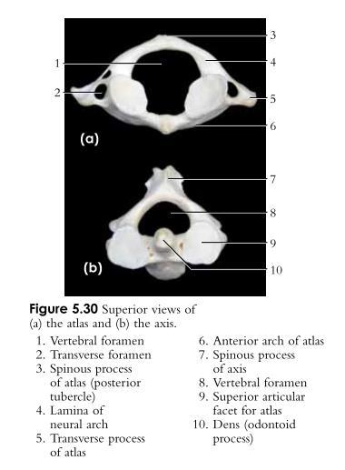
12 New General Structure oftypical Vertebrae 1. Body or centrum (disc shaped):weight-bearing region. 2. Vertebral arch:composed of pedicles and laminae that along with the centrum encloses the vertebral foramen. 3. Laminae: the flat continuation of the body posteriorly. ****laminae has anterior connection with pedicle 4. pedicle: thick round connection between arch and body 5. Vertebral foramina:make up the vertebral canal through which the spinal cord passes. 6. Spinous process:2 laminae fuse together & project posterior and usually downward, some are bifid which provide muscle insertion. Just bifid in the cervical region

13
14 New 7. Transverse processes:pedicle meets with lamina then projects laterally. They are for muscle attachment of the back. In the cervical vertebrae, there are vertebral artery foramen through which vertebral artery pass. In thoracic vertebrae, these processes are present with facets or demi-facet (demi-facet also present on the body), facets exist on the apex of the trans. process.
15 8. Superior and inferior articular processes: protrude superiorly and inferiorly from the "pediclelamina" junctions. All vertebrae have 2 superior & 2 inferior articular processes except the sacrum & coccyx bones except The first sacral vertebra has superior one to actriculate with the 5 th lumabar vertebra.. These processes depend on the movement range, they differ in orientation (laterally, anteriorly, more or less lateral & anterior), so they differ from region to another,to provide more movement according to that region.
16 7.Inter-vertebral foramina:lateral openings formed from notched areas on the superior and inferior borders of adjacent pedicles. Lacated anterior to the transverse process and more or less immediately underneath the pedicle. The spinal nerves exist through it from the spinal cord. The atlas & axis vertebrae are different from all other vertebrae, although they contain same features of other vertebrae,but still they have additional processes so therefore they are considered atypical vertebrae. Sacral and coccyx vertebrae are typical;they are fused together so we don t consider them as atypical
17 ** Vertebrae are the same(arch& body), but the difference from one region to another can be in: 1- the prominence of the spinous process, 2- size of the body 3- the shape in the vertebral canal. New
18 CervicalVertebrae Seven vertebrae (C1-C 7) are the smallest & lightest vertebrae. C3-C7 are distinguished with an 1. Oval body. 2. Long spinous processes. 3. Large and triangular vertebral foramina. Each transverse process contains a transverse foramen which vertebral artery passes through. Superior and inferior articular facets are oriented superior & inferior.
19 New ATYPICAL C ERVICAL VERTEBRAE 1. Atlas (C 1):it is nothing but a round arch. Has no body and no spinous process. Consists of anterior and posterior arches, and two lateral masses (masses are elongated more toward lateral aspect). The superior and inferior surface of lateral masses has articular surfaces (depressions) to articulate with the occipital condyles and with the axis Lateral to the lateral masses there are transverse processes, and it has transverse foramen.


20
21 Notice the anterior & posterior tubercle made by meeting of arches posteriorly & anteriorly. Atlanto-occipital joint: an important joint, only flexion & extension movement occurs ; where at the rest of the joints in vertebrae such as (atlanto-axial, axial-c3),the movement is rotation. The superior articular facets are divided into 2 facets separated by a ridge ; this ridge is important in the flex.& ext. maneuver. It will stop the posterior sliding of condyles of the occipital bone on atlas. The ridge will limit the movement anterior & posterior.
22 With age the flex. & ext. maneuver will be reduced; because the intervertebral layer (hyaline cartilage) in the joint is eroded,it won't be moving like before, exercise will help in keeping good movement. The inferior articular facets are more rounded and more medially oriented to fit the superior aspect of the axis. On the anterior arch, notice the fovea dentis which is the facet for the dens, odontoid ligament holds the dens to lateral borders of fovea dentis.
23 2.Axis (C 2): The axis has a body (very small),spine, and vertebral arches as do other cervical vertebrae. Unique to the axis is the dens,or odontoid process,which projects superiorly from the body and is attached to the anterior surface of the arch of the atlas. The dens is a pivot for the rotation of the atlas.

24
25 Transvers processes comes from similar masses (pedicles & laminae), they are very small with transverse foramen where vertebral artery passes. Masses in axis are called pedicles & laminae while in atlas called lateral masses
26 The pedicles are more or less connected directly; because of the presence of the masses, the transverse process comes out before the pedicle,that when you go down to C3 it is pushed anteriorly & the pedicle is more posterior. That's why we call it more or less atypical vertebra; because it doesn't present the typical aspect of any vertebra. The spinous process is posterior & it is bifidic. Irregular vertebral canal. The atlanto-occipital joint has limited movement due to the ridge on the facets & the spinous processes all the way down in the cervical region.

27 Pivot joint: Projection (dens) of axis articulates within ring of atlas. Movements: Rotation Around one Axis.

28


29 the doctor pointed to all the labeled parts New
30 Thoracic vertebrae H ow do you know this is a thoracic vertebra? The spinous process is long & inclined inferiorly, heart shape body. The body has a mass, that is full of spongy bone). The trans. process has articular facets & the body have demi facet so that 2 demi facets of adjacent vertebrae forms one facet which articulate with the head of rib.that s why the rib takes the name of the above vertebrae. Superior articular facets are lateral & posterior, the inferior ones are medially & anterior.

31
32 General features There are twelve vertebrae (T 1 -T 12 ) all of which articulate with ribs. Major markings include two demi-facets on the heart-shaped body for the head of the rib. Circular vertebral foramen, transverse processes with articular costal facets for the rib tubercles. Long spinous process that is inclined downward. The location of the articulate facets prevent flexion and extension, but allow rotation of this area of the spine. Superior articular processes are oriented backward and laterally.
33 LumbarVertebrae The five lumbar vertebrae (L 1 -L 5 ) are located in the small region of the back and have an enhanced weight-bearing function. Body is large and kidney-shaped. They have short, thick pedicles and lamina. Flat hatchet-shaped spinous processes. Triangular-shaped vertebral foramen. Orientation of the sup. articular facets face medially to lock the lumbar vertebrae together to provide stability

34 *Spinous process is more shorter and more square in shape. *The bodies are more bulky than thoracic and cervical


35 New **Why it is a lumbar vertebrae?? 1- the spinous processes are more shorter 2- The body is more bulky **The intervertebral disc appear as a gap between the vertebrae.

36 VERTEBRAL C H A R ACTERISTICS:

37

38

39
40 Below & above the 2 pedicles, there is a vertebral notch (sup & inf);it makes the intervertebral foramen between 2 vertebrae which spinal nerves exit from it. Usually (especially thoracic vertebrae) the transverse processes will grow longer,they will incline inferiorly like the wings & this resembles the ribs, it occurs mainly at the level of C 7 (you can palpate it at the end of your neck), we call it cervical rib, it will compress the nerve root leading to difficulty in moving the upper extremities.
41 Cervical rib The arm & forearm muscles will be tired & will take their tiredness to the shoulder muscle. Cervical ribs are very common; because of the computer, games,driving & desk sitting, we compress ourselves while sitting, then the cervical rib will compress the nerve & you will have headache & shoulder stiffness. If someone told you I have headache after working,you will examine 2 things: blood pressure & x-ray of cervical region. Spinal nerves go to muscles, so any compression will lead to defects in movement skeletal muscles.
42 Inter-vertebral discs Cushion-like pad Joint between 2 bodies of the vertebrae It s composed of two parts 1. Nucleus pulposus: inner gelatinous nucleus that gives the disc is elasticity and compressibility 2. Annulus fibrosus:surrounds the nucleus pulposus with a collar composed of collagen and Fibro cartilage.

43
44 Pathological condition Sometimes, annulus fibrosus bulge or rupture leading to movement of nucleus pulposus toward the edge,so it will exit the concentric layer of the fibrous tissue, and that's what we call herniation. Herniation varies in bulging,so bulging of annulus fibrosus can lead to reduction in distance between 2 vertebrae;because you lose the power of annulus fibrosus.
45 The whole vertebrae have longitudinal ligaments to hold them in place,but they cannot help in preventing the herniation of the nucleus polposus. There are anterior and posterior longitudinal ligament; Allow them not to move in the rotational movement, instead, they will move bending left and right. New Notice that the posterior ligament is anterior to the spinal cord>>> give protection
46 Any degeneration of annulus fibrosus can lead to herniation. Spine surgeon has a ruler to measure the intervertebral discs or by naked eye, the measurement will indicate the age of compression on this disc.

47 We have inter-spinous ligaments between the spinous processes, which hold the spinous processes in their position.

48 M OVEMENTS:occurs at the level of vertebral column
49 **when we rotate the hole vertebrae, the rotation occur at the hip joint, not within the vertebral column. New
50 ATLANTO -O C C IPITAL JOINT In slides Often considered to be a Hinge joint because of its primary uniaxial range of movement (as in shaking your head yes ). The atlanto-occipital joints occur between the reciprocally curved surfaces of the two occipital condyles and the articular facets on the lateral masses of the atlas. Movements: While the primary axis of movement is the nodding movements (flexion and extension of the head) in the antero-posterior plane,a small amount of side to side bending (lateral flexion),and rotation is possible at this joint surface. Ligaments of the joint: Are the fibrous membrane of the joint capsule (Ant and Post), the anterior atlanto-occipital membrane,and the posterior atlantooccipital membrane.
51 LIGAMENTS OF THE JOINT In slides Most important are anterior & posterior longitudinal ligament,they will hold the atlas with the occipital bone. Anterior atlanto-occipital membrane goes from anterior arch of the atlas into the base of the occipital bone anteriorly.

52 In slides

53 In slides
54 In slides Posterior longitudinal ligament doesn't appear from posterior view because it is anterior to posterior atlanto-occipital membrane & below inferior nuchal line. At the lateral aspect you will be able to see an articular capsule (image above). There is always a capsule between the 2 joints, especially synovial joints (most of joints are synovial).
55 In slides The bodies of the vertebrae are articulated with a separate articular joint rather if you compare it with the superior & inferior articular surface,so whenever you look at any slide you will be able to see that articular capsule for the atlanto-occipital joint in the lateral aspect, because this articular capsule is surrounding the outer area of the condyles and the superior articular surface.
56 In slides Ligaments between the atlas & the axis are important, one of them is the cruciate ligament,it has 2 branches one superior & one inferior,they are called superior & inferior longitudinal band & 2 transverse ligaments.

57 Cruciate ligament of atlas In slides
58 In slides In the above image we have removed the posterior aspect of the occipital bone & the spinous processes, in the posterior surface of the dens after the annular ligament of the dens you will be able to see this. Parcticaly you will be able to see the anterior arch & dens & the body of the vertebrae then posterior to that the annular ligament & posterior to that & anterior to the spinal cord you will be able to see the cruciate ligament,and lateral to it we have alar ligament.
59 In slides Alar ligaments: They connect the sides of the dens (on the axis, or the second cervical vertebra) to the tubercles on the medial side of the occipital condyle. Legamentum flavum: They connect the laminae of adjacent vertebrae, extending from the second CERVICAL vertebra, the axis, to the first segment of the sacrum Cruciform ligament of atlas (cruciform): is a cruciate ligament in the neck forming part of the atlanto-axial joint. صليب = Cruciform Cruciate =
60 In slides Ligamentum nuchae:comes from the occipital protubrance extending down till the level of the spinous process of C7,it is a very tough membrane seperating the right & left sides of the neck

61 In slides
62 Sacrum and Coccyx Sacrum : - 5 fused bones
63 Cont Sacrum - shape the posterior wall of the pelvis - It articulates with L 5 superiorly (by the only two superior processes), and with the auricular surfaces of the hip bones - Major markings include the sacral promontory, transverse lines, alae,dorsal sacral foramina, sacral canal, and sacral hiatus New - Upper anterior aspect of the body protrose anteriorly, create the concavity of sacrum convexity
64 Anterior : - Sacral promontory -concave curve - vertebral bodies -anterior/ventral sacral foraminae - transverse lines laterally : -Ala : formed by fused transverse processes. - (here the transverse processes are more bulk and tortious compared with the thoracic one)

65 New the posterior aspect of ala articulate with the ilium of the hip at the inferior part of the median crest divided into two right and left processes Dorsal sacral foramina continuated to the anterior sacral foramina(they are lateral to the bodies).

66 Posterior : -superior articular process ( betw S1-L5) -Sacral canal (contain fillum Terminale, open in Sacral hiatus at S5) - Median sacral crest - Lateral sacral crest Between them is : -Intermediate crest. -Dorsal Sacral foramina (betw lateral and intermediate) -sacral cornuae : belong to S4,near thesacral hiatus

67 Coccyx S1 S2 Coccyx (Tailbone) The coccyx is made up of four (in some cases three to five) fused vertebrae that articulate directly Superiorly with the sacrum Coccyx S3 S4 S
68 Disk problems At the level of the vertebra : - Fractures of the vertebra At the level of the intervertebral disk : - 1.D isk Herniation - 2.D egeneration (slipped) Most common sites for disc problems: C5 - C6 L4 - L5 L5 - S1 Laminectomy ( IS a surgical removal vertebral arch by shaving laminae to access disc)
69 Cont Disk problems At the level of the vertebra : * Fractures of the vertebra,could be : -Fragmented & nonfragmented -Complete & incomplete - distance between the broken vertebrae and the adjacent one above or below will be minimized

70
71 Cont Disk problems At the level of the intervertebral disk : - 1.Disk Herniation :complete or incomplete bulging - 2.Degeneration :degenerative changes in the structure of the annulus fibrosis

72
73 Thoracic cage 1) Sternum 2) Ribs ** The thoracic cage is composed of the thoracic vertebrae dorsally,the ribs laterally,and the sternum and costal cartilages anteriorly New **The movement of thorax will be only at the anterior aspect (during respiration) by action of cartilages. But posterior part will never move ;because it linked by ligaments to the vertebrae.
74 Thoracic cage Functions Forms a protective cage around the heart,lungs, and great blood vessels Supports the shoulder girdles and upper limbs Provides attachment for many neck,back,chest, and shoulder muscles Uses intercostal muscles to lift and depress the thorax during breathing

75 Subcostal angle is the angle in between the two Costal margins which are the inferior borders of the 7th & false ribs costal cartilages
76 Sternum dagger-shaped Flat bone made up of 3 parts : 1 Manubrium 2 Sternal Body 3 Xiphoid process Anatomical landmarks include the jugular (suprasternal) notch, the sternal angle,and the xiphisternal joint

77 1- Manubrium
78 1- Manubrium square shape part that has superior and inferior articular boarders Important landmarks : ** "angle of Louis = sternal angle **superiorly,and lateral for clavicular sternal end **lateral,and inferior for 1 st rib **Jugular notch = suprasternal notch, between the two clavicular facets used for ((tracheostomy)) cause behind it no veins or arteries, & the trachea lies directly under the skin of this region.
79 New Sternal(lewis) angle: it lies between manubrium and body very important clinically ; because you can count the ribs from the second rib legation.
80 2- Sternal Body The biggest part Presents on the lateral sides depressions serves as an articulation surface for the 2nd to the 7th rib 8th to the 10 th ribs are false ribs,they don t attach to the sternal body directly. 11 and 12 are floating ribs first rib is atypical because it's surfaces orientations are different from the typical.it has superiorinferior (horizontal) rather than anterior posterior surfaces(vertical)
81 3- Xiphoid process Till the age of 17 to 19 it's an flexible cartilaginous & Elastic tissue
82 Ribs (12) composed of two parts : -One anterior "the end" continues to the sternum as the costal cartilage -One posterior,and "the head" articulate with the lateral surface "2 demifacets" on the superior and inferior boarders ofthe body of the adjacent thoracic vertebrae,as well as the anterior part of the transverse processes for the lower vertebra articulate with the tubercale so : the head with the body the tubercle transeverse process
83 Ribs & sternum articulations: All ribs are incomplete bones ; because they need cartilages to connect with lateral boarder of sternum. 1-7 ribs : articulate directly with the sternum by costal cartilages False ribs (8-12) : normal ribs but they never reach the sternum (8-10) ; adhere to the 7th cartilages (11-12) ; Floating ribs, they don't articulate with any thing anteriorly. New
84 Cont Ribs All the typical ribs are oriented anterior & inferior ribs have 2 borders & two surfaces : -One outer surface "external(lateral) surface". facing the fascia,and One inner surface(medial) facing the pleura - The superior border is rounded,andthe inferior border is more sharp (with subcoastal groove),contain Subcostal vein,artery & nerve from superior to inferior respectively on the internal surface (( clinically )),Pleural puncture we do it in the superior border..if done on inferior border it may cause nerve injury which is called in medicine Intercostal neuralgia

85

86 the head with the body the tubercle with the transeverse process So, rib= bowed, flat bone consisting of a head, neck, tubercle, and shaft


87 Superior facet Inferior facet Articular facet of tubercle


88

89

90
Copyright 2010 Pearson Education, Inc. Copyright 2010 Pearson Education, Inc. Figure Sectioned spinous process. Interspinous.
 PowerPoint Lecture Slides prepared by Janice Meeking, Mount Royal College C H A P T E R 7 The Skeleton: Part B Vertebral Column Transmits weight of trunk to lower limbs Surrounds and protects spinal cord
Chapter 7 Part B The Skeleton
 Chapter 7 Part B The Skeleton 7.2 The Vertebral Column General Characteristics Extends from skull to pelvis Also called spine or spinal column Functions to transmit weight of trunk to lower limbs, surround
Copyright 2010 Pearson Education, Inc.
 E. VERTEBRAL COLUMN 1. The vertebral column extends from the skull to the pelvis and forms the vertical axis of the skeleton. 2. The vertebral column is composed of vertebrae that are separated by intervertebral
Vertebral Column. Backbone consists of 26 vertebrae. Five vertebral regions. Cervical
 Vertebral Column Backbone consists of 26 vertebrae. Five vertebral regions Cervical vertebrae (7) in the neck. Thoracic vertebrae (12) in the thorax. Lumbar vertebrae (5) in the lower back. Sacrum (5,
Axial Skeleton: Vertebrae and Thorax
 Axial Skeleton: Vertebrae and Thorax Function of the vertebral column (spine or backbone): 1) 2) 3) Composition of Vertebral column The vertebral column is formed by 33 individual vertebrae (some of which
2. The vertebral arch is composed of pedicles (projecting from the body) and laminae (uniting arch posteriorly).
 and laminae (uniting arch posteriorly).") VERTEBRAL COLUMN 2018zillmusom I. VERTEBRAL COLUMN - functions to support weight of body and protect spinal cord while permitting movements of trunk and providing for muscle attachments. A. Typical vertebra
VERTEBRAL COLUMN 2018zillmusom I. VERTEBRAL COLUMN - functions to support weight of body and protect spinal cord while permitting movements of trunk and providing for muscle attachments. A. Typical vertebra
VERTEBRAL COLUMN VERTEBRAL COLUMN
 VERTEBRAL COLUMN FUNCTIONS: 1) Support weight - transmits weight to pelvis and lower limbs 2) Houses and protects spinal cord - spinal nerves leave cord between vertebrae 3) Permits movements - *clinical
THE VERTEBRAL COLUMN. Average adult length: In male: about 70 cms. In female: about 65 cms.
 THE VERTEBRAL COLUMN Average adult length: In male: about 70 cms. In female: about 65 cms. 1 Vertebral Column (Regions and Curvatures) Curvatures of the vertebral column: A. Primary curvature: C-shaped;
AXIAL SKELETON FORM THE VERTICAL AXIS OF THE BODY CONSISTS OF 80 BONES INCLUDES BONES OF HEAD, VERTEBRAL COLUMN, RIBS,STERNUM
 AXIAL SKELETON FORM THE VERTICAL AXIS OF THE BODY CONSISTS OF 80 BONES INCLUDES BONES OF HEAD, VERTEBRAL COLUMN, RIBS,STERNUM APPENDICULAR SKELETON BONES OF THE FREE APPENDAGES & THEIR POINTS OF ATTACHMENTS
The Thoracic Cage ANATOMY 2: THORACIC CAGE AND VERTEBRAL COLUMN
 ANATOMY 2: THORACIC CAGE AND VERTEBRAL COLUMN PSK 4U Mr. S. Kelly North Grenville DHS The Thoracic Cage 7 true ribs 3 false ribs 2 floating ribs Clavicle = collarbone Manubrium Sternum Xiphoid Process
human anatomy 2015 lecture four Dr meethak ali ahmed neurosurgeon
 The Vertebral Column the vertebral columnis central pillar of the body.it serve to protect the spinal cord and support the weight of the head trunk, which it transmits to the hip bones & the lower limbs.
THE THORACIC WALL. Boundaries Posteriorly by the thoracic part of the vertebral column. Anteriorly by the sternum and costal cartilages
 THE THORACIC WALL Boundaries Posteriorly by the thoracic part of the vertebral column Anteriorly by the sternum and costal cartilages Laterally by the ribs and intercostal spaces Superiorly by the suprapleural
Anatomy Lecture #19 AN INTRODUCTION TO THE THORAX April 3, 2012
 Page 1 بسم الله الرحمن الرحيم The Thoracic Wall Firstly, when we talk about thorax, we should begin with the thorax wall which means not only bones that construct the thorax but also the muscles which
The Thoracic Cage. Role of the Thoracic Cage 2/13/2019. Anatomy 2: Thoracic Cage and Vertebral Column
 PSK 4U Mr. S. Kelly North Grenville DHS Anatomy 2: Thoracic Cage and Column The Thoracic Cage 7 true ribs 3 false ribs 2 floating ribs Clavicle = collarbone Manubrium Sternum Xiphoid Process 12 thoracic
Overview of the Skeleton: Bone Markings
 Name Overview of the Skeleton: Bone Markings Match the terms in column B with the appropriate description in column A. Column A 1. sharp, slender process* 2. small rounded projection* 3. narrow ridge of
Ligaments of the vertebral column:
 In the last lecture we started talking about the joints in the vertebral column, and we said that there are two types of joints between adjacent vertebrae: 1. Between the bodies of the vertebrae; which
Yara saddam & Dana Qatawneh. Razi kittaneh. Maher hadidi
 1 Yara saddam & Dana Qatawneh Razi kittaneh Maher hadidi LECTURE 10 THORAX The thorax extends from the root of the neck to the abdomen. The thorax has a Thoracic wall Thoracic cavity and it is divided
Skeletal System. Axial Division
 Skeletal System Axial Division The Axial Skeleton You will see that each bone has special features (overviewed in section I below) that provide Sites of Attachment (for muscles, ligaments, tendons, etc.)
Chapter 7 The Skeletal System:The Axial Skeleton
 Chapter 7 The Skeletal System:The Axial Skeleton Axial Skeleton 80 bones lie along longitudinal axis skull, hyoid, vertebrae, ribs, sternum, ear ossicles Appendicular Skeleton 126 bones upper & lower limbs
Anatomy notes-thorax.
 Anatomy notes-thorax. Thorax: the part extending from the root of the neck to the abdomen. Parts of the thorax: - Thoracic cage (bones). - Thoracic wall. - Thoracic cavity. ** The thoracic cavity is covered
Bones of the wrist and ankle Bones that form within tendons (e.g., patella)
") Skeletal System Review Surface Anatomy Dr. Gary Mumaugh Function of Bones Support form the framework that supports the body and cradles soft organs Protection provide a protective case for the brain, spinal
Skeletal System Review Surface Anatomy Dr. Gary Mumaugh Function of Bones Support form the framework that supports the body and cradles soft organs Protection provide a protective case for the brain, spinal
THEME 2. VERTEBRAE (GENERAL DATA). CERVICAL, THORACIC AND LUMBAR VERTEBRAE. SACRUM. COCCYX. THE VERTEBRAL COLUMN AS A WHOLE
. CERVICAL, THORACIC AND LUMBAR VERTEBRAE. SACRUM. COCCYX. THE VERTEBRAL COLUMN AS A WHOLE") THEME 2. VERTEBRAE (GENERAL DATA). CERVICAL, THORACIC AND LUMBAR VERTEBRAE. SACRUM. COCCYX. THE VERTEBRAL COLUMN AS A WHOLE Osteology of the Vertebral Column Bone Description vertebra Notes a vertebra
THEME 2. VERTEBRAE (GENERAL DATA). CERVICAL, THORACIC AND LUMBAR VERTEBRAE. SACRUM. COCCYX. THE VERTEBRAL COLUMN AS A WHOLE Osteology of the Vertebral Column Bone Description vertebra Notes a vertebra
Crafton Hills College Human Anatomy & Physiology Axial Skeleton
 A. Major Divisions Crafton Hills College Human Anatomy & Physiology Axial keleton 1. Axial: Part of skeleton lies along long axis of body 2. Appendicular: Bones & features of the appendages B. AXIAL KELETON
Anatomy and Physiology II. Spine
 Anatomy and Physiology II Spine Bones and Other Structures Vertibrae Contains Cervical, Thoracic, Lumbar, Sacral and Coccygeal regions We use Capital letters to refer to these (C, T, L, S, and Co) and
ANATOMY & PHYSIOLOGY I Laboratory Version B Name Section. REVIEW SHEET Exercise 10 Axial Skeleton
 ANATOMY & PHYSIOLOGY I Laboratory Version B Name Section REVIEW SHEET Exercise 10 Axial Skeleton 1 POINT EACH. THE SKULL MULTIPLE CHOICE 1. The major components of the axial skeleton include the 7. The
Human Anatomy and Physiology - Problem Drill 07: The Skeletal System Axial Skeleton
 Human Anatomy and Physiology - Problem Drill 07: The Skeletal System Axial Skeleton Question No. 1 of 10 Which of the following statements about the axial skeleton is correct? Question #01 A. The axial
Clarification of Terms
 Clarification of Terms The Spine, Spinal Column, and Vertebral Column are synonymous terms referring to the bony components housing the spinal cord Spinal Cord = made of nervous tissue Facet = a small,
Bony framework of the vertebral column Structure of the vertebral column
 5.1: Vertebral column & back. Overview. Bones o vertebral column. o typical vertebra. o vertebral canal. o spinal nerves. Joints o Intervertebral disc. o Zygapophyseal (facet) joint. Muscles o 2 compartments:
Clarification of Terms
 Clarification of Terms The Spine, Spinal Column, and Vertebral Column are synonymous terms referring to the bony components housing the spinal cord Spinal Cord = made of nervous tissue Facet = a small,
Clarification of Terms
 Clarification of Terms The Spine, Spinal Column, and Vertebral Column are synonymous terms referring to the bony components housing the spinal cord Spinal Cord = made of nervous tissue Facet = a small,
Main Menu. Trunk and Spinal Column click here. The Power is in Your Hands
 1 The Trunk and Spinal Column click here Main Menu K.9 http://www.handsonlineeducation.com/classes/k9/k9entry.htm[3/27/18, 2:00:55 PM] The Trunk and Spinal Column Vertebral column complex 24 intricate
STERNUM. Lies in the midline of the anterior chest wall It is a flat bone Divides into three parts:
 STERNUM Lies in the midline of the anterior chest wall It is a flat bone Divides into three parts: 1-Manubrium sterni 2-Body of the sternum 3- Xiphoid process The body of the sternum articulates above
Anatomy of the Thorax
 Anatomy of the Thorax A) THE THORACIC WALL Boundaries Posteriorly by the thoracic part of the vertebral column Anteriorly by the sternum and costal cartilages Laterally by the ribs and intercostal spaces
River North Pain Management Consultants, S.C., Axel Vargas, M.D., Regional Anesthesiology and Interventional Pain Management.
 River North Pain Management Consultants, S.C., Axel Vargas, M.D., Regional Anesthesiology and Interventional Pain Management. Chicago, Illinois, 60611 Phone: (888) 951-6471 Fax: (888) 961-6471 Clinical
Structure and Function of the Vertebral Column
 Structure and Function of the Vertebral Column Posture Vertebral Alignment Does it really matter? Yes it does! Postural Curves The vertebral column has a series of counterbalancing curves posterior anterior
Chapter 7. Skeletal System
 Chapter 7 Skeletal System 1 Skull A. The skull is made up of 22 bones: 8 cranial bones, 13 facial bones, and the mandible. B. The Cranium encloses and protects the brain, provides attachments for muscles,
The vault bones Frontal Parietals Occiput Temporals Sphenoid Ethmoid
 The Vertebral Column Head, Neck and Spine Bones of the head Some consider the bones of the head in terms of the vault bones and the facial bones hanging off the front of them The vault bones Frontal Parietals
Anatomy of the Spine. Figure 1. (left) The spine has three natural curves that form an S-shape; strong muscles keep our spine in alignment.
 The spine has three natural curves that form an S-shape; strong muscles keep our spine in alignment.") 1 2 Anatomy of the Spine Overview The spine is made of 33 individual bony vertebrae stacked one on top of the other. This spinal column provides the main support for your body, allowing you to stand upright,
1 2 Anatomy of the Spine Overview The spine is made of 33 individual bony vertebrae stacked one on top of the other. This spinal column provides the main support for your body, allowing you to stand upright,
THE SKELETAL SYSTEM. Focus on the Skull
 THE SKELETAL SYSTEM Focus on the Skull Review Anatomical Terms Anterior/Posterior Dorsal/Ventral Medial/Lateral Superior/Inferior Bone Markings - Review Projections for attachment of muscles, ligaments
The Trunk and Spinal Column Kinesiology Cuneyt Mirzanli Istanbul Gelisim University
 The Trunk and Spinal Column Kinesiology Cuneyt Mirzanli Istanbul Gelisim University The Trunk and Spinal Column Vertebral column 24 articulating vertebrae 31 pairs of spinal nerves Abdominal muscles some
Chapter 7: Skeletal System: Gross Anatomy
 Chapter 7: Skeletal System: Gross Anatomy I. General Considerations A. How many bones in an average adult skeleton? B. Anatomic features of bones are based on II. Axial Skeleton A. Skull 1. Functionally
Human Anatomy - Problem Drill 06: The Skeletal System Axial Skeleton & Articualtions
 Human Anatomy - Problem Drill 06: The Skeletal System Axial Skeleton & Articualtions Question No. 1 of 10 Instructions: (1) Read the problem and answer choices carefully, (2) Work the problems on paper
The Vertebral Column
 The Vertebral Column The vertebral column (also called the backbone, spine, or spinal column) consists of a series of 33 irregularly shaped bones, called vertebrae. These 33 bones are divided into five
VERTEBRAL COLUMN ANATOMY IN CNS COURSE
 VERTEBRAL COLUMN ANATOMY IN CNS COURSE Vertebral body Sections of the spine Atlas (C1) Axis (C2) What type of joint is formed between atlas and axis? Pivot joint What name is given to a fracture of both
Chest cavity, vertebral column and back muscles. Respiratory muscles. Sándor Katz M.D., Ph.D.
 Chest cavity, vertebral column and back muscles. Respiratory muscles. Sándor Katz M.D., Ph.D. Chest cavity - bony structures Chest cavity- bony structures Sternum Ribs True ribs: first seven pairs connect
Skeletal system. Prof. Abdulameer Al-Nuaimi. E. mail:
 Skeletal system Prof. Abdulameer Al-Nuaimi E-mail: a.al-nuaimi@sheffield.ac.uk E. mail: abdulameerh@yahoo.com Functions of Bone and The Skeletal System Support: The skeleton serves as the structural framework
INDEPENDENT LEARNING: DISC HERNIATION IN THE NATIONAL FOOTBALL LEAGUE: ANATOMICAL FACTORS TO CONSIDER IN REVIEW
 INDEPENDENT LEARNING: DISC HERNIATION IN THE NATIONAL FOOTBALL LEAGUE: ANATOMICAL FACTORS TO CONSIDER IN REVIEW CDC REPORT - CAUSES OF DISABILITY, 2005 REVIEW QUESTIONS ABOUT DISC HERNIATION IN THE NATIONAL
PELVIS & SACRUM Dr. Jamila El-Medany Dr. Essam Eldin Salama
 PELVIS & SACRUM Dr. Jamila El-Medany Dr. Essam Eldin Salama Learning Objectives At the end of the lecture, the students should be able to : Describe the bony structures of the pelvis. Describe in detail
LAB Notes#1. Ahmad Ar'ar. Eslam
 LAB Notes#1 Ahmad Ar'ar Eslam 1 P a g e Anatomy lab Notes Lower limb bones :- Pelvic girdle: It's the connection between the axial skeleton and the lower limb; it's made up of one bone called the HIP BONE
Sports Medicine Part I : ANATOMY OF THE SPINE, ABDOMEN AND SHOULDER COMPLEX
 Sports Medicine 25 1.1 Part I : ANATOMY OF THE SPINE, ABDOMEN AND SHOULDER COMPLEX c.w.p. Wagner High School, Sports Medicine, A. Morgan, T. Morgan 2008 Anatomy of the Upper Body In this section of the
11/25/2012. Chapter 7 Part 2: Bones! Skeletal Organization. The Skull. Skull Bones to Know Cranium
 Chapter 7 Part 2: Bones! 5) Distinguish between the axial and appendicular skeletons and name the major parts of each 6) Locate and identify the bones and the major features of the bones that compose the
Any of the vertebra in the cervical (neck) region of the spinal column. The cervical vertebra are the smallest vertebra in the spine, reflective of th
 region of the spinal column. The cervical vertebra are the smallest vertebra in the spine, reflective of th") Any of the vertebra in the cervical (neck) region of the spinal column. The cervical vertebra are the smallest vertebra in the spine, reflective of the fact that they support the least load. In humans,
Any of the vertebra in the cervical (neck) region of the spinal column. The cervical vertebra are the smallest vertebra in the spine, reflective of the fact that they support the least load. In humans,
BOGOMOLETS NATIONAL MEDICAL UNIVERSITY. Department of human anatomy
 BOGOMOLETS NATIONAL MEDICAL UNIVERSITY Department of human anatomy GUIDELINES Academic discipline дисципліна HUMAN ANATOMY Module 1 Content module 2 The theme of the lesson Sacrum. Coccyx. Ribs. Chest.
Osteology of the Thorax. Prof Oluwadiya K S
 Osteology of the Thorax Prof Oluwadiya K S www.oluwadiya.com The thoracic skeleton consists of the following: 12 pairs of ribs and associated costal cartilages 12 thoracic vertebrae and their intervertebral
Human Anatomy & Physiology I Dr. Sullivan Unit VIIIa The Axial Skeleton Chapter 8 (Sections )
") Human Anatomy & Physiology I Dr. Sullivan Unit VIIIa The Axial Skeleton Chapter 8 (Sections 8.1-8.3) I. Divisions of the skeletal system a) An adult human skeleton has 206 named bones b) Most are paired
Human Anatomy & Physiology I Dr. Sullivan Unit VIIIa The Axial Skeleton Chapter 8 (Sections 8.1-8.3) I. Divisions of the skeletal system a) An adult human skeleton has 206 named bones b) Most are paired
Bones of Thorax (Rib Cage)
") Musculoskeletal System (Part A-2) Module 7 -Chapter 10 Overview Muscles Attachments Bones Bone types Surface features of bones Divisions of the skeletal system Joints or Articulations Susie Turner, M.D.
Musculoskeletal System (Part A-2) Module 7 -Chapter 10 Overview Muscles Attachments Bones Bone types Surface features of bones Divisions of the skeletal system Joints or Articulations Susie Turner, M.D.
A. Incorrect! The axial skeleton includes bones of the skull, inner ear, chest and spinal column.
 Anatomy and Physiology - Problem Drill 07: The Skeletal System II No. 1 of 10 1. Which of the following statements about the axial skeleton is correct? A. The axial skeleton includes bones of the skull,
1TRUNK: BODY WALL AND SPINE
 TRUNK: BODY WALL AND SPINE SURFACE ANATOMY SKELETON JOINTS & LIGAMENTS MUSCLES VASCULATURE NERVES SPINAL CORD & VERTEBRAL CANAL ANTERIOR BODY WALL & MAMMARY GLAND LATERAL BODY WALL INGUINAL REGION SUPERFICIAL
Cranium Facial bones. Sternum Rib
 Figure 7.1 The human skeleton. Skull Thoracic cage (ribs and sternum) Cranium Facial bones Sternum Rib Bones of pectoral girdle Vertebral column Sacrum Vertebra Bones of pelvic girdle (a) Anterior view
It consist of two components: the outer, laminar fibrous container (or annulus), and the inner, semifluid mass (the nucleus pulposus).
, and the inner, semifluid mass (the nucleus pulposus).") Lumbar Spine The lumbar vertebrae are the last five vertebrae of the vertebral column. They are particularly large and heavy when compared with the vertebrae of the cervical or thoracicc spine. Their bodies
Lumbar Spine The lumbar vertebrae are the last five vertebrae of the vertebral column. They are particularly large and heavy when compared with the vertebrae of the cervical or thoracicc spine. Their bodies
The Skeletal System. Parts of the skeletal system. Bones (Skeleton) Joints Cartilages Ligaments
 Joints Cartilages Ligaments") The Skeletal System Parts of the skeletal system Bones (Skeleton) Joints Cartilages Ligaments Functions of the Bones Support Internal framework of the body Protection Skull and vertebrae protect brain
The Skeletal System Parts of the skeletal system Bones (Skeleton) Joints Cartilages Ligaments Functions of the Bones Support Internal framework of the body Protection Skull and vertebrae protect brain
Dr Ajit Singh Moderator Dr P S Chandra Dr Rajender Kumar
 BIOMECHANICS OF SPINE Dr Ajit Singh Moderator Dr P S Chandra Dr Rajender Kumar What is biomechanics? Biomechanics is the study of the consequences of application of external force on the spine Primary
The Biomechanics of the Human Spine. Basic Biomechanics, 6 th edition By Susan J. Hall, Ph.D.
 Chapter 9 The Biomechanics of the Human Spine Structure of the Spine The spine is a curved stack of 33 vertebrae structurally divided into five regions: cervical region - 7 vertebrae thoracic region -
2 skull, vertebral column, thoracic cage
 CHAPTER 7-SKELTON FILL-IN NOTES 2 skull, vertebral column, thoracic cage 3 Fig. 7.1 pg. 199 4 I. Skull: A. : Encloses and the brain - 8 bones B. : 14 bones Cranium A. Forehead (brain) Anterior part of
Exercise 10. The Axial Skeleton
 Exercise 10 The Axial Skeleton The Axial Skeleton Consists of the skeletal structures found along the midline of the body. Includes the skull, hyoid, vertebrae, ribs, sternum, and sacrum. The cartilages
Biology 218 Human Anatomy. Adapted from Martini Human Anatomy 7th ed. Chapter 6 The Skeletal System: Axial Division
 Adapted from Martini Human Anatomy 7th ed. Chapter 6 The Skeletal System: Axial Division Introduction The axial skeleton: Composed of bones along the central axis of the body Divided into three regions:
You have 24 vertebrae in your spinal column. Two are special enough to be individually named.
 You have 24 vertebrae in your spinal column. Two are special enough to be individually named. Your atlas (C01) and axis (C02) are very important vertebrae. Without them, head and neck movement would be
NOTES SKELETAL SYSTEM
 NOTES for the SKELETAL SYSTEM Anatomy & Physiology 2016 Johnson The Skeletal System I. System includes 4 basic parts: A. Bones (206 of em) B. Joints C. Cartilages D. Ligaments II. Bones have 5 basic functions:
Chapter 7: Skeletal System
 Chapter 7: Skeletal System The Skeletal System Introduction P. 182 Bone is an organ made up of tissues: It is made up of the following components. Cartilage Blood Nerves Bone Connective Bone Classification
Anatomy & Physiology II. Trunk
 Anatomy & Physiology II Trunk Bones and Landmarks of the Vertebral column 24 vertebrae Sacrum - consists of 5 vertebrae that fuse into one bone Median sacral crest Sacral hiatus 4 sacral foramina Coccyx
 P R E S E N T S Dr. Mufa T. Ghadiali is skilled in all aspects of General Surgery. His General Surgery Services include: General Surgery Advanced Laparoscopic Surgery Surgical Oncology Gastrointestinal
APPENDICULAR SKELETON 126 AXIAL SKELETON SKELETAL SYSTEM. Cranium. Skull. Face. Skull and associated bones. Auditory ossicles. Associated bones.
 SKELETAL SYSTEM 206 AXIAL SKELETON 80 APPENDICULAR SKELETON 26 Skull Skull and associated s 29 Cranium Face Auditory ossicles 8 4 6 Associated s Hyoid Thoracic cage 25 Sternum Ribs 24 Vertebrae 24 column
Cervical Spine Anatomy and Biomechanics. Typical Cervical Vertebra C3 6. Typical Cervical Vertebra Anterior 10/5/2017
 Cervical Spine Anatomy and Biomechanics Typical Cervical Vertebra C3 6 Small, relatively broad body Bifid SpinousProcess Long and narrow laminae Spinal Canal: large, triangular; remarkably consistent dimensions
Copyright 2004 Lippincott Williams & Wilkins. 2. Bone Structure. Copyright 2004 Lippincott Williams & Wilkins
 Chapter 7 The Skeleton: Bones and Joints The Skeleton Skeletal system is made up of bones and joints and supporting connective tissue. 1. Bone Functions 1. To store calcium salts 2. To protect delicate
MD Bones & Joints of the Back. A/Prof Chris Briggs Department of Anatomy & Neuroscience
 MD 2017 Bones & Joints of the Back A/Prof Chris Briggs Department of Anatomy & Neuroscience WARNING This material has been provided to you pursuant to section 49 of the Copyright Act 1968 (the Act) for
The Thoracic Cage. OpenStax College
 OpenStax-CNX module: m46350 1 The Thoracic Cage OpenStax College This work is produced by OpenStax-CNX and licensed under the Creative Commons Attribution License 3.0 By the end of this section, you will
o Diaphysis o Area where red marrow is found o Area where yellow marrow is found o Epiphyseal plate AXIAL SKELETON Skull
 64 Anatomy & Physiology Coloring Workbook 7. Figure 5-2A is a midlevel, cross-sectional view of the diaphysis of the femur. Label the membrane that lines the cavity and the membrane that covers the outside
To classify the joints relative to structure & shape
 To classify the joints relative to structure & shape To describe the anatomy of the hip joint To describe the ankle joint To memorize their blood & nerve supply JOINTS: Joints are sites where skeletal
LEARN * DREAM * AWAKEN* DISCOVER * ENLIGHTEN * INVESTIGATE * QUESTION * EXPLORE
 S C I E N C E H U M A N Spine & Vertebrate The Spine & Vertebrate in Focus This Enrichment4You e-guide provides a brief overview of the spine and vertebrate. In this e-guide you will: Human Body Spine/Vertebrate
Chapter 8A. The Skeletal System: The Axial Skeleton. The Skeletal System: The Axial Skeleton. Types of Bones. Types of Bones
 Chapter 8A The Skeletal System: The Axial Skeleton The Skeletal System: The Axial Skeleton 206 named bones Axial Skeleton 80 bones lie along longitudinal axis skull, hyoid, vertebrae, ribs, sternum, ear
The Skeletal System: Axial Skeleton
 The Skeletal System: Axial Skeleton The Big Idea The Axial Skeleton & Homeostasis The bones of the axial skeleton contribute to homeostasis by protecting many of the body s organs such as the brain, spinal
Spring Written By: J. E. Sutton. Contents: I. Overview of the Skeleton: II. Appendicular Skeleton III. Axial Skeleton IV.
 Spring 2012 Written By: J. E. Sutton Contents: I. Overview of the Skeleton: II. Appendicular Skeleton III. Axial Skeleton IV. Articulations Overview of the Skeleton: I. Orientation to Human Skeleton: a.
CHAPTER 7, PART II (BONES)
") Anatomy Name: CHAPTER 7, PART II (BONES) Entry #: INSTRUCTIONS: 1) READ Chapter 7, pg. 140-161. 2) Using the outline, make a note card for each underlined bone name or phrase. 3) On each note card, put
Anatomy Name: CHAPTER 7, PART II (BONES) Entry #: INSTRUCTIONS: 1) READ Chapter 7, pg. 140-161. 2) Using the outline, make a note card for each underlined bone name or phrase. 3) On each note card, put
Bony Thorax. Anatomy and Procedures of the Bony Thorax Edited by M. Rhodes
 Bony Thorax Anatomy and Procedures of the Bony Thorax 10-526-191 Edited by M. Rhodes Anatomy Review Bony Thorax Formed by Sternum 12 pairs of ribs 12 thoracic vertebrae Conical in shape Narrow at top Posterior
The Back. Anatomy RHS 241 Lecture 9 Dr. Einas Al-Eisa
 The Back Anatomy RHS 241 Lecture 9 Dr. Einas Al-Eisa The spine has to meet 2 functions Strength Mobility Stability of the vertebral column is provided by: Deep intrinsic muscles of the back Ligaments
Skeletal System. It s all about the bones!!!
 Skeletal System It s all about the bones!!! The Skeletal System in Action!! The Skeletal System in Action! https://www.youtube.com/watch?v=icwllrqkv cg&list=plzile25upgebvru0jneppcabh0fhktgt Q 1. FYI 5
بسم الله الرحمن الرحيم
 بسم الله الرحمن الرحيم * Last lecture we talked about : thoracic wall sternum ribs (according to their features they are divided into typical and atypical) vertebral column ( which is made of 33 vertebrae
Transitioning to the Suboccipital Triangle. Suboccipital Triangle
 Transitioning to the Suboccipital Triangle Syllabus p. 14-15 Suboccipital Triangle Borders -Rectus capitis posterior major -Obliquus capitis superior -Obliquus capitis inferior Contents -Vertebral artery
TEST YOURSELF- Chapter 7
 TEST YOURSELF- Chapter 7 Cranial Bones 1. Give the name of the bone for each of the following markings. Some of the markings are found on more than one bone. List all that apply. Cranium a. Frontal squama:
The trunk and spinal column. Functions of Spine. Bones 6/5/2017. Chapter 10. Consider the complexity of functions. 33 bones of the spine
 The trunk and spinal column Chapter 10 Functions of Spine Consider the complexity of functions provides stability to a cylinder permits movement in all directions supports structures of considerable weight
Biology 2401 The Skeletal System
 Biology 2401 The Skeletal System Purpose: The lab will describe the microscopic and gross anatomy of bone, identify bones of the body, and identify important bone markings. I. Overview of the Skeleton
8/4/2012. Causes and Cures. Nucleus pulposus. Annulus fibrosis. Vertebral end plate % water. Deforms under pressure
 Causes and Cures Intervertebral discs Facet (zygopophyseal) joints Inter body joints Spinal nerve roots Nerve compression Pathological conditions Video Causes of back pain Nucleus pulposus Annulus fibrosis
Note : I put the sheet's info within the slides to easily understand this lecture Done by : Zaid Al-Ghnaneem
 Note : I put the sheet's info within the slides to easily understand this lecture Done by : Zaid Al-Ghnaneem Thoracic Wall Lecture Objectives Describe the shape and outline of the thoracic cage including
Diaphragm and intercostal muscles. Dr. Heba Kalbouneh Associate Professor of Anatomy and Histology
 Diaphragm and intercostal muscles Dr. Heba Kalbouneh Associate Professor of Anatomy and Histology Skeletal System Adult Human contains 206 Bones 2 parts: Axial skeleton (axis): Skull, Vertebral column,
Copyright 2003 Pearson Education, Inc. publishing as Benjamin Cummings. Dr. Nabil Khouri MD, MSc, Ph.D
 Dr. Nabil Khouri MD, MSc, Ph.D Pelvic Girdle (Hip) Organization of the Lower Limb It is divided into: The Gluteal region The thigh The knee The leg The ankle The foot The thigh and the leg have compartments
The Skeletal System. Dr. Naim Kittana. Faculty of Medicine & Health Sciences An-Najah National University
 The Skeletal System Dr. Naim Kittana Faculty of Medicine & Health Sciences An-Najah National University 1 Declaration The content and the figures of this seminar were directly adopted from the text book
Anatomy of thoracic wall
 Anatomy of thoracic wall Topographic Anatomy of the Thorax 1 Bones of Thoracic wall ribs 1-7"true" ribs -those which attach directly to the sternum true ribs actually attach to the sternum by means of
MEDICAL IMAGING OF THE VERTEBRAE
 MEDICAL IMAGING OF THE VERTEBRAE Vertebrae are your friends Matthew Harper MS-IV LECTURE OBJECTIVES INTRODUCE THE MOST COMMON MODALITIES OF MEDICAL IMAGING AND BASIC TECHNIQUES FOR READING THESE IMAGES
Anatomy. Anatomy deals with the structure of the human body, and includes a precise language on body positions and relationships between body parts.
 Anatomy deals with the structure of the human body, and includes a precise language on body positions and relationships between body parts. Proper instruction on safe and efficient exercise technique requires
Yoga Anatomy & Physiology
 Yoga Anatomy & Physiology Anatomy & Physiology Anatomy- One of the basic essential sciences of medicine that studies the structure of an organism. Physiology- The biological study of the functions of living