Written Response #1. 1. Based on what you know about Latin root words, what do you think these terms refer to?
|
|
|
- Eugenia Robertson
- 5 years ago
- Views:
Transcription


1 Muscular System
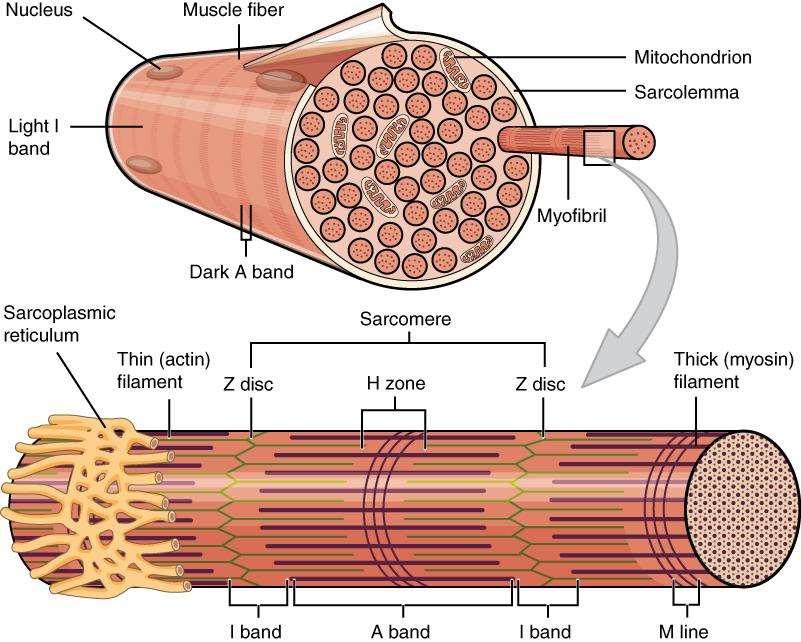
2 Written Response #1 1. Based on what you know about Latin root words, what do you think these terms refer to? Sarcomere Sarcoplasm Myofibril Epimysium Perimysium Endomysium 2. What structure connects muscle to bone?
3 Muscles muscle = myo- or mys- sarco- = flesh - also refers to muscles
4 Main Functions of Muscles 1. Produce movement 2. Maintain posture & body position 3. Stabilize joints 4. Generate heat Additional functions: protect organs, valves, dilate pupils, raise hairs
5 Written Response #2 1. Describe how connective tissue is part of a skeletal muscle. 2. Describe the general structure of a skeletal muscle fiber. 3. Explain why skeletal muscle fibers appear striated. 4. Explain the relationship between the sarcoplasmic reticulum and the transverse tubules.
, nonstriated, involuntary Cardiac (heart) striated,")
6 Types of Muscle Skeletal voluntary, striated, multinucleated The word striated means striped. Smooth visceral (lines hollow organs), nonstriated, involuntary Cardiac (heart) striated, involuntary
7 Special Characteristics of Muscle Excitability can receive and respond to stimuli Contractility can shorten forcibly Extensibility can be stretched or extended Elasticity can recoil and resume resting length after being stretched
8 Gross Anatomy of Skeletal Muscle 1 muscle = 1 organ Each muscle served by a nerve, artery and vein Rich blood supply: need energy & oxygen Connective tissue sheaths: wraps each cell and reinforce whole muscle Attachment is either 1. directly to bone 2. by tendons or aponeuroses to bone, cartilage, or other muscles

9 Muscles and Muscle Fiber Structure Muscles are composed of many FIBERS that are arranged in bundles called FASCICLES


10 Individual muscles are separated by FASCIA, which also forms tendons
 ENDOMYSIUM = surrounds each individual muscle fiber The model to the right uses")
11 Muscle Structure EPIMYSIUM = outermost layer, surrounds entire muscle. PERIMYSIUM = separates and surrounds fascicles (bundles of muscle fibers) ENDOMYSIUM = surrounds each individual muscle fiber The model to the right uses straws to represent fibers: Green = endomysium Yellow/Brown = perimysium Blue = epimysium

12 Muscle Layers

13 Perimysium Epimysium Endomysium


14 Muscles / Cells Sarcolemma = muscle fiber membrane Sarcoplasm = inner material surrounding fibers (like cytoplasm) Myofibrils = individual parallel muscle fibers, within sarcoplasm

15 Sarcoplasmic Reticulum (SR): specialized smooth ER, surrounds each myofibril Stores and releases calcium T Tubule: part of sarcolemma, conducts nerve impulses to every sarcomere Triggers release of calcium from SR
16 Myofibrils are made of ACTIN = thin filaments MYOSIN = thick filaments Fibers found in muscles.
 I band = light thin")
17 Myofilaments Myofilaments: ACTIN (thin) and MYOSIN (thick) form dark and light bands A band = dark thick (myosin) I band = light thin (actin)
18

19 Sacromere The function unit of the muscle fiber. Bundled within the myofibril that runs the entire length of the muscle fiber and attaches to the sarcolemma at its end.

20
21 Skeletal Muscle Structures Written Response #3 1. What are the names of the junction points between sarcomeres? 2. What are the names of the subunits within the myofibrils that run the length of skeletal muscle fibers? 3. What is the double strand of pearls described in the video? 4. What gives a skeletal muscle fiber its striated appearance?


22
23 Written Response #4: It is important to remember the hierarchy fascicles myofibrils myofilaments actin myosin
24 Sarcomere Filament Coloring - Handout
25 Written Response #5 1. What is the definition of a motor unit? 2. What is the structural and functional difference between a large motor unit and a small motor unit? 3. Give an example of a large motor unit and a small motor unit. 4. Why is the neurotransmitter acetylcholine degraded after binding to its receptor?
26 NEUROMUSCULAR JUNCTION - where a nerve and muscle fiber come together MOTOR END PLATE - folded area where muscle and neuron communicate SYNAPTIC CLEFT - gap between the neuron and motor end plate SYNAPTIC VESICLES - where neurotransmitters are stored these are released into the cleft and tell the muscle to contract

27 How Muscles Work with the Nervous System
 3. Vesicle 4. Synapse 5. Mitochondria 6. Receptors 7.")
28 Motor Unit or Neuromuscular Junction Motor Unit (Neuromuscular Junction) 1. Neuron 2. Sarcolemma (or motor end plate) 3. Vesicle 4. Synapse 5. Mitochondria 6. Receptors 7. Acetylcholine

29 The neurotransmitter that crosses the gap is ACETYLCHOLINE. ACH is broken down by CHOLINESTERASE Acetylcholine is stored in vesicles This is what activates the muscle.

30
31 Written Response #6 1. Which biochemical provides the energy to regenerate ATP? 2. What are the sources of oxygen for aerobic respiration? 3. How are lactic acid, oxygen debt, and muscle fatigue related? 4. What is the relationship between cellular respiration and heat production?

32 Sliding Filament Theory (Model) The theory of how muscle contracts is the sliding filament theory. The contraction of a muscle occurs as the thin filament slide past the thick filaments. What is needed: ATP Calcium Myosin & Actin Acetylcholine Cholinesterase

33
34 Contraction of Muscle Cell 1. Action potential travels down sarcolemma along T-Tubules 2. Calcium is released from SR 3. Calcium binds to troponin changes shape myosin binding sites exposed on actin 4. Myosin cross-bridge forms with actin 5. Myosin head pivots and pulls actin filament toward M line 6. ATP attaches to myosin and cross-bridge detaches 7. Myosin can be reactivated

35
36 Lab: Muscle Fatigue

37 Hank explains muscles and the sliding filament model.

38 Energy Source ATP is produced by CELLULAR RESPIRATION occurs in the mitochondria Creatine phosphate increases regeneration of ATP Only 25% of energy produced during cellular respiration is used in metabolic processes - the rest is in the form of HEAT. maintains body temperature.

39 Written Response #7: Why might products like Pro-Creatine claim to increase energy? ATP = adenosine triphosphate ADP = adenosine diphosphate

40 Other Muscle Terms 1. Threshold Stimulus 2. All-or-None Response 3. Motor Unit 4. Recruitment 5. Muscle Tone 6. Muscular Hypertrophy 7. Muscular Atrophy 8. Muscle Fatigue 9. Muscle Cramp 10. Oxygen Debt
41 Threshold Stimulus: Minimal strength required to cause a contraction Motor neuron releases enough acetylcholine to reach threshold All-or-None Response Fibers do not contract partially, they either do or don't


42 Motor Unit The muscle fiber + the motor neuron Recruitment more and more fibers contract as the intensity of the stimulus increases Muscle Tone Sustained contraction of individual fibers, even when muscle is at rest
 Atrophy muscles become small and weak due to")
43 Hypertrophy Muscles enlarge (working out or certain disorders) Atrophy muscles become small and weak due to disuse
44 Muscle Fatigue muscle loses ability to contract after prolonged exercise or strain Muscle Cramp a sustained involuntary contraction Oxygen Debt oxygen is used to create ATP, -- not having enough oxygen causes Lactic Acid to accumulate in the muscles Soreness Magic School Bus
45 Case Study: The Tired Swimmer Handout Pick up a case study from the front counter. Read and complete independently. Turn in when you have completed.
46 Other Handouts to Complete (Notebook): Muscle Groups Coloring Naming of Muscles Crosswords Muscle Physiology and
47 Written Response #8 1. What is rigor mortis? 2. Explain why rigor mortis occurs after death at the physiological level. Think about what we have learned with the sliding filament theory and respiration to answer this question.


48 Rigor Mortis A few hours after a person or animal dies, the joints of the body stiffen and become locked in place. This stiffening is called rigor mortis. Depending on temperature and other conditions, rigor mortis lasts approximately 72 hours. Crime Scene Investigation

49 Disorders of Muscular System What is tetanus? Tetanus causes cholinesterase to not break down the acetylcholine in the synapse. This results in a person's muscles contracting and not relaxing. A tetanus shot must be administered shortly after exposure to the bacteria. Once you develop tetanus, there is no cure.
50 Muscular System Disorders What is Myotonia? Delayed relaxation of the skeletal muscles after voluntary contraction, electrical stimulation, or even being startled. These fainting goats have myotonia congenita

51 Muscular System Disorders What is myasthenia gravis? Means "grave muscular weakness." Autoimmune disease Acetylcholine receptors are damaged Symptoms A drooping eyelid Blurred vision Slurred speech Difficulty swallowing Weakness / Fatigue


52 Muscular System Disorders What is muscular dystrophy? Muscles progressively get weaker, often resulting in inability to walk, talk or breathe. Duchenne MD occurs in boys (sex-linked inheritance pattern) Video: Gower s Sign

53 Muscular System Disorders ALS, or amyotrophic lateral sclerosis, is a progressive neurodegenerative disease. A-myo-trophic comes from the Greek language. "A" means no. "Myo" refers to muscle, and "Trophic" means nourishment "No muscle nourishment." When a muscle has no nourishment, it "atrophies" or wastes away. The motor nerves that are affected are the motor neurons (motor unit) that provide voluntary movements and muscle control.
 enters nerve cells and eventually interferes with the release of acetylcholine so the nerve cannot stimulate the muscle to contract")
54 Poisons that Affect the Neuromuscular Junction Botulism Botulism: rare but serious illness caused by a toxin (from bacteria) that attacks the body s nerves and causes difficulty breathing, muscle paralysis, and even death. Neurotoxin (botulinum) enters nerve cells and eventually interferes with the release of acetylcholine so the nerve cannot stimulate the muscle to contract


55 Poisons that Affect the Neuromuscular Junction Botox? Botox: injectable treatment derived from botulinum. Neurotoxin (botulinum) blocks the release of acetylcholine. Remember botulism
56 Article: If Looks Could Kill Handout Read the article. Complete a summary about the article in 1-2 paragraphs. In another paragraph, choose a stance on whether botox is safe for humans or not and explain your reasoning. This means you should have a total of 2-3 paragraphs.

57 Poisons that Affect the Neuromuscular Junction Strychnine Lowers the threshold level for an action potential, making it more likely the muscles will contract Death occurs from convulsions and asphyxia

 in skeletal")

58 Poisons that Affect the Neuromuscular Junction Curare Classified as a neuromuscular blocking agent: produces flaccidity (limpness) in skeletal muscle by competing with the neurotransmitter acetylcholine at the neuromuscular junction
59 Pushing the Limits
 Muscle")

; smooth muscle")

60 Developmental Aspects Muscles develop from myoblasts (embryonic cells) Muscle fibers formed when myoblasts fuse Newborn: uncoordinated movements, reflexive Regeneration: skeletal & cardiac (very limited); smooth muscle (throughout life) Muscle in Men and Women Women: 36% body mass Men: 42% body mass difference due to testosterone


61 Aging & Muscles With age, muscle mass decreases & become more sinewy (braided and thin) Strength decreases by 50% by age 80 Exercise helps retain muscle mass and strength
62 Types of Contractions Isotonic same tension Muscle length changes Concentric: shortens Eccentric: lengthens Eg. bicep curl, bend knee, smiling Isometric same length Muscle length stays same Tension increases Moving against heavy load or immovable object Eg. lifting heavy weights


63 Isotonic Contractions

64
65 How Do Muscles Grow? TEDed
66 Five Golden Rules of Skeletal Muscle 1. All muscles cross at least one joint (a few exceptions). 2. The bulk of muscle lies proximal to the joint crossed. 3. All muscles have at least two attachments: origin and insertion 4. Muscle can only pull; they never push. 5. During contraction, the muscle insertion moves toward origin.
 bone Insertion: attached to movable")
67 Muscle Origin & Insertion Every skeletal muscle is attached to bone or connective tissue at two or more points Origin: attached to immovable (or less movable) bone Insertion: attached to movable bone
 of the muscle")
68 Muscle Origin & Insertion Origin and Insertion Example: the biceps brachii has two origins (or heads) Action Potential the change in electrical potential, passage of an impulse along the membrane (sarcolemma) of the muscle cell

69 Muscles and Body Movements


70 Types of Ordinary Body Movements Flexion Decreases the angle of the joint Brings two bones closer together Typical of hinge joints like knee and elbow Extension Opposite of flexion Increases angle between two bones

71 Types of Ordinary Body Movements - Hyperextension
")
72 Types of Ordinary Body Movements Rotation Movement of a bone around its longitudinal axis Common in ball-andsocket joints Example is when you move atlas around the axis vertebra (shake your head no )

73 Types of Ordinary Body Movements Abduction Movement of a limb away from the midline Adduction Opposite of abduction Movement of a limb toward the midline Circumduction Combination of flexion, extension, abduction, and adduction Common in ball-andsocket joints

74 Special Movements Dorsiflexion Lifting the foot so that the superior surface approaches the shin Plantar flexion Depressing the foot (pointing the toes)

75 Special Movements Inversion Turn sole of foot medially Eversion Turn sole of foot laterally
 Pronation Forearm rotates medially so palm faces down")
76 Special Movements Supination Forearm rotates laterally so palm faces up (anterior) Pronation Forearm rotates medially so palm faces down (posterior)

77 Special Movements Opposition Move thumb to touch the tips of other fingers on the same hand

78 Major Muscles of the Head and Face A. Epicranius frontalis B. Temporalis C. Epicranius occipitalis D. Orbicularis oculi E. Orbicularis oris F. Zygomaticus G. Masseter H. Sternocleidomastoid I. Platysma J. Trapezius


79 Epicranius frontalis O: Galea aponeurotica I: skin superior to eyebrows F: raise eyebrows and wrinkle forehead Temporalis O: temporal bone I: mandible F: raises the jaw


80 Platysma O: pectoralis major fascia I: mandible F: draws corners of mouth downward and backward Orbicularis oculi O: I: F: closes eyelid

81 Orbicularis oris O: muscle surrounding opening of mouth I: skin at corner of mouth F: compresses and closes lips Zygomaticus O: zygomatic I: fascia at corner of mouth and upper lip F: raises corners of mouth


82 Masseter O: zygomatic bone I: mandible F: closes the jaw Sternocleidomastoid O: clavicle, sternum I: temporal bone, occipital bone F: flexes head; rotates head toward opposite side from muscle
 C. Pectoralis major D.")
83 Major Muscles of the Chest A. Deltoid B. Triceps brachii (arms) C. Pectoralis major D. Serratus anterior

84 Pectoralis major O: clavicle, sternum, ribs 1-6 I: humerus F: flexes and adducts the upper arm

85 Major Muscles of the Abdomen 1. Serratus anterior 2. External oblique 3. Rectus abdominus 4. Transverse abdominus 5. Internal oblique


86 Serratus anterior O: upper 8 ribs I: scapula F: abduct scapula, depress scapula, hold scapula against rib cage External oblique O: 5 th -12 th ribs I: iliac, abdominal aponeurosis F: depresses ribs, flexes spinal column

87 Rectus abdominus O: pubis I: 5 th -7 th rib cartilage F: compresses the abdomen Transverse abdominus O: iliac, inguinal ligament, lower 6 ribs I: abdominal aponeurosis F:

88 Internal oblique O: iliac, inguinal ligament I: lower 3 ribs, abdominal aponeurosis F: depresses ribs, flexes spinal column
 10.")
89 Major Muscles of the Back 1. Levator Scapulae 2. Trapezius 3. Deltoid 4. Rhomboid Major 5. Supraspinatus 6. Infraspinatus 7. Teres Minor 8. Teres Major 9. Triceps brachii (arms) 10. Latissimus Dorsi


90 Levator scapulae O: C1-C4 vertebrae I: scapula F: elevate scapula, laterally flex head and neck, rotate head and neck to same side Trapezius O: occipital, C7-T12 vertebrae I: lateral clavicle, spine of scapula F: moves shoulder, extends head


91 Deltoid O: lateral clavicle, spine of scapula I: humerus F: abducts the upper arm Rhomboid major O: T2-T5 vertebrae I: scapula F: adduct scapula, elevate scapula

92 Supraspinatus O: scapula I: humerus F: abduct shoulder Infraspinatus O: scapula I: humerus F: laterally rotate shoulder, adduct shoulder

93 Teres minor O: scapula I: humerus F: adduct shoulder, laterally rotate shoulder Teres major O: scapula I: humerus F: extend shoulder, adduct shoulder
94 Latissimus dorsi O: inferior scapula, T6- T12 vertebrae, last three ribs, iliac I: humerus F: extend shoulder, adduct shoulder, medially rotate shoulder

95 Major Muscles of the Arms (Flexors) A. Biceps brachii B. Pronator teres C. Palmaris longus D. Flexor carpi ulnaris E. Flexor carpi radialis F. Brachioradialis


96 Biceps brachii O: scapula I: radius F: flexes the elbow, flexes the shoulder, supinate forearm Pronator teres O: humerus and ulna I: radius F: pronate forearm

97 Palmaris longus O: humerus I: flexor retinaculum F: tense palmar fascia, flex wrist Brachioradialis O: humerus I: radius F: flex elbow

98 Flexor carpi ulnaris O: humerus and ulna I: pisiform, hamate, 5 th metacarpal F: flex wrist, adduct wrist Flexor carpi radialis O: humerus I: base of 2-3 metacarpals F: flex wrist, abduct wrist

99 Major Muscles of the Arms (Extensors) A. Triceps brachii B. Extensor carpi radialis longus C. Extensor digitorum D. Extensor carpi ulnaris


100 Triceps brachii O: scapula, humerus I: ulna F: extends the lower arm Extensor carpi radialis longus O: humerus I: 2 nd metacarpal F: extend wrist, abduct wrist


101 Extensor digitorum O: humerus I: base of 2-5 fingers F: extend 2-5 fingers Extensor carpi ulnaris O: humerus I: 5 th metacarpal F: extend wrist, adduct wrist

102 Major Muscles of the Upper Leg (Quads) A. Sartorius B. Don t need to know C. Rectus femoris D. Vastus lateralis E. Vastus medialis F. Adductor longus G. Gracilis Rectus femoris, vastus lateralis, vastus medialis make up the quadriceps group

103 Sartorius O: iliac I: proximal, medial shaft of tibia F: flexes hip, laterally rotate hip, abduct hip, flex knee Rectus femoris O: anterior inferior iliac I: tibia F: extends knee and flexes hip

104 Vastus lateralis O: gluteal tuberosity and greater trochanter I: patella F: extend knee Vastus medialis O: medial lip of linea aspera I: patella F: extend knee


105 Adductor longus O: pubic tubercle I: medial linea aspera F: adduct hip, medially rotate hip, flex hip Gracilis O: pubis I: proximal, medial shaft of tibia F: flexes knee, adduct hip, medially rotate hip

106 Major Muscles of the Upper Leg (Hamstrings) A. Gluteus medius B. Gluteus maximus C. Biceps femoris D. Semitendinosus E. Semimembranosus Biceps femoris, semitendinosus and semimembranosus make up the hamstrings group


107 Gluteus maximus O: coccyx, edge of sacrum, iliac I: iliotibial track and gluteal tuberosity F: extends hip, abduct the hip, laterally rotate hip Biceps femoris O: ischium I: fibula head F: flex knee, laterally rotate knee, extend hip, tilt pelvis posterior

108 Semitendinosus O: ischium I: proximal, medial tibia F: flex knee, extend hip, tilt pelvis posterior Semimembranosus O: ischium I: posterior, medial tibia F: flex knee, extend hip, tilt pelvis posterior

109 Major Muscles of the Lower Leg A. Fibularis longus B. Tibialis anterior C. Extensor digitorum longus D. Gastrocnemius E. Soleus F. Flexor digitorum longus

110 Fibularis (peroneus) longus O: head of fibula and lateral fibula I: base of 1 st metatarsal, medial cuneiform F: evert foot Tibialis anterior O: tibia, proximal, lateral tibia I: cuneiform, base of first metatarsal F: invert food, dorsiflex ankle


111 Extensor digitorum longus O: tibia, proximal, anterior fibula I: middle and distal phalanges 2-5 F: extend toes 2-5, dorsiflex ankle, evert foot Gastrocnemius O: inferior femur I: calcaneus F: points toes and flexes the lower leg


112 Soleus O: posterior tibia and fibula I: calcaneus F: extends foot Flexor digitorum longus O: middle, posterior tibia I: distal phalanges 2-5 F: flex toes 2-5, invert foot

113 Frawg Dissections Labs to Complete: 4.1 Observing Skeletal Muscle Through the Microscope 4.2 Identifying The Frog s Muscles 4.3 The Muscles of the Human Body Introduction 4.5 Naming Muscle Movements
Types of Muscle. Skeletal striated & voluntary Smooth involuntary Cardiac - heart
 Muscular System Types of Muscle Skeletal striated & voluntary Smooth involuntary Cardiac - heart The word striated means striped. Skeletal muscle appears striped under a microscope. Muscles and Muscle
Types of Muscle. Skeletal striated & voluntary Smooth involuntary Cardiac - heart
 Muscular System Types of Muscle Skeletal striated & voluntary Smooth involuntary Cardiac - heart The word striated means striped. Skeletal muscle appears striped under a microscope. Muscles and Muscle
Test Bank for The Human Body in Health and Illness 4th Edition by Herlihy
 Test Bank for The Human Body in Health and Illness 4th Edition by Herlihy Chapter 9: Muscular System Test Bank MULTIPLE CHOICE 1. Which of the following muscles is described as striated and involuntary?
2/4/2018. Identify the two reasons why muscle cells may go through muscle fatigue. Ch.7 Review. Sternocleidomastoid.
 Ch.7 Review Identify the two reasons why muscle cells may go through muscle fatigue Temporalis Depressor anguli oris Sternocleidomastoid Tibialis anterior 1 Gluteus medius Deltoid Adducts & rotates scapula
Match the types of muscle tissues with the words and phrases. 1) Skeletal 2) Smooth 3) Cardiac 2 Walls of blood vessels. 2 Walls of digestive tract
 Skeletal 2) Smooth 3) Cardiac 2 Walls of blood vessels. 2 Walls of digestive tract") S T U D Y G U I D E. Types of Muscle Tissues Match the types of muscle tissues with the words and phrases. ) Skeletal ) Smooth ) Cardiac, Striated Walls of blood vessels, Single nucleus Heart muscle, Involuntary
S T U D Y G U I D E. Types of Muscle Tissues Match the types of muscle tissues with the words and phrases. ) Skeletal ) Smooth ) Cardiac, Striated Walls of blood vessels, Single nucleus Heart muscle, Involuntary
The Muscular System PART C. PowerPoint Lecture Slide Presentation by Patty Bostwick-Taylor, Florence-Darlington Technical College
 PowerPoint Lecture Slide Presentation by Patty Bostwick-Taylor, Florence-Darlington Technical College The Muscular System 6 PART C Five Golden Rules of Skeletal Muscle Activity Table 6.2 Muscles and Body
Lab 9: Learn origin and insertion for each of the listed muscles. For Exercise 15, do Activities 1-6 in 9 th edition, Activities 1-4 in 10 th edition
 The Muscular System Exercises 14, 15, and 16 (begins: page 187 in 9 th and 10 th editions) Exercises 12, 13, and 14 (begins: page 185 in 11 th edition, page 189 in 12 th edition) Lab 8 and 9 Objectives
Chapter 9. The Muscular System
 1 Chapter 9 The Muscular System 2 Introduction Skeletal muscles: movement in environment Smooth muscles: intestines, ureters, veins and arteries Cardiac muscle: pumps blood through heart and blood vessels
Warm-Up. 2. What structure connects muscle to bone?
 Warm-Up 1. Based on what you know about Latin root words, what do you think these terms refer to? Sarcomere Sarcoplasm Myofibril Epimysium Perimysium Endomysium 2. What structure connects muscle to bone?
Unit 4: The Muscular System REVIEW GUIDE
 NPHS Anatomy & Physiology Questions to answer: 1) List the three functions of the muscular system. Unit 4: The Muscular System REVIEW GUIDE 2) What are the four characteristics of muscle tissue? Briefly
or Everything you ever wanted to know about Muscles, but were afraid to ask!!!
 The Muscular System or Everything you ever wanted to know about Muscles, but were afraid to ask!!! Did you know that? - more than 50% of body weight is muscle! - And muscle is made up of proteins and water
Types of Muscle: Skeletal- muscle involved in movement of the skeleton. Striated, has alternating bands of light and dark due to overlapping
 Types of Muscle: Skeletal- muscle involved in movement of the skeleton. Striated, has alternating bands of light and dark due to overlapping filaments within the muscle cell. Skeletal muscle can be consciously
A. All movements require muscle which are organs using chemical energy to contract.
 Ch 8 Muscles Introduction: A. All movements require muscle which are organs using chemical energy to contract. B. The three types of muscle in the body are skeletal, smooth, and cardiac muscle. C. This
Energy for Muscle Contractions: Direct phosphorylation. Creatine phosphate loses a phosphate to ADP to create ATP
 Energy for Muscle Contractions: Direct phosphorylation Aerobic respiration Anaerobic respiration (lactic acid fermentation) Creatine phosphate loses a phosphate to ADP to create ATP Requires oxygen to
The Muscular System The Muscular System Muscles are responsible for all types of body movement Three basic muscle types are found in the body
 The Muscular System The Muscular System Muscles are responsible for all types of body movement Three basic muscle types are found in the body Skeletal muscle Cardiac muscle Smooth muscle Characteristics
Monday, November 13, 2017 A & P 2401
 Monday, November 13, 2017 A & P 2401 Today you will complete the following handouts. Study the last part of the handout for this will be on your quiz, which will be on Wednesday. It is titled steps of
Due in Lab weeks because of Thanksgiving Prelab #10. Homework #8. Both sides! Both sides!
 Lab 8 MUSCLES Due in Lab 10 2 weeks because of Thanksgiving Prelab #10 Both sides! Homework #8 Both sides! Refer to Muscles 22-23 Naming of muscles Origin Site of muscle attachment that doesn t move during
The Muscular System. Myology the study of muscles
 The Muscular System Myology the study of muscles Functions of muscles: 1. Movement 2. Stability /support posture 3. Heat production 85% of our body heat 4. Communication 5. Constriction of organs and vessels
Muscular System. IB Sports, exercise and health science 1.2
 Muscular System IB Sports, exercise and health science 1.2 Characteristics Common to Contractility-ability to shorten the muscles length Extensibility-ability to lengthen the muscles length Elasticity-muscle
Unit 6 - The Muscular System 1
 Unit 6 - The Muscular System 1 I. Unit 6: The Muscular System A. The Muscular System 1. Muscles are responsible for all types of body movement 2. Three basic muscle types are found in the body a) Skeletal
Masseter- in front of ear Temporalis Mandible
 Frontal Belly (Epicranius) Occipital Belly (Epicranius) Orbicularis Oculi Orbicularis Oris Zygomaticus minor Zygomaticus major Buccinator Facial Expression Origin- stays still Raises eyebrows Galea aponeurotica
Unit 6: The Muscular System
 Unit 6: The Muscular System I. The Muscular System A. Muscles are responsible for all types of body movement B. Three basic muscle types are found in the body 1. Skeletal muscle 2. Cardiac muscle 3. Smooth
Lab Exercise #5 The Muscular System Student Performance Objectives
 Student Performance Objectives The material that you are required to learn in this exercise can be found in either the lecture text or the supplemental materials provided in lab. Prior to coming to class,
Essentials of Human Anatomy & Physiology. The Muscular System
 Essentials of Human Anatomy & Physiology The Muscular System The Muscular System Muscles are responsible for all types of body movement they contract or shorten and are the machine of the body Three basic
A&P 1 Muscle In-Lab Guide
 A&P 1 Muscle In-Lab Guide This lab guide includes a table with all the muscles you need to ID, along with their origins, insertions and actions Dashed lines means ignore. If several actions are listed,
Chapter 6- The Muscular System
 Chapter 6- The Muscular System I. The muscular system A. Muscles are responsible for all types of body movement B. Three basic muscle types are found in the body 1. Skeletal muscle 2. Cardiac muscle 3.
SKELETAL MUSCLE ANATOMY
 SKELETAL MUSCLE ANATOMY OUTLINE I. Anatomical Terms of Motion II. Head, Face & Neck Muscles III. Anterior Torso Muscles IV. Posterior Torso Muscles V. Arm & Shoulder Muscles VI. Leg & Hip Muscles 2 ANATOMICAL
Muscles are organs They provide tone, move body fluids & food, provide the heartbeat & distribute heat.
 The Muscular System Muscles are organs They provide tone, move body fluids & food, provide the heartbeat & distribute heat. There are 3 types of muscle: 1. Skeletal Muscle 2. Smooth Muscle 3. Cardiac Muscle
The Muscular System. - composed of mostly skeletal muscle tissue, nervous tissue, blood and connective tissue
 The Muscular System Every action the body takes utilizes a muscular activity. Some of the muscles of the body are under voluntary control (skeletal muscles), and by using these muscle, you are able to
Bio 103 Muscular System 61
 61 Lecture Outline: MUSCULAR SYSTEM [Chapter 9] A. Functions of Skeletal Muscle 1. Movement 2. Maintain posture 3. Support 4. Guard openings 5. Maintain body temperature (thermogenesis) B. Muscle Tissue
Muscle fiber (cell) Blood vessel. Perimysium. Epimysium. Fascicle (wrapped by perimysium) Endomysium (between fibers) Tendon. Bone
 Blood vessel. Perimysium. Epimysium. Fascicle (wrapped by perimysium) Endomysium (between fibers) Tendon. Bone") Figure 6.1 Connective tissue wrappings of skeletal muscle. Blood vessel Muscle fiber (cell) Perimysium Epimysium Fascicle (wrapped by perimysium) Tendon Endomysium (between fibers) Bone Figure 6.15 Superficial
Figure 6.1 Connective tissue wrappings of skeletal muscle. Blood vessel Muscle fiber (cell) Perimysium Epimysium Fascicle (wrapped by perimysium) Tendon Endomysium (between fibers) Bone Figure 6.15 Superficial
11/15/2018. Temporalis Elevates & retracts mandible. Masseter = Prime mover of jaw closure. Levator scapulae Supraspinatus Clavicle.
 Due in Lab 10 Lab 8 MUSCLES 2 weeks because of Thanksgiving Prelab #10 Both sides! Homework #8 Both sides! Refer to Muscles 22-23 Examples of Origin & Insertion Naming of muscles Origin Site of muscle
Human Anatomy and Physiology I Laboratory
 Human Anatomy and Physiology I Laboratory Gross Anatomy of the Muscular System (Two weeks) 1 This lab involves study of the laboratory exercise Gross Anatomy of the Muscular System. Complete the Review
Lab Exercise 8. BIOPAC Exercise. Muscle Tissue. Muscles. What you need to be able to do on the exam after completing this lab exercise:
 Lab Exercise 8 BIOPAC Exercise Muscle Tissue Muscles Textbook Reference: See Chapters 9 & 10 What you need to be able to do on the exam after completing this lab exercise: Be able to answer questions covering
Epicranius (frontal belly) Zygomaticus minor. Zygomaticus major Buccinator
 Zygomaticus minor. Zygomaticus major Buccinator") Epicranius (frontal belly) Zygomaticus minor Zygomaticus major Buccinator Masseter Digastric (posterior belly) Stylohyoid Sternocleidomastoid Trapezius Scalenus Omohyoid (inferior belly) Orbicularis oris
Epicranius (frontal belly) Zygomaticus minor Zygomaticus major Buccinator Masseter Digastric (posterior belly) Stylohyoid Sternocleidomastoid Trapezius Scalenus Omohyoid (inferior belly) Orbicularis oris
The Muscular System OBJECTIVES ACTIVITIES. A. Completion
 C H A P T E R 9 The Muscular System OBJECTIVES After studying this chapter, you should be able to: 1. Describe the gross and microscopic anatomy of skeletal muscle. 2. Describe and compare the basic differences
BIO130 Lab Practice Exam 2 Questions
 BIO130 Lab Practice Exam 2 Questions 1. Refer to Figure 1 and answer the following: Name the covering labeled Name the tubular portion labeled Name the hollow part labeled Name the material labeled Name
MUSCULAR TISSUE. Dr. Gary Mumaugh
 MUSCULAR TISSUE Dr. Gary Mumaugh MUSCLE OVERVIEW The three types of muscle tissue are skeletal, cardiac, and smooth These types differ in structure, location, function, and means of activation FUNCTIONAL
Naming Skeletal Muscles
 Naming Skeletal Muscles Direction of Muscle Fibers Action Location Origin & Insertion Skeletal Muscle Size Shape Number Of Origins Direction of Muscle Fibers Relative to the Midline RECTUS = parallel to
Bio 113 Anatomy and Physiology The Muscles. Muscles of the Head and Neck. Masseter. Orbicularis occuli. Orbicularis oris. Sternocleidomastoid
 Bio 113 Anatomy and Physiology The Muscles Muscles of the Head and Neck Masseter Orbicularis occuli Orbicularis oris Sternocleidomastoid Temporalis BIO 113 Fall 2011 Muscles Page 1 of 5 Muscles of the
OBJECTIVES. Unit 7:5 PROPERTIES OR CHARACTERISTICS OF MUSCLES. Introduction. 3 Kinds of Muscles. 3 Kinds of Muscles 4/17/2018 MUSCULAR SYSTEM
 OBJECTIVES Unit 7:5 MUSCULAR SYSTEM Compare the three main kinds of muscles by describing the action of each Differentiate between voluntary and involuntary muscles List at least three functions of muscles
10/30/2014 APPEARANCE
 APPEARANCE Striated: has a striped appearance due to the thickness of the protein fibers Smooth: protein fibers (which are arranged the same in striated muscle) is not as thick so you cannot see the pattern
1) The different types of muscle tissue differ from each other by
 The different types of muscle tissue differ from each other by") Chapters 10, 11 Practice Exam 1) The different types of muscle tissue differ from each other by a) microscopic anatomy. b) location. c) type of Control. d) both microscopic anatomy and location. e) All
Chapters 10, 11 Practice Exam 1) The different types of muscle tissue differ from each other by a) microscopic anatomy. b) location. c) type of Control. d) both microscopic anatomy and location. e) All
36 2 The Muscular System
 36 2 The Muscular System 1 Muscular System Functions Skeletal muscle pulls on the bones of the skeleton, creating movement. Even when not moving, skeletal muscle is partially contracted, maintaining tone
The Human Muscular System Required reading before beginning this lab: Saladin, KS: Human Anatomy 5th ed (2017) Chapters 10, 11, 12 INTRODUCTION
 Chapters 10, 11, 12 INTRODUCTION") Biology 322: Human Anatomy The Human Muscular System Required reading before beginning this lab: Saladin, KS: Human Anatomy 5 th ed (2017) Chapters 10, 11, 12 INTRODUCTION We will use a number of lab periods
Biology 322: Human Anatomy The Human Muscular System Required reading before beginning this lab: Saladin, KS: Human Anatomy 5 th ed (2017) Chapters 10, 11, 12 INTRODUCTION We will use a number of lab periods
Muscles & Muscle Tissue
 Muscles & Muscle Tissue Chapter 6 I. Overview of Muscle 1 A. MUSCLE TYPES SKELETAL: striated, voluntary CARDIAC: only in heart involuntary striated SMOOTH: walls of organs involuntary nonstriated All Muscle
NOTES MUSCULAR SYSTEM
 NOTES for the MUSCULAR SYSTEM Anatomy & Physiology 2016 Johnson I. Muscular System A. Specialized tissue that does one thing: shorten. B. Functions: 1. Movement (of body, blood, food, etc.). 2. Stabilize
Temporalis Elevates & retracts mandible. Masseter Elevates mandible. Sternocleidomastoid Neck flexion. Trapezius Elevates & depresses shoulders
 Anterior Posterior Temporalis Elevates & retracts mandible Masseter Elevates mandible Sternocleidomastoid Neck flexion Trapezius Elevates & depresses shoulders Masseter Elevates mandible Temporalis Elevates
Scapula Spine Lateral edge of clavicle. Medial border Scapula. Medial border of Scapula, between superior angle and root of spine. Scapula.
 Muscle attachments and actions answer sheet Muscle Origins insertions Movements Joints crossed Trapezius Base of skull Spinous process of C7 Thoracic Spine Lateral edge of clavicle Elevation Retraction
Exercise Science Section 3: The Muscular System
 Exercise Science Section 3: The Muscular System An Introduction to Health and Physical Education Ted Temertzoglou Paul Challen ISBN 1-55077-132-9 Major Functions of Muscles Movement Includes: breathing,
Exam 3 Self Quiz. Muscle
 Exam 3 Self Quiz Muscle ***Disclaimer- ALL lectured material is fair game for the exam. The instructor DOES NOT guarantee in any way that these questions will be on the exam. The instructor DOES guarantee
Skeletal Muscle. Smooth Muscle. Cardiac Muscle. I. 3 Types of Muscle Tissue. 1. Smooth 2. Cardiac 3. Skeletal
 I. 3 Types of Muscle Tissue 1. Smooth 2. Cardiac 3. Skeletal Smooth Muscle Found in body organs,vessels, respiratory passages Not striated, tapered, single cell nucleus involuntary, peristaltic contractions
Anatomy & Physiology B. Chapter 6: Muscles
 Anatomy & Physiology B Chapter 6: Muscles Warm-up What are the three types of muscle tissue? Where are each located? Which are voluntary and which are involuntary? Which are striated which are unstriated?
May 12, Three Types of Muscle
 Muscular System Three Types of Muscle Smooth Muscle Involuntary muscle Makes up the walls of the hollow body organs, blood vessels and respiratory pathways Responsible for Cardiac Muscle muscle Makes up
List of Muscles and Function. Region View Muscle Function Facial Anterior/Oblique Occipitofrontalis front belly Raises eyebrows
 List of Muscles and Function Region View Muscle Function Facial Anterior/Oblique Occipitofrontalis front belly Raises eyebrows Orbicularis oculi Closes eye Orbicularis oris Purses lips Zygomaticus minor/major
Anatomy & Physiology. Muscles of the Lower Limbs.
 Anatomy & Physiology Muscles of the Lower Limbs http://www.ishapeup.com/musclecharts.html Muscles of the Lower Limbs Among the strongest muscles in the body. Because pelvic girdle is composed of heavy,
Certified Personal Trainer Re-Certification Manual
 Certified Personal Trainer Re-Certification Manual Section II 1 Anatomy & Physiology Terms Anatomy and physiology are closely related fields of study: anatomy is the study of form, and physiology is the
Chapter 6 part 2. Skeletal Muscles of the Body
 Chapter 6 part 2 Skeletal Muscles of the Body Basic Principles 600 + muscles in the human body (you are required to learn 45, lucky kids)! Skeletal Muscles pull on bones Origin of a muscle = point of attachment
Muscles of the Cat. N Deltoid MUSCLES OF THE CHEST. Pectoralis major. (This muscle is superior to Pectoralis minor) MUSCLES OF THE CHEST
 MUSCLES OF THE CHEST") MUSCLES OF THE CHEST Pectoralis major (This muscle is superior to Pectoralis minor) 1. MUSCLES OF THE CHEST Pectoralis minor (This muscle is inferior to Pectoralis major) 2. MUSCLES OF THE ARM Deltoid
MUSCLES OF THE CHEST Pectoralis major (This muscle is superior to Pectoralis minor) 1. MUSCLES OF THE CHEST Pectoralis minor (This muscle is inferior to Pectoralis major) 2. MUSCLES OF THE ARM Deltoid
The Muscular System Lab Power Point
 The Muscular System Lab Power Point Myoneural Junction Sarcoplasm Nucleus Myofibrils Sarcomere (black line to black line) Sarcolemma Myoneural space Nucleus Endomysium Motor Neuron Muscles of Facial Expression
10/4/18. Muscular System. 1 Copyright 2016 by Elsevier Inc. All rights reserved. Introduction. Anatomy. Physiology. Skeletal Muscle Anatomy
 Introduction Muscular System Chapter 20 Shortening or lengthening of a muscle results from changes in relative positions of one small part of a muscle cell to another To understand contraction, we will
Functions of Muscle Tissue
 The Muscular System Functions of Muscle Tissue Movement Facilitation Thermogenesis Postural Support Regulation of Organ Volume Protects Internal Organs Pumps Blood (HEART) Characteristics of Muscle Tissue
PRELIMINARY HSC PDHPE. CQ1 How do the musculoskeletal and cardiorespiratory systems of the body influence and respond to movement?
 PRELIMINARY HSC PDHPE CQ1 How do the musculoskeletal and cardiorespiratory systems of the body influence and respond to movement? How do the musculoskeletal and cardiorespiratory systems of the body influence
1) A motor neuron and all the muscle cells that it stimulates are referred to as a motor end plate. 1)
 A motor neuron and all the muscle cells that it stimulates are referred to as a motor end plate. 1)") Chapter 6: Muscular System Test Study Guide CP Anatomy Mrs. Puzon Name TRUE/FALSE. Write 'T' if the statement is true and 'F' if the statement is false. Please mark "A" for True and "B" for false on your
Chapter 6: Muscular System Test Study Guide CP Anatomy Mrs. Puzon Name TRUE/FALSE. Write 'T' if the statement is true and 'F' if the statement is false. Please mark "A" for True and "B" for false on your
Three types of muscles
 The Muscular System Three types of muscles Smooth Cardiac Skeletal This chapter focuses on skeletal muscle walls of the viscera (organs), blood vessels, bronchioles Smooth muscle INVOLUNTARY muscle NONSTRIATED
The Muscular System. PowerPoint Lecture Slides C H A P T E R 6. Prepared by Patty Bostwick-Taylor, Florence-Darlington Technical College
 PowerPoint Lecture Slides Prepared by Patty Bostwick-Taylor, Florence-Darlington Technical College C H A P T E R 6 The Muscular System 2012 Pearson Education, Inc. The Muscular System Muscles are responsible
In which arm muscle are intramuscular injections most often given? (not in text)
") AP1 Lab 9 - Muscles of the Arms and Legs Locate the following muscles on the models and on yourself. Recall anatomical position. Directional terms such as anterior, posterior, lateral, etc. all assume
AP1 Lab 9 - Muscles of the Arms and Legs Locate the following muscles on the models and on yourself. Recall anatomical position. Directional terms such as anterior, posterior, lateral, etc. all assume
Because flexing muscles look like mice scurrying beneath the skin, scientists dubbed them, muscles, from the Latin word mus meaning little mouse
 Because flexing muscles look like mice scurrying beneath the skin, scientists dubbed them, muscles, from the Latin word mus meaning little mouse The Muscular System Muscles are responsible for all types
Muscles Built on the Maniken
 Muscles Built on the Maniken Facial Muscle Group 1. Temporalis O temporal fossa I anterior border of the ramus of the mandible A elevates the mandible (bite muscle) and holds jaw while at rest 2. Procerus
Exercise Science Section 3: The Muscular System
 Exercise Science Section 3: The Muscular System An Introduction to Health and Physical Education Ted Temertzoglou Paul Challen ISBN 1-55077-132-9 Major Functions of Muscles Movement v Includes: breathing,
Muscles Unit TEST and Final Exam Study Guide May 2017
 Muscles Unit TEST and Final Exam Study Guide May 2017 Part 1 of final exam is pictures, see bottom of the study guide Part 2 of the final exam is only going to cover muscles unit. If you do this study
VCE PHYSICAL EDUCATION WORKBOOK UNIT 1 BODIES IN MOTION NAME:
 VCE PHYSICAL EDUCATION WORKBOOK UNIT 1 BODIES IN MOTION NAME: SKELETAL SYSTEM List the 5 functions of the skeletal system and complete the following table. FUNCTION DESCRIPTION Label the following features
5/21/2013. Muscle Anatomy. Thursday January, 24 th, Skeletal Muscle. Smooth Muscle. Cardiac Muscle
 Muscle Anatomy Thursday January, 24 th, 2013 Skeletal Muscle Cardiac Muscle Smooth Muscle 1 Smooth Muscle 1. Found in the walls of the digestive system, bladder, uterus and blood vessels 2. Involuntary
MicroAnatomy Muscle Fiber Model
 MicroAnatomy Muscle Fiber Model Muscle fiber whole model (but model is only a fraction of a fiber) Sarcolemma 14 Myofibril 1 Nucleus 8 Mitochondria 2 Triad 16 Sarcoplasmic reticulum 17 T tubule 15 Thin
Muscle stations Answers
 Muscle Unit Muscle stations Answers A: What #is: C = 3 F = 5 E = 6 D = 1 B =4 A =2 B 5. superior 6. Inferior 4. anterior C: 1. What # is a,b,c,d 2. What muscle group #1? Quads 3. What muscle is #5? Gastrocnemius
Human Muscles (Anterior View) Model 3-44
 Model 3-44") Human Muscles (Anterior View) Model 3-44 Temporalis Frontalis Orbicularis Occuli Orbicularis Oris Masseter Sternocleidomastoid Orbicularis Occuli Human Muscles (Anterior View) Model 3-65 Temporalis Masseter
Human Muscles (Anterior View) Model 3-44 Temporalis Frontalis Orbicularis Occuli Orbicularis Oris Masseter Sternocleidomastoid Orbicularis Occuli Human Muscles (Anterior View) Model 3-65 Temporalis Masseter
VCE PHYSICAL EDUCATION WORKBOOK UNIT 1 BODIES IN MOTION NAME:
 VCE PHYSICAL EDUCATION WORKBOOK UNIT 1 BODIES IN MOTION NAME: SKELETAL SYSTEM List the 5 functions of the skeletal system and complete the following table. FUNCTION DESCRIPTION Label the following features
Anatomy and Physiology 141 Exam II November 6, Name Student Number
 Anatomy and Physiology 141 Exam II November 6, 2014 Name Student Number 1. In regards to the gross anatomy of muscle, which of the following is NOT TRUE? a. Perimysium is more superficial than the epimysium
Figure 11-1: The lever-fulcrum principle is illustrated by flexion of the forearm.
 Chapter 11: The Muscular System Read pages 325 to 399 NAME Topic Outline And Objectives: A. How skeletal muscles produce movement, and naming muscles 1. Describe the relationship between bones and skeletal
In-Depth Foundations: Anatomy Terms to Know
 Be familiar with / able to identify and define all the following parts. The Spine Cranium Vertebrae Cervical, Thoracic, Lumbar Sacrum Coccyx Bones of Upper Body Cranium Mastoid process; Occipital condyle,
Muscle. Dr. Carmen E. Rexach Anatomy 35 Mt San Antonio College
 Muscle Dr. Carmen E. Rexach Anatomy 35 Mt San Antonio College Functions Movements of bones and soft body parts Movements of fluids through a tube (blood, digestive) Functions Maintain posture Support soft
2/28/18. Muscular System. 1 Copyright 2016 by Elsevier Inc. All rights reserved. Introduction. Physiology. Anatomy. Muscle Fiber
 Introduction Muscular System Chapter 20 Shortening or lengthening of a muscle results from changes in relative positions of one small part of a muscle cell to another To understand contraction, we will
2/28/18. Muscular System. Introduction. Anatomy. Chapter 20
 Muscular System Chapter 20 1 Introduction Shortening or lengthening of a muscle results from changes in relative positions of one small part of a muscle cell to another To understand contraction, we will
Anatomy and Physiology 2016
 Anatomy and Physiology 2016 O = Temporal line I = coronoid process (Mandible) A = elevates mandible (chewing) O = galea aponeurotica (layer of dense fibrous tissue which covers the upper part of the cranium)
Structural Support and Movement. Chapter 33
 Structural Support and Movement Chapter 33 33.1 Skeletons and Muscles Most animals move when the force of muscle contraction is applied to skeletal elements Animal Skeletons Hydrostatic skeleton A confined
7/10/18. Introduction. Muscular System. Anatomy. Physiology. Skeletal Muscle Anatomy. Muscle Fiber
 Introduction Muscular System Chapter 20 Shortening or lengthening of a muscle results from changes in relative positions of one small part of a muscle cell to another To understand contraction, we will
Chapter 8 The Muscular System
 Chapter 8 The Muscular System Copyright 2015 Wolters Kluwer Health Lippincott Williams & Wilkins Overview Key Terms acetylcholine membrane potential synapse actin motor unit synergist action potential
Bell Work. How does the muscular system relate to the following organ systems, Respiratory Circulatory Digestive
 Muscular System Bell Work How does the muscular system relate to the following organ systems, Respiratory Circulatory Digestive Exercise Science Standards 8) Review the gross and cellular anatomy and physiology
Cardiac muscle Smooth muscle Skeletal muscle Endomysium Perimysium fascicle Epimysium tendons aponeuroses Fascia Sarcolemma Sarcoplasmic reticulum
 THE MUSCUALR SYSTEM I. Overview of muscle tissue A. Muscle Functions 1. Produce movement all muscle types 2. Maintain posture only skeletal 3. Stabilizing joints only skeletal 4. Generate heat only skeletal
Skeletal Muscle. Cardiac Muscle. Smooth Muscle. II. Muscular System. The Muscular System
 Chapter CHAPTER 8 8 The Muscular System College Prep NOTES Smooth Muscle Found in body organs,vessels, respiratory passages Not striated, tapered, single cell nucleus involuntary, peristaltic contractions
Hole s Human Anatomy and Physiology Eleventh Edition. Mrs. Hummer. Chapter 9 Muscular System
 Hole s Human Anatomy and Physiology Eleventh Edition Mrs. Hummer Chapter 9 Muscular System 1 Chapter 9 Muscular System Skeletal Muscle usually attached to bones under conscious control striated Three Types
CHAPTER 1: 1.1 Muscular skeletal system. Question - text book page 16. Question - text book page 20 QUESTIONS AND ANSWERS. Answers
 QUESTIONS AND ANSWERS CHAPTER 1: 1.1 Muscular skeletal system Question - text book page 16 Using the information on pages 12 to 14 above, complete the table below. joint joint type articulating bones associated
The Muscular System home study course
 The Muscular System home study course harmony house holistic therapy treatment centre and training academy www.harmony-house.org 1 Copyright 2010 by Mark and Katy Rogers All rights reserved. No part of
Chapter 10 Muscle Tissue Lecture Outline
 Chapter 10 Muscle Tissue Lecture Outline Muscle tissue types 1. Skeletal muscle = voluntary striated 2. Cardiac muscle = involuntary striated 3. Smooth muscle = involuntary nonstriated Characteristics
4) The muscle protein that binds calcium used for muscle contraction is the. a) G actin b) Troponin c) Tropomyosin d) calmodulin e) B and D
 The muscle protein that binds calcium used for muscle contraction is the. a) G actin b) Troponin c) Tropomyosin d) calmodulin e) B and D") BIOL 2401 PRACTICE EXAM 4 MULTIPLE CHOICE QUESTIONS: 1) When acetylcholine binds to receptors at the motor end plate, the muscle membrane becomes. a) more permeable to sodium ions b) more permeable to
BIOL 2401 PRACTICE EXAM 4 MULTIPLE CHOICE QUESTIONS: 1) When acetylcholine binds to receptors at the motor end plate, the muscle membrane becomes. a) more permeable to sodium ions b) more permeable to
CHAPTER 6 2/9/2016. Learning Objectives List the four traits that all muscle types have in common.
 Learning Objectives List the four traits that all muscle types have in common. CHAPTER 6 The Muscular System Demonstrate and explain the use of antagonistic muscle pairs. Describe the attachment of muscle
Anatomy & Physiology Muscular System Worksheet
 Anatomy & Physiology Muscular System Worksheet 1. What are the three categories of muscle tissue? a) b) c) 2. The smallest functional unit of a muscle fiber is called a. 3. What are the four characteristics
LEARN - INSPIRE - SUCCEED
 Anatomy and Physiology Workbook LEARN - INSPIRE - SUCCEED Label The Skeletal System Fibula Lumbar vertebrae Patella Sternum Ilium Femur Scapula Phalanges Sacrum Ischium Tarsals Cranium Clavicle Pubis Ribs
Name this muscle. Name this muscle
 this muscle this muscle Pectoralis Major Pectoralis Minor Serratus anterior Pectoralis minor Serratus anterior this muscle Deltoid: The major abductor of the upper limb this muscle this muscle this muscle
Cadaver Muscular System Practice Practical
 Cadaver Muscular System Practice Practical Station 1 Station 1 1. Specific structure 1. Rectus sheath 2. Red line 2. Linea alba Station 2 Station 2 3. Red muscle 1. Rectus abdominis 4. Red muscle actions