Masseter- in front of ear Temporalis Mandible
|
|
|
- Camilla Wilkerson
- 5 years ago
- Views:
Transcription

1 Frontal Belly (Epicranius) Occipital Belly (Epicranius) Orbicularis Oculi Orbicularis Oris Zygomaticus minor Zygomaticus major Buccinator Facial Expression Origin- stays still Raises eyebrows Galea aponeurotica Insertion- moves Skin of eyebrows Pulls scalp back Galea aponeurotica Occipital bones Masseter- in front of ear Temporalis Closes eyes Frontal bones Closes lips Maxilla Smile Zygomatic bone Smile Zygomatic bone Compresses cheeks Mandible The Jaw Closes jaw Zygomatic arch Closes jaw Temporal fossa Platysma The Neck Tenses skin of neck Deltoid Sternocleidomastoid Flexion of neck forward Medial of clavicle Thorax, Shoulder, Abdominal Wall Inserts of eyelid tissue Encircles mouth Skin at corner of mouth Skin at corner of mouth Orbicularis oris Angle of mandible Coronoid process of mandible Lower margin of mandible Mastoid process of temporal bone

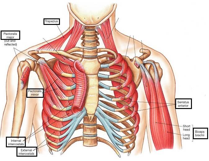
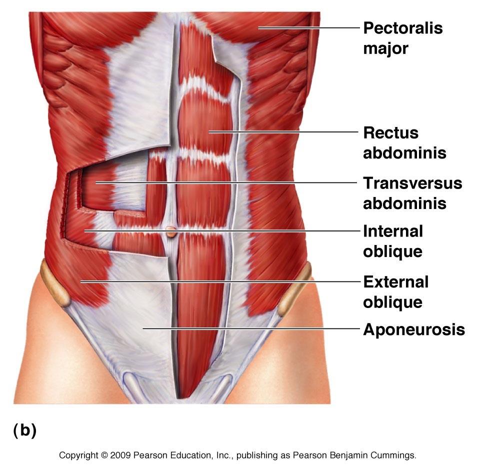
2 Origin Clavicle Pectoralis Minor Prime mover of arm flexion, rotates arm Draws forward Serratus Anterior Rotate Lateral aspects of ribs Deltoid Acromion External Intercostals Prime mover of arm abduction Elevate rib cage Internal Intercostals Depress rib cage Diaphragm Prime mover of inspiration, flattens on contraction Rotate vertebral column Compress abdominal wall Compress abdominal wall Compress abdominal contents Superior border of rib below Lumbar vertebrae Pectoralis Major Rectus Abdominis External Obliques Internal Obliques Transversus Abdominis Anterior surface of ribs Inferior border of rib Insertion Intertubercular sulcus in Coracoid process Anterior surface of Deltoid tuberosity of Superior border of rib below Inferior border of rib above Central tendon Pubic crest Anterior surface of 8 ribs Iliac crest Xiphoid process Pubic crest Iliac crest Pubic crest Pubic crest
3
 Lateral rotation of Infraspinous fossa of Lateral rotation of Supraspinous")
4 Trapezius Latissimus Dorsi Rhomboids Minor Rhomboids Major Infraspinatus Supraspinatus Teres Minor Teres Major Human Trunk- the Back Origin Rotates Thoracic vertebrae Prime mover of arm Lumbar vertebrae extension Stabilize Spinous processpointed tip of vertebrae Stabilize Spinous process s of Scapula (rotator cuff) Lateral rotation of Infraspinous fossa of Lateral rotation of Supraspinous fossa of Lateral rotation of Lateral margin of Rotation of Inferior angle of Humerus s act on Forearm Insertion Acromion of Floor of Medial border of Medial border of Greater tubercle of Greater tubercle Greater tubercle Intertubercular sulcus of
5 Triceps Brachii Forearm extensor Origin Inferior of glenoid cavity Biceps Brachii- Flexion of elbow Coracoid process Supraglenoid tubercle Distal of anterior Distal end of muscle closer to body Brachialis Flexion of forearm Brachioradialis Synergist (helper) in forearm flexion Pronator Teres-inner elbow Flexor Carpi Radialis Flexor Carpi Ulnaris Palmaris Longus 4. Extensor carpi radialis Longus 3. Extensor carpi radialis Brevis thumb side 2. Extensor Digitorum 1.Extensor carpi ulnaris Supinator on outside edge of elbow Humerus s act on Hands/ Fingers Pronate forearm Medial epicondyle of Flexor of wrist Medial epicondyle Flexor of wrist Tenses skin Extend wrist Insertion Olecranon processopposite end of ulna head Radial tuberosity- bump near the head Coronoid process- lower than olecranon of ulna Styloid process of radius- opposite end of head Midshaft of radius Base of metacarpals Medial epicondyle Medial epicondyle Lateral supracondylar ridge of Lateral epicondyle of Base of metacarpal 5 Skin of palm Base of metacarpal 2 Prime mover of finger extension Extend wrist Lateral epicondyle Distal phalanges Lateral epicondyle Base of metacarpal 5 Acts w/ biceps brachii to supinate forearm Lateral epicondyle Proximal end of radius Extend wrist Base of metacarpal 3 Radial head by the elbow, ulna head at other end by wrist Pronator teres touches flexor carpi radialis Under forearm: (thumb side) Brachioradialis- Flexor Carpi Radialis- Palmaris Longus- Flexor Carpi Ulnaris (pinky side) Above Forearm: (pinky side) Flexor Carpi Ulnaris- Extensor Carpi Ulnaris- Extensor Digitorum- extensor carpi radialis longus

6 Thigh and Leg

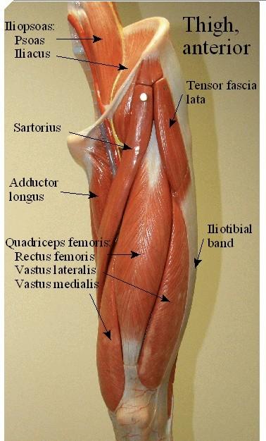
7 Iliopsoas Flex thigh Origin Iliac crest Sartorius Flex thigh Anterior superior iliac spine Ischial tuberosity Pubis Flex thigh Adductor Magnus Flex thigh Adductor Longusabove the magnus Gracilis- long muscle, Adducts thigh inner thigh Rectus Femorismain muscle on anterior leg Vastus Lateralis Vastus Medialis Vastus Intermedius Tensor Fasciae Latae Biceps Femoris Semitendinosus Semimembranous Gluteus Maximus Body of pubis Insertion Lesser trochanter of femur proximal tibia Linea aspera Linea aspera Medial of tibia Quadriceps (front of thigh) Extend knee Anterior inferior iliac spine Tibial tuberosity Extend knee Extend knee Extend knee Steadies trunk Tibial tuberosity Tibial tuberosity Tibial tuberosity Iliotibial tract Greater trochanter Linea aspera Lateral surface of femur Anterior aspect of iliac crest Hamstrings (back of thigh) Extend thigh Linea aspera Extend thigh Ischial tuberosity Extend thigh Ischial tuberosity Butt Powerful thigh extensor Coccyx and sacrum Head of fibula Upper tibial shaft Medial condyle of tibia Gluteal tuberosityfemur Medially rotates thigh Upper lateral surface of Greater trochanter of Gluteus Medius ilium femur Thigh Anterior: (left to right) Vastus Lateralis- Vastus Intermedius- Vastus Medialis [Rectus Femoris on top of V. Inter.]
 Plantar flexion Flexes leg")
8 Tibialis anterior Tibialis posterior EXTENSOR digitorum longus FLEXOR digitorum longus Gastrocnemius Soleus Popliteus Feet and Ankle Origin Prime mover of Lateral condyle- femur dorsiflexion Prime mover of foot Superior of tibia inversion Prime mover of toe Lateral condyle of tibia extension Flexes toes Posterior of tibia Plantar flexes foot (move foot up and down) Plantar flexion Flexes leg Insertion Metatarsal 1 Several tarsals Distal phalanges Distal phalanges Medial condyle of femur Calcaneus (heel bone) Proximal portion of tibia Lateral condyle of femur Calcaneus Proximal tibia


9 All muscles exert their force by pulling between at least two points of attachment. The movement that results from contraction is called the action of the muscle. Typically, one attachment remains stationary and is called the origin and the other attachment moves and is called the insertion. Almost all muscles cross at least one joint (moveable connection between two bones) and cause an action across that joint. The type of movement that results depends upon the nature of the joint. Common types of movement that result from muscle contraction include: Flexion A movement that decreases the angle between two bones at the joint. Extension A movement that increases the angle between two bones at the joint. However, if the angle goes past 180o (a straight line) in the direction opposite flexion the movement is called hyperextension. Rotation Rotation is a movement that results in movement of one bone around its longitudinal axis. Abduction A movement that results in the part moving away from the midline.
10 Adduction A movement that results in the part moving toward the midline. Special types of movement occur at particular joints and include: Dorsiflexion and plantar flexion These movements only apply to movements of the foot at the ankle joint. Dorsiflexion is when the foot is raised as when you dig in your heels. Plantar flexion is when you lower your foot as when you lift yourself onto the balls of your feet. Inversion and eversion These movements also apply only to the feet. Inversion is when you turn your feet inward so that your soles are facing one another. Eversion is when you turn your feet outward so that your soles are facing laterally. Supination and pronation These movements apply to the forearm. Pronation is rotation of the radius across the ulna that results is your palms facing backwards. Supination is movement in the opposite direction that uncrosses the radius from the ulna to cause the palms to face forward. Opposition This movement enables us to be skillful tool users. Opposition is the movement of the tip of the thumb that enables it to touch the tips of the other fingers. Chapter 12- anatomy of Skeletal Sarcolemma-plasma membrane of skeletal cells Myofibrils- longitudinally arranged, fill sarcoplasm, push nuclei peripherally Myofilaments- smaller threadlike structures make up the myofibrils - Composed of contractile proteins: actin and myosin (slide past each other during muscle activityshorten or contract muscle cells) Sarcomeres- actual contractile units of muscles extending from Z disc to next Z disc (middle of I band) Transverse tubule (T tubule)- formed by the sarcolemma indents in muscle cell (junction of A and I bands) - Tubules run deep btwn cross channels or terminal cisternae - Sarcoplasmic Reticulum- elaborate smooth endoplasmic reticulum - Triads- regions where SR terminal cisternae abut a T tubule on each side Endomysium- delicate areolar CT sheath wrapping muscle fiber Perimysium- collagenic membrane wrapping sheathed muscle fibers Fascicle- a bundle of fibers formed by perimysium wrapping the fibers together Epimysium- overcoat of dense CT which sheathes entire muscle Deep Fascia- coarse sheets of dense CT that bind muscle into function groups (tendons or aponeuroses) Aponeuroses- sheetlike functional group which attaches muscles to each other or indirectly to bones Insertion- movable attachment Origin- fixed/ immovable attachment Tendons fx- provide durability and conserve space Neuromuscular junction- junction btwn nerve fiber (Axon) and muscle cell - Neuron and muscle fiber membranes don t touch, are separated by small fluid filled gap called the synaptic cleft Triceps brachii- forearm extension- INFERIOR OF GLENOID CAVITY olecranon process of ulna Biceps brachii- elbow flexion- supraglenoid tubercle and coracoid process- radial tuberosity Brachialis- forearm flexion- distal end of -coronoid process ulna Brachioradialis- synergist arm flexion- distal end of - styloid process radius
11
A&P 1 Muscle In-Lab Guide
 A&P 1 Muscle In-Lab Guide This lab guide includes a table with all the muscles you need to ID, along with their origins, insertions and actions Dashed lines means ignore. If several actions are listed,
Lab Exercise #5 The Muscular System Student Performance Objectives
 Student Performance Objectives The material that you are required to learn in this exercise can be found in either the lecture text or the supplemental materials provided in lab. Prior to coming to class,
Lab 9: Learn origin and insertion for each of the listed muscles. For Exercise 15, do Activities 1-6 in 9 th edition, Activities 1-4 in 10 th edition
 The Muscular System Exercises 14, 15, and 16 (begins: page 187 in 9 th and 10 th editions) Exercises 12, 13, and 14 (begins: page 185 in 11 th edition, page 189 in 12 th edition) Lab 8 and 9 Objectives
Due in Lab weeks because of Thanksgiving Prelab #10. Homework #8. Both sides! Both sides!
 Lab 8 MUSCLES Due in Lab 10 2 weeks because of Thanksgiving Prelab #10 Both sides! Homework #8 Both sides! Refer to Muscles 22-23 Naming of muscles Origin Site of muscle attachment that doesn t move during
Bio 113 Anatomy and Physiology The Muscles. Muscles of the Head and Neck. Masseter. Orbicularis occuli. Orbicularis oris. Sternocleidomastoid
 Bio 113 Anatomy and Physiology The Muscles Muscles of the Head and Neck Masseter Orbicularis occuli Orbicularis oris Sternocleidomastoid Temporalis BIO 113 Fall 2011 Muscles Page 1 of 5 Muscles of the
SKELETAL MUSCLE ANATOMY
 SKELETAL MUSCLE ANATOMY OUTLINE I. Anatomical Terms of Motion II. Head, Face & Neck Muscles III. Anterior Torso Muscles IV. Posterior Torso Muscles V. Arm & Shoulder Muscles VI. Leg & Hip Muscles 2 ANATOMICAL
A. All movements require muscle which are organs using chemical energy to contract.
 Ch 8 Muscles Introduction: A. All movements require muscle which are organs using chemical energy to contract. B. The three types of muscle in the body are skeletal, smooth, and cardiac muscle. C. This
11/15/2018. Temporalis Elevates & retracts mandible. Masseter = Prime mover of jaw closure. Levator scapulae Supraspinatus Clavicle.
 Due in Lab 10 Lab 8 MUSCLES 2 weeks because of Thanksgiving Prelab #10 Both sides! Homework #8 Both sides! Refer to Muscles 22-23 Examples of Origin & Insertion Naming of muscles Origin Site of muscle
The Muscular System PART C. PowerPoint Lecture Slide Presentation by Patty Bostwick-Taylor, Florence-Darlington Technical College
 PowerPoint Lecture Slide Presentation by Patty Bostwick-Taylor, Florence-Darlington Technical College The Muscular System 6 PART C Five Golden Rules of Skeletal Muscle Activity Table 6.2 Muscles and Body
Human Anatomy and Physiology I Laboratory
 Human Anatomy and Physiology I Laboratory Gross Anatomy of the Muscular System (Two weeks) 1 This lab involves study of the laboratory exercise Gross Anatomy of the Muscular System. Complete the Review
Anatomy and Physiology 2016
 Anatomy and Physiology 2016 O = Temporal line I = coronoid process (Mandible) A = elevates mandible (chewing) O = galea aponeurotica (layer of dense fibrous tissue which covers the upper part of the cranium)
List of Muscles and Function. Region View Muscle Function Facial Anterior/Oblique Occipitofrontalis front belly Raises eyebrows
 List of Muscles and Function Region View Muscle Function Facial Anterior/Oblique Occipitofrontalis front belly Raises eyebrows Orbicularis oculi Closes eye Orbicularis oris Purses lips Zygomaticus minor/major
Muscle fiber (cell) Blood vessel. Perimysium. Epimysium. Fascicle (wrapped by perimysium) Endomysium (between fibers) Tendon. Bone
 Blood vessel. Perimysium. Epimysium. Fascicle (wrapped by perimysium) Endomysium (between fibers) Tendon. Bone") Figure 6.1 Connective tissue wrappings of skeletal muscle. Blood vessel Muscle fiber (cell) Perimysium Epimysium Fascicle (wrapped by perimysium) Tendon Endomysium (between fibers) Bone Figure 6.15 Superficial
Figure 6.1 Connective tissue wrappings of skeletal muscle. Blood vessel Muscle fiber (cell) Perimysium Epimysium Fascicle (wrapped by perimysium) Tendon Endomysium (between fibers) Bone Figure 6.15 Superficial
BIO130 Lab Practice Exam 2 Questions
 BIO130 Lab Practice Exam 2 Questions 1. Refer to Figure 1 and answer the following: Name the covering labeled Name the tubular portion labeled Name the hollow part labeled Name the material labeled Name
Scapula Spine Lateral edge of clavicle. Medial border Scapula. Medial border of Scapula, between superior angle and root of spine. Scapula.
 Muscle attachments and actions answer sheet Muscle Origins insertions Movements Joints crossed Trapezius Base of skull Spinous process of C7 Thoracic Spine Lateral edge of clavicle Elevation Retraction
Monday, November 13, 2017 A & P 2401
 Monday, November 13, 2017 A & P 2401 Today you will complete the following handouts. Study the last part of the handout for this will be on your quiz, which will be on Wednesday. It is titled steps of
In-Depth Foundations: Anatomy Terms to Know
 Be familiar with / able to identify and define all the following parts. The Spine Cranium Vertebrae Cervical, Thoracic, Lumbar Sacrum Coccyx Bones of Upper Body Cranium Mastoid process; Occipital condyle,
Epicranius (frontal belly) Zygomaticus minor. Zygomaticus major Buccinator
 Zygomaticus minor. Zygomaticus major Buccinator") Epicranius (frontal belly) Zygomaticus minor Zygomaticus major Buccinator Masseter Digastric (posterior belly) Stylohyoid Sternocleidomastoid Trapezius Scalenus Omohyoid (inferior belly) Orbicularis oris
Epicranius (frontal belly) Zygomaticus minor Zygomaticus major Buccinator Masseter Digastric (posterior belly) Stylohyoid Sternocleidomastoid Trapezius Scalenus Omohyoid (inferior belly) Orbicularis oris
ACTIVITIES 5 & 6: APPENDICULAR AND AXIAL MUSCLES
 ACTIVITIES 5 & 6: APPENDICULAR AND AXIAL MUSCLES Objectives: 1) How to get ready: Read Chapter 11 & 12, McKinley et al., Human Anatomy, 4e. All text references are for this textbook. Begin identifying
Muscles Built on the Maniken
 Muscles Built on the Maniken Facial Muscle Group 1. Temporalis O temporal fossa I anterior border of the ramus of the mandible A elevates the mandible (bite muscle) and holds jaw while at rest 2. Procerus
Temporalis Elevates & retracts mandible. Masseter Elevates mandible. Sternocleidomastoid Neck flexion. Trapezius Elevates & depresses shoulders
 Anterior Posterior Temporalis Elevates & retracts mandible Masseter Elevates mandible Sternocleidomastoid Neck flexion Trapezius Elevates & depresses shoulders Masseter Elevates mandible Temporalis Elevates
Anatomy and Physiology 141 Exam II November 6, Name Student Number
 Anatomy and Physiology 141 Exam II November 6, 2014 Name Student Number 1. In regards to the gross anatomy of muscle, which of the following is NOT TRUE? a. Perimysium is more superficial than the epimysium
2/4/2018. Identify the two reasons why muscle cells may go through muscle fatigue. Ch.7 Review. Sternocleidomastoid.
 Ch.7 Review Identify the two reasons why muscle cells may go through muscle fatigue Temporalis Depressor anguli oris Sternocleidomastoid Tibialis anterior 1 Gluteus medius Deltoid Adducts & rotates scapula
Muscles of the Cat. N Deltoid MUSCLES OF THE CHEST. Pectoralis major. (This muscle is superior to Pectoralis minor) MUSCLES OF THE CHEST
 MUSCLES OF THE CHEST") MUSCLES OF THE CHEST Pectoralis major (This muscle is superior to Pectoralis minor) 1. MUSCLES OF THE CHEST Pectoralis minor (This muscle is inferior to Pectoralis major) 2. MUSCLES OF THE ARM Deltoid
MUSCLES OF THE CHEST Pectoralis major (This muscle is superior to Pectoralis minor) 1. MUSCLES OF THE CHEST Pectoralis minor (This muscle is inferior to Pectoralis major) 2. MUSCLES OF THE ARM Deltoid
In which arm muscle are intramuscular injections most often given? (not in text)
") AP1 Lab 9 - Muscles of the Arms and Legs Locate the following muscles on the models and on yourself. Recall anatomical position. Directional terms such as anterior, posterior, lateral, etc. all assume
AP1 Lab 9 - Muscles of the Arms and Legs Locate the following muscles on the models and on yourself. Recall anatomical position. Directional terms such as anterior, posterior, lateral, etc. all assume
Muscles in the Shoulder, Chest, Arm, Stomach, and Back
 Muscles in the Shoulder, Chest, Arm, Stomach, and Back Shoulder Muscles Deltoid Supraspinatus Infraspinatus Teres Major Teres Minor Subscapularis Deltoid (Delts) Function: Raises the upper arm Origin:
Muscles of the Upper Limb
 Muscles of the Upper Limb anterior surface of ribs 3 5 coracoid process Pectoralis minor pectoral nerves protracts / depresses scapula Serratus anterior Subclavius ribs 1-8 long thoracic nerve rib 1 ----------------
Match the types of muscle tissues with the words and phrases. 1) Skeletal 2) Smooth 3) Cardiac 2 Walls of blood vessels. 2 Walls of digestive tract
 Skeletal 2) Smooth 3) Cardiac 2 Walls of blood vessels. 2 Walls of digestive tract") S T U D Y G U I D E. Types of Muscle Tissues Match the types of muscle tissues with the words and phrases. ) Skeletal ) Smooth ) Cardiac, Striated Walls of blood vessels, Single nucleus Heart muscle, Involuntary
S T U D Y G U I D E. Types of Muscle Tissues Match the types of muscle tissues with the words and phrases. ) Skeletal ) Smooth ) Cardiac, Striated Walls of blood vessels, Single nucleus Heart muscle, Involuntary
Muscles of Lesson Five. Muscular Nomenclature and Kinesiology - Two. Muscles of Lesson Five, cont. Chapter 16
 Chapter 16 Muscular Nomenclature and Kinesiology - Two Lessons 5-6 Muscles of Lesson Five Iliopsoas (psoas major, iliacus) Hip outward rotators (piriformis, gemellus superior, gemellus inferior, obturator
Chapter 9. The Muscular System
 1 Chapter 9 The Muscular System 2 Introduction Skeletal muscles: movement in environment Smooth muscles: intestines, ureters, veins and arteries Cardiac muscle: pumps blood through heart and blood vessels
The Muscular System Outline 10.1 For any movement, muscles can act in one of three ways (pp ) A. Muscles only pull; they never push, and as a
 A. Muscles only pull; they never push, and as a") The Muscular System Outline 10.1 For any movement, muscles can act in one of three ways (pp. 321 322) A. Muscles only pull; they never push, and as a muscle shortens, the insertion is pulled toward the
The Muscular System Outline 10.1 For any movement, muscles can act in one of three ways (pp. 321 322) A. Muscles only pull; they never push, and as a muscle shortens, the insertion is pulled toward the
The Human Muscular System Required reading before beginning this lab: Saladin, KS: Human Anatomy 5th ed (2017) Chapters 10, 11, 12 INTRODUCTION
 Chapters 10, 11, 12 INTRODUCTION") Biology 322: Human Anatomy The Human Muscular System Required reading before beginning this lab: Saladin, KS: Human Anatomy 5 th ed (2017) Chapters 10, 11, 12 INTRODUCTION We will use a number of lab periods
Biology 322: Human Anatomy The Human Muscular System Required reading before beginning this lab: Saladin, KS: Human Anatomy 5 th ed (2017) Chapters 10, 11, 12 INTRODUCTION We will use a number of lab periods
Prime movers provide the major force for producing a specific movement Antagonists oppose or reverse a particular movement Synergists
 Dr. Gary Mumaugh Prime movers provide the major force for producing a specific movement Antagonists oppose or reverse a particular movement Synergists Add force to a movement Reduce undesirable or unnecessary
Anatomy & Physiology B. Chapter 6: Muscles
 Anatomy & Physiology B Chapter 6: Muscles Warm-up What are the three types of muscle tissue? Where are each located? Which are voluntary and which are involuntary? Which are striated which are unstriated?
Exam 3 Self Quiz. Muscle
 Exam 3 Self Quiz Muscle ***Disclaimer- ALL lectured material is fair game for the exam. The instructor DOES NOT guarantee in any way that these questions will be on the exam. The instructor DOES guarantee
Lectures Muscular System 10-1
 Lectures 12-14 Muscular System 10-1 Properties of Muscle Ability of a muscle to shorten with force Capacity of muscle to respond to a stimulus Muscle can be stretched to its normal resting length and beyond
BLUE SKY SCHOOL OF PROFESSIONAL MASSAGE AND THERAPEUTIC BODYWORK. Musculoskeletal Anatomy & Kinesiology II REVIEW
 BLUE SKY SCHOOL OF PROFESSIONAL MASSAGE AND THERAPEUTIC BODYWORK Musculoskeletal Anatomy & Kinesiology II REVIEW MSAK101-II Session 4 LEARNING OBJECTIVES: By the end of this session, the student will be
lesser trochanter of femur lesser trochanter of femur iliotibial tract (connective tissue) medial surface of proximal tibia
 medial surface of proximal tibia") LOWER LIMB MUSCLES OF THE APPENDICULAR SKELETON The muscles that act on the lower limb fall into three groups: those that move the thigh, those that move the lower leg, and those that move the ankle, foot,
LOWER LIMB MUSCLES OF THE APPENDICULAR SKELETON The muscles that act on the lower limb fall into three groups: those that move the thigh, those that move the lower leg, and those that move the ankle, foot,
Biology 2401 Muscles List for CPC models
 Biology 2401 List for CPC models Italicized muscles are dissect and similar in the cat = Dissect and note the differences in human and cat Major of the Human Head Facial Expression Epicranius frontalis
Lab Activity 11: Group II
 Lab Activity 11: Group II Muscles Martini Chapter 11 Portland Community College BI 231 Origin and Insertion Origin: The place where the fixed end attaches to a bone, cartilage, or connective tissue. Insertion:
The Muscular System Lab Power Point
 The Muscular System Lab Power Point Myoneural Junction Sarcoplasm Nucleus Myofibrils Sarcomere (black line to black line) Sarcolemma Myoneural space Nucleus Endomysium Motor Neuron Muscles of Facial Expression
Muscles of the Cat Review Sheet
 MUSCLES F THE CHEST 1. Pectoralis major clavicle, sternum, costal cartilages greater tubercle of humerus Flexes, adducts and medially rotates arm (This muscle is superior to Pectoralis minor) MUSCLES F
Test Bank for The Human Body in Health and Illness 4th Edition by Herlihy
 Test Bank for The Human Body in Health and Illness 4th Edition by Herlihy Chapter 9: Muscular System Test Bank MULTIPLE CHOICE 1. Which of the following muscles is described as striated and involuntary?
The Muscular System. - composed of mostly skeletal muscle tissue, nervous tissue, blood and connective tissue
 The Muscular System Every action the body takes utilizes a muscular activity. Some of the muscles of the body are under voluntary control (skeletal muscles), and by using these muscle, you are able to
Exercise Science Section 3: The Muscular System
 Exercise Science Section 3: The Muscular System An Introduction to Health and Physical Education Ted Temertzoglou Paul Challen ISBN 1-55077-132-9 Major Functions of Muscles Movement Includes: breathing,
Cadaver Muscular System Practice Practical
 Cadaver Muscular System Practice Practical Station 1 Station 1 1. Specific structure 1. Rectus sheath 2. Red line 2. Linea alba Station 2 Station 2 3. Red muscle 1. Rectus abdominis 4. Red muscle actions
Muscles of the Hip 1. Tensor Fasciae Latae O: iliac crest I: lateral femoral condyle Action: abducts the thigh Nerve: gluteal nerve
 Muscles of the Hip 1. Tensor Fasciae Latae O: iliac crest I: lateral femoral condyle Action: abducts the thigh Nerve: gluteal nerve 2. Gluteus Maximus O: ilium I: femur Action: abduct the thigh Nerve:
Chapter 6 part 2. Skeletal Muscles of the Body
 Chapter 6 part 2 Skeletal Muscles of the Body Basic Principles 600 + muscles in the human body (you are required to learn 45, lucky kids)! Skeletal Muscles pull on bones Origin of a muscle = point of attachment
Lab Activity 11: Group I
 Lab Activity 11: Group I Muscles Martini Chapter 11 Portland Community College BI 231 Origin and Insertion Origin: The place where the fixed end attaches to a bone, cartilage, or connective tissue. Insertion:
Human Anatomy Biology 351
 Human Anatomy Biology 351 Lower Limb Please place your name on the back of the last page of this exam. You must answer all questions on this exam. Because statistics demonstrate that, on average, between
Connects arm to thorax 3 joints. Glenohumeral joint Acromioclavicular joint Sternoclavicular joint
 Connects arm to thorax 3 joints Glenohumeral joint Acromioclavicular joint Sternoclavicular joint Scapula Elevation Depression Protraction (abduction) Retraction (adduction) Downward Rotation Upward Rotation
Muscular Nomenclature and Kinesiology - One
 Chapter 16 Muscular Nomenclature and Kinesiology - One Lessons 1-3 (with lesson 4) 1 Introduction 122 major muscles covered in this chapter Chapter divided into nine lessons Kinesiology study of human
Practical 2 Worksheet
 Practical 2 Worksheet Upper Extremity BONES 1. Which end of the clavicle is on the lateral side (acromial or sternal)? 2. Describe the difference in the appearance of the acromial and sternal ends of the
Naming Skeletal Muscles
 Naming Skeletal Muscles Direction of Muscle Fibers Action Location Origin & Insertion Skeletal Muscle Size Shape Number Of Origins Direction of Muscle Fibers Relative to the Midline RECTUS = parallel to
Unit 4: The Muscular System REVIEW GUIDE
 NPHS Anatomy & Physiology Questions to answer: 1) List the three functions of the muscular system. Unit 4: The Muscular System REVIEW GUIDE 2) What are the four characteristics of muscle tissue? Briefly
Name this muscle. Name this muscle
 this muscle this muscle Pectoralis Major Pectoralis Minor Serratus anterior Pectoralis minor Serratus anterior this muscle Deltoid: The major abductor of the upper limb this muscle this muscle this muscle
Biology Human Anatomy February 28, 2000 Section # 005 Exam # Midterm Examination #2
 Biology 2050 - Human Anatomy Name: KEY February 28, 2000 Section # 005 Exam # Midterm Examination #2 Directions: This exam consists of 45 multiple choice questions worth 2 points each and 10 true/false
Types of Muscle: Skeletal- muscle involved in movement of the skeleton. Striated, has alternating bands of light and dark due to overlapping
 Types of Muscle: Skeletal- muscle involved in movement of the skeleton. Striated, has alternating bands of light and dark due to overlapping filaments within the muscle cell. Skeletal muscle can be consciously
The Muscular System The Muscular System Muscles are responsible for all types of body movement Three basic muscle types are found in the body
 The Muscular System The Muscular System Muscles are responsible for all types of body movement Three basic muscle types are found in the body Skeletal muscle Cardiac muscle Smooth muscle Characteristics
Anatomy & Physiology. Muscles of the Lower Limbs.
 Anatomy & Physiology Muscles of the Lower Limbs http://www.ishapeup.com/musclecharts.html Muscles of the Lower Limbs Among the strongest muscles in the body. Because pelvic girdle is composed of heavy,
Human Anatomy Lab #7: Muscles of the Cadaver
 Human Anatomy Lab #7: Muscles of the Cadaver Table of Contents: Expected Learning Outcomes.... 1 Introduction...... 1 Identifying Muscles on Yourself.... 2 Muscles of the Anterior Trunk and Arm.. 2 Muscles
Lower limb summary. Anterior compartment of the thigh. Done By: Laith Qashou. Doctor_2016
 Lower limb summary Done By: Laith Qashou Doctor_2016 Anterior compartment of the thigh Sartorius Anterior superior iliac spine Upper medial surface of shaft of tibia 1. Flexes, abducts, laterally rotates
Unit 6 - The Muscular System 1
 Unit 6 - The Muscular System 1 I. Unit 6: The Muscular System A. The Muscular System 1. Muscles are responsible for all types of body movement 2. Three basic muscle types are found in the body a) Skeletal
Human Anatomy Biology 255
 Human Anatomy Biology 255 Exam #4 Please place your name and I.D. number on the back of the last page of this exam. You must answer all questions on this exam. Because statistics demonstrate that, on average,
MicroAnatomy Muscle Fiber Model
 MicroAnatomy Muscle Fiber Model Muscle fiber whole model (but model is only a fraction of a fiber) Sarcolemma 14 Myofibril 1 Nucleus 8 Mitochondria 2 Triad 16 Sarcoplasmic reticulum 17 T tubule 15 Thin
medial half of clavicle; Sternum; upper six costal cartilages External surfaces of ribs 3-5
 MUSCLE ORIGIN INSERTION ACTION NERVE Pectoralis Major medial half of clavicle; Sternum; upper six costal cartilages Lateral lip of intertubercular groove of horizontal adduction Medial and lateral pectoral
Anatomy and Physiology II. Review Shoulder Girdle New Material Upper Extremities - Bones
 Anatomy and Physiology II Review Shoulder Girdle New Material Upper Extremities - Bones Anatomy and Physiology II Shoulder Girdle Review Questions From Last Lecture Can you identify the following muscles?
Unit 6: The Muscular System
 Unit 6: The Muscular System I. The Muscular System A. Muscles are responsible for all types of body movement B. Three basic muscle types are found in the body 1. Skeletal muscle 2. Cardiac muscle 3. Smooth
REFERENCE DIAGRAMS OF UPPER LIMB MUSCLES: NAMES, LOCATIONS, ATTACHMENTS, FUNCTIONS MUSCLES CONNECTING THE UPPER LIMB TO THE AXIAL SKELETON
 REFERENCE DIAGRAMS OF UPPER LIMB MUSCLES: NAMES, LOCATIONS, ATTACHMENTS, FUNCTIONS MUSCLES CONNECTING THE UPPER LIMB TO THE AXIAL SKELETON A25LAB EXERCISES: UPPER LIMB MUSCLES Page 1 MUSCLES CONNECTING
Chapter 6- The Muscular System
 Chapter 6- The Muscular System I. The muscular system A. Muscles are responsible for all types of body movement B. Three basic muscle types are found in the body 1. Skeletal muscle 2. Cardiac muscle 3.
5/21/2013. Muscle Anatomy. Thursday January, 24 th, Skeletal Muscle. Smooth Muscle. Cardiac Muscle
 Muscle Anatomy Thursday January, 24 th, 2013 Skeletal Muscle Cardiac Muscle Smooth Muscle 1 Smooth Muscle 1. Found in the walls of the digestive system, bladder, uterus and blood vessels 2. Involuntary
Human Anatomy Biology 351
 Human Anatomy Biology 351 Lower Limb Please place your name on the back of the last page of this exam. You must answer all questions on this exam. Because statistics demonstrate that, on average, between
Sports Medicine Part II : ANATOMY OF THE SPINE, ABDOMEN AND SHOULDER COMPLEX
 Sports Medicine 25 1.1 Part II : ANATOMY OF THE SPINE, ABDOMEN AND SHOULDER COMPLEX c.w.p. Wagner High School, Sports Medicine, A. Morgan, T. Morgan & A. Eastlake, 2008 Muscles of the Upper Limbs In this
Figure 11-1: The lever-fulcrum principle is illustrated by flexion of the forearm.
 Chapter 11: The Muscular System Read pages 325 to 399 NAME Topic Outline And Objectives: A. How skeletal muscles produce movement, and naming muscles 1. Describe the relationship between bones and skeletal
Exercise Science Section 3: The Muscular System
 Exercise Science Section 3: The Muscular System An Introduction to Health and Physical Education Ted Temertzoglou Paul Challen ISBN 1-55077-132-9 Major Functions of Muscles Movement v Includes: breathing,
Human Anatomy Biology 351
 1 Human Anatomy Biology 351 Upper Limb Exam Please place your name on the back of the last page of this exam. You must answer all questions on this exam. Because statistics demonstrate that, on average,
2. In regards to the bones of the arm and forearm, which of the following is TRUE?
 Anatomy and Physiology Fall Exam II: Form A Name: 1. Use the following table to answer Question 1. I II III IV V The nasal bone articulates with the frontal process of the maxilla The mastoid process is
Appendix. Useful Anatomical Data of Clinical Significance
 Appendix Useful Anatomical Data of Clinical Significance Appendix Outline Respiratory System 426 Table I. Important Airway Distances (Adult) 426 Table II. Important Data Concerning the Trachea 426 Musculoskeletal
Cat Muscles Flashcards Mt SAC
 1. MUSCLES OF THE CHEST Pectoralis major (This muscle is superior to Pectoralis minor) 2. MUSCLES OF THE CHEST Pectoralis minor (This muscle is inferior to Pectoralis major) 3. MUSCLES OF THE ARM AD CHEST
Anatomy and Physiology 1 Chapter 11 self quiz Pro, Dima Darwish,MD.
 Anatomy and Physiology 1 Chapter 11 self quiz Pro, Dima Darwish,MD. 1) The attachment of a muscle s tendon to the stationary bone is called the ; the attachment of the muscle s other tendon to the movable
1. In regards to the bones of the face, which of the following is NOT TRUE?
 Anatomy and Physiology Fall Exam II: Form B Name: 1. In regards to the bones of the face, which of the following is NOT TRUE? A. The vomer bone articulates with the dorsal surface of the palatine process
The Muscular System. Chapter 10 Part D. PowerPoint Lecture Slides prepared by Karen Dunbar Kareiva Ivy Tech Community College
 Chapter 10 Part D The Muscular System Annie Leibovitz/Contact Press Images PowerPoint Lecture Slides prepared by Karen Dunbar Kareiva Ivy Tech Community College Table 10.14: Muscles Crossing the Hip and
MUSCLES OF THE LOWER LIMBS
 MUSCLES OF THE LOWER LIMBS Naming, location and general function Dr. Nabil khouri ROLES THAT SHOULD NOT BE FORGOTTEN Most anterior compartment muscles of the hip and thigh Flexor of the femur at the hip
Written Response #1. 1. Based on what you know about Latin root words, what do you think these terms refer to?
 Muscular System Written Response #1 1. Based on what you know about Latin root words, what do you think these terms refer to? Sarcomere Sarcoplasm Myofibril Epimysium Perimysium Endomysium 2. What structure
Muscles of the lower extremities. Dr. Nabil khouri MD, MSc, Ph.D
 Muscles of the lower extremities Dr. Nabil khouri MD, MSc, Ph.D Posterior leg Popliteal fossa Boundaries Biceps femoris (superior-lateral) Semitendinosis and semimembranosis (superior-medial) Gastrocnemius
Class Outline: Posterior Anatomy
 Class Outline: Posterior Anatomy 5 minutes Breath of Arrival and Attendance 5 minutes Howdy Partner 35 minutes Posterior Anatomy using Power Point Presentation 5 minutes Overview of skeletal segments 5
Chiropractic Technician Class
 Chiropractic Technician Class Presentation By: Dr. Kay Miller. The Role of Exercise as it Relates to Our Musculoskeletal System Introduction to the topic and Preliminary Physical exam Musculoskeletal anatomy:
Lab Exercise 8. BIOPAC Exercise. Muscle Tissue. Muscles. What you need to be able to do on the exam after completing this lab exercise:
 Lab Exercise 8 BIOPAC Exercise Muscle Tissue Muscles Textbook Reference: See Chapters 9 & 10 What you need to be able to do on the exam after completing this lab exercise: Be able to answer questions covering
TABLES OF MUSCLE ACTIONS, INNERVATIONS, AND ATTACHMENTS
 TABLES OF MUSCLE ACTIONS, INNERVATIONS, AND ATTACHMENTS Table 1-1 ERECTOR SPINAE MUSCLES Intrinsic muscles producing extension and/or lateral of the spine Muscle Joint and Action Innervation Inferior Attachment
BIOH111. o Cell Module o Tissue Module o Skeletal system o Integumentary system o Muscle system o Nervous system o Endocrine system
 BIOH111 o Cell Module o Tissue Module o Skeletal system o Integumentary system o Muscle system o Nervous system o Endocrine system TEXTBOOK AND REQUIRED/RECOMMENDED READINGS o Principles of anatomy and
Synergist Muscles. Shoulder (glenohumeral joint) Flexion Deltoid (anterior fibers) Pectoralis major (upper fibers) Biceps Brachii Coracobrachialis
 Flexion Deltoid (anterior fibers) Pectoralis major (upper fibers) Biceps Brachii Coracobrachialis") Synergist Muscles Dr Gene Desepoli DrGeneLMT@gmail.com Shoulder (glenohumeral joint) Deltoid (anterior fibers) Pectoralis major (upper fibers) Biceps Brachii Coracobrachialis Deltoid (posterior fibers)
Synergist Muscles Dr Gene Desepoli DrGeneLMT@gmail.com Shoulder (glenohumeral joint) Deltoid (anterior fibers) Pectoralis major (upper fibers) Biceps Brachii Coracobrachialis Deltoid (posterior fibers)
Healing Hands School of Holistic Health. Advanced Circulatory & Sports Massage Class Handouts
 Class Handouts 1 Posterior Trepidations Torso Rock Torso Rock half-step Torso Rock both sides Torso Rock down body Torso Side Stretch Erector Rock Spinal Rock Lumbo Rock Cha Cha Leg Clay Snake Flop Leg
Because flexing muscles look like mice scurrying beneath the skin, scientists dubbed them, muscles, from the Latin word mus meaning little mouse
 Because flexing muscles look like mice scurrying beneath the skin, scientists dubbed them, muscles, from the Latin word mus meaning little mouse The Muscular System Muscles are responsible for all types
1) The different types of muscle tissue differ from each other by
 The different types of muscle tissue differ from each other by") Chapters 10, 11 Practice Exam 1) The different types of muscle tissue differ from each other by a) microscopic anatomy. b) location. c) type of Control. d) both microscopic anatomy and location. e) All
Chapters 10, 11 Practice Exam 1) The different types of muscle tissue differ from each other by a) microscopic anatomy. b) location. c) type of Control. d) both microscopic anatomy and location. e) All
Human Muscles (Anterior View) Model 3-44
 Model 3-44") Human Muscles (Anterior View) Model 3-44 Temporalis Frontalis Orbicularis Occuli Orbicularis Oris Masseter Sternocleidomastoid Orbicularis Occuli Human Muscles (Anterior View) Model 3-65 Temporalis Masseter
Human Muscles (Anterior View) Model 3-44 Temporalis Frontalis Orbicularis Occuli Orbicularis Oris Masseter Sternocleidomastoid Orbicularis Occuli Human Muscles (Anterior View) Model 3-65 Temporalis Masseter
LEARN - INSPIRE - SUCCEED
 Anatomy and Physiology Workbook LEARN - INSPIRE - SUCCEED Label The Skeletal System Fibula Lumbar vertebrae Patella Sternum Ilium Femur Scapula Phalanges Sacrum Ischium Tarsals Cranium Clavicle Pubis Ribs
BIOH111. o Cell Module o Tissue Module o Skeletal system o Integumentary system o Muscle system o Nervous system o Endocrine system
 BIOH111 o Cell Module o Tissue Module o Skeletal system o Integumentary system o Muscle system o Nervous system o Endocrine system TEXTBOOK AND REQUIRED/RECOMMENDED READINGS o Principles of anatomy and
3/27/2012. Muscle Classification: Functional Groups. Interactions of Skeletal Muscles. Naming Skeletal Muscles. Naming Skeletal Muscles
 Interactions of Skeletal Muscles Skeletal muscles work together or in opposition Muscles only pull (never push) As muscles shorten, the insertion generally moves toward the origin Whatever a muscle (or
Upper Limb Muscles Muscles of Axilla & Arm
 Done By : Saleh Salahat Upper Limb Muscles Muscles of Axilla & Arm 1) Muscles around the axilla A- Muscles connecting the upper to thoracic wall (4) 1- pectoralis major Origin:- from the medial half of
Electrode Placement. Skin Preparation. Frontalis (FRL) (Specific) Temporalis Anterior (TA) (Specific) Sternocleidomastoid (SCM) (Specific)
 (Specific) Temporalis Anterior (TA) (Specific) Sternocleidomastoid (SCM) (Specific)") Electrode Placement Skin Preparation 1) Removing the hair: Shave if necessary 2) Clean the skin: Use a towel or abrasive pad with conductive cleaning paste or alcohol to remove dead skin cells (high impedance)
Electrode Placement Skin Preparation 1) Removing the hair: Shave if necessary 2) Clean the skin: Use a towel or abrasive pad with conductive cleaning paste or alcohol to remove dead skin cells (high impedance)